evaluation of the xanthan-based film incorporated with
TRANSCRIPT
Research ArticleEvaluation of the Xanthan-Based Film Incorporated withSilver Nanoparticles for Potential Application in the NonhealingInfectious Wound
Jinjian Huang,1,2 Jianan Ren,1 Guopu Chen,1,3 Youming Deng,1,3
Gefei Wang,1 and XiuwenWu1
1Department of Surgery, Jinling Hospital, Nanjing, China2Medical School of Southeast University, Nanjing, China3Medical School of Nanjing University, Nanjing, China
Correspondence should be addressed to Jianan Ren; [email protected]
Received 29 April 2017; Accepted 18 July 2017; Published 27 August 2017
Academic Editor: Piersandro Pallavicini
Copyright © 2017 Jinjian Huang et al. This is an open access article distributed under the Creative Commons Attribution License,which permits unrestricted use, distribution, and reproduction in any medium, provided the original work is properly cited.
Xanthan gum is a highmolecular weight polysaccharide biocompatible to biological systems, so its products promise high potentialinmedicine. In this study, we crosslinked xanthan gumwith citric acid to develop a transparent film for protecting the wound. Silvernanoparticles (AgNPs) are incorporated into the film to enhance the antimicrobial property of our biomaterial.This paper discussedthe characteristics and manufacturing of this nanocomposite dressing. The safety of the dressing was studied using fibroblasts(L929) by the method of 3-(4,5-dimethylthiazol-2-yl)-2,5-diphenyltetrazolium bromide (MTT) assay and staining of ethidiumhomodimer (PI) and calcein AM. The bacterial inhibition test and application of the dressing to nonhealing wounds infectedwith methicillin-resistant S. aureus (MRSA) were performed to evaluate the antibacterial effects in vitro and in vivo, respectively.The results indicated that the dressing could restrict the formation of biofilms, reduce inflammatory reactions, and promote theangiogenesis of granulation tissues in infectious wounds. Therefore, this dressing has a great advantage over traditional clinicalproducts especially when administered under the condition of infections or for the purpose of infection prevention.
1. Introduction
The treatment of nonhealing wounds is an arduous taskin the case of repeated infections, severe skin damage, andaltered physiological conditions such as diabetes mellitus andmalnutrition [1–3]. This leads to a great financial burdenfor both patients and healthcare systems worldwide. Somecomplicated wound infections, to our great concern, arecaused by virulent bacteria resistant to most of antibiotics.Staphylococcus aureus is the most common cause of theseinfections, and methicillin-resistant S. aureus (MRSA) ranksfirst among all drug-resistant pathogens involved in compli-cated skin and soft-tissue infections (cSSTI) [4–6]. There-fore, the development of an innovative dressing capable offacilitating wound observation, absorbing wound exudates,and preventing or treating the skin infections effectively is a
challenging and meaningful work for the sake of acceleratingthe healing of complicated wounds [7].
Xanthan gum is a macromolecular polysaccharide pro-duced mainly from sugarcane, corn, or their derivatives byXanthomonas campestris in aerobic conditions [8, 9]. It isextensively applied as thickener agent in the food industry,cosmetics, drilling fluids, and so on [10–12]. Hydroxyl groupsin the backbone and carboxyl groups in the branched chainscan be crosslinked with citric acid to form ester bonds,generating a chemical hydrogel with porous structure. Thepresence of hydrophilic moieties in the three-dimensionalcrosslinked networks of this hydrogel allows it to retain largeamounts of water.The good biocompatibility of xanthan gumdue to its natural origin could minimize allergic reactionsagainst its products in human tissues [13, 14]. Moreover, thecitric acid, besides being a crosslinker, can also play a role of
HindawiJournal of NanomaterialsVolume 2017, Article ID 6802397, 10 pageshttps://doi.org/10.1155/2017/6802397
2 Journal of Nanomaterials
an antioxidative agent to stabilize other materials, especiallynanoparticles [15]. Hence, it is theoretically practical forxanthan-based hydrogel to function as the wound-protectingdressing.
In recent years, silver nanoparticles have been recognizedas a powerful weapon against infectious diseases caused byvarious bacteria [16, 17]. Three main methods have beenreported to facilitate the synthesis of silver nanoparticles.Evaporation-condensation and laser ablation are the mostcommonphysical approaches, but consume toomuch energy.The most important approach for synthesis of AgNPs ischemical reduction by organic and inorganic reducing agentssuch as sodium citrate and sodium borohydride (NaBH4).However, the solvent contamination and uniformity of AgNPdistribution are problems in the reactive process that needto be considered. The last is bio-based method, in whichorganisms like bacteria and plants play an important role.It is worth mentioning that this is an environmentally andeconomically friendly synthesis protocol [18].
Owing to the success of AgNP techniques, investigationsabout its potential medical practice are continuously con-ducted on behalf of antibacterial or anticancer agents. Tagli-etti et al. [19, 20] coated AgNPs using glutathione or pectin tomodify the antibacterial effects with less cytotoxicity. Zhanget al. [21] combined silver nanoparticles with hydrogel topromote bone regeneration in infected bone defects. Basicresearch in oncology demonstrated that AgNPs had apoptoticeffects on MCF-7 human breast cancer cells and could blockthe growth of prostate cancer (PC-3) cells [22, 23].Therefore,AgNPs have recently become an indispensable componentfor biomaterials.
In order to explore a novel dressing to prevent infectionor treat nonhealing wounds more effectively than before, thexanthan-based film was assembled with AgNPs to bring theirsuperiority into full play.The safety and effects of this dressingfor infectious wounds will be a major topic in this paper.
2. Experiment
2.1. Materials. The materials are as follows: commercialxanthan gum (pharmaceutical grade, viscosity of 1% solutionat 20∘C: 1450–2000mPa⋅s, Shanghai JiangLai Co. Ltd); citricacid (Sigma-Aldrich); commercial AgNPs (size: ∼60 nm,Shanghai Jianglai CO. LTD); 3-(4,5-dimethylthiazolyl-2)-2,5-diphenyltetrazoliumbromide (MTT) (Biotium Inc); live/deadcell staining kit (Invitrogen); ultrasonic homogenizer (model:XO-400XD; Nanjing Xianou CO. LTD); Teflon-coated plate(Nanjing Ruinike Tech Co. Ltd); UV-Visible spectropho-tometry (model: UV-5200, Shanghai Metash InstrumentCO. LTD); IL-6, CD68 and TNF-𝛼 antibodies (providedby Wuhan Goodbio Tech Co. Ltd); CD31 primer sequence(Forward: AGTAGCATCCTGGTCAACATAACA, Reverse:ATACTGTGACAACACCGTCTCTTC, designed by Primer5 software). All other chemicals and reagents were of thehighest purity grade commercially available.
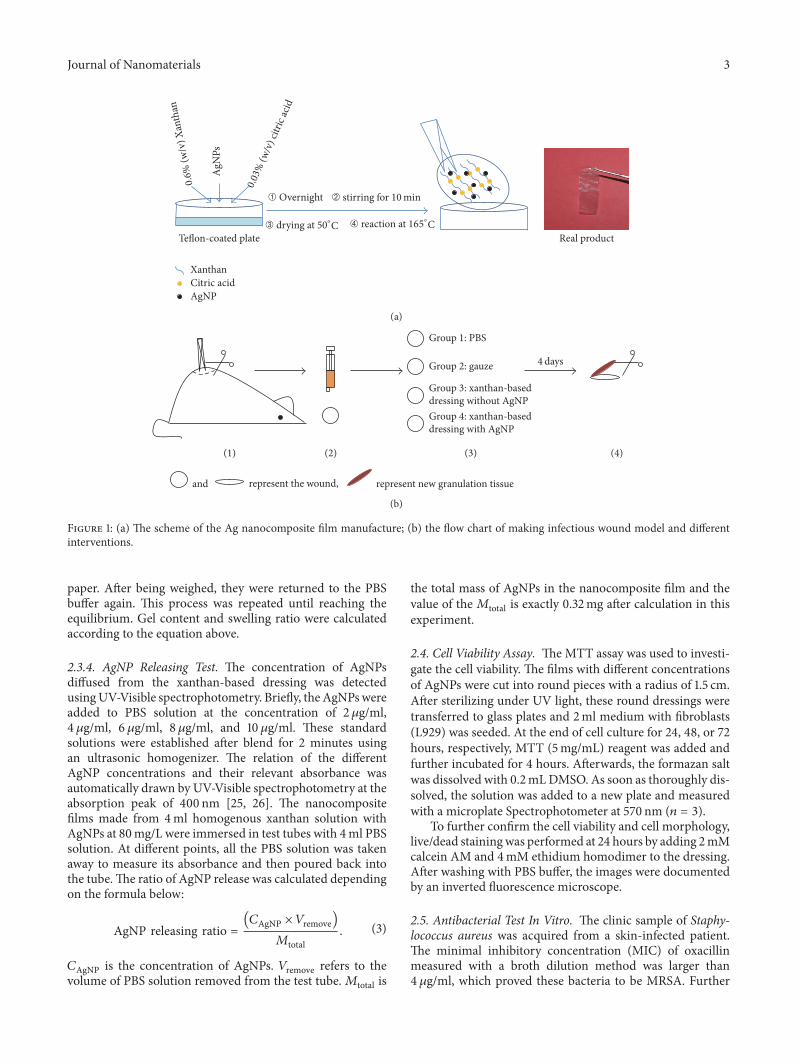
2.2. Preparation of Ag Nanocomposite Films. 6 g/L xanthanaqueous solution was mixed with AgNPs at different concen-tration gradients in the presence of citric acid at 300mg/L.This concentration of citric acidwas chosen because it workedwell for xanthan crosslinking [24].The homogenous solutionwas well prepared after being thoroughly stirred overnightand blended by ultrasonic homogenizer at 50% power for 10minutes. The solution was then poured onto Teflon-coatedplates and treated in a hot air oven at 50∘C until the films(∼0.2mm) were casted. The casted films were directly peeledfrom the plates and put into the hot air oven again at 165∘Cfor 7 minutes. As soon as the oven cooled down, all sampleswere collected and then stored in a sealed plastic bag at roomtemperature for further research. The working process andthe product were simply illustrated in the Figure 1(a).
2.3. Characterization of Ag Nanocomposite Films
2.3.1. Microstructure. Themicromorphology of the xanthan-based Ag filmwas assessed by Scanning ElectronMicroscopy(SEM). The films were immersed in deionized water at 37∘Cfor 0.5 h to remove unreacted residues and then freeze-dried(in a freeze dryer). Subsequently, the freeze-dried sampleswere investigated by a scanning electron microscope S-4800at a voltage of 5 kV, through which longitudinal sections ofthe film were studied and images were available.
2.3.2. Identification of AgNPs. Theblurry photograph of silvernanoparticles was acquired by the SEM at a high magnifi-cation of 100K. To obtain clear description of AgNPs, weutilized a transmission electron microscope (TEM). Samplesfor the transmission electron microscope were prepared byplacing the original solution on a carbon-coated copper grid.The grid was air-dried and then reacted at 165∘C for 7minutesbefore TEM scanning.
2.3.3. Gel Content and Swelling Ratio. Gel content and hydro-gel swelling ratio are determined by the formula as follows:
Gel content (%) =𝑀driedgel
𝑀pol× 100; (1)
Swelling ratio (%) =(𝑀swollengel −𝑀driedgel)𝑀driedgel
× 100. (2)
𝑀pol represents the initial mass of polymer. 𝑀driedgel and𝑀swollengel stand for the mass of dried hydrogel and swollenhydrogel at equilibrium, respectively.
The study of the gel content started with dried films,which then were soaked in an excess of PBS buffer for 48hours to rinse away all unreacted reagents and by-products.Subsequently, these wet samples were removed from PBSbuffer and dried under vacuumuntil the weight was constant.With regard to measurement of swelling ratio, several freeze-dried hydrogels were prepared first and then immersed inPBS buffer. At each setting time, they were removed andthe surface moisture was immediately wiped off using tissue
Journal of Nanomaterials 3
Te�on-coated plate
AgN
Ps
Citric acidAgNP
Xanthan
Real product0.0
3% (w
/v) c
itric
acid
0.6%
(w/v
) Xan
than
reaction at 165∘#➃drying at 50∘#➂
Overnight ➀ stirring for➁ 10min
(a)
Group 1: PBS
Group 2: gauze
Group 3: xanthan-based dressing without AgNPGroup 4: xanthan-based dressing with AgNP
and represent the wound, represent new granulation tissue
4 days
(2)(1) (3) (4)
(b)
Figure 1: (a) The scheme of the Ag nanocomposite film manufacture; (b) the flow chart of making infectious wound model and differentinterventions.
paper. After being weighed, they were returned to the PBSbuffer again. This process was repeated until reaching theequilibrium. Gel content and swelling ratio were calculatedaccording to the equation above.
2.3.4. AgNP Releasing Test. The concentration of AgNPsdiffused from the xanthan-based dressing was detectedusingUV-Visible spectrophotometry. Briefly, theAgNPswereadded to PBS solution at the concentration of 2 𝜇g/ml,4 𝜇g/ml, 6𝜇g/ml, 8 𝜇g/ml, and 10 𝜇g/ml. These standardsolutions were established after blend for 2 minutes usingan ultrasonic homogenizer. The relation of the differentAgNP concentrations and their relevant absorbance wasautomatically drawn by UV-Visible spectrophotometry at theabsorption peak of 400 nm [25, 26]. The nanocompositefilms made from 4ml homogenous xanthan solution withAgNPs at 80mg/L were immersed in test tubes with 4ml PBSsolution. At different points, all the PBS solution was takenaway to measure its absorbance and then poured back intothe tube.The ratio of AgNP release was calculated dependingon the formula below:
AgNP releasing ratio =(𝐶AgNP × 𝑉remove)𝑀total
. (3)
𝐶AgNP is the concentration of AgNPs. 𝑉remove refers to thevolume of PBS solution removed from the test tube.𝑀total is
the total mass of AgNPs in the nanocomposite film and thevalue of the𝑀total is exactly 0.32mg after calculation in thisexperiment.
2.4. Cell Viability Assay. TheMTT assay was used to investi-gate the cell viability. The films with different concentrationsof AgNPs were cut into round pieces with a radius of 1.5 cm.After sterilizing under UV light, these round dressings weretransferred to glass plates and 2ml medium with fibroblasts(L929) was seeded. At the end of cell culture for 24, 48, or 72hours, respectively, MTT (5mg/mL) reagent was added andfurther incubated for 4 hours. Afterwards, the formazan saltwas dissolved with 0.2mLDMSO. As soon as thoroughly dis-solved, the solution was added to a new plate and measuredwith a microplate Spectrophotometer at 570 nm (𝑛 = 3).
To further confirm the cell viability and cell morphology,live/dead stainingwas performed at 24 hours by adding 2mMcalcein AM and 4mM ethidium homodimer to the dressing.After washing with PBS buffer, the images were documentedby an inverted fluorescence microscope.
2.5. Antibacterial Test In Vitro. The clinic sample of Staphy-lococcus aureus was acquired from a skin-infected patient.The minimal inhibitory concentration (MIC) of oxacillinmeasured with a broth dilution method was larger than4 𝜇g/ml, which proved these bacteria to be MRSA. Further
4 Journal of Nanomaterials
investigation in the department of laboratory medicine usingPCR technology indicated the mecA gene of penicillinbinding protein was positive [27]. Bacterial suspension wasprepared by adding the S. aureus isolates until the turbiditywas around 0.5 based onMcFarland standards. Subsequently,isolates of S. aureus were inoculated onto mannitol salt agarin the condition of 37∘C. Simultaneously, the xanthan-baseddressing loaded with or without AgNPs was put onto the agarsurface. After 24 hours, the results were recorded by camera.
2.6. Acute Wound Infection Model for Assessing Efficiency ofthe Dressing. Twenty-four adult male Sprague-Dawley rats(weight ranges from 180 g to 220 g, Mergene Co. Ltd) wereused for this experiment. All the rats were raised at thetemperature of 25∘C and the relative humidity of 50–60%in regular day-and-night cycles and allowed free access tofood and water. All the research protocols were approved byAnimal Investigation Ethics Committee of Jinling Hospitaland conducted in accordance with international standards onanimal welfare.
All the rats got general anesthesia through intraperitonealinjections of 10%chloral hydrate at 0.4ml/100 g andwere thenrandomly divided into four groups with six per group. Theinfectious wound was created by removing circular skin at1 cm in diameter on the back of these animals and injecting0.1ml bacterial suspension onto the defected area. Afterthat, different interventions were performed on the wound.Specifically, each group received PBS solution, gauze, simplexanthan-based dressing, and xanthan-based dressing withAgNPs, respectively. All the rats were put back into their cagesand allowed free access to water and food. Four days later,these animals were sacrificed and fresh granulation tissueswere excised completely. Promptly, one-half of granulationtissues was immersed in 10% neutral formaldehyde for thehistological staining and the other half was stored in −80∘Cfor the qPCR. All the procedures were briefly introduced inFigure 1(b).
The samples were taken out of formaldehyde, dehydrated,and subsequently embedded in paraffin. Serial sections with5 𝜇m thickness for each granulation tissue were made with amicrotome. These sections were stained with H&E and Mas-son trichrome staining. After drying overnight, these slideswere microscopically examined and then photographed. IL-6, TNF-𝛼, and macrophages were analysed by immunohis-tochemical staining of IL-6, TNF-𝛼, and CD68, respectively,according to standard protocols. Three images each slidewere acquired at 200xmagnification randomly formeasuringtheir integral optical density (IOD) using image-pro plus 6.0software. SPSS16.0 was used to analyse the data via One-WayANOVA test.
3. Results
3.1. Physical Properties of Ag Nanocomposite Films
3.1.1. Micromorphology. SEM photographs of the freeze-dried dressing were investigated to observe the microstruc-ture and assess its porous property. In our study, the pore
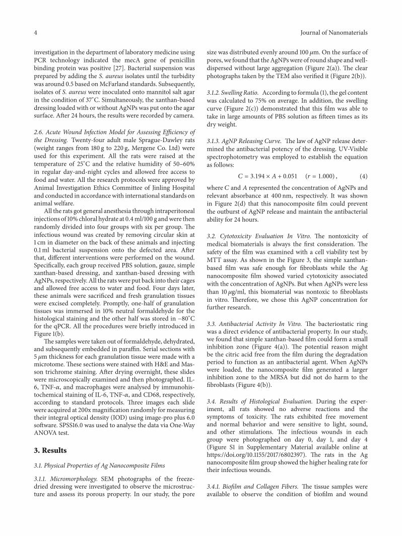
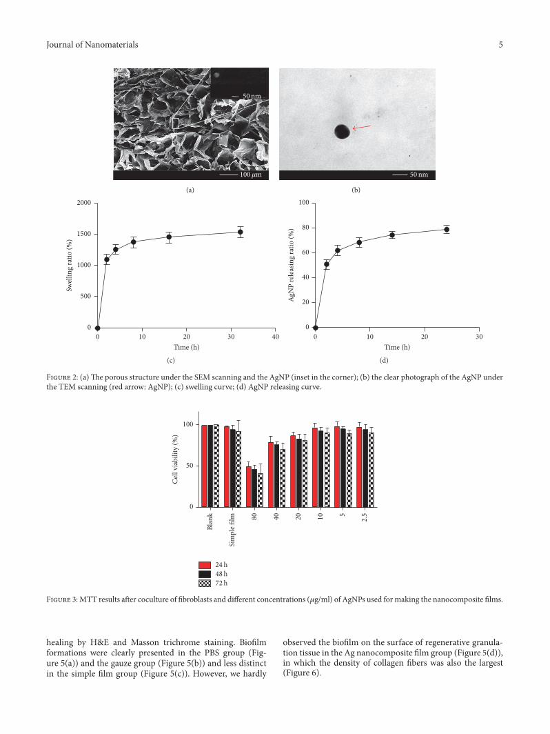
size was distributed evenly around 100 𝜇m. On the surface ofpores, we found that theAgNPswere of round shape andwell-dispersed without large aggregation (Figure 2(a)). The clearphotographs taken by the TEM also verified it (Figure 2(b)).
3.1.2. Swelling Ratio. According to formula (1), the gel contentwas calculated to 75% on average. In addition, the swellingcurve (Figure 2(c)) demonstrated that this film was able totake in large amounts of PBS solution as fifteen times as itsdry weight.
3.1.3. AgNP Releasing Curve. The law of AgNP release deter-mined the antibacterial potency of the dressing. UV-Visiblespectrophotometry was employed to establish the equationas follows:
𝐶 = 3.194 × 𝐴 + 0.051 (𝑟 = 1.000) , (4)
where 𝐶 and 𝐴 represented the concentration of AgNPs andrelevant absorbance at 400 nm, respectively. It was shownin Figure 2(d) that this nanocomposite film could preventthe outburst of AgNP release and maintain the antibacterialability for 24 hours.
3.2. Cytotoxicity Evaluation In Vitro. The nontoxicity ofmedical biomaterials is always the first consideration. Thesafety of the film was examined with a cell viability test byMTT assay. As shown in the Figure 3, the simple xanthan-based film was safe enough for fibroblasts while the Agnanocomposite film showed varied cytotoxicity associatedwith the concentration of AgNPs. But when AgNPs were lessthan 10 𝜇g/ml, this biomaterial was nontoxic to fibroblastsin vitro. Therefore, we chose this AgNP concentration forfurther research.
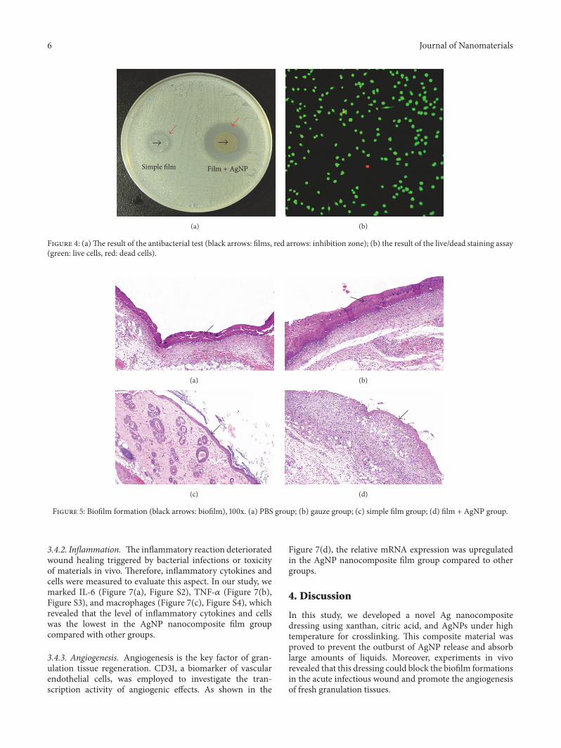
3.3. Antibacterial Activity In Vitro. The bacteriostatic ringwas a direct evidence of antibacterial property. In our study,we found that simple xanthan-based film could form a smallinhibition zone (Figure 4(a)). The potential reason mightbe the citric acid free from the film during the degradationperiod to function as an antibacterial agent. When AgNPswere loaded, the nanocomposite film generated a largerinhibition zone to the MRSA but did not do harm to thefibroblasts (Figure 4(b)).
3.4. Results of Histological Evaluation. During the exper-iment, all rats showed no adverse reactions and thesymptoms of toxicity. The rats exhibited free movementand normal behavior and were sensitive to light, sound,and other stimulations. The infectious wounds in eachgroup were photographed on day 0, day 1, and day 4(Figure S1 in Supplementary Material available online athttps://doi.org/10.1155/2017/6802397). The rats in the Agnanocomposite film group showed the higher healing rate fortheir infectious wounds.
3.4.1. Biofilm and Collagen Fibers. The tissue samples wereavailable to observe the condition of biofilm and wound
Journal of Nanomaterials 5
100 m
50nm
(a)
50nm
(b)
4020 30100
Time (h)
0
500
1000
1500
2000
Swel
ling
ratio
(%)
(c)
3020100
Time (h)
0
20
40
60
80
100
AgN
P re
leas
ing
ratio
(%)
(d)
Figure 2: (a)The porous structure under the SEM scanning and the AgNP (inset in the corner); (b) the clear photograph of the AgNP underthe TEM scanning (red arrow: AgNP); (c) swelling curve; (d) AgNP releasing curve.
24 h48 h72 h
5
Blan
k
Sim
ple �
lm 2.510204080
0
50
100
Cel
l via
bilit
y (%
)
Figure 3:MTT results after coculture of fibroblasts and different concentrations (𝜇g/ml) of AgNPs used for making the nanocomposite films.
healing by H&E and Masson trichrome staining. Biofilmformations were clearly presented in the PBS group (Fig-ure 5(a)) and the gauze group (Figure 5(b)) and less distinctin the simple film group (Figure 5(c)). However, we hardly
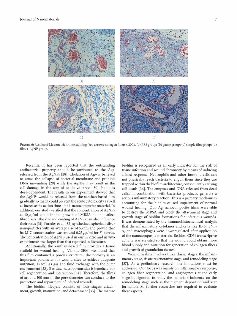
observed the biofilm on the surface of regenerative granula-tion tissue in the Ag nanocomposite film group (Figure 5(d)),in which the density of collagen fibers was also the largest(Figure 6).
6 Journal of Nanomaterials
&CFG + !A.0Simple film
(a) (b)
Figure 4: (a)The result of the antibacterial test (black arrows: films, red arrows: inhibition zone); (b) the result of the live/dead staining assay(green: live cells, red: dead cells).
(a) (b)
(c) (d)
Figure 5: Biofilm formation (black arrows: biofilm), 100x. (a) PBS group; (b) gauze group; (c) simple film group; (d) film + AgNP group.
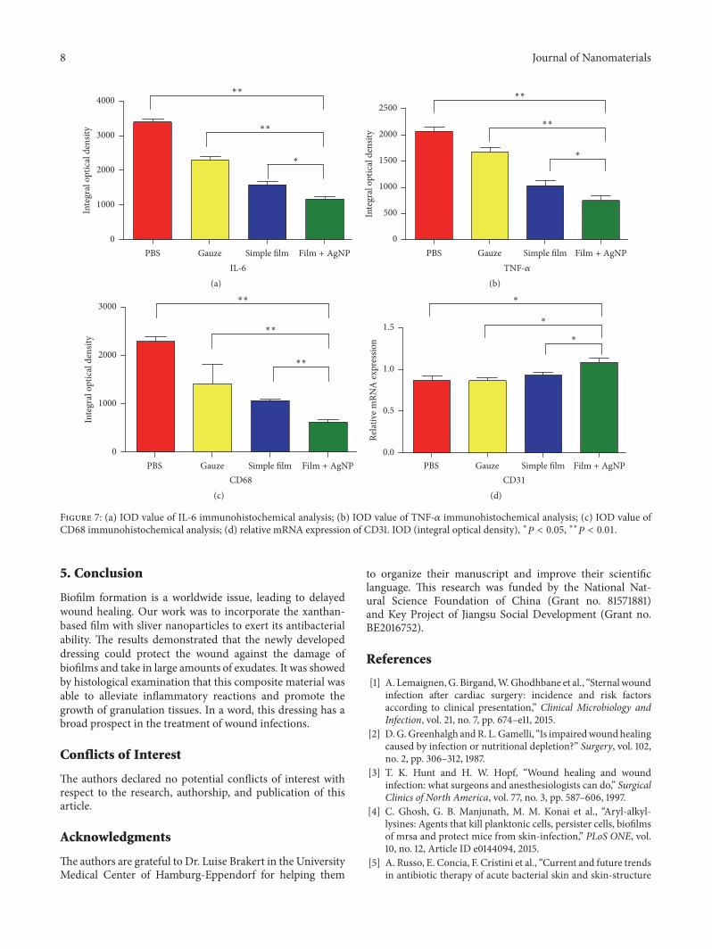
3.4.2. Inflammation. The inflammatory reaction deterioratedwound healing triggered by bacterial infections or toxicityof materials in vivo. Therefore, inflammatory cytokines andcells were measured to evaluate this aspect. In our study, wemarked IL-6 (Figure 7(a), Figure S2), TNF-𝛼 (Figure 7(b),Figure S3), and macrophages (Figure 7(c), Figure S4), whichrevealed that the level of inflammatory cytokines and cellswas the lowest in the AgNP nanocomposite film groupcompared with other groups.
3.4.3. Angiogenesis. Angiogenesis is the key factor of gran-ulation tissue regeneration. CD31, a biomarker of vascularendothelial cells, was employed to investigate the tran-scription activity of angiogenic effects. As shown in the
Figure 7(d), the relative mRNA expression was upregulatedin the AgNP nanocomposite film group compared to othergroups.
4. Discussion
In this study, we developed a novel Ag nanocompositedressing using xanthan, citric acid, and AgNPs under hightemperature for crosslinking. This composite material wasproved to prevent the outburst of AgNP release and absorblarge amounts of liquids. Moreover, experiments in vivorevealed that this dressing could block the biofilm formationsin the acute infectious wound and promote the angiogenesisof fresh granulation tissues.
Journal of Nanomaterials 7
(a) (b)
(c) (d)
Figure 6: Results of Masson trichrome staining (red arrows: collagen fibers), 200x. (a) PBS group; (b) gauze group; (c) simple film group; (d)film + AgNP group.
Recently, it has been reported that the outstandingantibacterial property should be attributed to the Ag+released from the AgNPs [28]. Chelation of Ag+ is believedto cause the collapse of bacterial membrane and prohibitDNA unwinding [29] while the AgNPs may result in thecell damage in the way of oxidative stress [30], but it isdose-dependent. The results in our experiment showed thatthe AgNPs would be released from the xanthan-based filmgradually so that it could prevent the acute cytotoxicity as wellas increase the action time of this nanocompositematerial. Inaddition, our study verified that the concentration of AgNPsat 10 𝜇g/ml could inhibit growth of MRSA but not affectfibroblasts. The size and coating of AgNPs can also influencetheir roles [31]. Paredes et al. [32] synthesized spherical silvernanoparticles with an average size of 55 nm and proved thatits MIC concentration was around 0.25 𝜇g/ml for S. aureus.The concentration of AgNPs used in our in vitro and in vivoexperiments was larger than that reported in literature.
Additionally, the xanthan-based film provides a tissuescaffold for wound healing. Via the SEM, we found thatthis film contained a porous structure. The porosity is animportant parameter for wound sites to achieve adequatenutrition, as well as gas and fluid exchange with the outerenvironment [33]. Besides, macroporous size is beneficial forcell regeneration and interaction [34]. Therefore, the filmsof around 100mm in the pore diameter can conduce to theprotection and repairment of infected wounds.
The biofilm lifecycle consists of four stages: attach-ment, growth, maturation, and detachment [35]. The mature
biofilm is recognized as an early indicator for the risk oftissue infection and wound chronicity by means of inducinga host response. Neutrophils and other immune cells cannot physically reach bacteria to engulf them since they aretrappedwithin the biofilm architecture, consequently causingcell death [36]. The enzymes and DNA released from deadcells, in combination with bacteria’s products, generate aserious inflammatory reaction. This is a primary mechanismaccounting for the biofilm-caused impairment of normalwound healing. Our Ag nanocomposite films were ableto destroy the MRSA and block the attachment stage andgrowth stage of biofilm formations for infectious wounds.It was demonstrated by the immunohistochemical analysisthat the inflammatory cytokines and cells like IL-6, TNF-𝛼, and macrophages were downregulated after applicationof the nanocomposite materials. Besides, CD31 transcriptiveactivity was elevated so that the wound could obtain moreblood supply and nutrition for generation of collagen fibersand growth of granulation tissues.
Wound healing involves three classic stages: the inflam-matory stage, tissue regenerative stage, and remodeling stage[37]. As a preliminary research, the limitations must beaddressed. Our focus was mainly on inflammatory response,collagen fiber regeneration, and angiogenesis at the earlystage but ignored to study the material’s influence on theremodeling stage such as the pigment deposition and scarformation. So further researches are required to evaluatethese aspects.
8 Journal of Nanomaterials
∗
PBS Gauze Simple film &CFG + !A.0
IL-6
∗∗
∗∗
0
1000
2000
3000
4000
Inte
gral
opt
ical
den
sity
(a)
PBS Gauze Simple film &CFG + !A.0
TNF-
∗
∗∗
∗∗
0
500
1000
1500
2000
2500
Inte
gral
opt
ical
den
sity
(b)
PBS Gauze Simple film &CFG + !A.0
CD68
∗∗
∗∗
∗∗
0
1000
2000
3000
Inte
gral
opt
ical
den
sity
(c)
PBS Gauze Simple film &CFG + !A.0
∗
∗
∗
CD31
0.0
0.5
1.0
1.5
Relat
ive m
RNA
expr
essio
n
(d)
Figure 7: (a) IOD value of IL-6 immunohistochemical analysis; (b) IOD value of TNF-𝛼 immunohistochemical analysis; (c) IOD value ofCD68 immunohistochemical analysis; (d) relative mRNA expression of CD31. IOD (integral optical density), ∗𝑃 < 0.05, ∗∗𝑃 < 0.01.
5. Conclusion
Biofilm formation is a worldwide issue, leading to delayedwound healing. Our work was to incorporate the xanthan-based film with sliver nanoparticles to exert its antibacterialability. The results demonstrated that the newly developeddressing could protect the wound against the damage ofbiofilms and take in large amounts of exudates. It was showedby histological examination that this composite material wasable to alleviate inflammatory reactions and promote thegrowth of granulation tissues. In a word, this dressing has abroad prospect in the treatment of wound infections.
Conflicts of Interest
The authors declared no potential conflicts of interest withrespect to the research, authorship, and publication of thisarticle.
Acknowledgments
The authors are grateful to Dr. Luise Brakert in the UniversityMedical Center of Hamburg-Eppendorf for helping them
to organize their manuscript and improve their scientificlanguage. This research was funded by the National Nat-ural Science Foundation of China (Grant no. 81571881)and Key Project of Jiangsu Social Development (Grant no.BE2016752).
References
[1] A. Lemaignen,G. Birgand,W.Ghodhbane et al., “Sternal woundinfection after cardiac surgery: incidence and risk factorsaccording to clinical presentation,” Clinical Microbiology andInfection, vol. 21, no. 7, pp. 674–e11, 2015.
[2] D.G.Greenhalgh andR. L.Gamelli, “Is impairedwoundhealingcaused by infection or nutritional depletion?” Surgery, vol. 102,no. 2, pp. 306–312, 1987.
[3] T. K. Hunt and H. W. Hopf, “Wound healing and woundinfection: what surgeons and anesthesiologists can do,” SurgicalClinics of North America, vol. 77, no. 3, pp. 587–606, 1997.
[4] C. Ghosh, G. B. Manjunath, M. M. Konai et al., “Aryl-alkyl-lysines: Agents that kill planktonic cells, persister cells, biofilmsof mrsa and protect mice from skin-infection,” PLoS ONE, vol.10, no. 12, Article ID e0144094, 2015.
[5] A. Russo, E. Concia, F. Cristini et al., “Current and future trendsin antibiotic therapy of acute bacterial skin and skin-structure
Journal of Nanomaterials 9
infections,” Clinical Microbiology and Infection, vol. 22, pp. S27–S36, 2016.
[6] L. J. Bessa, P. Fazii, M. Di Giulio, and L. Cellini, “Bacterialisolates from infected wounds and their antibiotic susceptibilitypattern: some remarks about wound infection,” InternationalWound Journal, 2013.
[7] C. Ghobril and M. W. Grinstaff, “The chemistry and engi-neering of polymeric hydrogel adhesives for wound closure: Atutorial,” Chemical Society Reviews, vol. 44, no. 7, pp. 1820–1835,2015.
[8] P. Li, T. Li, Y. Zeng et al., “Biosynthesis of xanthan gum byXanthomonas campestris LRELP-1 using kitchen waste as thesole substrate,” Carbohydrate Polymers, vol. 151, pp. 684–691,2016.
[9] V. Pegos, R. Canevarolo, A. Sampaio, A. Balan, and A. Zeri,“Xanthan Gum Removal for 1H-NMR Analysis of the Intracel-lular Metabolome of the Bacteria Xanthomonas axonopodis pv.citri 306,”Metabolites, vol. 4, no. 2, pp. 218–231, 2014.
[10] S. A. Rather, F. A. Masoodi, R. Akhter et al., “Application ofguar–xanthan gum mixture as a partial fat replacer in meatemulsions,” Journal of Food Science and Technology, vol. 53, no.6, pp. 2876–2886, 2016.
[11] C. V. Nikiforidis and V. Kiosseoglou, “Physicochemical stabilityof maize germ oil body emulsions as influenced by oil bodysurface-xanthan gum interactions,” Journal of Agricultural andFood Chemistry, vol. 58, no. 1, pp. 527–532, 2010.
[12] X. Chen, M. Wang, F. Yang, W. Tang, and X. Li, “Isolationand characterization of xanthan-degrading Enterobacter sp.nov. LB37 for reducing the viscosity of xanthan in petroleumindustry,”World Journal of Microbiology and Biotechnology, vol.30, no. 5, pp. 1549–1557, 2014.
[13] J. F. Llamas-Moreno, L. M. Baiza-Duran, L. R. Saucedo-Rodrıguez, and J. Felix Alanız-De la O, “Efficacy and safetyof chondroitin sulfate/xanthan gum versus polyethylene gly-col/propylene glycol/hydroxypropyl guar in patients with dryeye,” Clinical Ophthalmology, vol. 7, pp. 995–999, 2013.
[14] N. Vilardell, L. Rofes, V. Arreola, R. Speyer, and P. Clave,“A Comparative Study Between Modified Starch and XanthanGum Thickeners in Post-Stroke Oropharyngeal Dysphagia,”Dysphagia, vol. 31, no. 2, pp. 169–179, 2016.
[15] G. A. Sotiriou, A. Meyer, J. T. N. Knijnenburg, S. Panke, and S.E. Pratsinis, “Quantifying the origin of released Ag+ ions fromnanosilver,” Langmuir, vol. 28, no. 45, pp. 15929–15936, 2012.
[16] T. M. Tolaymat, A. M. El Badawy, A. Genaidy, K. G. Scheckel, T.P. Luxton, and M. Suidan, “An evidence-based environmentalperspective of manufactured silver nanoparticle in synthesesand applications: a systematic review and critical appraisal ofpeer-reviewed scientific papers,” Science of the Total Environ-ment, vol. 408, no. 5, pp. 999–1006, 2010.
[17] S. Chernousova and M. Epple, “Silver as antibacterial agent:ion, nanoparticle, andmetal,”Angewandte Chemie InternationalEdition, vol. 52, no. 6, pp. 1636–1653, 2013.
[18] S. Iravani, “Synthesis of silver nanoparticles: chemical, physicaland biological methods,” Research in Pharmaceutical Sciences,vol. 9, no. 6, pp. 385–406, 2014.
[19] A. Taglietti, Y. A. Diaz Fernandez, E. Amato et al., “Antibacterialactivity of glutathione-coated silver nanoparticles against grampositive and gram negative bacteria,” Langmuir, vol. 28, no. 21,pp. 8140–8148, 2012.
[20] P. Pallavicini, C. R. Arciola, F. Bertoglio et al., “Silver nanopar-ticles synthesized and coated with pectin: An ideal compro-mise for anti-bacterial and anti-biofilm action combined with
wound-healing properties,” Journal of Colloid and InterfaceScience, vol. 498, pp. 271–281, 2017.
[21] S. Zhang, Y. Guo, Y. Dong et al., “ANovel Nanosilver/NanosilicaHydrogel for Bone Regeneration in Infected BoneDefects,”ACSApplied Materials and Interfaces, vol. 8, no. 21, pp. 13242–13250,2016.
[22] Y. He, Z. Du, S. Ma et al., “Biosynthesis, Antibacterial Activityand Anticancer Effects Against Prostate Cancer (PC-3) Cellsof Silver Nanoparticles Using Dimocarpus Longan Lour. PeelExtract,” Nanoscale Research Letters, vol. 11, no. 1, article 300,2016.
[23] S. J. Jang, I. J. Yang, C. O. Tettey, K. M. Kim, and H. M.Shin, “In-vitro anticancer activity of green synthesized silvernanoparticles on MCF-7 human breast cancer cells,” MaterialsScience and Engineering C, vol. 68, pp. 430–435, 2016.
[24] N. Reddy and Y. Yang, “Citric acid cross-linking of starch films,”Food Chemistry, vol. 118, no. 3, pp. 702–711, 2010.
[25] N. E. El-Naggar and N. A. M. Abdelwahed, “Applicationof statistical experimental design for optimization of silvernanoparticles biosynthesis by a nanofactory Streptomyces viri-dochromogenes,” Journal of Microbiology, vol. 52, no. 1, pp. 53–63, 2014.
[26] P. Lodeiro, E. P. Achterberg, J. Pampın, A. Affatati, and M. S.El-Shahawi, “Silver nanoparticles coated with natural polysac-charides as models to study AgNP aggregation kinetics usingUV-Visible spectrophotometry upon discharge in complexenvironments,” Science of the Total Environment, vol. 539, pp.7–16, 2016.
[27] W. Pu, Y. Su, J. Li et al., “High incidence of oxacillin-susceptiblemecA-positive Staphylococcus aureus (OS-MRSA) associatedwith bovine mastitis in China,” PLoS ONE, vol. 9, no. 2, ArticleID e88134, 2014.
[28] G. A. Sotiriou and S. E. Pratsinis, “Antibacterial activity ofnanosilver ions and particles,” Environmental Science and Tech-nology, vol. 44, no. 14, pp. 5649–5654, 2010.
[29] K. I. Batarseh, “Anomaly and correlation of killing in thetherapeutic properties of siliver (I) chelation with glutamic andtartaric acids,” Journal of Antimicrobial Chemotherapy, vol. 54,no. 2, pp. 546–548, 2004.
[30] M. J. Piao, K. A. Kang, I. K. Lee et al., “Silver nanoparticlesinduce oxidative cell damage in human liver cells through inhi-bition of reduced glutathione and induction of mitochondria-involved apoptosis,” Toxicology Letters, vol. 201, no. 1, pp. 92–100, 2011.
[31] N. R. Chowdhury, M. MacGregor-Ramiasa, P. Zilm, P. Majew-ski, and K. Vasilev, “‘Chocolate’ silver nanoparticles: Synthesis,antibacterial activity and cytotoxicity,” Journal of Colloid andInterface Science, vol. 482, pp. 151–158, 2016.
[32] D. Paredes, C. Ortiz, and R. Torres, “Synthesis, characterization,and evaluation of antibacterial effect of Ag nanoparticles againstescherichia coli O157:H7 andmethicillin-resistant staphylococ-cus aureus (MRSA),” International Journal of Nanomedicine, vol.9, no. 1, pp. 1717–1729, 2014.
[33] C. M. Murphy and F. J. O’Brien, “Understanding the effect ofmean pore size on cell activity in collagen-glycosaminoglycanscaffolds,” Cell Adhesion and Migration, vol. 4, no. 3, pp. 377–381, 2010.
[34] N. Huebsch, E. Lippens, K. Lee et al., “Matrix elasticity of void-forming hydrogels controls transplanted-stem-cell-mediatedbone formation,”NatureMaterials, vol. 14, no. 12, pp. 1269–1277,2015.
10 Journal of Nanomaterials
[35] H. M. Lappin-Scott and C. Bass, “Biofilm formation: attach-ment, growth, and detachment of microbes from surfaces,”American Journal of Infection Control, vol. 29, no. 4, pp. 250-251,2001.
[36] C. R. Arciola, D. Campoccia, P. Speziale, L. Montanaro, andJ. W. Costerton, “Biofilm formation in Staphylococcus implantinfections. A review ofmolecular mechanisms and implicationsfor biofilm-resistant materials,” Biomaterials, vol. 33, no. 26, pp.5967–5982, 2012.
[37] Y. Deng, J. Ren, G. Chen et al., “Evaluation of polypropylenemesh coated with biological hydrogels for temporary closure ofopen abdomen,” Journal of Biomaterials Applications, vol. 31, no.2, pp. 302–314, 2016.

Submit your manuscripts athttps://www.hindawi.com
ScientificaHindawi Publishing Corporationhttp://www.hindawi.com Volume 2014
CorrosionInternational Journal of
Hindawi Publishing Corporationhttp://www.hindawi.com Volume 2014
Polymer ScienceInternational Journal of
Hindawi Publishing Corporationhttp://www.hindawi.com Volume 2014
Hindawi Publishing Corporationhttp://www.hindawi.com Volume 2014
CeramicsJournal of
Hindawi Publishing Corporationhttp://www.hindawi.com Volume 2014
CompositesJournal of
NanoparticlesJournal of
Hindawi Publishing Corporationhttp://www.hindawi.com Volume 2014
Hindawi Publishing Corporationhttp://www.hindawi.com Volume 2014
International Journal of
Biomaterials
Hindawi Publishing Corporationhttp://www.hindawi.com Volume 2014
NanoscienceJournal of
TextilesHindawi Publishing Corporation http://www.hindawi.com Volume 2014
Journal of
NanotechnologyHindawi Publishing Corporationhttp://www.hindawi.com Volume 2014
Journal of
CrystallographyJournal of
Hindawi Publishing Corporationhttp://www.hindawi.com Volume 2014
The Scientific World JournalHindawi Publishing Corporation http://www.hindawi.com Volume 2014
Hindawi Publishing Corporationhttp://www.hindawi.com Volume 2014
CoatingsJournal of
Advances in
Materials Science and EngineeringHindawi Publishing Corporationhttp://www.hindawi.com Volume 2014
Smart Materials Research
Hindawi Publishing Corporationhttp://www.hindawi.com Volume 2014
Hindawi Publishing Corporationhttp://www.hindawi.com Volume 2014
MetallurgyJournal of
Hindawi Publishing Corporationhttp://www.hindawi.com Volume 2014
BioMed Research International
MaterialsJournal of
Hindawi Publishing Corporationhttp://www.hindawi.com Volume 2014