evaluationofmelanogenesisin a ...downloads.hindawi.com/journals/tswj/2012/854096.pdf · melanins...
TRANSCRIPT

The Scientific World JournalVolume 2012, Article ID 854096, 7 pagesdoi:10.1100/2012/854096
The cientificWorldJOURNAL
Research Article
Evaluation of Melanogenesis inA-375 Cells in the Presence of DMSO and Analysis ofPyrolytic Profile of Isolated Melanin
Ewa Chodurek,1 Arkadiusz Orchel,1 Joanna Orchel,2 Sławomir Kurkiewicz,3 Natalia Gawlik,4
Zofia Dzierzewicz,1 and Krystyna Stepien3
1 Department of Biopharmacy, Medical University of Silesia, Narcyzow 1, 41-200 Sosnowiec, Poland2 Department of Molecular Biology, Medical University of Silesia, Narcyzow 1, 41-200 Sosnowiec, Poland3 Department of Instrumental Analysis, Medical University of Silesia, Narcyzow 1, 41-200 Sosnowiec, Poland4 Department of Biotechnology and Genetic Engineering, Medical University of Silesia, Narcyzow 1, 41-200 Sosnowiec, Poland
Correspondence should be addressed to Ewa Chodurek, [email protected]
Received 26 October 2011; Accepted 19 December 2011
Academic Editor: Jahn M. Nesland
Copyright © 2012 Ewa Chodurek et al. This is an open access article distributed under the Creative Commons Attribution License,which permits unrestricted use, distribution, and reproduction in any medium, provided the original work is properly cited.
The increase of a skin malignant melanoma (melanoma malignum) incidence in the world has been observed in recent years. Thetumour, especially in advanced stadium with metastases, is highly resistant to conventional treatment. One of the strategies is tomodulate melanogenesis using chemical compounds. In this study, the processes of differentiation and melanogenesis induced bydimethylsulfoxide (DMSO) in human melanoma cells (A-375) were investigated. Natural melanin isolated from A-375 melanomacell line treated with 0.3% DMSO was analyzed by pyrolysis-gas chromatography-mass spectrometry (Py-GC/MS) method. Theproducts derived from pheomelanin have not been stated in the pyrolytic profile of analyzed melanin. Within all products derivedfrom eumelanins, 1,2-benzenediol has been predominated. It has been shown that in the melanoma cells stimulated with 0.3% and1% DMSO, the increase of transcriptional activity of the tyrosinase gene took place. It was accompanied by the rise of tyrosinaseactivity and an accumulation of melanin in the cells. The better knowledge about the structure of melanins can contribute toestablish the uniform criteria of malignant melanoma morbidity risk.
1. Introduction
In recent years, the increase of a skin malignant melanoma(melanoma malignum) incidence in the world has beenobserved. Though this type of carcinoma comprises just 5%of all malignant skin cancers, it is also responsible for 75–80% of deaths caused by these tumors. The highest morbidityrate of this cancer was noted in Australia (40–60/100 000), inthe United States of America (10–20/100 000), and in Europe(10–15/100 000) [1, 2].
The morbidity rate of skin melanoma malignum inPoland, according to the National Registry of Cancer,amounted 4.4/100 000 and 5.1/100 000 in 2002, for men andwomen, respectively. It was observed that the amount ofnew cases of the melanoma in Poland from 1982 to 2002
increased 3 times and the rate of death in the same periodincreased over 2 times, what was attested to a better efficiencyof therapy of this cancer in recent years [3].
The high morbidity rate is also associated with rapidlygrowing mortality rate. The malignant melanoma at higherstages of malignancy is highly resistant to conventionaltreatment. Therefore, there is a demand for the developmentof alternative antitumor therapies and better understandingof the mechanisms responsible for the malignant phenotypes[4].
Melanoma malignum is one of the most malignant and atthe same time the most frequently occurring aggressive skintumor, the origin of which is the malignant transformationof melanocytes, cells which produce the melanin pigment [4,5]. Melanocytes of mammals produce two types of melanin

2 The Scientific World Journal
pigments, the black to brown eumelanin and the yellow toreddish pheomelanin [6–9].
In contrast to other biopolymers such as proteins ornucleic acids, melanin is characterized by the presence ofnonhydrolyzable carbon-carbon bonds linking its mono-mers. This fact makes difficult to elucidate melanin structuredue to the lack of effective analytical methods [10, 11].
As yet, the mechanisms regulating the pathogenesis ofmelanoma malignum have not been fully elucidated. Ithas not been established which exogenous (e.g., UV) orendogenous (genetic) factors play a key role in the melanomaetiopathogenesis. However, it is well known that melanoma isa highly metabolically active tumor that produces numeroussubstances recognized as neoplastic markers (e.g., cytokines,growth factors, apoptotic factors) used in laboratory diag-nostics. The markers used for the detection of melanomacells included some antigenic proteins as well as precursorsand metabolites of melanin (assayed in serum and urine)[12].
The synthesis and accumulation of the melanins inthe malignant melanoma cells are essential factors whichdeterminate the efficacy of therapy. There are differentopinions about the role of melanins in melanoma malignumetiopathogenesis. On the one hand, the presence of melaninscan cause the negative effects in malignant cells. Firstly,melanins can bind some cancer medicines (including cyto-static drugs) and, in this way, reduce the efficiency ofchemotherapy. Secondly, the excess of melanins can leadto hypoxia and reduction of the efficiency of radiother-apy. Moreover, indirect products of melanogenesis and theproducts arising as a result of melanins degradation areall cytotoxic. The cytotoxicity, instead of inhibiting thedevelopment of malignant cells, can turn against the immunesystem cells [4].
On the other hand, melanogenesis is the marker ofmelanocytes’ differentiation, which is the opposite process tocarcinogenesis [13]. One of the chemical compounds, whichcan suppress the proliferation and induce the differentiationof malignant cells in vitro, is dimethyl sulfoxide (DMSO).This highly polar organic liquid is often used as a chemicalsolvent. It also shows a range of pharmacological activity, likeanti-inflammatory properties. DMSO induces melanogene-sis and differentiation of HO melanoma cell line. However,it does not induce the dendrite-like structures in malignantcells [14].
In the present study, we developed an experimentalmodel useful for investigations of factors controlling theprocesses of differentiation and melanogenesis in humanmelanoma cells. DMSO was used as a model compoundstimulating differentiation and melanin synthesis in humanA375 cell line.
The aim of the study was to characterize the chemicalstructure of melanin isolated from DMSO-treated humanmelanoma cells (A-375), using pyrolysis-gas chromatogra-phy/mass spectrometry (Py-GC/MS). In the future, structureelucidation of melanin pigments isolated from melanomamalignum may be helpful in the establishment of criteria forprediction of the risk of melanoma skin cancer.
2. Experimental
2.1. Materials
2.1.1. Tumor Cells. The human malignant melanoma cellline A-375 was purchased from LGC Promochem (Lomianki,Poland). Malignant cell line was grown in the mediumcontaining the following composition: 90% MinimumEssential Medium Eagle (MEM, Sigma-Aldrich), 10% fetalbovine serum (FBS, PAA), 100 U/mL penicillin, 100 µg/mLstreptomycin (Sigma-Aldrich), and 10 mM HEPES (Sigma-Aldrich). The cell culture was cultivated in the standardconditions: temperature was 37◦C, and atmosphere was con-taining 95% air and 5% CO2. To study the cell proliferation,melanocytes were plated at an initial density of 103 cells perwell in 200 µL of culture medium in 96-well plates. Cells wereallowed to attach and grow for 24 h prior to exposure to testreagents. Cells were incubated with DMSO for 72 h.
2.1.2. Melanin Isolation from A-375 Melanoma Cells. 1 g ofmelanoma cells (A-375 cell line) was mixed with 5 mL of 1%Triton X-100 (Sigma) and incubated for 1 h at room tem-perature [15]. Next the sample was centrifuged (10000×g,15 min), and the cell pellet was washed with phosphate bufferand once again centrifuged. The pellet was mixed with 5 mLof (5 mg/mL) sodium dodecyl sulfate (SDS) in Tris-HClbuffer (50 mM, pH = 7.4) with proteinase K (Sigma) tothe final solution of 0.33 mg/mL. The mixture was incubatedfor 3 h in 37◦C. After centrifugation (10000×g, 15 min), themelanin pigment was successively washed with 0.9% NaCl,methanol, and hexane and every time centrifuged (10000×g,15 min). The melanin was dried at 37◦C and stored in glassdesiccator over P2O5.
2.1.3. Synthesis of Melanin from Tyrosine Catalyzed by Tyrosi-nase (Tyr-Melanin). 0.004 g tyrosine hydrochloride (Sigma)was dissolved in 50 mL sodium phosphate buffer (pH6.8), then tyrosinase 100 U/mL (Sigma, 5370 U/mg) wasadded, and the reaction mixtures were incubated for 48 hat 37◦C with vigorous stirring and protection from light.The obtained Tyr-melanin pigment formed was collected bycentrifugation (5000×g, 10 min) and washed several timeswith deionized water. To remove possible traces of tyrosinase,Tyr-melanin standard was treated with SDS and methanol,NaCl, then rewashed with deionized water, and dried to aconstant weight at 37◦C.
2.2. Methods
2.2.1. Tyrosinase Activity Assay. The tyrosinase activity wasassayed by the method of Slominski et al. [16]. A-375 cellswere seeded in 100 mm culture dishes at a density of 1× 10⁶
cells/dish in 12 mL of above-mentioned medium. The cellswere allowed to attach and grow for 24 h. Subsequently,the culture medium was changed and the cells were treatedwith 0.3% and 1% DMSO for 3 and 7 days. At the endof the incubation periods, cells were washed with PBS andcollected by trypsinization. Detached cells were centrifuged

The Scientific World Journal 3
at 4000 g for 5 min. Subsequently, the cell pellet was lysedin 1% Triton X-100 (Sigma) in 0.1 M phosphate buffer(pH 6.8) for 30 min. Lysate was incubated with an equalvolume of DOPA (3 mg/mL in 0.1 M phosphate buffer pH6.8) for 3 h at 37◦C and the absorbance was measuredat 490 nm (spectrophotometer Hewlett Packard 8452A).The stimulation of tyrosinase activity following the DMSOtreatment was estimated as fold increase in treated cells withrespect to vehicle-treated control cells.
2.2.2. Tyrosinase Expression (RT-PCR). A-375 cells wereseeded and treated as it was described previously. The tran-scriptional activity of tyrosinase gene was determined using areal-time RT-PCR technique. Total RNA was extracted fromA-375 cells treated with DMSO for 3 and 7 days. RNA wasextracted with NucleoSpin RNA II Kit (Macherey-Nagel)according to the manufacturer’s instructions.
All RNA samples were treated with DNase I (Macherey-Nagel). The RNA concentration was determined usingQuant-iT RiboGreen RNA Assay Kit (Invitrogen) accord-ing to the manufacturer’s instructions. The primers forPCR amplification of tyrosinase transcript were designedusing Primer Express 2.0 software on a sequence attainedfrom GenBank (ref. no U01873). Primer sequences wereas follows: TF 5′-CTTCGATTTGAGTGCCCCAGA-3′ andTR 5′-CCAAGCAGTGCATCCATTGAC-3′. Glyceraldehyde-3-phosphate dehydrogenase (GAPDH) RNA was used asan endogenous control. GAPDH primer pair was as fol-lows: GF 5′-GAAGGTGAAGGTCGGAGTC-3′ and GR 5′-GAAGATGGTGATGGGATTTC-3′ [17].
The reverse transcription and amplification reactionswere performed by use of the Power SYBR Green RNA-to-CT 1-Step Kit (Invitrogen). The reaction was performed ina volume of 20 µL, containing 50 ng of RNA and 0,2 µMprimers. The cycle parameters were as follows: 30 min at48◦C, 10 min at 95◦C followed by 40 cycles of 15 s at95◦C, 30 s at 54◦C, and 30 s at 72◦C. Under these reactionconditions, the amplification efficiencies were between 95and 100%. The specificity of PCR reaction was confirmedby melting curve analysis and by the use of electrophoresison 2% agarose gels, stained with ethidium bromide. Thethreshold cycle (Ct) values were used to determine therelative expression ratios between the controlled and treatedcells. The relative expression calculations and statisticalanalyses were performed using the REST 2009 software [18].Real-time RT-PCR was run in triplicate for both genes ineach sample.
2.2.3. Py-GC/MS of Melanin. Melanin samples were placedon the tips of ferromagnetic wires and inserted immediatelyinto a Curie Point pyrolyser (type 795050, Pye-Unicam),coupled directly to a gas chromatograph (5890 Series II,Hewlett-Packard), and interfaced with a mass spectrometer(5989A, Hewlett-Packard). The pyrolysis (thermolysis) wascarried out at 770◦C for 8 s. The pyrolysis cell was kept at220◦C. GC separations of the products formed were per-formed on Rtx-5MS (Restek) fused-silica capillary column(5% diphenyl, 95% dimethyl polysiloxane, 60 m × 0.32 mm
i.d., 0.5 µm film thickness). The GC column outlet wasconnected directly to the ion source of a mass spectrometer.The GC/MS interface was kept at 250◦C. Helium was used asthe carrier gas at a flow rate of 1.8 mL/min. The GC oventemperature was programmed from 40◦C (isothermal for5 min) to 220◦C at a rate of 10◦C/min; the final temperaturewas held for 15 min. The MS conditions were as follows:electron energy, 70 eV; ion source temperature, 200◦C; andquadrupole analyzer temperature, 100◦C. All the mass spec-tra were recorded at m/z 25–200 (0–5 min) and m/z 33–500(above 5 min). A Hewlett-Packard, ChemStation G1034Cversion C.02.00 software was used for data collection andmass spectra processing. The mass spectra of the thermolysisproducts obtained were compared with standard spectra ofthe Wiley Registry of Mass Spectral Data 8th Edition.
3. Results and Discussion
It is now well known that changes in the histone acetylationlevels are involved in melanoma pathogenesis. Especially, his-tone hypoacetylation influences the transcriptional activityof numerous genes controlling cell cycle progression, cellsignaling, differentiation, DNA repair, apoptosis, invasion,and immune response. The acetylation reaction is catalyzedby the histone acetyltransferase (HAT) and the oppositeprocess—deacetylation—is catalyzed by histone deacetylases(HDACs). Histone deacetylase inhibitors (HDACis) inhibitcell proliferation and induce apoptosis in numerous neo-plastic cell lines. They are considered as a class of newpromising antineoplastic drugs, possessing chemopreventivepotential. So far, clinical studies were performed with thesome HDACis, for example, valproic acid, FR901228, MS-275, and SAHA [19]. DMSO is also HDAC inhibitor andhence it influences on proliferation and differentiation ofvarious cancer cell lines.
Dimethyl sulfoxide is one of the factors, which caninduce the melanogenesis. DMSO and the other factor, 6,7-dimethoxycoumarin, can induce melanogenesis even with-out increased tyrosine concentration in the culture medium[20, 21]. Similar effects were obtained in the experimentperformed by Huberman and coworkers, where humanmalignant melanoma cell line HO was exposed to DMSOat concentrations of 0.5%–2%. The authors have also shownthat DMSO at concentrations of 1.5% and 2% significantlyinhibited proliferation of malignant cells [13]. It has beenconfirmed during our preliminary experiments that DMSOat concentrations of 3% and 10% has significantly decreasedthe proliferation of A-375 cell line and exerted strongcytotoxic effect (data not shown). We have also foundthat DMSO in concentrations of 0.3% and 1% did notsignificantly influence on the A375 cells growth.
It has been proved that DMSO can induce the differentia-tion of pineal cells of 8-day embryonic quail. Those cells haveability to transform into skeletal muscle fibers, pigmentedepithelial cells, lens cells, and neurons. Melanogenesis isone of the earliest markers of multipotential pineal cellsdifferentiation. In 8-day pineal, three levels of differentiationare observed. The first level includes melanin and tyrosinase;the second owns tyrosinase without pigment. Third level
4 The Scientific World Journal
is tyrosinase-negative. DMSO transforms pineal cells intopigment cells including all 3 levels [22].
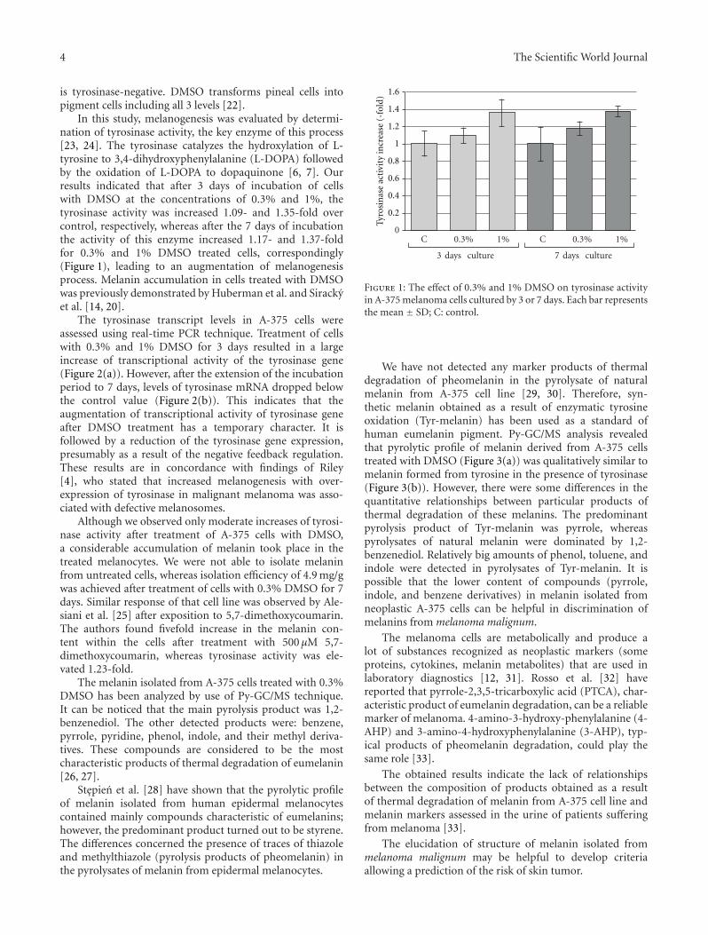
In this study, melanogenesis was evaluated by determi-nation of tyrosinase activity, the key enzyme of this process[23, 24]. The tyrosinase catalyzes the hydroxylation of L-tyrosine to 3,4-dihydroxyphenylalanine (L-DOPA) followedby the oxidation of L-DOPA to dopaquinone [6, 7]. Ourresults indicated that after 3 days of incubation of cellswith DMSO at the concentrations of 0.3% and 1%, thetyrosinase activity was increased 1.09- and 1.35-fold overcontrol, respectively, whereas after the 7 days of incubationthe activity of this enzyme increased 1.17- and 1.37-foldfor 0.3% and 1% DMSO treated cells, correspondingly(Figure 1), leading to an augmentation of melanogenesisprocess. Melanin accumulation in cells treated with DMSOwas previously demonstrated by Huberman et al. and Sirackyet al. [14, 20].
The tyrosinase transcript levels in A-375 cells wereassessed using real-time PCR technique. Treatment of cellswith 0.3% and 1% DMSO for 3 days resulted in a largeincrease of transcriptional activity of the tyrosinase gene(Figure 2(a)). However, after the extension of the incubationperiod to 7 days, levels of tyrosinase mRNA dropped belowthe control value (Figure 2(b)). This indicates that theaugmentation of transcriptional activity of tyrosinase geneafter DMSO treatment has a temporary character. It isfollowed by a reduction of the tyrosinase gene expression,presumably as a result of the negative feedback regulation.These results are in concordance with findings of Riley[4], who stated that increased melanogenesis with over-expression of tyrosinase in malignant melanoma was asso-ciated with defective melanosomes.
Although we observed only moderate increases of tyrosi-nase activity after treatment of A-375 cells with DMSO,a considerable accumulation of melanin took place in thetreated melanocytes. We were not able to isolate melaninfrom untreated cells, whereas isolation efficiency of 4.9 mg/gwas achieved after treatment of cells with 0.3% DMSO for 7days. Similar response of that cell line was observed by Ale-siani et al. [25] after exposition to 5,7-dimethoxycoumarin.The authors found fivefold increase in the melanin con-tent within the cells after treatment with 500 µM 5,7-dimethoxycoumarin, whereas tyrosinase activity was ele-vated 1.23-fold.
The melanin isolated from A-375 cells treated with 0.3%DMSO has been analyzed by use of Py-GC/MS technique.It can be noticed that the main pyrolysis product was 1,2-benzenediol. The other detected products were: benzene,pyrrole, pyridine, phenol, indole, and their methyl deriva-tives. These compounds are considered to be the mostcharacteristic products of thermal degradation of eumelanin[26, 27].
Stepien et al. [28] have shown that the pyrolytic profileof melanin isolated from human epidermal melanocytescontained mainly compounds characteristic of eumelanins;however, the predominant product turned out to be styrene.The differences concerned the presence of traces of thiazoleand methylthiazole (pyrolysis products of pheomelanin) inthe pyrolysates of melanin from epidermal melanocytes.
0C 0.3% 1% C 0.3% 1%
3 days culture 7 days culture
Tyro
sin
ase
acti
vity
incr
ease
(-f
old)
0.2
0.4
0.6
0.8
1
1.2
1.4
1.6
Figure 1: The effect of 0.3% and 1% DMSO on tyrosinase activityin A-375 melanoma cells cultured by 3 or 7 days. Each bar representsthe mean ± SD; C: control.
We have not detected any marker products of thermaldegradation of pheomelanin in the pyrolysate of naturalmelanin from A-375 cell line [29, 30]. Therefore, syn-thetic melanin obtained as a result of enzymatic tyrosineoxidation (Tyr-melanin) has been used as a standard ofhuman eumelanin pigment. Py-GC/MS analysis revealedthat pyrolytic profile of melanin derived from A-375 cellstreated with DMSO (Figure 3(a)) was qualitatively similar tomelanin formed from tyrosine in the presence of tyrosinase(Figure 3(b)). However, there were some differences in thequantitative relationships between particular products ofthermal degradation of these melanins. The predominantpyrolysis product of Tyr-melanin was pyrrole, whereaspyrolysates of natural melanin were dominated by 1,2-benzenediol. Relatively big amounts of phenol, toluene, andindole were detected in pyrolysates of Tyr-melanin. It ispossible that the lower content of compounds (pyrrole,indole, and benzene derivatives) in melanin isolated fromneoplastic A-375 cells can be helpful in discrimination ofmelanins from melanoma malignum.
The melanoma cells are metabolically and produce alot of substances recognized as neoplastic markers (someproteins, cytokines, melanin metabolites) that are used inlaboratory diagnostics [12, 31]. Rosso et al. [32] havereported that pyrrole-2,3,5-tricarboxylic acid (PTCA), char-acteristic product of eumelanin degradation, can be a reliablemarker of melanoma. 4-amino-3-hydroxy-phenylalanine (4-AHP) and 3-amino-4-hydroxyphenylalanine (3-AHP), typ-ical products of pheomelanin degradation, could play thesame role [33].
The obtained results indicate the lack of relationshipsbetween the composition of products obtained as a resultof thermal degradation of melanin from A-375 cell line andmelanin markers assessed in the urine of patients sufferingfrom melanoma [33].
The elucidation of structure of melanin isolated frommelanoma malignum may be helpful to develop criteriaallowing a prediction of the risk of skin tumor.

The Scientific World Journal 5
0
5
10
15
20
25
30
35
40
45
C 0.3 1
Fold
incr
ease
(a)
0
0.5
1
1.5
2
2.5
1C 0.3
(b)
Figure 2: The effect of 0.3% and 1% DMSO on the transcriptional activity of tyrosinase gene in A-375 cells cultured by 3 (a) or 7 (b) days.Each bar represents the mean ± SE; Control value was taken as 1; ∗P < 0.05.
Time (min)
20
40
60
80
100
6 8 10 12 14 16 18 20 22
1
2
3
4
5 6
7
8
9
1011
12
15
17
19
2021
2223
24Rel
ativ
e ab
un
dan
ce (
%)
(a)
1
2
3
4
5
67
8910
11
12
1314
15
16
17
1819
22
23
24
Time (min)
6 8 10 12 14 16 18 20 22
20
40
60
80
100
Rel
ativ
e ab
un
dan
ce (
%)
(b)
Figure 3: Chromatogram of the products formed during pyrolysis of (a) melanin isolated from the human melanoma malignum (A-375cell line) treated by DMSO, and (b) model synthetic eumelanin (Tyr-melanin). Peak designation: (1) acetic acid, (2) benzene, (3) pyridine,(4) pyrrole, (5) toluene, (6) acetamide, (7) pyridine, 2-methyl-, (8) furfural, (9) pyrrole, 2-methyl-, (10) pyrrole, 3-methyl-, (11) pyridine,4-methyl-, (12) styrene, (13) furanone, (14) cyclohexanone, (15) phenol, (16) n.i., (17) phenol, 2-methyl-, (18) phenol, 4-methyl-, (19)1,2-benzenediol, (20) n.i., (21) 1,2-benzenediol, 4-methyl-, (22) indole, (23) 2-methoxy-4-vinylphenol, (24) indole, 3-methyl-.
4. Conclusions
Malignant melanoma is one of the most malicious tumors.The average survival time of patients with this stage ofmelanoma usually does not exceed 1 year. Chemotherapyor immunotherapy lengthens the survival time only toabout 5 years and only in a small group of patients. Inthis case, there is a need to look for the new therapeuticand diagnostic solutions. Melanogenesis is a marker of themelanocyte differentiation, which is an opposite process tothe carcinogenesis. Therefore, there is a possibility to evaluate
the stage of malignancy by determination of the intensity ofmelanogenesis in malignant melanoma cells. In our study weproved that DMSO induces melanogenesis in A-375 cell line,thereby it stimulates the differentiation of melanoma cellsand increases the activity of tyrosinase enzyme. However,this compound does not induce the formation of dendritic-like cells (e.g., 5,7-dimethoxycoumarin). We also provedthat the predominant melanin isolated from melanomacells is eumelanin. The results presented in our work showthat DMSO is able to induce the differentiation of humanmelanoma cells in vitro. Thereby, DMSO can be used as a

6 The Scientific World Journal
research model of other HDACis, which have application inthe treatment of malignant melanoma.
Acknowledgments
This work was supported by SUM Grant KNW-1-002/P/1/0.This work was supported by the European Community fromthe European Social Fund within the RFSD 2 project.
References
[1] A. Jemal, R. Siegel, E. Ward et al., “Cancer statistics, 2008,” CACancer Journal for Clinicians, vol. 58, no. 2, pp. 71–96, 2008.
[2] T. N. T. Tran, J. Schulman, and D. E. Fisher, “UV and pig-mentation: molecular mechanisms and social controversies,”Pigment Cell and Melanoma Research, vol. 21, no. 5, pp. 509–516, 2008.
[3] M. Michalska-Jakubus, T. Jakubus, and D. Krasowska,“Czerniak—epidemiologia, etiopatogeneza i rokowanie,”Medycyna Rodzinna, vol. 2, pp. 45–53, 2006.
[4] P. A. Riley, “Melanogenesis and melanoma,” Pigment CellResearch, vol. 16, no. 5, pp. 548–552, 2003.
[5] M. S. Blois, R. W. Sagebiel, and R. M. Abarbanel, “Malignantmelanoma of the skin. I. The association of tumor depth andtype, and patient sex, age, and site with survival,” Cancer, vol.52, no. 7, pp. 1330–1341, 1983.
[6] K. Wakamatsu, D. N. Hu, S. A. McCormick, and S. Ito,“Characterization of melanin in human iridal and choroidalmelanocytes from eyes with various colored irides,” PigmentCell and Melanoma Research, vol. 21, no. 1, pp. 97–105, 2008.
[7] S. Ito, “IFPCS presidential lecture: a chemist’s view ofmelanogenesis,” Pigment Cell Research, vol. 16, no. 3, pp. 230–236, 2003.
[8] S. S. Sulaimon and B. E. Kitchell, “Review article the biology ofmelanocytes,” Veterinary Dermatology, vol. 14, no. 2, pp. 57–65, 2003.
[9] Y. Liu, L. Hong, K. Wakamatsu et al., “Comparison ofstructural and chemical properties of black and red humanhair melanosomes,” Photochemistry and Photobiology, vol. 81,no. 1, pp. 135–144, 2005.
[10] S. Ito, K. Wakamatsu, and H. Ozeki, “Chemical analysis ofmelanins and its application to the study of the regulation ofmelanogenesis,” Pigment Cell Research, vol. 13, no. 8, pp. 103–109, 2000.
[11] G. Zonios and A. Dimou, “Optical properties of humanmelanocytic nevi in vivo,” Photochemistry and Photobiology,vol. 85, no. 1, pp. 298–303, 2009.
[12] D. Becker, M. C. Mihm, S. M. Hewitt, V. K. Sondak, J. W.Fountain, and M. Thurin, “Markers and tissue resources formelanoma: meeting report,” Cancer Research, vol. 66, no. 22,pp. 10652–10657, 2006.
[13] A. Slominski, J. Wortsman, B. Nickoloff, K. McClatchey, M.C. Mihm, and J. S. Ross, “Molecular pathology of malignantmelanoma,” American Journal of Clinical Pathology, vol. 110,no. 6, pp. 788–794, 1998.
[14] E. Huberman, C. Heckman, and R. Langenbach, “Stimulationof differentiated functions in human melanoma cells bytumor-promoting agents and dimethyl sulfoxide,” CancerResearch, vol. 39, no. 7 I, pp. 2618–2624, 1979.
[15] E. Chodurek, D. Kusmierz, A. Dzierzega-Lecznar, S.Kurkiewicz, K. Stepien, and Z. Dzierzewicz, “Thermochemol-ysis as the useful method to assess the purity of melanin
isolated from the human melanoma malignum,” Acta PoloniaePharmaceutica, vol. 65, no. 6, pp. 731–734, 2008.
[16] A. Slominski, P. Jastreboff, and J. Pawelek, “L-Tyrosine stim-ulates induction of tyrosinase activity by MSH and reducescooperative interactions between MSH receptors in hamstermelanoma cells,” Bioscience Reports, vol. 9, no. 5, pp. 579–586,1989.
[17] I. Lima-Couy, A. Cervero, F. Bonilla-Musoles, A. Pellicer, andC. Simon, “Endometrial leptin and leptin receptor expressionin women with severe/moderate endometriosis,” MolecularHuman Reproduction, vol. 10, no. 11, pp. 777–782, 2004.
[18] M. W. Pfaffl, G. W. Horgan, and L. Dempfle, “Relative expres-sion software tool (REST) for group-wise comparison andstatistical analysis of relative expression results in real-timePCR,” Nucleic Acids Research, vol. 30, no. 9, article e36, 2002.
[19] L. Sigalotti, A. Covre, E. Fratta et al., “Epigenetics of humancutaneous melanoma: setting the stage for new therapeuticstrategies,” Journal of Translational Medicine, vol. 8, article 56,2010.
[20] J. Siracky, M. Blasko, and J. Borovansky, “Stimulation of dif-ferentiation in human melanoma cells by dimethyl sulphoxide(DMSO),” Neoplasma, vol. 32, no. 6, pp. 685–688, 1985.
[21] J. Y. Yang, J. H. Koo, Y. G. Song et al., “Stimulation ofmelanogenesis by scoparone in B16 melanoma cells,” ActaPharmacologica Sinica, vol. 27, no. 11, pp. 1467–1473, 2006.
[22] H. Orii, M. Hyuga, M. Mochii, J. Kosaka, G. Eguchi, and K.Watanabe, “Predominant melanogenesis and lentoidogenesisin vitro from multipotent pineal cells by dimethyl sulfoxideand hexamethylene bisacetamide,” International Journal ofDevelopmental Biology, vol. 38, no. 2, pp. 397–404, 1994.
[23] A. Slominski, D. J. Tobin, S. Shibahara, and J. Wortsman,“Melanin pigmentation in mammalian skin and its hormonalregulation,” Physiological Reviews, vol. 84, no. 4, pp. 1155–1228, 2004.
[24] K. U. Schallreuter, S. Kothari, B. Chavan, and J. D. Spencer,“Regulation of melanogenesis-controversies and new con-cepts,” Experimental Dermatology, vol. 17, no. 5, pp. 395–404,2008.
[25] D. Alesiani, R. Ciccon, M. Mattei, R. Bei, and A. Canini,“Inhibition of Mek 1/2 kinase activity and stimulationof melanogenesis by 5,7-dimethoxycoumarin treatment ofmelanoma cells,” International Journal of Oncology, vol. 34, no.6, pp. 1727–1735, 2009.
[26] A. Dzierzega-Lecznar, E. Chodurek, K. Stepien, and T.Wilczok, “Pyrolysis-gas chromatography-mass spectrometryof synthetic neuromelanins,” Journal of Analytical and AppliedPyrolysis, vol. 62, no. 2, pp. 239–248, 2002.
[27] E. Chodurek, B. Pilawa, A. Dzierzega-Lecznar, S. Kurkiewicz,L. Swiatkowska, and T. Wilczok, “Effect of Cu2+ and Zn2+
ions on DOPA-melanin structure as analyzed by pyrolysis-gaschromatography-mass spectrometry and EPR spectroscopy,”Journal of Analytical and Applied Pyrolysis, vol. 70, no. 1, pp.43–54, 2003.
[28] K. Stepien, A. Dzierzega-Lecznar, S. Kurkiewicz, and I. Tam,“Melanin from epidermal human melanocytes: study bypyrolytic GC/MS,” Journal of the American Society for MassSpectrometry, vol. 20, no. 3, pp. 464–468, 2009.
[29] J. P. Dworzanski and M. Debowski, “Pyrolysis-gas chromatog-raphy of pheomelanins,” Journal of Analytical and AppliedPyrolysis, vol. 8, pp. 463–472, 1985.
[30] A. Dzierzega-Lecznar, K. Stepien, E. Chodurek, S. Kurkiewicz,L. Swiatkowska, and T. Wilczok, “Pyrolysis-gas chromatog-raphy/mass spectrometry of peroxynitrite-treated melanins,”

The Scientific World Journal 7
Journal of Analytical and Applied Pyrolysis, vol. 70, no. 2, pp.457–467, 2003.
[31] A. Kyrgidis, T. G. Tzellos, and S. Triaridis, “Melanoma: stemcells, sun exposure and hallmarks for carcinogenesis, molec-ular concepts and future clinical implications,” Journal ofCarcinogenesis, vol. 9, article 3, pp. 2–15, 2010.
[32] S. Rosso, R. Zanetti, M. J. Sanchez et al., “Is 2,3,5-pyrroletri-carboxylic acid in hair a better risk indicator for melanomathan traditional epidemiologic measures for skin phenotype?”American Journal of Epidemiology, vol. 165, no. 10, pp. 1170–1177, 2007.
[33] D. Nezirevic Dernroth, A. Rundstrom, and B. Kagedal, “Gaschromatography-mass spectrometry analysis of pheomelanindegradation products,” Journal of Chromatography A, vol.1216, no. 30, pp. 5730–5739, 2009.

Submit your manuscripts athttp://www.hindawi.com
Stem CellsInternational
Hindawi Publishing Corporationhttp://www.hindawi.com Volume 2014
Hindawi Publishing Corporationhttp://www.hindawi.com Volume 2014
MEDIATORSINFLAMMATION
of
Hindawi Publishing Corporationhttp://www.hindawi.com Volume 2014
Behavioural Neurology
EndocrinologyInternational Journal of
Hindawi Publishing Corporationhttp://www.hindawi.com Volume 2014
Hindawi Publishing Corporationhttp://www.hindawi.com Volume 2014
Disease Markers
Hindawi Publishing Corporationhttp://www.hindawi.com Volume 2014
BioMed Research International
OncologyJournal of
Hindawi Publishing Corporationhttp://www.hindawi.com Volume 2014
Hindawi Publishing Corporationhttp://www.hindawi.com Volume 2014
Oxidative Medicine and Cellular Longevity
Hindawi Publishing Corporationhttp://www.hindawi.com Volume 2014
PPAR Research
The Scientific World JournalHindawi Publishing Corporation http://www.hindawi.com Volume 2014
Immunology ResearchHindawi Publishing Corporationhttp://www.hindawi.com Volume 2014
Journal of
ObesityJournal of
Hindawi Publishing Corporationhttp://www.hindawi.com Volume 2014
Hindawi Publishing Corporationhttp://www.hindawi.com Volume 2014
Computational and Mathematical Methods in Medicine
OphthalmologyJournal of
Hindawi Publishing Corporationhttp://www.hindawi.com Volume 2014
Diabetes ResearchJournal of
Hindawi Publishing Corporationhttp://www.hindawi.com Volume 2014
Hindawi Publishing Corporationhttp://www.hindawi.com Volume 2014
Research and TreatmentAIDS
Hindawi Publishing Corporationhttp://www.hindawi.com Volume 2014
Gastroenterology Research and Practice
Hindawi Publishing Corporationhttp://www.hindawi.com Volume 2014
Parkinson’s Disease
Evidence-Based Complementary and Alternative Medicine
Volume 2014Hindawi Publishing Corporationhttp://www.hindawi.com