evaporation-induced monolayer compression improves droplet...
TRANSCRIPT

Evaporation-induced monolayer compression improves droplet interface bilayerformation using unsaturated lipidsGuru A. Venkatesan, Graham J. Taylor, Colin M. Basham, Nathan G. Brady, C. Patrick Collier, and Stephen A.Sarles
Citation: Biomicrofluidics 12, 024101 (2018); doi: 10.1063/1.5016523View online: https://doi.org/10.1063/1.5016523View Table of Contents: http://aip.scitation.org/toc/bmf/12/2Published by the American Institute of Physics
Articles you may be interested inDroplet confinement and leakage: Causes, underlying effects, and amelioration strategiesBiomicrofluidics 9, 024119 (2015); 10.1063/1.4917343
Engineering plant membranes using droplet interface bilayersBiomicrofluidics 11, 024107 (2017); 10.1063/1.4979045
Fabrication of truly 3D microfluidic channel using 3D-printed soluble moldBiomicrofluidics 12, 014105 (2018); 10.1063/1.5012548
Evaporation-induced monolayer compression improvesdroplet interface bilayer formation using unsaturated lipids
Guru A. Venkatesan,1 Graham J. Taylor,1,2,3 Colin M. Basham,1
Nathan G. Brady,4 C. Patrick Collier,1,3,5 and Stephen A. Sarles1
1Department of Mechanical, Aerospace, and Biomedical Engineering,University of Tennessee, Knoxville, Tennessee 37996, USA2Bredesen Center, University of Tennessee, Knoxville, Tennessee 37996, USA3Joint Institute for Biological Sciences, Oak Ridge National Laboratory, Oak Ridge,Tennessee 37831, USA4Department of Biochemistry, Cellular and Molecular Biology, University of Tennessee,Knoxville, Tennessee 37996, USA5Center for Nanophase Materials Sciences, Oak Ridge National Laboratory, Oak Ridge,Tennessee 37831, USA
(Received 20 November 2017; accepted 13 February 2018; published online 1 March 2018)
In this article, we report on a new experimental methodology to enable reliable
formation of droplet interface bilayer (DIB) model membranes with two types of
unsaturated lipids that have proven difficult for creating stable DIBs. Through the
implementation of a simple evaporation technique to condition the spontaneously
assembled lipid monolayer around each droplet, we increased the success rates of
DIB formation for two distinct unsaturated lipids, namely 1,2-dioleoyl-sn-glycero-3-
phosphocholine (DOPC) and 1-palmitoyl-2-oleoyl-sn-glycero-3-phosphocholine
(POPC), from less than 10% to near 100%. Separately, using a pendant drop
tensiometer, we learned that: (a) DOPC and POPC monolayers do not spontaneously
assemble into their tightest possible configurations at an oil-water interface, and (b)
reducing the surface area of a water droplet coated with a partially packed monolayer
leads to a more tightly packed monolayer with an interfacial tension lower than that
achieved by spontaneous assembly alone. We also estimated from Langmuir com-
pression isotherms obtained for both lipids that the brief droplet evaporation proce-
dure prior to DIB formation resulted in a 6%–16% reduction in area per lipid for
DOPC and POPC, respectively. Finally, the increased success rates of formation for
DOPC and POPC DIBs enabled quantitative characterization of unsaturated lipid
membrane properties including electrical resistance, rupture potential, and specific
capacitance. Published by AIP Publishing. https://doi.org/10.1063/1.5016523
INTRODUCTION
The structure and composition of cellular membranes play essential roles in maintaining
healthy cell and organelle functions, which is why cells actively regulate which lipid types are
present in their various membranes.1 Changing the lipid composition of a cell membrane can
affect many physical properties, including thickness, bending modulus, intramembrane potential,
and permeability of the bilayer, to name just a few. Variations in the lipid composition also
directly affect the interaction with and function of transmembrane proteins.2,3 These dependen-
cies thus motivate careful consideration of lipid choice for the assembly and study of simplified
model membranes, including liposomes and planar lipid bilayers.
The droplet interface bilayer (DIB) approach for creating planar model membranes yields a
planar lipid bilayer at the interface between adjoined lipid-coated water droplets in oil.4,5 The
DIB method has gained interest in recent years because of key experimental advantages, includ-
ing: a relatively simple experimental setup (i.e., no nano-porous substrate is required) and meth-
odology (i.e., bringing droplets into contact does not require significant skill), the ability to con-
trol lipid composition for each leaflet of the membrane, and access to both sides of the bilayer
1932-1058/2018/12(2)/024101/13/$30.00 Published by AIP Publishing.12, 024101-1
BIOMICROFLUIDICS 12, 024101 (2018)
for quantitative electrophysiological measurements.6–8 These advantages have enabled research-
ers to utilize DIBs to study various properties of membranes such as water permeability,7 lipid
phase behavior,9,10 and lateral diffusion of lipids and proteins,11 and to develop various types
of bio-inspired and bio-derived sensors using lipids (both synthetic and natural extracts10,12),
proteins, and synthetic polymers.12–14
The formation of a stable DIB hinges on spontaneous self-assembly of lipids to form a
“well-packed” monolayer at the oil-water interface around each droplet. Failure to achieve suffi-
ciently packed lipid monolayers leads to coalescence upon droplet contact, rather than bilayer
formation.15,16 Results from the few published attempts to characterize the conditions required
to successfully assemble a DIB suggest that, in addition to using a sufficiently thermodynami-cally poor (i.e., energetically less favorable) organic solvent for the acyl chains,17 a “well-
packed” monolayer with an interfacial tension (IFT) of 1–2 mN/m is necessary.16,18
While achieving low IFT is proven to be a prerequisite, anecdotal evidence obtained in our
own work suggest that monolayers formed by certain lipids, even those with IFT values of
�1–2 mN/m in suitable solvents, fail to form a stable DIB. For instance, it is well known from
literature that 1,2-diphytanoyl-sn-glycero-3-phosphocholine (DPhPC) phospholipids quickly
self-assemble (<5 min) at a water-alkane (e.g., decane, hexadecane) interface to form “well-
packed” monolayers with IFTs of �1.2 mN/m and yield a near 100% DIB formation success
rate in the same oils.16 However, 1,2-dioleoyl-sn-glycero-3-phosphocholine (DOPC) lipids,
which also self-assemble in only a few minutes to form monolayers with similar values of IFT
(<2 mN/m), rarely form stable DIBs (5%–10% success rates) after the same amount of time for
incubation in oil.16
Owing to its high repeatability and subsequent bilayer stability, DPhPC—a saturated lipid
native to archaeal organisms—has been the most common lipid of choice for DIB formation to-
date.12 In contrast, DOPC—an unsaturated phospholipid containing oleic acyl chains common
to eukaryotes and often used to model mammalian cell membrane compositions1—is used less
often in DIB formation. Interestingly, nearly all studies on DOPC DIBs have utilized microflui-
dic environments and droplet volumes less than 100 nl to achieve consistent DIB formation
(see Table S1 in the supplementary material). In a recent exception,19 Barlow et al. reported
stable DIB formation between large, 900 nl DOPC-coated droplets, though only after more than
40 min of incubation in oil.
To improve DIB formation with DOPC, other groups have recently shown that amphiphilic
compounds (such as SDS and SPAN) used as co-surfactants with lipids promote sufficient
monolayer formation and enable the assembly of stable DOPC DIBs even with large drop-
lets.20–22 These findings were explained in the context of lipid-shape factor; addition of co-
surfactants with a conical shape that compliments the inverted conical shape of DOPC enables
tighter monolayer packing and a better coverage of the oil-water interface. However, the pres-
ence of surface active polymers in a lipid-based monolayer or bilayer is known to alter the
transport properties of the membrane and, more importantly, may not accurately mimic the
membranes of living cells.23,24 Thus, there remains a need for understanding why unsaturated
phospholipids like DOPC fail in DIB formation despite their low equilibrium surface tensions,
as well as new experimental methods to improve DIB formation from biologically relevant
unsaturated lipids such as DOPC and with droplets that can be easily dispensed with a standard
micropipette (>100 nl)—a key in the simplicity of the DIB method.25
Here, we report on a quick and simple droplet evaporation technique that provides lateral
compression to the spontaneously assembled monolayers around the droplets to improve DIB
formation with unsaturated phospholipids. We show that this approach achieved stable DIBs
with near 100% success. The two unsaturated lipids chosen for this study included DOPC and
POPC (1-palmitoyl-2-oleoyl-sn-glycero-3-phosphocholine); both are in the liquid-disordered
phase at room temperature and are commonly used to model eukaryote cell membranes.
Importantly, application of this evaporation technique obviates the usage of polymer-based co-
surfactants in a biomimetic system that is highly sensitive to composition, instead yielding sta-
ble, phospholipid-only DIBs. To understand how this technique improved bilayer formation, we
quantified the effects of evaporation-induced lateral compression on unsaturated lipid
024101-2 Venkatesan et al. Biomicrofluidics 12, 024101 (2018)

monolayers using pendant drop tensiometry and Langmuir isotherms and made comparisons to
identical measurements of DPhPC monolayers. Lastly, we measured membrane electrical resis-
tance, specific capacitance, and rupture potential with stable DOPC, POPC, and DPhPC DIBs
to show that these unsaturated model membranes are suitable for a variety of electrophysiology
studies.
METHODS
DIB formation
Lipid-in DIBs were formed on the ends of wire-type electrodes as described in the supple-
mentary material. A minimum of 5 DIB assembly trials were performed for each lipid-oil com-
bination to assess the effect of intentional droplet evaporation on the bilayer formation success
rate. Stable bilayers were electrically characterized following previously reported techni-
ques26,27 to obtain the resistance, rupture potential, and specific capacitance (CM).
Pendant drop tensiometry
Interfacial tension (IFT) measurements were used to quantitatively characterize the self-
assembly of lipid monolayers at an oil-water interface. Two types of IFT measurements—fixed-
volume IFT followed by volume reduction IFT step-response—were performed in sequence
using pendant drop tensiometry (PDT). In fixed-volume IFT measurements, equilibrium IFT
values of unperturbed (i.e., spontaneously assembled) monolayers were measured following an
established procedure reported elsewhere.16 Next, the automated volume control device of the
tensiometer was used to reduce the volume of pendant drop in small (�0.08 to 0.50 ll) steps
while continuously measuring the IFT. These volume reductions were performed to emulate the
evaporation-induced volume shrinkage performed in DIB experiments.
Monolayer compression isotherm
Monolayer compression isotherms were measured for each lipid type to quantify the area
occupied by each lipid molecule and identify the differences in their compressibility properties.
Five trials were recorded for each lipid type using steps described in the supplementary material
and the averages of surface pressure (P) and area (A) were plotted.
RESULTS
Improved DIB formation with evaporation-induced monolayer compression
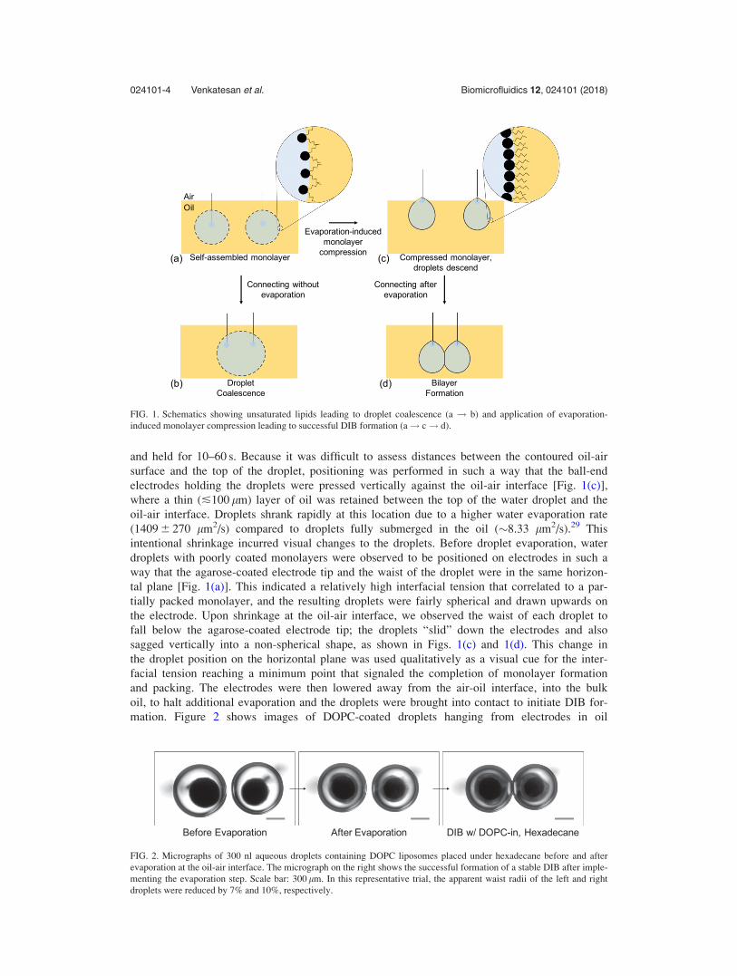
In a conventional lipid-in DIB experiment, droplets containing liposomes at a concentration
of 1–10 mg/ml are incubated in oil at a temperature above the phase transition temperature of
the lipid(s)10,12 for several minutes to promote monolayer formation prior to bringing the drop-
lets into contact [Fig. 1(a)]. However, we and others21,22 have observed that some types of lip-
ids, including unsaturated lipids such as DOPC and POPC, fail to form well-packed monolayers
capable of yielding a stable bilayer—i.e., droplets placed in contact coalesce [Fig. 1(b)] rather
than form an interfacial bilayer.
Herein, a controlled droplet evaporation procedure was performed prior to bringing droplets
into contact under oil. The premise for improving the success rate of DIB formation is that a loss
of volume of an aqueous droplet pre-coated with a partially packed monolayer reduces the surface
area of the droplet, which will result in lateral compression and tighter packing of the adsorbed
phospholipids. In turn, this increased packing should aid or enable bilayer formation upon droplet
contact. Thus, while Sandison et al. used evaporation of the organic solvent to accelerate thinning
of substrate supported black lipid membranes (BLMs),28 our approach harnesses evaporation to
condense the monolayer on each droplet, prior to the process of bilayer formation.
The sequence of our technique is illustrated in panels (a), (c), and (d) in Fig. 1.
Specifically, after incubating droplets in oil for 5 min to facilitate the formation of droplet surfa-
ces partially packed with lipids [Fig. 1(a)], droplets were brought close to the oil-air interface
024101-3 Venkatesan et al. Biomicrofluidics 12, 024101 (2018)
and held for 10–60 s. Because it was difficult to assess distances between the contoured oil-air
surface and the top of the droplet, positioning was performed in such a way that the ball-end
electrodes holding the droplets were pressed vertically against the oil-air interface [Fig. 1(c)],
where a thin (�100 lm) layer of oil was retained between the top of the water droplet and the
oil-air interface. Droplets shrank rapidly at this location due to a higher water evaporation rate
(1409 6 270 lm2/s) compared to droplets fully submerged in the oil (�8.33 lm2/s).29 This
intentional shrinkage incurred visual changes to the droplets. Before droplet evaporation, water
droplets with poorly coated monolayers were observed to be positioned on electrodes in such a
way that the agarose-coated electrode tip and the waist of the droplet were in the same horizon-
tal plane [Fig. 1(a)]. This indicated a relatively high interfacial tension that correlated to a par-
tially packed monolayer, and the resulting droplets were fairly spherical and drawn upwards on
the electrode. Upon shrinkage at the oil-air interface, we observed the waist of each droplet to
fall below the agarose-coated electrode tip; the droplets “slid” down the electrodes and also
sagged vertically into a non-spherical shape, as shown in Figs. 1(c) and 1(d). This change in
the droplet position on the horizontal plane was used qualitatively as a visual cue for the inter-
facial tension reaching a minimum point that signaled the completion of monolayer formation
and packing. The electrodes were then lowered away from the air-oil interface, into the bulk
oil, to halt additional evaporation and the droplets were brought into contact to initiate DIB for-
mation. Figure 2 shows images of DOPC-coated droplets hanging from electrodes in oil
FIG. 1. Schematics showing unsaturated lipids leading to droplet coalescence (a ! b) and application of evaporation-
induced monolayer compression leading to successful DIB formation (a! c! d).
FIG. 2. Micrographs of 300 nl aqueous droplets containing DOPC liposomes placed under hexadecane before and after
evaporation at the oil-air interface. The micrograph on the right shows the successful formation of a stable DIB after imple-
menting the evaporation step. Scale bar: 300 lm. In this representative trial, the apparent waist radii of the left and right
droplets were reduced by 7% and 10%, respectively.
024101-4 Venkatesan et al. Biomicrofluidics 12, 024101 (2018)
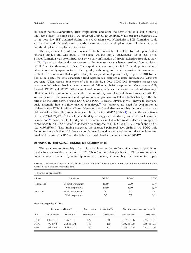
collected: before evaporation, after evaporation, and after the formation of a stable droplet
interface bilayer. In some cases, we observed droplets to completely fall off the electrodes due
to the very low IFT obtained during the evaporation step. Nonetheless, DIB formation could
still be assessed: electrodes were gently re-inserted into the droplets using micromanipulators
and the droplets were placed into contact.
The experimental result was concluded to be successful if a DIB formed upon contact
between droplets and was found to be stable, without droplet coalescence, for at least 1 min.
Bilayer formation was determined both by visual confirmation of droplet adhesion (see right panel
in Fig. 2) and via electrical measurement of the increase in capacitance resulting from exclusion
of oil from the thinning interface. The experiment was noted to fail if the droplets coalesced
either immediately upon contact or during bilayer thinning and radial expansion. As summarized
in Table I, we observed that implementing the evaporation step drastically improved DIB forma-
tion success rates for both unsaturated lipid types in two different alkanes: hexadecane (C16) and
dodecane (C12). Across both types of oils and lipids, a 90%–100% DIB formation success rate
was recorded when droplets were connected following brief evaporation. Once successfully
formed, DOPC and POPC DIBs were found to remain intact for longer periods of time (e.g.,
30–40 min at the minimum, which is the duration of a typical electrical characterization test). The
values for membrane resistance and rupture potential provided in Table I further testify to the sta-
bilities of the DIBs formed using DOPC and POPC. Because DPhPC is well known to spontane-
ously assemble into a tightly packed monolayer,16 we observed no need for evaporation to
achieve stable DIBs in either alkane. However, we found that performing the evaporation step
did not reduce the ability to achieve a stable DIB with DPhPC (Table I). A specific capacitance
of c.a. 0.63–0.69 lF/cm2 for all three lipid types suggested similar hydrophobic thicknesses in
hexadecane,27 however POPC bilayers in dodecane exhibited a far smaller decrease in specific
capacitance to c.a. 0.55 lF/cm2 in dodecane as compared to DPhPC (c.a. 0.39 lF/cm2) and DOPC
(c.a. 0.36 lF/cm2). This finding suggested the saturated palmitoyl acyl chain of the POPC lipid
favors greater exclusion of dodecane upon bilayer formation compared to both the double unsatu-
rated acyl chains of DOPC and the bulky and methylated saturated chains of DPhPC.
DYNAMIC INTERFACIAL TENSION MEASUREMENTS
The spontaneous assembly of a lipid monolayer at the surface of a water droplet in oil
results in a measurable reduction in IFT. Therefore, we also performed IFT measurements to
quantitatively compare dynamic spontaneous monolayer assembly for unsaturated lipids
TABLE I. Number of successful DIB formation trials with and without the evaporation step and the electrical measure-
ments obtained from the successful trials.
DIB formation success rate
Alkane Condition DPhPC DOPC POPC
Hexadecane Without evaporation 10/10 2/20 0/10
With evaporation 10/10 9/10 9/10
Dodecane Without evaporation 5/5 2/6 0/6
With evaporation 3/3 7/7 9/12
Electrical properties of DIBs
Lipid
Resistance (MX cm2) Max. rupture potential (mV) Specific capacitance (lF cm�2)
Hexadecane Dodecane Hexadecane Dodecane Hexadecane Dodecane
DPhPC 8.04 6 3.6 6.47 6 1.1 275 200 0.685 6 0.07 0.386 6 0.07
DOPC 1.99 6 0.88 2.78 6 0.71 150 100 0.652 6 0.08 0.357 6 0.07
POPC 1.03 6 0.68 3.53 6 2.2 100 125 0.626 6 0.05 0.553 6 0.13
024101-5 Venkatesan et al. Biomicrofluidics 12, 024101 (2018)
compared to DPhPC and, separately, to estimate the additional reduction in IFT incurred by
evaporation as applied to improve DIB formation success rates. First, IFT measurements per-
formed using fixed-volume (1 ll) PDT revealed that DPhPC and DOPC lipids, when placed as
liposomes in aqueous droplets (i.e., lipid-in Ref. 6), self-assembled at a hexadecane-water inter-
face to form monolayers with equilibrium IFT (c) values of 1.18 6 0.2 mN/m and
1.99 6 0.5 mN/m, respectively, within 5 min [Fig. 3(a)]. The surface pressures (P¼ 44 - c;
>42 mN/m) of both these monolayers were above the bilayer-monolayer equivalence pressure
of 40 mN/m,30 and, thus, were both expected to be suitable for DIB formation. While Table I
shows that this surface pressure correlated to sufficiently packed monolayers for bilayer forma-
tion in the case of DPhPC-coated droplets, we recorded an 80% failure rate in attempts to
assemble DIBs from DOPC monolayers after 5 min of monolayer assembly in hexadecane
(Table I). In the case of POPC, the IFT did not reach a stable value even after 15 min of spon-
taneous self-assembly (a quasi-equilibrium value of 12.68 6 1.9 mN/m is recorded after 15 min),
indicating the formation of a sparsely packed monolayer. This finding explains why POPC-
coated droplets that were not intentionally evaporated coalesce (100% failure rate) instead of
forming a stable DIB (Table I). The fact that intentional droplet evaporation improved DIB for-
mation for both DOPC and POPC indicates that it is possible, and perhaps required, to further
reduce the IFT of a partially packed monolayer to a point at which DIB formation is possible.
Next, we performed successive, step-wise reductions in pendant droplet volume while con-
tinuously measuring IFT to investigate how droplet shrinkage at the air-oil interface in DIB
experiments affected the IFT of a pre-assembled monolayer [see Fig. 3(b)]. Note that in this
technique, while the volume of the pendant droplet exposed to the oil decreased, the concentra-
tion of lipids remained constant. Figures 3(c)–3(e) shows sample IFT responses for volume
reduction steps performed on monolayers after reaching an equilibrium IFT (�5 min for DPhPC
and DOPC) or after 15 min of incubation of droplets in oil for POPC. Because POPC-coated
droplets exhibited the smallest spontaneous reduction in IFT, each volume reduction step per-
formed on a POPC monolayer further reduced the IFT, likely by the lateral compression of pre-
adsorbed lipids at the interface. Eventually, a minimum IFT value of less than 1 mN/m was
consistently observed as shown in Fig. 4(a) (i.e., c15min� csaturation), which shows how IFT
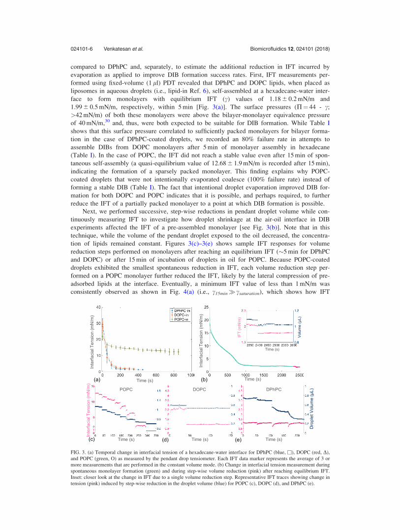
FIG. 3. (a) Temporal change in interfacial tension of a hexadecane-water interface for DPhPC (blue, �), DOPC (red, D),
and POPC (green, O) as measured by the pendant drop tensiometer. Each IFT data marker represents the average of 3 or
more measurements that are performed in the constant volume mode. (b) Change in interfacial tension measurement during
spontaneous monolayer formation (green) and during step-wise volume reduction (pink) after reaching equilibrium IFT.
Inset: closer look at the change in IFT due to a single volume reduction step. Representative IFT traces showing change in
tension (pink) induced by step-wise reduction in the droplet volume (blue) for POPC (c), DOPC (d), and DPhPC (e).
024101-6 Venkatesan et al. Biomicrofluidics 12, 024101 (2018)
decreased with shrinking droplet volume for multiple measurements on DOPC and POPC-
coated pendant droplets. In the case of DOPC [Fig. 4(b)], the volume reduction step was per-
formed once the IFT reached a value within 1 mN/m of the average value of IFT at equilibrium
recorded using fixed-volume PDT. We observed the volume reduction steps to result in a stable
value of IFT below 1 mN/m, suggesting even tighter lateral packing of lipids than achieved by
spontaneous assembly alone (i.e., cspontaneous> csaturation). A similar effect of reduction in DOPC
monolayer IFT (from equilibrium 4.8 to 1.2 mN/m) was reported upon adsorption of a globular
protein, lysozyme.31 In the case of DPhPC, however, volume reduction steps performed after
reaching equilibrium did not result in a stable reduction of IFT. Instead, the measured tension
rebounded to its equilibrium value after a transient response to the volume reduction [Fig.
3(e)]. This finding suggests that the spontaneously assembled DPhPC monolayer was already in
its tightest packing configuration (i.e., cspontaneous � csaturation). As a result, evaporation did not
yield a sustained increase in the lateral packing of lipids; i.e., exclusion of excess lipid mole-
cules from the compressed monolayer through buckling forced a rebound in transient IFT.32,33
This incompressibility was further supported by the higher stiffness values exhibited by com-
pressed DPhPC monolayers at high surface pressures as shown in Fig. S1(b), supplementary
material.
The data shown in Fig. 4 provide strong evidence that spontaneously assembled DOPC and
POPC monolayers can be compressed further by reducing drop volume, and thus drop surface
area available to the monolayer lipids, to achieve tighter lipid packing and lower values of IFT
than was achieved through spontaneous assembly alone. The resulting increase in lipid packing
is in direct agreement with our findings (Table I) that controlled droplet evaporation greatly
enhances DIB success for DOPC and POPC.
COMPRESSION ISOTHERM MEASUREMENTS
The observed decreases in IFT caused by reductions in droplet volume motivate the need
for further characterizing changes in lipid packing. Compression isotherms of pure DPhPC,
DOPC, and POPC monolayers at an air-water interface are shown in Fig. S1 (supplementary
material), and each isotherm shows a smooth response with surface pressure continuously
increasing as lipids were compressed. Surface pressure reaches a maximum and plateaus, a phe-
nomenon associated with buckling of the monolayer lipids compressed to their maximum allow-
able extent, for all lipid cases.33 The surface pressure-area isotherms contain no sharp transi-
tions like those expected in the case of a compression-induced transition from the liquid
expanded to the liquid condensed phase, proving that all three types of monolayers remained in
the liquid expanded phase during compression to the point of buckling. These measured iso-
therms and collapse pressure values (Table S2, supplementary material) are in good agreement
with previous reports,21,34–36 and they highlight differences in the average area occupied per
lipid at a given surface pressure. Specifically, a single DPhPC molecule takes up a larger area
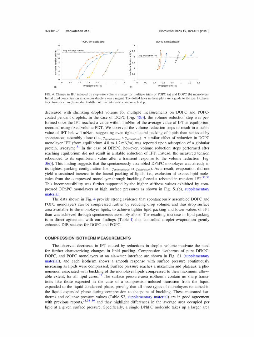
FIG. 4. Change in IFT induced by step-wise volume change for multiple trials of POPC (a) and DOPC (b) monolayers.
Initial lipid concentration in aqueous droplets was 2 mg/ml. The dotted lines in these plots are a guide to the eye. Different
trajectories seen in (b) are due to different time intervals between each step.
024101-7 Venkatesan et al. Biomicrofluidics 12, 024101 (2018)

(63.9 A2) compared to both DOPC (53.6 A2) and POPC (50.3 A2) at their respective collapse
pressures (i.e., maximum pressure before monolayer buckling).
DISCUSSION
Self-assembled lipid monolayers are essential for creating droplet interface bilayers,4 and it
is well established that both the kinetics and equilibrium packing density of monolayer forma-
tion at an oil-water interface are directly influenced by the concentration of the lipids.
Generally, the number of amphiphiles at the interface increases as the lipid concentration
increases in the bulk. However, Needham et al.37 demonstrated that increasing the lipid concen-
tration in the bulk beyond the critical micelle concentration (CMC) does not yield further
increases in surface density (i.e., decreases in interfacial tension). In this work, we studied
monolayer formation with three types of lipids introduced into the aqueous phase at an initial
concentration of 2 mg/ml, which is well above the CMCs (�0.1–10 lM) for all three lipids
(CMC measurements are described in the supplementary material and data are presented in
Figs. S2 and S3, and Table S3). A starting concentration above CMC implies that lipid assem-
bly in these experiments was not diffusion limited, and it also suggests that a higher lipid con-
centration, achieved either by a higher initial concentration in the droplet or incurred passively
upon evaporation of water, is insufficient to achieve a higher equilibrium packing density in the
monolayer. As a simple test of this theory, we also attempted DIB formation between POPC-
coated droplets containing 10 mg/ml of lipids and observed similar difficulty (without evapora-
tion) and success (with evaporation) as reported in Table I for 2 mg/ml.
To create a DIB, two or more aqueous droplets each coated with a well-packed lipid mono-
layer are connected under a suitable organic solvent. If the droplet interfaces are bare or
sparsely coated with lipids, the interfacial tension between the oil-water interface remains high
(as high as 44 mN/m). When two or more such droplets are brought into contact, the high ten-
sion creates a thermodynamic drive to fuse the droplets to minimize the total surface energy of
the system, since the surface area of a fused droplet is considerably lower than the total surface
area of two separate droplets. In contrast, the presence of sufficiently packed lipid monolayers
around both droplets enables the system to reduce its free energy by the amount DF¼ A� 2cm � cbð Þ through the formation of an oil-depleted interface bilayer, where A is the area
of the bilayer, cm is the tension of the monolayer, and cb is the bilayer tension.38 The stable
adhesive state of the droplets represents a local energy minimum with a total energy greater
than that of coalesced droplets.17 While several lipid types are known to readily self-assemble
to form well-packed monolayers at room temperature, there is also substantial evidence in the
literature that monolayers formed by certain lipids are more suitable than others for DIB forma-
tion.12 Specifically, unsaturated lipids such as DOPC and POPC often fail to form DIBs
between droplets larger than 100 nl in volume. In contrast, several groups have reported the
assembly and study of stable DOPC DIBs when droplet volumes are <100 nl (Table S1, sup-
plementary material).
The IFT measurements provided in Figs. 3 and 4 provide insights into the effectiveness of
monolayer formation for the three lipids studied herein and help to explain the DIB formation
success rates presented in Table I. DPhPC lipids spontaneously assembled to form a tightly
packed monolayer with an IFT that could not be further reduced via reduction in the droplet
surface area. This result confirmed our ability to form DPhPC DIBs without performing droplet
evaporation. On the other hand, POPC lipids self-assembled to form a sparsely packed mono-
layer that was further compressed by reducing the volume (and area) of the aqueous droplet.
Our measurements using PDT demonstrated that aqueous volume reductions stably reduced the
IFT of the monolayer-coated interface to below 1 mN/m. Lastly, while DOPC lipids spontane-
ously assembled to form monolayers with an average equilibrium IFT of �2 mN/m—a value
that we expected to be sufficient for DIB formation—we found that these monolayers can also
be further compressed to reach a minimum IFT of <1 mN/m. These findings showing
compression-induced reduction in IFT with both POPC and DOPC correlate well to our obser-
vations that evaporation improved DIB formation in both hexadecane and dodecane. Given
024101-8 Venkatesan et al. Biomicrofluidics 12, 024101 (2018)
these outcomes, it was necessary to understand what about the structure of a lipid prevents
spontaneous assembly of a densely packed monolayer, as well as how much lateral compression
of the monolayer occurred when droplets were prescribed this new droplet shrinking protocol.
Differences in equilibrium packing densities (and IFT values) between the three lipids stud-
ied can possibly be explained by structural differences brought about by the lipid shape and the
extent to which alkane molecules from the surrounding oil partition in between the tail groups.
All three lipids under consideration are made up of identical phosphatidylcholine (PC) head
groups. However, their tail groups vary in length and composition. DPhPC has two fully satu-
rated 16C fatty acid chains with 4 methyl groups attached to each chain, whereas DOPC is
made up of two 18C fatty acid chains with a single double-bond (D9-Cis) in each chain. Lastly,
POPC is a hybrid-monounsaturated lipid that is made up of one fully saturated 16C chain and
one mono-unsaturated 18C (D9-Cis) fatty acid chain. These double bonds found in the tail
groups of DOPC and POPC are known to induce a bend (often referred to as “kink”) in the
tail—a restricted rotation about the double-bound that is not found in fully saturated tail groups
of DPhPC. These kink(s) give both DOPC and POPC molecules conical shapes (DOPC being
more conical than POPC3). Our data suggests the conical shapes of DOPC and POPC prevent
efficient lateral packing and favor the persistence of defects in monolayers and bilayers. DPhPC
molecules, on the other hand, take on a more cylindrical shape that allows them to efficiently
pack to form defect-free planar monolayers and bilayer membranes.3,39–42
As lipids self-assemble to form a monolayer at an oil-water or air-water interface, the
intrinsic shapes of the amphiphilic molecules dictate the extent to which the IFT is reduced. In
the case of cylindrically shaped DPhPC, the lipid molecules spontaneously assembled to their
tightest packing state at equilibrium, retaining minimal packing defects [Fig. 5(a)] and giving
rise to low IFT. However, due to the steric hindrance posed by the conical-shape of neighboring
unsaturated lipids, DOPC and POPC molecules failed to form a planar monolayer that is packed
to its tightest packing state.43 Instead, we believe self-assembled monolayers of these unsatu-
rated lipids retained a significant degree of packing defects at equilibrium, which in turn led to
an equilibrium IFT higher than the lowest possible IFT achievable by these monolayers.31,44
While lipid shape describes why unsaturated lipids are unable to pack as tight as saturated
DPhPC lipids at equilibrium, it does not explain why POPC lipids, despite being more cylindri-
cal than DOPC,3 self-assembled the slowest of the three lipids. In addition to lipid shape, other
factors such as stability of liposomes and solubility of lipid tails in alkane may play roles in
determining the rate of monolayer formation.
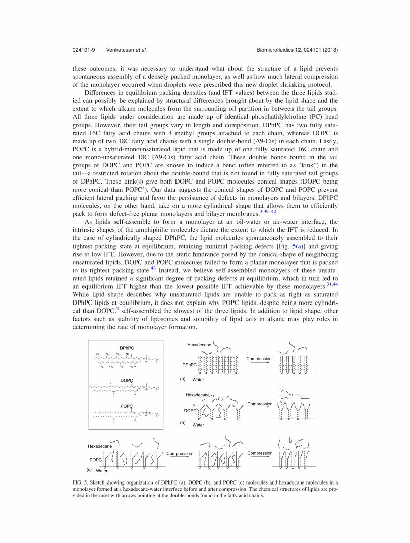
FIG. 5. Sketch showing organization of DPhPC (a), DOPC (b), and POPC (c) molecules and hexadecane molecules in a
monolayer formed at a hexadecane-water interface before and after compression. The chemical structures of lipids are pro-
vided in the inset with arrows pointing at the double-bonds found in the fatty acid chains.
024101-9 Venkatesan et al. Biomicrofluidics 12, 024101 (2018)

The excess area (i.e., packing defects) created by the kinks of unsaturated tail groups at a
water-oil interface are expected to be occupied by alkane molecules found at the interface.
Moreover, the presence of alkanes in the acyl chain regions of the monolayer may even provide
an additional impediment to tight monolayer packing. X-ray diffraction studies have revealed
that non-surface active molecules such as alkanes partition in between the hydrocarbon tail
groups in a monolayer and change its packing and thermodynamic properties.45–49 Compression
isotherm experiments by Thoma and M€ohwald revealed that higher amounts of alkanes partition
among the lipid tail groups when the length of hydrocarbon chains is comparable to that of
lipid tails.50 Therefore, we suspect that self-assembled monolayers of DOPC and POPC possess
packing defects that favor the partitioning of alkane molecules between the tail groups, as
shown in Figs. 5(b) and 5(c). Therefore, reducing the area available for each lipid by laterally
compressing the monolayer: (a) forces the lipids to pack closer to one another, and (b) squeezes
out residual solvent molecules found in the defects.50 The combination of these two processes
reduces the IFT of the monolayer laden oil-water interface by lowering the water-acyl chain
and water-solvent molecule interactions. As the initial lipid concentrations for all experiments
were significantly higher than their respective CMC values, the increase in packing density due
to evaporation is expected to be dominated by the decrease in surface area and not due to the
increase in lipid concentration in bulk. While it is possible that lateral compression of a mono-
layer could also lead to cis-trans isomerization of unsaturated acyl chains as seen during mono-
layer phase change, results from our experiments are inadequate to evaluate this effect.51
Next, we attempted to quantify the extent to which evaporation-induced droplet shrinkage
prior to DIB formation decreased the area occupied by each lipid molecule. Figure S1 (supple-
mentary material) shows how decreasing the average area available for lipid molecules changes
the surface pressure of the monolayer formed at an air-water interface for all three lipid types.
Because a direct comparison of IFT measured at the oil-water interface (using PDT) and sur-
face pressure measured at the air-water interface (using Langmuir compression isotherm) was
not possible, we made our comparison by equating measured changes in pendant droplet inter-
facial tension (Dc) upon droplet shrinkage with changes in surface pressure (DP) upon lateral
monolayer compression. Assuming the minimum tension (csaturation) reached in PDT measure-
ments (Fig. 4) corresponded to the collapse pressure (Pc) in the compression isotherm (Fig. S1,
supplementary material) for the given lipid type, we estimated the change in area per lipid
induced by the volume reduction steps. The filled circles in Fig. 6(a) represent the collapse
pressures, which were assumed to correspond to the average minimum c in PDT measurements
(�1 mN/m for both DOPC and POPC, Fig. 4). The total change in tension attained by volume
reduction steps is given by Dc¼ cspontaneous - csaturation. Subtracting DIFT from collapse pressure
should give us the surface pressure that was achieved by spontaneous assembly alone, i.e.,
Pspontaneous¼Pc - Dc. These values of Pspontaneous for DOPC and POPC are marked by open
circles in Fig. 6(a). Performing this calculation enabled us to: (a) estimate the area per lipid
molecule in the monolayer that was attained by spontaneous assembly alone, and (b) calculate
the change in the area per lipid molecule brought about by the step-wise volume reduction.
According to this analysis, each DOPC molecule in a spontaneously assembled monolayer
occupied about 50.6 A2, which is �3 A2 (6%) higher than the area at maximum packing (col-
lapse pressure). In the case of POPC, each molecule in the monolayer attained at the end of
15 min of incubation occupied approximately 42.3 A2, which is �8 A2 (16%) more area than at
its maximum packing.
The area compression analysis discussed above can also be used to estimate the extent of
droplet volume shrinkage required to successfully form DIBs with DOPC and POPC.
According to this analysis, to achieve the tightest packing configuration, a maximum of 16%
(6% for DOPC and 16% for POPC) reduction in area per molecule was predicted to be suffi-
cient to bring about the required packing for DIB formation. Assuming a lipid-coated aqueous
droplet to have a spherical shape, a 300 nl droplet has a surface area of 2.17 mm2. A 16%
reduction in surface area leads to a droplet with a final surface area of 1.82 mm2 (231 nl),
which is nearly a 23% reduction in volume. Depending on the initial packing state of mono-
layer before evaporation, the level of humidity in the local environment, and proximity of water
024101-10 Venkatesan et al. Biomicrofluidics 12, 024101 (2018)

droplets to the oil-air interface during the evaporation step, a mere 10–60 s evaporation at the
oil-air interface was found to be sufficient to achieve the packing required for successful DIB
formation. Figure 6(b) shows the change in droplet volume and percentage reduction in the sur-
face area of a spherical droplet with an initial volume of 300 nl that is subjected to evaporation
at different rates of volume loss; see Fig. S4 (supplementary material) for percentage change in
the surface area for different initial volumes. This simulation gives an insight to the period of
time a droplet should be held at the oil-air interface in order to surpass the threshold percentage
reduction in the surface area. Beyond this threshold (denoted by green line), the droplet is
expected to retain a monolayer in which the lipids are packed in their tightest configuration and
suitable for DIB formation. Excessive droplet shrinkage beyond the threshold is expected to
lead to monolayer buckling, by which surplus lipids are excluded to maintain maximal packing
in the monolayer.32,33
Finally, a few comments regarding the methodologies used herein. First, the IFT data mea-
sured using PDT were obtained with droplet volumes of only 1–2 ll. Because the accuracy of
computing tension values can be compromised by small droplets that do not exhibit significant
deformation, we computed the Worthington number,52 Wo, given by Wo ¼ DqgVd=pcmD to ver-
ify the accuracy of the measurements. In this expression, Dq represents the density difference
droplet phase and the external oil phase, Vd is the volume of droplet, g is the acceleration due
to gravity, cm is the monolayer IFT, and D represents the diameter of the dispensing needle. A
value of Wo close to 1 indicates high precision in measuring IFT, while a value close to 0 indi-
cates low precision. We estimated Wo to be between 0.6 and 0.9.
Second, the comparative analysis of IFT and compression isotherm data in Fig. 6(a) was
only performed to obtain an estimate of possible area change obtained by lateral compression.
The absolute values of the estimated change in area are subject to some variability based on
the accuracy of either measurements. Primarily, values of molecular areas for lipids presented
in the compression isotherms are sensitive to experimental procedures and are found to be
highly variable in literature.53 Third, the evaporation period estimated in Fig. 6(b) is likely
overestimated, due to our assumption that droplets remain spherical. In reality, we know and
observe that a droplet hanging from a slender electrode has a pendant-like shape [Fig. 1(c)],
which causes it to undergo faster reductions in surface area compared to a spherical volume
losing volume at the same rate. However, because it is difficult to accurately calculate the vol-
ume change using only the radius (at the droplet waist), as seen through an inverted micro-
scope, we simply used the visual cue of the droplets sagging as qualitative confirmation of a
tightly packed, minimum tension monolayer. Lastly, we expect the principle of increasing
monolayer packing density by decreasing the droplet surface area to be applicable for both
lipid-in and lipid-out methods (see Fig. S5, supplementary material). However, as identified in
FIG. 6. (a) Area reduction analysis. Compression isotherms indicating collapse pressures (shaded circles) and estimated
pressure and area per molecule corresponding to spontaneously assembled monolayer (unshaded circles). (b) Estimated per-
centage decrease in the droplet surface area caused by evaporation of a 300 nl spherical droplet at different rates of evapo-
ration for 5 min. The reference plane shown in gray indicates the recommended 16% decrease in surface area required to
achieve a tightly packed monolayer. Time value indicated next to each trend is the length of time required for the droplet
surface area to reduce by 16%.
024101-11 Venkatesan et al. Biomicrofluidics 12, 024101 (2018)

our previous work,16 other factors, such as the formation of swollen, inverse micelles in the oil
subphase near a partially packed monolayer, can also prevent successful DIB formation when
certain lipids are placed in oil.
CONCLUSION
Herein, we demonstrated a simple evaporation-induced monolayer compression technique
that enables improved and faster assembly of DIBs, including those comprised of unsaturated
lipids such as DOPC and POPC. The technique leverages controlled evaporation to condense
the spontaneously self-assembled monolayers, which we found increased the DOPC and POPC
bilayer formation success rate from <20% to nearly 100%. It is feasible that this approach
could also be used to improve DIB formation from charged lipids such as 1,2-dioleoyl-sn-
glycero-3-phosphoglycerol (DOPG). To understand how this method improved bilayer forma-
tion, the effects of lateral compression of DOPC and POPC monolayers were studied in com-
parison to DPhPC using pendant drop tensiometry and Langmuir compression isotherms. Our
measurements confirmed that, unlike DPhPC, DOPC and POPC lipids do not spontaneously
assemble into maximally packed monolayers. However, controlled evaporation was used to
reduce the IFT of DOPC and POPC monolayers to values lower than 1 mN/m, at which point
DIB formation became highly repeatable. The approximate reduction in area per lipid induced
by 10–60 s of evaporation was found to be c.a. 6% for DOPC and c.a. 16% for POPC lipids.
Lastly, the measured electrical properties such as membrane resistance and rupture potential of
DIBs formed with DOPC and POPC were found to be in desirable ranges (and comparable to
values obtained with DPhPC), thus making them useful platforms for studying transmembrane
proteins and ion channels relevant to mammals.
SUPPLEMENTARY MATERIAL
See supplementary material for a tabular summary of prior DOPC DIB studies, material
preparations, and experimental details for DIB formation, pendant droplet tensiometry, Langmuir
trough experiments, and measuring lipid CMC. Supplementary data are also provided.
ACKNOWLEDGMENTS
The authors acknowledge funding from the Air Force Office of Scientific Research Basic
Research Initiative Grant No. FA9550-12-1-0464, and we thank Mr. Subhadeep Koner and Ms.
Megan Pitz for their assistance with the POPC DIB experiments. Langmuir Trough compression
isotherm measurements were conducted in Dr. Barry Bruce’s lab at the Biochemistry, Cellular and
Molecular Biology at the University of Tennessee. Pendant drop measurements of interfacial
tensions of lipid monolayers were conducted at the Center for Nanophase Materials Sciences, which
is a DOE Office of Science User Facility. This manuscript has been authored by UT-Battelle, LLC,
under Contract No. DE-AC0500OR22725 with the U.S. Department of Energy. The United States
Government retains and the publisher, by accepting the article for publication, acknowledges that
the United States Government retains a non-exclusive, paid-up, irrevocable, world-wide license to
publish or reproduce the published form of this manuscript, or allow others to do so, for the United
States Government purposes.
1A. A. Spector and M. A. Yorek, J. Lipid Res. 26(9), 1015–1035 (1985).2G. Van Meer, D. R. Voelker, and G. W. Feigenson, Nat. Rev. Mol. Cell Biol. 9(2), 112–124 (2008).3S. Vanni, H. Hirose, H. Barelli, B. Antonny, and R. Gautier, Nat. Commun. 5, 4916 (2014).4H. Bayley, B. Cronin, A. Heron, M. A. Holden, W. L. Hwang, R. Syeda, J. Thompson, and M. Wallace, Mol. BioSyst.4(12), 1191–1208 (2008).
5K. Funakoshi, H. Suzuki, and S. Takeuchi, Anal. Chem. 78(24), 8169–8174 (2006).6W. L. Hwang, M. Chen, B. Cronin, M. A. Holden, and H. Bayley, J. Am. Chem. Soc. 130(18), 5878–5879 (2008).7P. J. Milianta, M. Muzzio, J. Denver, G. Cawley, and S. Lee, Langmuir 31(44), 12187–12196 (2015).8M. R. R. de Planque, S. Aghdaei, T. Roose, and H. Morgan, ACS Nano 5(5), 3599–3606 (2011).9N. Rojko, B. Cronin, J. S. H. Danial, M. A. B. Baker, G. Anderluh, and M. I. Wallace, Biophys. J. 106(8), 1630–1637(2014).
024101-12 Venkatesan et al. Biomicrofluidics 12, 024101 (2018)
10G. J. Taylor, F. A. Heberle, J. S. Seinfeld, J. Katsaras, C. P. Collier, and S. A. Sarles, Langmuir 33(38), 10016–10026(2017).
11J. R. Thompson, A. J. Heron, Y. Santoso, and M. I. Wallace, Nano Lett. 7(12), 3875–3878 (2007).12G. J. Taylor and S. A. Sarles, Langmuir 31(1), 325–337 (2015).13N. Tamaddoni, E. C. Freeman, and S. A. Sarles, Smart Mater. Struct. 24(6), 065014 (2015).14N. Tamaddoni, G. Taylor, T. Hepburn, S. M. Kilbey, and S. A. Sarles, Soft Matter 12(23), 5096–5109 (2016).15S. Leptihn, O. K. Castell, B. Cronin, E.-H. Lee, L. C. M. Gross, D. P. Marshall, J. R. Thompson, M. Holden, and M. I.
Wallace, Nat. Protocols 8(6), 1048–1057 (2013).16G. A. Venkatesan, J. Lee, A. B. Farimani, M. Heiranian, C. P. Collier, N. R. Aluru, and S. A. Sarles, Langmuir 31(47),
12883–12893 (2015).17P. Poulin and J. Bibette, Langmuir 14(22), 6341–6343 (1998).18S. Thutupalli, J.-B. Fleury, A. Steinberger, S. Herminghaus, and R. Seemann, Chem. Commun. 49(14), 1443–1445
(2013).19N. E. Barlow, G. Bolognesi, S. Haylock, A. J. Flemming, N. J. Brooks, L. M. C. Barter, and O. Ces, Sci. Rep. 7(1), 17551
(2017).20D.-W. Jeong, H. Jang, S. Q. Choi, and M. C. Choi, Sci. Rep. 6, 38158 (2016).21S. Nakata, A. Deguchi, Y. Seki, K. Fukuhara, M. Goto, and M. Denda, Thin Solid Films 615, 215–220 (2016).22H. Kim, K. Kim, H.-R. Lee, H. Jo, D.-w. Jeong, J. Ryu, D.-G. Gweon, and S. Q. Choi, J. Ind. Eng. Chem. 55, 198–203
(2017).23K. J. Seu, A. P. Pandey, F. Haque, E. A. Proctor, A. E. Ribbe, and J. S. Hovis, Biophys. J. 92(7), 2445–2450 (2007).24T. Baumgart and A. Offenh€ausser, Biophys. J. 83(3), 1489–1500 (2002).25M. A. Holden, D. Needham, and H. Bayley, J. Am. Chem. Soc. 129(27), 8650–8655 (2007).26G. A. Venkatesan and S. A. Sarles, Lab Chip 16(11), 2116–2125 (2016).27G. J. Taylor, G. A. Venkatesan, C. P. Collier, and S. A. Sarles, Soft Matter 11(38), 7592–7605 (2015).28M. E. Sandison, M. Zagnoni, and H. Morgan, Langmuir 23(15), 8277–8284 (2007).29P. Mruetusatorn, J. B. Boreyko, G. A. Venkatesan, S. A. Sarles, D. G. Hayes, and C. P. Collier, Soft Matter 10(15),
2530–2538 (2014).30A. Yasmann and S. Sukharev, Langmuir 31(1), 350–357 (2015).31M. Ohno, T. Toyota, T. Nomoto, and M. Fujinami, Colloids Surf. A 480, 85–90 (2015).32K. Y. C. Lee, Annu. Rev. Phys. Chem. 59(1), 771–791 (2008).33S. Baoukina, L. Monticelli, H. J. Risselada, S. J. Marrink, and D. P. Tieleman, Proc. Natl. Acad. Sci. U.S.A. 105(31),
10803–10808 (2008).34S. F. Gilmore, A. I. Yao, Z. Tietel, T. Kind, M. T. Facciotti, and A. N. Parikh, Langmuir 29(25), 7922–7930 (2013).35A. Tsanova, A. Jordanova, and Z. Lalchev, J. Membr. Biol. 249(3), 229–238 (2016).36Y. Ishitsuka, D. S. Pham, A. J. Waring, R. I. Lehrer, and K. Y. C. Lee, Biochim. Biophys. Acta, Biomembr. 1758(9),
1450–1460 (2006).37S. Lee, D. H. Kim, and D. Needham, Langmuir 17(18), 5537–5543 (2001).38D. Needham and D. A. Haydon, Biophys. J. 41(3), 251–257 (1983).39J. Bigay and B. Antonny, Dev. Cell 23(5), 886–895 (2012).40T. G. Pomorski, T. Nylander, and M. C�ardenas, Adv. Colloid Interface Sci. 205, 207–220 (2014).41M. Garten, C. Pr�evost, C. Cadart, R. Gautier, L. Bousset, R. Melki, P. Bassereau, and S. Vanni, Phys. Chem. Chem. Phys.
17(24), 15589–15597 (2015).42B. Kollmitzer, P. Heftberger, M. Rappolt, and G. Pabst, Soft Matter 9(45), 10877–10884 (2013).43M. Jurak, J. Phys. Chem. B 117(13), 3496–3502 (2013).44K. Nag, J. G. Munro, K. Inchley, S. Sch€urch, N. O. Petersen, and F. Possmayer, Am. J. Physiol. 277(6), L1179–L1189
(1999).45F. Giebel, M. Paulus, J. Nase, I. Kiesel, S. Bieder, and M. Tolan, Colloids Surf., A 504, 126–130 (2016).46V. Fainerman, E. Aksenenko, and R. Miller, Adv. Colloid Interface Sci. 244, 100–112 (2017).47Q. Lei and C. D. Bain, Phys. Rev. Lett. 92(17), 176103 (2004).48S. Yefet, E. Sloutskin, L. Tamam, Z. Sapir, M. Deutsch, and B. M. Ocko, Langmuir 30(27), 8010–8019 (2014).49J. Marqusee and K. A. Dill, J. Chem. Phys. 85(1), 434–444 (1986).50M. Thoma and H. M€ohwald, J. Colloid Interface Sci. 162(2), 340–349 (1994).51C. Roach, S. E. Feller, J. A. Ward, S. R. Shaikh, M. Zerouga, and W. Stillwell, Biochemistry 43(20), 6344–6351 (2004).52J. D. Berry, M. J. Neeson, R. R. Dagastine, D. Y. Chan, and R. F. Tabor, J. Colloid Interface Sci. 454, 226–237 (2015).53P. B. Welzel, I. Weis, and G. Schwarz, Colloids Surf., A 144(1), 229–234 (1998).
024101-13 Venkatesan et al. Biomicrofluidics 12, 024101 (2018)