evasion of cell senescence leads to medulloblastoma ... · cell reports article evasion of cell...
TRANSCRIPT
Article
Evasion of Cell Senescenc
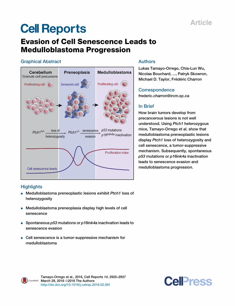
e Leads toMedulloblastoma ProgressionGraphical Abstract
Highlights
d Medulloblastoma preneoplastic lesions exhibit Ptch1 loss of
heterozygosity
d Medulloblastoma preneoplasia display high levels of cell
senescence
d Spontaneous p53mutations or p16ink4a inactivation leads to
senescence evasion
d Cell senescence is a tumor-suppressive mechanism for
medulloblastoma
Tamayo-Orrego et al., 2016, Cell Reports 14, 2925–2937March 29, 2016 ª2016 The Authorshttp://dx.doi.org/10.1016/j.celrep.2016.02.061
Authors
Lukas Tamayo-Orrego, Chia-Lun Wu,
Nicolas Bouchard, ..., Patryk Skowron,
Michael D. Taylor, Frederic Charron
In Brief
How brain tumors develop from
precancerous lesions is not well
understood. Using Ptch1 heterozygous
mice, Tamayo-Orrego et al. show that
medulloblastoma preneoplastic lesions
display Ptch1 loss of heterozygosity and
cell senescence, a tumor-suppressive
mechanism. Subsequently, spontaneous
p53 mutations or p16ink4a inactivation
leads to senescence evasion and
medulloblastoma progression.
Cell Reports
Article
Evasion of Cell SenescenceLeads to Medulloblastoma ProgressionLukas Tamayo-Orrego,1,2 Chia-Lun Wu,1,3 Nicolas Bouchard,1,3 Ahmed Khedher,1 Shannon M. Swikert,1,2 Marc Remke,4
Patryk Skowron,5 Michael D. Taylor,5 and Frederic Charron1,2,3,6,*1Molecular Biology of Neural Development, Institut de Recherches Cliniques de Montreal, 110 Pine Avenue West, Montreal, QC H2W 1R7,
Canada2Integrated Program in Neuroscience, McGill University, Montreal, QC H3A 2K6, Canada3Department of Medicine, University of Montreal, Montreal, QC H3T 1J4, Canada4Research Group ‘‘Non-coding RNAs in Pediatric Cancers,’’ German Cancer Consortium, University Hospital D€usseldorf, Department of
Pediatric Oncology, Hematology, and Clinical Immunology, 40225 D€usseldorf, Germany5Division of Neurosurgery and The Arthur and Sonia Labatt Brain Tumour Research Centre, Hospital for Sick Children, 555 University Avenue,Toronto, ON M5G 1X8, Canada6Division of Experimental Medicine, Department of Medicine, Department of Anatomy and Cell Biology, Department of Biology, McGill
University, Montreal, QC H3A 2B2, Canada
*Correspondence: [email protected]://dx.doi.org/10.1016/j.celrep.2016.02.061
This is an open access article under the CC BY-NC-ND license (http://creativecommons.org/licenses/by-nc-nd/4.0/).
SUMMARY
How brain tumors progress from precancerous le-sions to advanced cancers is not well understood.Using Ptch1+/� mice to study medulloblastoma pro-gression, we found that Ptch1 loss of heterozygosity(LOH) is an early event that is associated with highlevels of cell senescence in preneoplasia. In con-trast, advanced tumors have evaded senescence.Remarkably, we discovered that the majority ofadvanced medulloblastomas display either sponta-neous, somatic p53mutations or Cdkn2a locus inac-tivation. Consistent with senescence evasion, thesep53 mutations are always subsequent to Ptch1LOH. Introduction of a p53mutation prevents senes-cence, accelerates tumor formation, and increasesmedulloblastoma incidence. Altogether, our resultsshow that evasion of senescence associated withPtch1 LOH allows progression to advanced tumors.
INTRODUCTION
For a certain number of human malignancies, it has been
possible to examine different stages of tumor progression and
correlate different histopathological stages with specific genetic
events (Fearon and Vogelstein, 1990). More recently, an
emerging paradigm proposes that oncogenic changes associ-
ated with the formation of precancerous lesions lead to cell
senescence, a mechanism that restrains tumor progression
in vivo (Braig et al., 2005; Chen et al., 2005; Michaloglou et al.,
2005). Brain tumors constitute a challenge for investigating
tumor progression because precancerous lesions are rarely de-
tected. Medulloblastoma is the most common brain tumor in
children, and deregulation of hedgehog signaling characterizes
Cell
25% of human medulloblastomas (Taylor et al., 2012). Although
genomic studies have revealed recurrent genetic and epigenetic
alterations in human sonic hedgehog (SHH) medulloblastoma
(Hovestadt et al., 2014; Kool et al., 2014; Pugh et al., 2012; Rob-
inson et al., 2012), these studies can only be done on advanced
tumors and thus they do not illuminate the process of medullo-
blastoma progression.
Ptch1 heterozygous mice (Ptch1+/LacZ, designated here as
Ptch1+/� for simplicity) constitute one of the best-studied
models of medulloblastoma (Goodrich et al., 1997). It is currently
accepted thatmedulloblastoma development inPtch1+/�mice is
a two-step process in which the Ptch1+/� germline mutation
leads to preneoplasia formation and the subsequent Ptch1
loss of heterozygosity (LOH) is sufficient to promote medullo-
blastoma progression (Ayrault et al., 2009; Pazzaglia et al.,
2006). Consistent with this, human genomic studies indicate
that medulloblastoma, similar to other pediatric cancers, dis-
plays fewmolecular changes compared to other types of tumors
(Pugh et al., 2012). Nevertheless, one study reported that pre-
neoplastic cells with Ptch1 LOH can differentiate into granule
neurons (Kessler et al., 2009), raising the possibility that Ptch1
LOH is not sufficient to promote medulloblastoma progression
and that there may be additional genetic or epigenetic events
governing the transition from preneoplasia to medulloblastoma.
Using the Ptch1 model of medulloblastoma, we discovered
that Ptch1 LOH is a very early event during medulloblastoma
development and is associated with high levels of cell senes-
cence in preneoplastic lesions. Additionally, we found that
advanced tumors have evaded cell senescence as a result of
p53 mutations or Cdkn2a locus inactivation, which occurs via
methylation of p16Ink4a/Cdkn2a regulatory sequences. Using or-
thotopic transplantation and genetic experiments, we show that
p53 point mutations prevent cell senescence, accelerate tumor
formation, and increase medulloblastoma incidence. Altogether,
we propose that medulloblastoma formation requires at least
three genetic events, where p53 or Cdkn2a inactivation disables
Reports 14, 2925–2937, March 29, 2016 ª2016 The Authors 2925

Figure 1. Cell Senescence Is Disabled during Medulloblastoma Progression
(A) p21 (Cdkn1a) immunohistochemistry (IHC) images representative of P7 Ptch1+/� EGL, P14 preneoplasia, and advanced medulloblastoma (Adv. MB.).
(B) Number of p21-positive cells per 10,000 mm2 in Ptch1+/� EGL, preneoplasia, and Adv. MB (n R 5 animals).
(C and D) p16Ink4a IHC staining (C) and (D) number of p16Ink4a-positive cells per 10,000 mm2 in Ptch1+/� EGL, P14 preneoplasia, and Adv. MB (D) (nR 5 animals).
Red arrowheads indicate p21- (A) or p16Ink4a-positive (C) cells.
(E and F) Ki67 IHC staining (E) and number of Ki67+ cells in Ptch1+/� EGL, P14 preneoplasia, and Adv. MB (F) (n = 6 animals). Red rectangles (A, C, and E) indicate
the magnified regions.
(G and H) pH3 immunofluorescence (G) and number of pH3+ cells in Ptch1+/� EGL, preneoplasia, and Adv. MB (H) (n = 6 animals).
(legend continued on next page)
2926 Cell Reports 14, 2925–2937, March 29, 2016 ª2016 The Authors

cell senescenceassociatedwithPtch1LOH.Our results alsopro-
videamechanistic explanation for thepresenceof recurrent inac-
tivation of TP53 or CDKN2A in human SHH medulloblastoma.
RESULTS
Cell Senescence Is Disabled during MedulloblastomaProgression in Ptch1+/� MiceWe were puzzled by the fact that while most Ptch1+/� mice
develop preneoplastic lesions, only a fraction of them acquire
advanced medulloblastoma (Mille et al., 2014; Oliver et al.,
2005). In some cancers, oncogene activation in precancerous
lesions leads to oncogene-induced senescence (OIS), an anti-
cancer mechanism that restrains tumor progression (Braig
et al., 2005; Chen et al., 2005; Michaloglou et al., 2005). It is
not known whether medulloblastoma precancerous lesions
display features of senescence and whether cell senescence
plays a tumor-suppressive role during medulloblastoma forma-
tion. For example, although p53 deletion in Ptch1+/� mice in-
creases medulloblastoma incidence (Wetmore et al., 2001), the
mechanism responsible for this effect was not studied. Similarly,
inactivation of important cell-cycle regulators such as p18Ink4c
(Uziel et al., 2005) or p27Kip1 (Ayrault et al., 2009) increases
medulloblastoma incidence in Ptch1+/� mice, but the cellular
processes responsible for these effects are largely unknown.
To test whether cell senescence plays a role duringmedulloblas-
toma progression, we assessed the levels of the senescence
markers p16Ink4a and p21 at different histopathological stages
of medulloblastoma in Ptch1+/� mice: postnatal day 7 (P7)
external granule-cell layer (EGL), preneoplasia (obtained at
P14, the earliest stage preneoplastic lesions can be distin-
guished), and advanced medulloblastoma (tumors obtained
from mice with terminal illness). As expected, the EGL in
Ptch1+/� cerebella displayed very low levels of p16Ink4a and
p21 (Figures 1A–1D and S1C). Interestingly, we found that pre-
neoplastic lesions displayed high numbers of p16Ink4a and p21
positive cells, while advanced medulloblastoma (Adv. MB.) had
lost these senescence markers.
We next examined the possible impact of the senescence
phenotype on the levels of proliferation at different stages of me-
dulloblastoma. Interestingly, while the EGL and advanced me-
dulloblastoma display a very high proportion of Ki67-positive
cells, preneoplastic lesions have more heterogeneous Ki67
staining, with a significantly lower average proliferation (Figures
1E and 1F). We also observed reduced levels of proliferation in
preneoplastic lesions using a second proliferation marker, phos-
pho-histone H3 (pH3; Figures 1G and 1H). As expected, cells in
preneoplastic lesions that are positive for p16Ink4 are negative for
proliferation (Figure S1B).
To test whether apoptosis also plays a tumor-suppressive role
during medulloblastoma progression, we measured the levels of
(I and J) Cleaved caspase-3 (C.-caspase 3) IHC staining (I) and number of C.-cas
12-mm cryosections were processed for IHC (DAB) and counterstained with cres
Scale bars in (A)–(C) represent 50 mm (top), 20 mm (middle and bottom); scale bar
50 mm; and scale bars in (I) represent 20 mm. Each data point represents the mean
randomly selected sections; error bars indicate SEM; one-way ANOVA test with
section (E and F) and >50 cells/section (A–D) were counted. *, p % 0.05; **, p %
Cell
cleaved-caspase 3 and found that while preneoplastic le-
sions display higher levels of apoptosis compared to the
EGL, advanced medulloblastomas sustain the same levels of
apoptosis as preneoplasia, suggesting that apoptosis does not
play a major role in limiting the progression from preneoplasia
to advanced medulloblastoma (Figures 1I and 1J). Altogether,
these results provide evidence that medulloblastoma preneo-
plastic lesions exhibit a senescent phenotype that negatively im-
pacts their proliferative capacity and may limit their progression
to advanced tumors.
p53 Mutations Are Frequent in Ptch1+/�
Medulloblastoma Displaying Ptch1 LOHOIS during cancer development leads to selection pressure for
the loss of tumor suppressor genes, such as TP53, causing
senescence evasion and tumor progression (Narita and Lowe,
2005). Since we observed loss of cell senescence markers at
late stages of medulloblastoma, we next looked at the status
of p53 in advanced medulloblastoma tumor samples from
Ptch1+/� mice. We sequenced the full coding frame of p53
(exons 2 to 11) and, strikingly, we found p53 somatic mutations
in 36% (7/19) of advanced medulloblastomas (Figures 2A and
2B). Most mutations found were heterozygous missense substi-
tutions located in the p53 DNA binding domain (Figures 2C, 2D,
and 2F) that have previously been reported in other cancers and
are known to abrogate p53 transcriptional activity and act in a
dominant-negative manner (Kato et al., 2003). To determine
the status of Ptch1 in these tumors, we designed a qPCR
approach on genomic DNA that detects the wild-type allele of
Ptch1 (Figure 2A). 95% (18/19) of advanced Ptch1 medulloblas-
tomas display Ptch1 LOH as a result of a DNA loss (Figure 2A),
consistent with previous reports indicating that inactivation of
the Ptch1wild-type allele is required for medulloblastoma forma-
tion (Oliver et al., 2005; Pazzaglia et al., 2006). Importantly, all
p53mutant tumors also display Ptch1 LOH (Figure 2A), suggest-
ing that spontaneous p53 mutations cooperate with Ptch1 LOH
for medulloblastoma development. Overall, the results show that
while virtually all advanced Ptch1 medulloblastomas display
Ptch1 LOH, at least one-third of these tumors also display p53
inactivation via a mutational mechanism. Therefore, three ge-
netic events are required for medulloblastoma formation in at
least one-third of all Ptch1 medulloblastomas: Ptch1 heterozy-
gosity, Ptch1 LOH, and p53 mutation. These results challenge
the idea that Ptch1 LOH is sufficient for medulloblastoma forma-
tion (Pazzaglia et al., 2006).
We next investigated whether other Shh subgroup medullo-
blastoma mouse models also display spontaneous p53 muta-
tions. Interestingly, we discovered p53 mutations in 38% (3/8)
of Olig1-Gnas murine medulloblastomas (Figure 2E), a mouse
model of the Shh subgroup where the Gas subunit is inactivated
in Olig1+ neuronal progenitors and leads to high levels of
pase-3+ cells in Ptch1+/� EGL, preneoplasia, and Adv. MB (J) (n = 6 animals).
yl violet (Nissl).
s in (E) represent 200 mm (top) and 20 mm (bottom); scale bars in (G) represent
number of positive cells/area (per animal) quantified from two or three different
Bonferroni group comparisons was performed (B, D, F, H, and J). >300 cells/
0.01; ***, p % 0.001; n.s., non-significant. See also Figure S1.
Reports 14, 2925–2937, March 29, 2016 ª2016 The Authors 2927
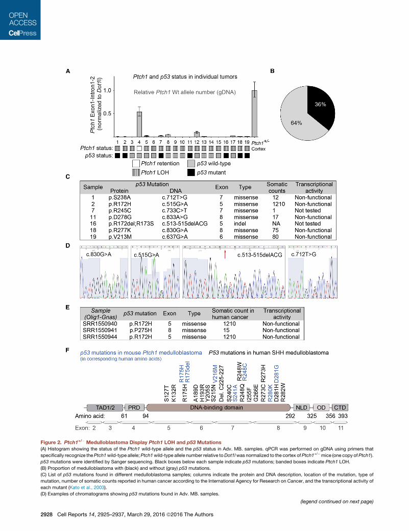
Figure 2. Ptch1+/� Medulloblastoma Display Ptch1 LOH and p53 Mutations
(A) Histogram showing the status of the Ptch1 wild-type allele and the p53 status in Adv. MB. samples. qPCR was performed on gDNA using primers that
specifically recognize thePtch1wild-type allele;Ptch1wild-type allele number relative toDot1lwas normalized to the cortex ofPtch1+/�mice (one copy ofPtch1).
p53 mutations were identified by Sanger sequencing. Black boxes below each sample indicate p53 mutations; banded boxes indicate Ptch1 LOH.
(B) Proportion of medulloblastoma with (black) and without (gray) p53 mutations.
(C) List of p53 mutations found in different medulloblastoma samples; columns indicate the protein and DNA description, location of the mutation, type of
mutation, number of somatic counts reported in human cancer according to the International Agency for Research on Cancer, and the transcriptional activity of
each mutant (Kato et al., 2003).
(D) Examples of chromatograms showing p53 mutations found in Adv. MB. samples.
(legend continued on next page)
2928 Cell Reports 14, 2925–2937, March 29, 2016 ª2016 The Authors
hedgehog signaling (He et al., 2014). Thus, spontaneous p53
mutations are not only present in the Ptch1 model but can also
be found in other Shh medulloblastoma mouse models.
TP53 mutations have recently been reported in human SHH
and WNT primary medulloblastomas (Kool et al., 2014; Zhukova
et al., 2013). Interestingly, the frequency of p53 mutations in the
Ptch1 andOlig1-Gnasmouse models and in human SHHmedul-
loblastomas is comparable (36%–38% in mice versus 13%–
21% in humans); moreover, the nature of the P53 mutations
and their location in the TP53 gene are remarkably similar
(Figure 2F).
p53 Pathway Dysfunction in a Large Proportion ofAdvanced MedulloblastomaSince mutations that compromise the P53 pathway are
frequent in human medulloblastoma (Kool et al., 2014), we
investigated whether some Ptch1 medulloblastomas that do
not bear p53 mutations also exhibit deregulation of the p53
pathway. p53 mutations alter p53 transcriptional activity and
lead to p53 protein accumulation because p53 mutants can
no longer induce the expression of its negative regulator
Mdm2. As expected, when we assessed p53 levels in Ptch1+/�
medulloblastomas, we found nuclear accumulation of p53 in
all advanced tumors bearing missense p53 mutations (Fig-
ure S2A). Remarkably, we also found p53 nuclear accumula-
tion in 50% (3/6) of p53 wild-type medulloblastomas, indi-
cating that the p53 pathway is also altered in tumors without
p53 mutations.
To further test whether p53 signaling is dysfunctional in
advanced medulloblastomas, we evaluated the integrity of p53
transcriptional activity in response to UV radiation in tumor cells
freshly isolated from primary Ptch1+/� medulloblastomas with
and without p53 somatic mutations. We assessed p53 activity
by measuring the mRNA levels of the p53 targets Pai-1, p21,
and Noxa. As expected, p53 mutant medulloblastoma cells
were deficient in upregulating Pai-1, p21, and Noxa in response
to UV (Figure S2B). Interestingly, while many p53 wild-type tu-
mors were capable of strongly activating these p53 target genes,
50% (7/14) of p53 wild-type tumors were deficient in transacti-
vating these key p53 target genes in response to UV (tumors
below themedian dashed line in Figure S2C and encircled in Fig-
ure S2B). Furthermore, when plotted to simultaneously compare
the expression of these three p53 targets, many p53 wild-type
tumors behave similarly to p53 mutant tumors (Figure S2D).
Therefore, one-half of p53 wild-type medulloblastomas display
p53 signaling defects.
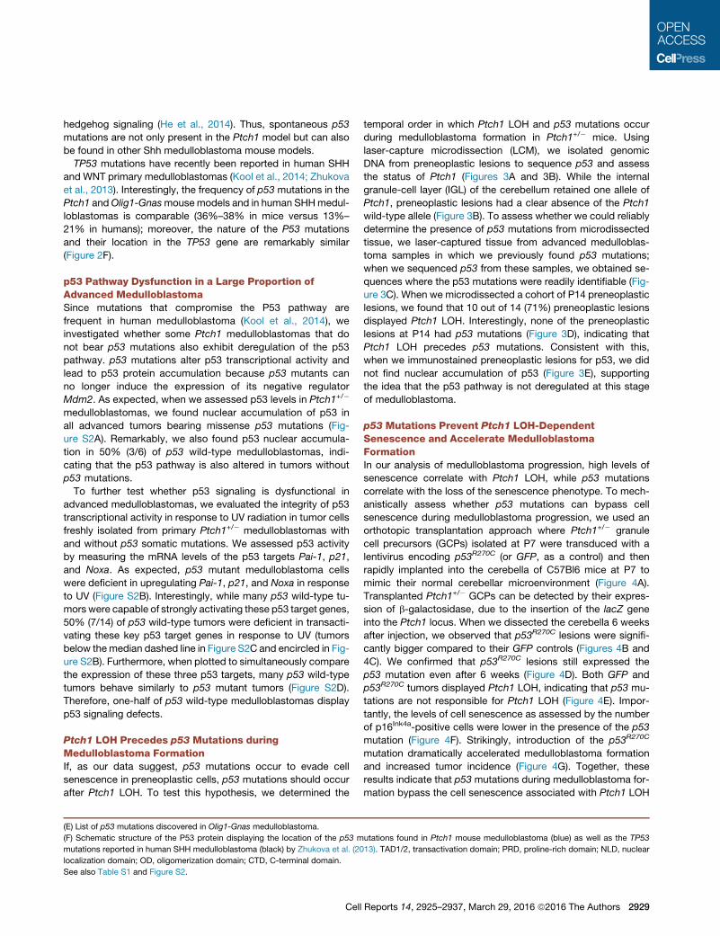
Ptch1 LOH Precedes p53 Mutations duringMedulloblastoma FormationIf, as our data suggest, p53 mutations occur to evade cell
senescence in preneoplastic cells, p53 mutations should occur
after Ptch1 LOH. To test this hypothesis, we determined the
(E) List of p53 mutations discovered in Olig1-Gnas medulloblastoma.
(F) Schematic structure of the P53 protein displaying the location of the p53 m
mutations reported in human SHH medulloblastoma (black) by Zhukova et al. (20
localization domain; OD, oligomerization domain; CTD, C-terminal domain.
See also Table S1 and Figure S2.
Cell
temporal order in which Ptch1 LOH and p53 mutations occur
during medulloblastoma formation in Ptch1+/� mice. Using
laser-capture microdissection (LCM), we isolated genomic
DNA from preneoplastic lesions to sequence p53 and assess
the status of Ptch1 (Figures 3A and 3B). While the internal
granule-cell layer (IGL) of the cerebellum retained one allele of
Ptch1, preneoplastic lesions had a clear absence of the Ptch1
wild-type allele (Figure 3B). To assess whether we could reliably
determine the presence of p53 mutations from microdissected
tissue, we laser-captured tissue from advanced medulloblas-
toma samples in which we previously found p53 mutations;
when we sequenced p53 from these samples, we obtained se-
quences where the p53 mutations were readily identifiable (Fig-
ure 3C). When we microdissected a cohort of P14 preneoplastic
lesions, we found that 10 out of 14 (71%) preneoplastic lesions
displayed Ptch1 LOH. Interestingly, none of the preneoplastic
lesions at P14 had p53 mutations (Figure 3D), indicating that
Ptch1 LOH precedes p53 mutations. Consistent with this,
when we immunostained preneoplastic lesions for p53, we did
not find nuclear accumulation of p53 (Figure 3E), supporting
the idea that the p53 pathway is not deregulated at this stage
of medulloblastoma.
p53 Mutations Prevent Ptch1 LOH-DependentSenescence and Accelerate MedulloblastomaFormationIn our analysis of medulloblastoma progression, high levels of
senescence correlate with Ptch1 LOH, while p53 mutations
correlate with the loss of the senescence phenotype. To mech-
anistically assess whether p53 mutations can bypass cell
senescence during medulloblastoma progression, we used an
orthotopic transplantation approach where Ptch1+/� granule
cell precursors (GCPs) isolated at P7 were transduced with a
lentivirus encoding p53R270C (or GFP, as a control) and then
rapidly implanted into the cerebella of C57Bl6 mice at P7 to
mimic their normal cerebellar microenvironment (Figure 4A).
Transplanted Ptch1+/� GCPs can be detected by their expres-
sion of b-galactosidase, due to the insertion of the lacZ gene
into the Ptch1 locus. When we dissected the cerebella 6 weeks
after injection, we observed that p53R270C lesions were signifi-
cantly bigger compared to their GFP controls (Figures 4B and
4C). We confirmed that p53R270C lesions still expressed the
p53 mutation even after 6 weeks (Figure 4D). Both GFP and
p53R270C tumors displayed Ptch1 LOH, indicating that p53 mu-
tations are not responsible for Ptch1 LOH (Figure 4E). Impor-
tantly, the levels of cell senescence as assessed by the number
of p16Ink4a-positive cells were lower in the presence of the p53
mutation (Figure 4F). Strikingly, introduction of the p53R270C
mutation dramatically accelerated medulloblastoma formation
and increased tumor incidence (Figure 4G). Together, these
results indicate that p53 mutations during medulloblastoma for-
mation bypass the cell senescence associated with Ptch1 LOH
utations found in Ptch1 mouse medulloblastoma (blue) as well as the TP53
13). TAD1/2, transactivation domain; PRD, proline-rich domain; NLD, nuclear
Reports 14, 2925–2937, March 29, 2016 ª2016 The Authors 2929
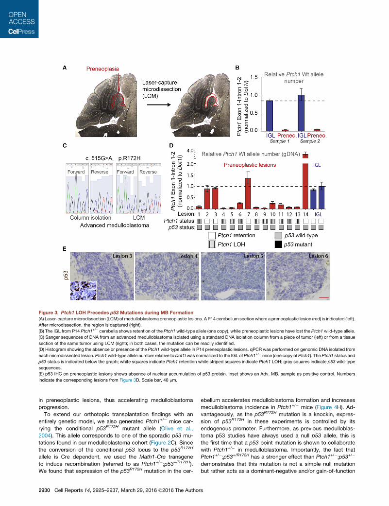
Figure 3. Ptch1 LOH Precedes p53 Mutations during MB Formation
(A) Laser-capturemicrodissection (LCM) of medulloblastoma preneoplastic lesions. A P14 cerebellum sectionwhere a preneoplastic lesion (red) is indicated (left).
After microdissection, the region is captured (right).
(B) The IGL from P14 Ptch1+/� cerebella shows retention of the Ptch1wild-type allele (one copy), while preneoplastic lesions have lost the Ptch1wild-type allele.
(C) Sanger sequences of DNA from an advanced medulloblastoma isolated using a standard DNA isolation column from a piece of tumor (left) or from a tissue
section of the same tumor using LCM (right); in both cases, the mutation can be readily identified.
(D) Histogram showing the absence or presence of the Ptch1 wild-type allele in P14 preneoplastic lesions. qPCR was performed on genomic DNA isolated from
eachmicrodissected lesion. Ptch1wild-type allele number relative toDot1lwas normalized to the IGL of Ptch1+/�mice (one copy of Ptch1). The Ptch1 status and
p53 status is indicated below the graph; white squares indicate Ptch1 retention while striped squares indicate Ptch1 LOH; gray squares indicate p53 wild-type
sequences.
(E) p53 IHC on preneoplastic lesions shows absence of nuclear accumulation of p53 protein. Inset shows an Adv. MB. sample as positive control. Numbers
indicate the corresponding lesions from Figure 3D. Scale bar, 40 mm.
in preneoplastic lesions, thus accelerating medulloblastoma
progression.
To extend our orthotopic transplantation findings with an
entirely genetic model, we also generated Ptch1+/� mice car-
rying the conditional p53R172H mutant allele (Olive et al.,
2004). This allele corresponds to one of the sporadic p53 mu-
tations found in our medulloblastoma cohort (Figure 2C). Since
the conversion of the conditional p53 locus to the p53R172H
allele is Cre dependent, we used the Math1-Cre transgene
to induce recombination (referred to as Ptch1+/�;p53+/R172H).We found that expression of the p53R172H mutation in the cer-
2930 Cell Reports 14, 2925–2937, March 29, 2016 ª2016 The Author
ebellum accelerates medulloblastoma formation and increases
medulloblastoma incidence in Ptch1+/� mice (Figure 4H). Ad-
vantageously, as the p53R172H mutation is a knockin, expres-
sion of p53R172H in these experiments is controlled by its
endogenous promoter. Furthermore, as previous medulloblas-
toma p53 studies have always used a null p53 allele, this is
the first time that a p53 point mutation is shown to collaborate
with Ptch1+/� in medulloblastoma. Importantly, the fact that
Ptch1+/�;p53+/R172H has a stronger effect than Ptch1+/�;p53+/�
demonstrates that this mutation is not a simple null mutation
but rather acts as a dominant-negative and/or gain-of-function
s
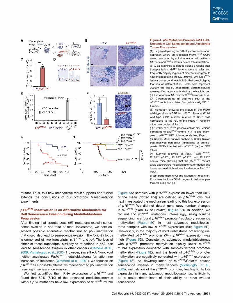
Figure 4. p53Mutations Prevent Ptch1 LOH-
Dependent Cell Senescence and Accelerate
Tumor Progression
(A)Diagramdepicting the orthotopic transplantation
approach where preneoplastic Ptch1+/lacZ GCPs
were transduced by spin-inoculation with either a
GFP or a p53R70C lentivirus before transplantation.
(B) X-gal stainings to detect lesions 6 weeks after
transplantation. GFP+ lesions were smaller and
frequently display regions of differentiated granule
neurons populating the IGL (arrows), while p53R70C
lesions correspond to Adv.MBs that do not display
features of differentiation. Scale bars represent
200 mm (top) and 50 mm (bottom). Bottom pictures
aremagnified regions indicated by theblackboxes.
(C) Tumor area ofGFP and p53R70C lesions (nR 4).
(D) Chromatograms of wild-type p53 or the
p53R70C mutation isolated from advanced p53R70C
tumors.
(E) Histogram showing the status of the Ptch1
wild-type allele in GFP and p53R70C lesions. Ptch1
wild-type allele number relative to Dot1l was
normalized to the IGL of the Ptch1+/+ recipient
mice (two copies of Ptch1).
(F) Number of p16Ink4a-positive cells inGFP lesions
compared to p53R70C tumors (n R 4) and exam-
ples of p16ink4a IHC pictures; scale bar, 20 mm.
(G) Kaplan-Meier survival analysis of C56BL6 mice
that received cerebellar transplants of preneo-
plastic GCPs infected with p53R70C (red) or GFP
(blue).
(H) Survival analysis of Ptch1+/�;p53+/R172H,Ptch1+/�;p53+/�, Ptch1+/�;p53+/+, and Ptch1+/+
control mice showing that the p53R172H mutant
allele accelerates medulloblastoma formation and
increases medulloblastoma incidence in Ptch1+/�
mice.
U test performed in (C) and Student’s t test in (F).
Error bars indicate SEM. Log-rank test was per-
formed in (G) and (H).
mutant. Thus, this new mechanistic result supports and further
extends the conclusions of our orthotopic transplantation
experiments.
p16ink4a Inactivation Is an Alternative Mechanism forCell Senescence Evasion during MedulloblastomaProgressionAfter finding that spontaneous p53 mutations explain senes-
cence evasion in one-third of medulloblastoma, we next as-
sessed possible alternative mechanisms to p53 inactivation
that could also lead to senescence evasion. The Cdkn2a locus
is comprised of two transcripts: p16ink4a and Arf. The loss of
either of these transcripts, similarly to mutations in p53, can
lead to senescence evasion in other cancers (Carnero et al.,
2000; Michaloglou et al., 2005). However, since the Arf knockout
neither accelerates Ptch1+/� medulloblastoma formation nor
increases its incidence (Wetmore et al., 2001), we focused on
p16ink4a as a possible alternative mechanism to p53 inactivation
resulting in senescence evasion.
We first quantified the mRNA expression of p16ink4a and
found that 60% (6/10) of the advanced medulloblastomas
without p53 mutations have low expression of p16ink4a mRNA
Cell
(Figure 5A; samples with p16ink4a expression lower than 50%
of the mean [dotted line] are defined as p16ink4a low). We
next investigated the mechanism leading to this low expression
of p16ink4a. We did not detect gene copy-number changes
in p16ink4a (exon 1a of Cdkn2a) (Figure 5B); in addition, we
did not find p16ink4a mutations. Interestingly, using bisulfite
sequencing, we found p16ink4a promoter/regulatory sequence
methylation (Figure 5C) in most advanced medulloblas-
toma samples with low p16ink4a expression (5/6; Figure 5D).
Conversely, in the majority of medulloblastoma presenting un-
methylated p16ink4a promoter (3/4), p16ink4a expression was
high (Figure 5D). Consistently, advanced medulloblastomas
with p16ink4a promoter methylation display lower p16ink4a
mRNA expression compared with samples without promoter
methylation (Figure 5E), and the levels of p16ink4a promoter
methylation are negatively correlated with p16ink4a expression
(Figure 5F). As downregulation of p16ink4a/Cdkn2a causes
senescence evasion in many cancers (Michaloglou et al.,
2005), methylation of the p16ink4a promoter, leading to its low
expression in many advanced medulloblastomas, is likely to
be a major determinant of their ability to have evaded
senescence.
Reports 14, 2925–2937, March 29, 2016 ª2016 The Authors 2931
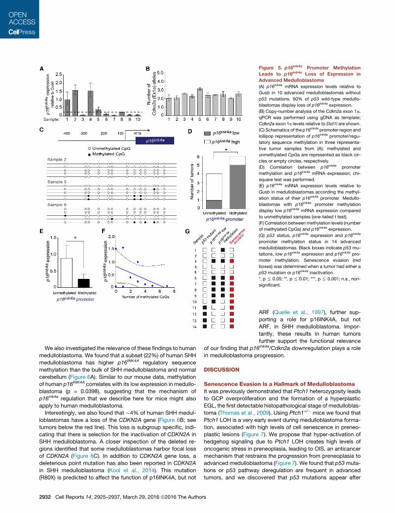
Figure 5. p16ink4a Promoter Methylation
Leads to p16ink4a Loss of Expression in
Advanced Medulloblastoma
(A) p16ink4a mRNA expression levels relative to
Gusb in 10 advanced medulloblastomas without
p53 mutations. 60% of p53 wild-type medullo-
blastomas display loss of p16ink4a expression.
(B) Copy-number analysis of the Cdkn2a exon 1a.
qPCR was performed using gDNA as template;
Cdkn2a exon 1a levels relative to Dot1l are shown.
(C) Schematics of the p16ink4a promoter region and
lollipop representation of p16ink4a promoter/regu-
latory sequence methylation in three representa-
tive tumor samples from (A); methylated and
unmethylated CpGs are represented as black cir-
cles or empty circles, respectively.
(D) Correlation between p16ink4a promoter
methylation and p16ink4a mRNA expression; chi-
square test was performed.
(E) p16ink4a mRNA expression levels relative to
Gusb in medulloblastomas according the methyl-
ation status of their p16ink4a promoter. Medullo-
blastomas with p16ink4a promoter methylation
display low p16ink4a mRNA expression compared
to unmethylated samples (one-tailed t test).
(F) Correlation betweenmethylation levels (number
of methylated CpGs) and p16ink4a expression.
(G) p53 status, p16ink4a expression and p16ink4a
promoter methylation status in 14 advanced
medulloblastomas. Black boxes indicate p53 mu-
tations, low p16ink4a expression and p16ink4a pro-
moter methylation. Senescence evasion (red
boxes) was determined when a tumor had either a
p53 mutation or p16ink4a inactivation.
*, p % 0.05; **, p % 0.01; ***, p % 0.001; n.s., non-
significant.
We also investigated the relevance of these findings to human
medulloblastoma. We found that a subset (22%) of human SHH
medulloblastoma has higher p16INK4A regulatory sequence
methylation than the bulk of SHH medulloblastoma and normal
cerebellum (Figure 6A). Similar to our mouse data, methylation
of human p16INK4A correlates with its low expression in medullo-
blastoma (p = 0.0398), suggesting that the mechanism of
p16ink4a regulation that we describe here for mice might also
apply to human medulloblastoma.
Interestingly, we also found that �4% of human SHH medul-
loblastomas have a loss of the CDKN2A gene (Figure 6B; see
tumors below the red line). This loss is subgroup specific, indi-
cating that there is selection for the inactivation of CDKN2A in
SHH medulloblastoma. A closer inspection of the deleted re-
gions identified that some medulloblastomas harbor focal loss
of CDKN2A (Figure 6C). In addition to CDKN2A gene loss, a
deleterious point mutation has also been reported in CDKN2A
in SHH medulloblastoma (Kool et al., 2014). This mutation
(R80X) is predicted to affect the function of p16INK4A, but not
2932 Cell Reports 14, 2925–2937, March 29, 2016 ª2016 The Authors
ARF (Quelle et al., 1997), further sup-
porting a role for p16INK4A, but not
ARF, in SHH medulloblastoma. Impor-
tantly, these results in human tumors
further support the functional relevance
of our finding that p16ink4a/Cdkn2a downregulation plays a role
in medulloblastoma progression.
DISCUSSION
Senescence Evasion Is a Hallmark of MedulloblastomaIt was previously demonstrated that Ptch1 heterozygosity leads
to GCP overproliferation and the formation of a hyperplastic
EGL, the first detectable histopathological stage of medulloblas-
toma (Thomas et al., 2009). Using Ptch1+/� mice we found that
Ptch1 LOH is a very early event during medulloblastoma forma-
tion, associated with high levels of cell senescence in preneo-
plastic lesions (Figure 7). We propose that hyper-activation of
hedgehog signaling due to Ptch1 LOH creates high levels of
oncogenic stress in preneoplasia, leading to OIS, an anticancer
mechanism that restrains the progression from preneoplasia to
advanced medulloblastoma (Figure 7). We found that p53muta-
tions or p53 pathway deregulation are frequent in advanced
tumors, and we discovered that p53 mutations appear after
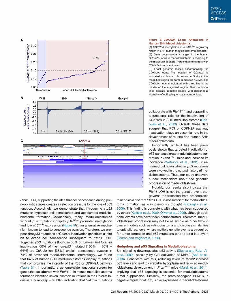
Figure 6. CDKN2A Locus Alterations in
Human SHH Medulloblastoma
(A) CDKN2A methylation at a p16ink4a regulatory
region in SHH human medulloblastoma samples.
(B) Gene copy-number changes in the human
CDKN2A locus in medulloblastoma, according to
the molecular subtype. Percentage of tumors with
CDKN2A loss is indicated.
(C) Focal genomic losses encompassing the
CDKN2A locus. The location of CDKN2A is
indicated on human chromosome 9 (top); this
magnified region (bottom) comprises 4.3 Mb. The
CDKN2A gene is indicated with a red line in the
middle of the magnified region. Blue horizontal
lines indicate genomic losses, with darker blue
intensity reflecting higher copy-number loss.
Ptch1 LOH, supporting the idea that cell senescence during pre-
neoplastic stages creates a selection pressure for the loss of p53
function. Accordingly, we demonstrate that introducing a p53
mutation bypasses cell senescence and accelerates medullo-
blastoma formation. Additionally, many medulloblastomas
without p53 mutations display p16ink4a promoter methylation
and low p16ink4a expression (Figure 5G), an alternative mecha-
nism known to lead to senescence evasion. Therefore, we pro-
pose that p53mutations orCdkn2a inactivation constitute a third
hit to evade cell senescence subsequent to Ptch1 LOH.
Together, p53 mutations (found in 36% of tumors) and Cdkn2a
inactivation (60% of the non-p53 mutated [100% � 36% =
64%] are Cdkn2a low [38%]) explain senescence evasion in
74% of advanced medulloblastoma. Interestingly, we found
that 64% of human SHH medulloblastomas display mutations
that compromise the integrity of the P53 or CDKN2A pathway
(Table S1). Importantly, a genome-wide functional screen for
genes that collaborate with Ptch1+/� in mouse medulloblastoma
formation identified seven insertion mutations in the Cdkn2a lo-
cus in 85 tumors (p = 0.0067), indicating that Cdkn2a mutations
Cell Reports 14, 2925–2937
collaborate with Ptch1+/� and supporting
a functional role for the inactivation of
CDKN2A in SHH medulloblastoma (Gen-
ovesi et al., 2013). Overall, these data
suggest that P53 or CDKN2A pathway
inactivation plays an essential role in the
development of murine and human SHH
medulloblastoma.
Importantly, while it has been previ-
ously shown that targeted inactivation of
p53 can accelerate medulloblastoma for-
mation in Ptch1+/� mice and increase its
incidence (Wetmore et al., 2001), it re-
mained unknown whether p53 mutations
were involved in the natural history of me-
dulloblastoma. Thus, our study uncovers
a new mechanism about the genomic
progression of medulloblastoma.
Notably, our results also indicate that
Ptch1 LOH is not the genetic event that
governs the transition from preneoplasia
to neoplasia and thatPtch1 LOH is not sufficient formedulloblas-
toma formation, as was previously thought (Pazzaglia et al.,
2006). This finding is consistent with what has been suggested
by others (Kessler et al., 2009; Oliver et al., 2005), although addi-
tional events have never been demonstrated. Therefore, medul-
loblastoma progression may not be as simple as other two-hit
cancer models such as retinoblastoma and displays similarities
to epithelial cancers, where multiple genetic events are required
for tumor formation and p53 mutations tend to be a late event
(Fearon and Vogelstein, 1990).
Hedgehog and p53 Signaling in MedulloblastomaShh signaling downregulates p53 activity (Stecca and Ruiz i Al-
taba, 2009), possibly by Gli1 activation of Mdm2 (Abe et al.,
2008). Consistent with this, reducing levels of Mdm2 increase
p53 levels and lead to cerebellar hypoplasia and reducedmedul-
loblastoma development in Ptch1+/� mice (Malek et al., 2011),
implying that p53 signaling is essential for medulloblastoma
tumor suppression. Similarly, the proto-oncogene PPM1D, a
negative regulator of P53, is overexpressed inmedulloblastomas
, March 29, 2016 ª2016 The Authors 2933
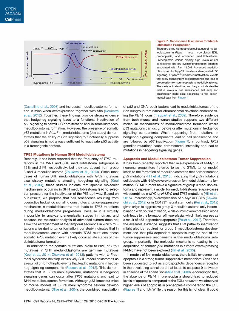
Figure 7. Senescence Is a Barrier for Medul-
loblastoma Progression
There are three histopathological stages of medul-
loblastoma in Ptch1+/� mice: hyperplastic EGL,
preneoplasia, and advanced medulloblastoma.
Preneoplastic lesions display high levels of cell
senescence and low levels of proliferation, changes
associated with Ptch1 LOH. Advanced medullo-
blastomas display p53mutations, deregulated p53
signaling, or p16ink4a promoter methylation, events
that allow escape from cell senescence and lead to
progression frompreneoplasia tomedulloblastoma.
Thexaxis indicates time, and theyaxis indicates the
relative levels of cell senescence (left axis) and
proliferation (right axis) according to the experi-
mental data from Figure 1.
(Castellino et al., 2008) and increases medulloblastoma forma-
tion in mice when overexpressed together with Shh (Doucette
et al., 2012). Together, these findings provide strong evidence
that hedgehog signaling leads to a functional inactivation of
p53 signaling to permit GCP proliferation and, in some instances,
medulloblastoma formation. However, the presence of somatic
p53 mutations in Ptch1+/� medulloblastoma (this study) demon-
strates that the ability of Shh signaling to functionally suppress
p53 signaling is not always sufficient to inactivate p53 activity
in a tumorigenic context.
TP53 Mutations in Human SHH MedulloblastomaRecently, it has been reported that the frequency of TP53 mu-
tations in the WNT and SHH medulloblastoma subgroups is
16% and 21%, respectively, but they are absent from group
3 and 4 medulloblastoma (Zhukova et al., 2013). Since most
cases of human SHH medulloblastoma with TP53 mutations
also display mutations affecting hedgehog signaling (Kool
et al., 2014), these studies indicate that specific molecular
mechanisms occurring in SHH medulloblastoma lead to selec-
tion pressure for the inactivation of the P53 pathway. Based on
our results, we propose that cell senescence resulting from
overactive hedgehog signaling constitutes a tumor-suppressive
mechanism in medulloblastoma that leads to TP53 mutations
during medulloblastoma progression. Because it is virtually
impossible to analyze preneoplastic stages in human, and
because the molecular analysis of advanced tumors does not
allow the establishment of the temporal sequence in which mu-
tations arise during tumor formation, our study indicates that in
medulloblastoma cases with somatic TP53 mutations, these
somatic TP53 mutation events likely occur at late stages of me-
dulloblastoma formation.
In addition to the somatic mutations, close to 50% of TP53
mutations in SHH medulloblastoma are germline mutations
(Kool et al., 2014; Zhukova et al., 2013); patients with Li-Frau-
meni syndrome develop exclusively SHH medulloblastomas as
a result of chromothriptic events that lead to mutations in hedge-
hog signaling components (Rausch et al., 2012). This demon-
strates that in Li-Fraumeni syndrome, mutations in hedgehog
signaling genes can occur after TP53 mutations and lead to
SHH medulloblastoma formation. Although p53 knockout mice
or mouse models of Li-Fraumeni syndrome seldom develop
medulloblastoma (Olive et al., 2004), the combined inactivation
2934 Cell Reports 14, 2925–2937, March 29, 2016 ª2016 The Author
of p53 and DNA repair factors lead to medulloblastomas of the
Shh subgroup that harbor chromosomal deletions encompass-
ing the Ptch1 locus (Frappart et al., 2009). Therefore, evidence
from both mouse and human studies supports two different
molecular mechanisms of medulloblastoma formation where
p53 mutations can occur before or after mutations in hedgehog
signaling components. When happening first, mutations in
hedgehog signaling components lead to cell senescence and
are followed by p53 inactivation (Figure 7); in contrast, TP53
germline mutations cause chromosomal instability and lead to
mutations in hedgehog signaling genes.
Apoptosis and Medulloblastoma Tumor SuppressionIt has been recently reported that mis-expression of N-Myc in
neuronal progenitors (referred to as the GTML tumor model)
leads to the formation of medulloblastomas that harbor somatic
p53 mutations (Hill et al., 2015), indicating that p53 mutations
collaborate with N-Myc overexpression formedulloblastoma for-
mation. GTML tumors have a signature of group 3 medulloblas-
toma and represent a model for medulloblastoma relapse cases
with combined c-MYC or N-MYC and TP53mutations (Hill et al.,
2015). Interestingly, overexpression of c-Myc in GCPs (Kawau-
chi et al., 2012) or in CD133+ neural stem cells (Pei et al., 2012)
gives origin to aggressive group 3medulloblastoma only in com-
bination with p53 inactivation, while c-Myc overexpression alone
only leads to the formation of hyperplasia, which likely regress as
a result of p53-dependent apoptosis (Pei et al., 2012). Therefore,
the available evidence suggests that P53 pathway inactivation
might also be required for group 3 medulloblastoma develop-
ment and that p53-dependent apoptosis may be one of the
tumor-suppressive mechanisms in this medulloblastoma sub-
group. Importantly, the molecular mechanisms leading to the
acquisition of somatic p53 mutations in tumors overexpressing
N-Myc have not been explored (Hill et al., 2015).
In models of Shh medulloblastoma, there is little evidence that
apoptosis is a strong tumor-suppressive mechanism. Ptch1 has
been suggested to act as a proapoptotic dependence receptor
in the developing spinal cord that leads to caspase-9 activation
in absence of the ligand Shh (Mille et al., 2009). According to this,
the absence of Ptch1 in preneoplasia should lead to reduced
levels of apoptosis compared to the EGL; however, we observed
higher levels of apoptosis in preneoplasia compared to the EGL
(Figures 1I and 1J). While the reason for this is not clear, it could
s
be due to tissue-specific differences between spinal cord and
cerebellum.
The fact that we did not observe changes in apoptosis levels
as preneoplastic lesions progress from preneoplasia to
advanced medulloblastoma (Figures 1I and 1J) supports the
notion that apoptosis is not an essential tumor-suppressive bar-
rier to Shh medulloblastoma progression. Consistent with this,
human SHH medulloblastomas display a paucity of mutations
involved in apoptosis (Kool et al., 2014). Therefore, cell senes-
cence appears to be amore important tumor-suppressivemech-
anism than apoptosis for Shh medulloblastoma, while the
converse might be true for group 3medulloblastoma. Consistent
with this, p53 mutations are known to have context-specific
effects. For example, it has been shown that in tumors such
as lymphomas p53 mutations abrogate apoptosis but not cell
senescence, while in sarcomas p53 mutations abrogate cell
senescence but not apoptosis (Ventura et al., 2007). The fact
that p53 dysfunction does not reduce apoptosis in Shh medullo-
blastoma suggests that p53 preferentially regulates cell senes-
cence over apoptosis in these tumors.
Cell Senescence Is a Tumor-Suppressive Mechanismfor MedulloblastomaHere, we provide evidence that medulloblastoma preneoplastic
lesions display a senescent phenotype that limits their prolif-
erative capacity and their progression to advanced tumors.
Although we describe this senescence response as a form of
OIS, cell senescence in medulloblastoma preneoplasia likely re-
sults from the oncogenic stress subsequent to the loss of Ptch1,
a tumor suppressor gene; analogously, loss of PTEN, another
tumor suppressor, has been shown to induce senescence in
prostate cancer (Chen et al., 2005). Loss of Ptch1 leads to hyper-
activation of hedgehog signaling, which can lead to high levels of
N-Myc and CyclinD1, two candidates that can mediate Ptch1
LOH-dependent oncogenic stress and senescence. Because
Ptch1 has also been shown to interact with and to sequester
CyclinB1 at the cell membrane (Barnes et al., 2001), the
increased nuclear availability of CyclinB1 after Ptch1 LOH may
also lead to oncogenic stress.
Basal cell carcinoma (BCC), a skin tumor caused by deregula-
tion of hedgehog signaling, is characterized by mutations in
PTCH1 (Gailani et al., 1996). Similar to medulloblastoma,
PTCH1 LOH is required for BCC formation (Gailani et al.,
1996). Importantly, TP53 mutations occur in BCCs together
with PTCH1 mutations (Zhang et al., 2001) even in the presence
of PTCH1 LOH. This indicates that p53 pathway inactivation also
cooperates with hedgehog signaling deregulation during BCC
formation. However, whether TP53 mutations precede or follow
PTCH1 LOH in BCC remains unknown (Ling et al., 2001). In view
of our data, we speculate that at least in some cases, BCC pre-
neoplastic lesions may experience cell senescence, leading to
P53 inactivation.
In conclusion, our data suggest that cell senescence in medul-
loblastoma preneoplastic lesions creates selection pressure for
p53 and p16ink4a inactivation. We propose that human SHH
medulloblastomas as well as BCCs with PTCH1 mutations
may experience similar molecular changes that also lead to
TP53 mutations and senescence evasion. In addition to TP53
Cell
and CDKN2A, mutations in other genes important for cell senes-
cence such as P63 and ATM have also been reported in SHH
medulloblastoma (Kool et al., 2014), further supporting the idea
that cell senescence constitutes a tumorigenesis barrier during
medulloblastoma precancerous stages.
EXPERIMENTAL PROCEDURES
Additional experimental procedures for qRT-PCR, bioinformatic analyses,
virus preparation, orthotopic transplantation, and bisulfite sequencing can
be found in Supplemental Experimental Procedures.
Mouse Lines
All animal work was performed according to the Canadian Council on Animal
Care guidelines. Ptch1+/lacZ (referred to as Ptch1+/�) mice (Goodrich et al.,
1997) were backcrossed with C57BL/6 mice (Harlan). p53R172H mutant mice
(p53LSL-R172H) were described previously (Olive et al., 2004).
Immunohistochemistry
12-mm cryosections were processed for immunohistochemistry or immunoflu-
orescence as described previously (Izzi et al., 2011; Mille et al., 2014). Anti-
bodies and dilutions are described in Supplemental Experimental Procedures.
p53 sequencing
To screen for p53mutations, we isolated genomicDNA fromadvancedmedullo-
blastomas and sequenced the full coding frame of the p53 gene. Each sequence
was inspected tofindmutations. IndelsweredetectedusingCodonCodeAligner.
See Supplemental Experimental Procedures for primer sequences.
Laser Capture Microdissection
Preneoplastic lesions were microdissected using an Arcturus Laser Capture
microscope (Life Technologies). 12-mm cerebellum cryosections were
collected on Super Frost slides (Fisher). After dehydrating the tissue, the areas
of interest were captured on CapSure High Sensitivity (HS) caps using the
infrared laser. DNA was isolated from the lesions using a DNA Pico Pure Isola-
tion kit (Life Technologies).
Assessment of the Status of the Ptch1 Wild-Type Allele
We performed qPCR on total genomic DNA isolated using a DNeasy Kit
(QIAGEN). The levels of the wild-type allele of Ptch1 relative to Dot1l were
normalized to the IGL (for preneoplasia; Figure 3) or to the cortex (for advanced
medulloblastoma; Figure 2) of Ptch1 mice (one copy of Ptch1) and were ob-
tained using the delta-delta-CT method and Sybrgreen reagents on a Viia 7
system (Life Technologies); reactions were carried in triplicate, and the amount
of gDNA per reaction was 20 ng. We designed primers recognizing Ptch1
Exon1-Intron1-2. Since this region is deleted in the Ptch1mutant allele (Good-
rich et al., 1997), these primers are specific for the wild-type allele of Ptch1.
SUPPLEMENTAL INFORMATION
Supplemental Information includes Supplemental Experimental Procedures,
two figures, and one table and can be found with this article online at http://
dx.doi.org/10.1016/j.celrep.2016.02.061.
AUTHOR CONTRIBUTIONS
F.C. and L.T.-O. designed the study and wrote the article. N.B., C.-L.W., A.K.,
S.M.S., and L.T.-O. conducted the experiments. M.R., P.S., and M.D.T. per-
formed bioinformatics analyses of human data.
ACKNOWLEDGMENTS
We thank Frederic Mille, Nada Jabado, Daniel Durocher, Francis Rodier, Ger-
ardo Ferbeyre, and Elliot Drobetsky for helpful advice and Patricia T. Yam for
comments on the manuscript. We thank J. Barthe for animal husbandry and J.
Cardin and J.-F. Michaud for technical expertise. We thank A. Dumont and O.
Reports 14, 2925–2937, March 29, 2016 ª2016 The Authors 2935
Neyret for help with bisulfite sequencing. We thank Tyler Jacks and the NCI
Animal Repository for kindly providing the p53LSL-R172H mice. This work was
supported by grants from the Canadian Institutes of Health Research
(CIHR), the Fonds de Recherche du Quebec-Sante (FRQS), and the Canada
Foundation for Innovation (CFI). L.T.-O. is recipient of the Caldas fellowship
(Colciencias). F.C. holds the Canada Research Chair in Developmental
Neurobiology.
Received: August 3, 2015
Revised: December 28, 2015
Accepted: February 11, 2016
Published: March 17, 2016
REFERENCES
Abe, Y., Oda-Sato, E., Tobiume, K., Kawauchi, K., Taya, Y., Okamoto, K.,
Oren, M., and Tanaka, N. (2008). Hedgehog signaling overrides p53-mediated
tumor suppression by activatingMdm2. Proc. Natl. Acad. Sci. USA 105, 4838–
4843.
Ayrault, O., Zindy, F., Rehg, J., Sherr, C.J., and Roussel, M.F. (2009). Two
tumor suppressors, p27Kip1 and patched-1, collaborate to prevent medullo-
blastoma. Mol. Cancer Res. 7, 33–40.
Barnes, E.A., Kong, M., Ollendorff, V., and Donoghue, D.J. (2001). Patched1
interacts with cyclin B1 to regulate cell cycle progression. EMBO J. 20,
2214–2223.
Braig, M., Lee, S., Loddenkemper, C., Rudolph, C., Peters, A.H., Schlegel-
berger, B., Stein, H., Dorken, B., Jenuwein, T., and Schmitt, C.A. (2005). Onco-
gene-induced senescence as an initial barrier in lymphoma development.
Nature 436, 660–665.
Carnero, A., Hudson, J.D., Price, C.M., and Beach, D.H. (2000). p16INK4A and
p19ARF act in overlapping pathways in cellular immortalization. Nat. Cell Biol.
2, 148–155.
Castellino, R.C., De Bortoli, M., Lu, X., Moon, S.H., Nguyen, T.A., Shepard,
M.A., Rao, P.H., Donehower, L.A., and Kim, J.Y. (2008). Medulloblastomas
overexpress the p53-inactivating oncogene WIP1/PPM1D. J. Neurooncol.
86, 245–256.
Chen, Z., Trotman, L.C., Shaffer, D., Lin, H.K., Dotan, Z.A., Niki, M., Koutcher,
J.A., Scher, H.I., Ludwig, T., Gerald, W., et al. (2005). Crucial role of p53-
dependent cellular senescence in suppression of Pten-deficient tumorigen-
esis. Nature 436, 725–730.
Doucette, T.A., Yang, Y., Pedone, C., Kim, J.Y., Dubuc, A., Northcott, P.D.,
Taylor, M.D., Fults, D.W., and Rao, G. (2012). WIP1 enhances tumor formation
in a sonic hedgehog-dependent model of medulloblastoma. Neurosurgery 70,
1003–1010, discussion 1010.
Fearon, E.R., and Vogelstein, B. (1990). A genetic model for colorectal tumor-
igenesis. Cell 61, 759–767.
Frappart, P.O., Lee, Y., Russell, H.R., Chalhoub, N., Wang, Y.D., Orii, K.E.,
Zhao, J., Kondo, N., Baker, S.J., and McKinnon, P.J. (2009). Recurrent
genomic alterations characterize medulloblastoma arising from DNA double-
strand break repair deficiency. Proc. Natl. Acad. Sci. USA 106, 1880–1885.
Gailani, M.R., Leffell, D.J., Ziegler, A., Gross, E.G., Brash, D.E., and Bale, A.E.
(1996). Relationship between sunlight exposure and a key genetic alteration in
basal cell carcinoma. J. Natl. Cancer Inst. 88, 349–354.
Genovesi, L.A., Ng, C.G., Davis, M.J., Remke, M., Taylor, M.D., Adams, D.J.,
Rust, A.G., Ward, J.M., Ban, K.H., Jenkins, N.A., et al. (2013). Sleeping Beauty
mutagenesis in a mouse medulloblastoma model defines networks that
discriminate between human molecular subgroups. Proc. Natl. Acad. Sci.
USA 110, E4325–E4334.
Goodrich, L.V., Milenkovi�c, L., Higgins, K.M., and Scott, M.P. (1997). Altered
neural cell fates and medulloblastoma in mouse patched mutants. Science
277, 1109–1113.
He, X., Zhang, L., Chen, Y., Remke,M., Shih, D., Lu, F., Wang, H., Deng, Y., Yu,
Y., Xia, Y., et al. (2014). The G protein a subunit Gas is a tumor suppressor in
Sonic hedgehog-driven medulloblastoma. Nat. Med. 20, 1035–1042.
2936 Cell Reports 14, 2925–2937, March 29, 2016 ª2016 The Author
Hill, R.M., Kuijper, S., Lindsey, J.C., Petrie, K., Schwalbe, E.C., Barker, K.,
Boult, J.K., Williamson, D., Ahmad, Z., Hallsworth, A., et al. (2015). Combined
MYC and P53 defects emerge at medulloblastoma relapse and define rapidly
progressive, therapeutically targetable disease. Cancer Cell 27, 72–84.
Hovestadt, V., Jones, D.T., Picelli, S., Wang, W., Kool, M., Northcott, P.A., Sul-
tan, M., Stachurski, K., Ryzhova, M., Warnatz, H.J., et al. (2014). Decoding the
regulatory landscape of medulloblastoma using DNAmethylation sequencing.
Nature 510, 537–541.
Izzi, L., Levesque, M., Morin, S., Laniel, D., Wilkes, B.C., Mille, F., Krauss, R.S.,
McMahon, A.P., Allen, B.L., and Charron, F. (2011). Boc and Gas1 each form
distinct Shh receptor complexes with Ptch1 and are required for Shh-medi-
ated cell proliferation. Dev. Cell 20, 788–801.
Kato, S., Han, S.Y., Liu,W., Otsuka, K., Shibata, H., Kanamaru, R., and Ishioka,
C. (2003). Understanding the function-structure and function-mutation rela-
tionships of p53 tumor suppressor protein by high-resolution missense muta-
tion analysis. Proc. Natl. Acad. Sci. USA 100, 8424–8429.
Kawauchi, D., Robinson, G., Uziel, T., Gibson, P., Rehg, J., Gao, C., Finkel-
stein, D., Qu, C., Pounds, S., Ellison, D.W., et al. (2012). A mouse model of
the most aggressive subgroup of human medulloblastoma. Cancer Cell 21,
168–180.
Kessler, J.D., Hasegawa, H., Brun, S.N., Emmenegger, B.A., Yang, Z.J., Dut-
ton, J.W., Wang, F., and Wechsler-Reya, R.J. (2009). N-myc alters the fate of
preneoplastic cells in a mouse model of medulloblastoma. Genes Dev. 23,
157–170.
Kool, M., Jones, D.T., Jager, N., Northcott, P.A., Pugh, T.J., Hovestadt, V.,
Piro, R.M., Esparza, L.A., Markant, S.L., Remke, M., et al.; ICGC PedBrain Tu-
mor Project (2014). Genome sequencing of SHH medulloblastoma predicts
genotype-related response to smoothened inhibition. Cancer Cell 25,
393–405.
Ling, G., Ahmadian, A., Persson, A., Unden, A.B., Afink, G.,Williams, C., Uhlen,
M., Toftgard, R., Lundeberg, J., and Ponten, F. (2001). PATCHED and p53
gene alterations in sporadic and hereditary basal cell cancer. Oncogene 20,
7770–7778.
Malek, R., Matta, J., Taylor, N., Perry, M.E., and Mendrysa, S.M. (2011). The
p53 inhibitor MDM2 facilitates Sonic Hedgehog-mediated tumorigenesis and
influences cerebellar foliation. PLoS ONE 6, e17884.
Michaloglou, C., Vredeveld, L.C., Soengas, M.S., Denoyelle, C., Kuilman, T.,
van der Horst, C.M., Majoor, D.M., Shay, J.W., Mooi, W.J., and Peeper, D.S.
(2005). BRAFE600-associated senescence-like cell cycle arrest of human
naevi. Nature 436, 720–724.
Mille, F., Thibert, C., Fombonne, J., Rama, N., Guix, C., Hayashi, H., Corset, V.,
Reed, J.C., and Mehlen, P. (2009). The Patched dependence receptor triggers
apoptosis through a DRAL-caspase-9 complex. Nat. Cell Biol. 11, 739–746.
Mille, F., Tamayo-Orrego, L., Levesque,M., Remke,M., Korshunov, A., Cardin,
J., Bouchard, N., Izzi, L., Kool, M., Northcott, P.A., et al. (2014). The Shh recep-
tor Boc promotes progression of early medulloblastoma to advanced tumors.
Dev. Cell 31, 34–47.
Narita, M., and Lowe, S.W. (2005). Senescence comes of age. Nat. Med. 11,
920–922.
Olive, K.P., Tuveson, D.A., Ruhe, Z.C., Yin, B., Willis, N.A., Bronson, R.T.,
Crowley, D., and Jacks, T. (2004). Mutant p53 gain of function in two mouse
models of Li-Fraumeni syndrome. Cell 119, 847–860.
Oliver, T.G., Read, T.A., Kessler, J.D., Mehmeti, A., Wells, J.F., Huynh, T.T.,
Lin, S.M., and Wechsler-Reya, R.J. (2005). Loss of patched and disruption
of granule cell development in a pre-neoplastic stage of medulloblastoma.
Development 132, 2425–2439.
Pazzaglia, S., Tanori, M., Mancuso, M., Gessi, M., Pasquali, E., Leonardi, S.,
Oliva, M.A., Rebessi, S., Di Majo, V., Covelli, V., et al. (2006). Two-hit model
for progression of medulloblastoma preneoplasia in Patched heterozygous
mice. Oncogene 25, 5575–5580.
Pei, Y., Moore, C.E., Wang, J., Tewari, A.K., Eroshkin, A., Cho, Y.J., Witt, H.,
Korshunov, A., Read, T.A., Sun, J.L., et al. (2012). An animal model of
MYC-driven medulloblastoma. Cancer Cell 21, 155–167.
s
Pugh, T.J., Weeraratne, S.D., Archer, T.C., Pomeranz Krummel, D.A., Auclair,
D., Bochicchio, J., Carneiro, M.O., Carter, S.L., Cibulskis, K., Erlich, R.L., et al.
(2012). Medulloblastoma exome sequencing uncovers subtype-specific
somatic mutations. Nature 488, 106–110.
Quelle, D.E., Cheng, M., Ashmun, R.A., and Sherr, C.J. (1997). Cancer-associ-
ated mutations at the INK4a locus cancel cell cycle arrest by p16INK4a but not
by the alternative reading frame protein p19ARF. Proc. Natl. Acad. Sci. USA
94, 669–673.
Rausch, T., Jones, D.T., Zapatka, M., St€utz, A.M., Zichner, T., Weischenfeldt,
J., Jager, N., Remke, M., Shih, D., Northcott, P.A., et al. (2012). Genome
sequencing of pediatric medulloblastoma links catastrophic DNA rearrange-
ments with TP53 mutations. Cell 148, 59–71.
Robinson, G., Parker, M., Kranenburg, T.A., Lu, C., Chen, X., Ding, L., Phoenix,
T.N., Hedlund, E., Wei, L., Zhu, X., et al. (2012). Novel mutations target distinct
subgroups of medulloblastoma. Nature 488, 43–48.
Stecca, B., and Ruiz i Altaba, A. (2009). A GLI1-p53 inhibitory loop controls
neural stem cell and tumour cell numbers. EMBO J. 28, 663–676.
Taylor, M.D., Northcott, P.A., Korshunov, A., Remke, M., Cho, Y.J., Clifford,
S.C., Eberhart, C.G., Parsons, D.W., Rutkowski, S., Gajjar, A., et al. (2012).
Molecular subgroups of medulloblastoma: the current consensus. Acta Neu-
ropathol. 123, 465–472.
Cell
Thomas, W.D., Chen, J., Gao, Y.R., Cheung, B., Koach, J., Sekyere, E., Norris,
M.D., Haber, M., Ellis, T., Wainwright, B., and Marshall, G.M. (2009). Patched1
deletion increases N-Myc protein stability as a mechanism of medulloblas-
toma initiation and progression. Oncogene 28, 1605–1615.
Uziel, T., Zindy, F., Xie, S., Lee, Y., Forget, A., Magdaleno, S., Rehg, J.E., Cal-
abrese, C., Solecki, D., Eberhart, C.G., et al. (2005). The tumor suppressors
Ink4c and p53 collaborate independently with Patched to suppress medullo-
blastoma formation. Genes Dev. 19, 2656–2667.
Ventura, A., Kirsch, D.G., McLaughlin, M.E., Tuveson, D.A., Grimm, J., Lintault,
L., Newman, J., Reczek, E.E.,Weissleder, R., and Jacks, T. (2007). Restoration
of p53 function leads to tumour regression in vivo. Nature 445, 661–665.
Wetmore, C., Eberhart, D.E., and Curran, T. (2001). Loss of p53 but not ARF
accelerates medulloblastoma in mice heterozygous for patched. Cancer
Res. 61, 513–516.
Zhang, H., Ping, X.L., Lee, P.K., Wu, X.L., Yao, Y.J., Zhang, M.J., Silvers, D.N.,
Ratner, D., Malhotra, R., Peacocke, M., and Tsou, H.C. (2001). Role of PTCH
and p53 genes in early-onset basal cell carcinoma. Am. J. Pathol. 158,
381–385.
Zhukova, N., Ramaswamy, V., Remke, M., Pfaff, E., Shih, D.J., Martin, D.C.,
Castelo-Branco, P., Baskin, B., Ray, P.N., Bouffet, E., et al. (2013). Sub-
group-specific prognostic implications of TP53 mutation in medulloblastoma.
J. Clin. Oncol. 31, 2927–2935.
Reports 14, 2925–2937, March 29, 2016 ª2016 The Authors 2937
Cell Reports, Volume 14
Supplemental Information
Evasion of Cell Senescence
Leads to Medulloblastoma Progression
Lukas Tamayo-Orrego, Chia-Lun Wu, Nicolas Bouchard, Ahmed Khedher, Shannon M.Swikert, Marc Remke, Patryk Skowron, Michael D. Taylor, and Frédéric Charron

Figure S1 (Related to Figure 1). p21 and p16ink4a are co-expressed in non-proliferating preneoplastic cells. The EGL of the cerebellum does not express p16ink4a or p19Arf. (A) p21 and p16ink4a immunofluorescence of preneoplastic lesions shows that p16ink4a is expressed in cells that are positive for p21. (B) Ki67 and p16ink4a immunostaining shows that proliferating cells (Ki67+) are negative for p16ink4a. (C) mRNA expression levels of Gli1, Ptch1, Boc, Cdon, CyclinD1, p19Arf, p16ink4a, Bmp3 and IL6 relative to Gusb in the EGL of the cerebellum at postnatal day 7. Gli1, Ptch1, Boc and CyclinD1 are highly expressed in the EGL, but not Cdon (previously characterized to not be expressed in EGL (Izzi et al., 2011)). Similarly, p19Arf, p16ink4a, BMP3 (as a negative control), and IL6 (as a negative control) are not expressed in the EGL.
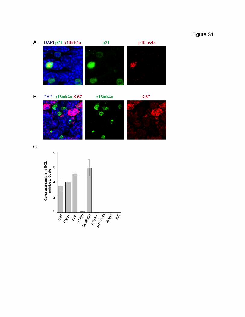

Table S1 (Related to Figure 2). P53 and CDKN2A pathway mutations in human SHH medulloblastoma. This table lists the genes belonging to the P53 and CDKN2A pathways that were found mutated by Kool et al. (2014) in human SHH medulloblastoma. Abbreviations: * In cases where a gene is found mutated several times, the asterisk indicates the most frequent or most deleterious mutation. Not reported: data not provided. Not applicable (na): the mutation is located outside the coding region. Unknown: information not provided.
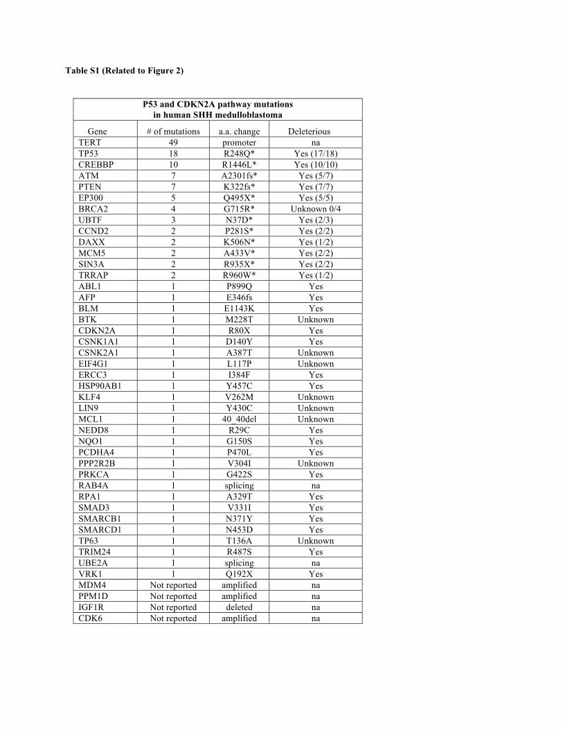
Table S1 (Related to Figure 2)
P53 and CDKN2A pathway mutations in human SHH medulloblastoma
Gene # of mutations a.a. change Deleterious TERT 49 promoter na TP53 18 R248Q* Yes (17/18) CREBBP 10 R1446L* Yes (10/10) ATM 7 A2301fs* Yes (5/7) PTEN 7 K322fs* Yes (7/7) EP300 5 Q495X* Yes (5/5) BRCA2 4 G715R* Unknown 0/4 UBTF 3 N37D* Yes (2/3) CCND2 2 P281S* Yes (2/2) DAXX 2 K506N* Yes (1/2) MCM5 2 A433V* Yes (2/2) SIN3A 2 R935X* Yes (2/2) TRRAP 2 R960W* Yes (1/2) ABL1 1 P899Q Yes AFP 1 E346fs Yes BLM 1 E1143K Yes BTK 1 M228T Unknown CDKN2A 1 R80X Yes CSNK1A1 1 D140Y Yes CSNK2A1 1 A387T Unknown EIF4G1 1 L117P Unknown ERCC3 1 I384F Yes HSP90AB1 1 Y457C Yes KLF4 1 V262M Unknown LIN9 1 Y430C Unknown MCL1 1 40_40del Unknown NEDD8 1 R29C Yes NQO1 1 G150S Yes PCDHA4 1 P470L Yes PPP2R2B 1 V304I Unknown PRKCA 1 G422S Yes RAB4A 1 splicing na RPA1 1 A329T Yes SMAD3 1 V331I Yes SMARCB1 1 N371Y Yes SMARCD1 1 N453D Yes TP63 1 T136A Unknown TRIM24 1 R487S Yes UBE2A 1 splicing na VRK1 1 Q192X Yes MDM4 Not reported amplified na PPM1D Not reported amplified na IGF1R Not reported deleted na CDK6 Not reported amplified na

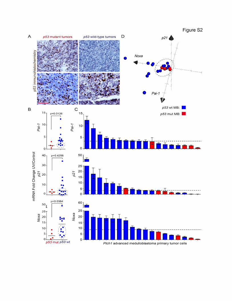
Figure S2 (Related to Figure 2). p53 pathway dysfunction in a large proportion of advanced medulloblastomas. (A) p53 IHC staining in p53 mutant (left) and p53 wild-type (right) Ptch1 medulloblastoma samples. (B) Pai-1, p21 and Noxa mRNA fold change in UV-treated relative to control samples for p53 mutant and p53 wild-type tumors; lines indicate the median. Dashed circles indicate p53 wild-type samples that behave like p53 mutant samples, i.e. lie in the inter-quartile range of the p53 mutant samples. The UV dose was 60 J/m2 UV, and total RNA was isolated 24h after UV treatment. (C) Fold change of Pai-1, p21 and Noxa mRNA levels between UV-treated and control primary medulloblastoma tumor cells; dashed lines indicate the median. (D) Three-dimensional graph of Pai-1, p21 and Noxa mRNA fold change for each tumor shows that 50% (7/14) of p53 wild-type medulloblastomas cluster close to p53 mutant tumors. Scale bar (A), 50 µm. Mann-Whitney test was performed in B.

Supplemental Experimental Procedures Antibody information Antibody Catalog Number/Company Dilution P21 ab7960 Abcam 1:200 p16Ink4a ab54210 Abcam 1:200 Ki67 550609 BD Pharmingen 1:100 Phospho histone H3 (ser10) 06-570 Millipore 1:1000 Cleaved-caspase 3 (D175) 9661S Cell Signaling 1:1000 P53 FL393 Santa Cruz 1:100 Plasmids and constructs The murine p53 cDNA (pMXs-p53) (Addgene plasmid 22725 (Hong et al., 2009)) was amplified by PCR; the product was digested using NheI and EcoRI and subcloned into the lentiviral vector pPrime-CMV (Stegmeier et al., 2005). Packaging vectors (pLp1, pLp2 and pVSVG) were purchased from Invitrogen. Mouse p53 sequencing, primer sequences p53-Ex2 For: TACTGGATGTCCCACCTTCT p53-Ex2 Rev: TTACAGACACCCAACACCATAC p53-Ex3-4 For (Ampl. only): CCTTAAGATACATCCCGCCATAC p53-Ex3-4 Rev: AGGTCACACGAAAGACAACTC p53-Ex3-4 F (Seq): CAGCCTGGGATAAGTGAGATTC p53-Ex5-6 For: CCT TGA CAC CTG ATC GTT ACT C p53-Ex5-6 Rev: TCT CCC AGA GAC TGC TGT TA p53-Ex7-9 For: TGC CGA ACA GGT GGA ATA TC p53-Ex7-9 Rev: GCG AGA GAC AGA GGC AAT AAT p53-Ex10 For: AAATCGTGAAAGTGGTTGTGTG p53-Ex10 Rev: CAAGAAGTCAGTTCTCGTAGGG p53-Ex11 For: GTGTTCTGTGAATATCCCTACCC p53-Ex11 Rev: AGACCTGACAACTATCAACCTATTC Primers to assess Ptch1 loss of heterozygosity Dot1l F: TAG TTG GCA TCC TTA TGC TTC ATC Dot1l R: GCC CCA GCA CGA CCA TT Ptch1 ex1 int1-2 F: CCT TCG CTC TGG AGC AGA TT Ptch1 ex1 int1-2 R: GGA TCC CAA GGA GGA AGA AGA Virus preparation Lentiviruses were generated by cotransfecting 6 µg of lentiviral vector, 5.7 µg of pLp1, 2.7 µg of pLp2 and 3.8 µg of pVSVG packaging vector into 293T cells in 10 cm dishes using PEI (Sigma). Supernatants were collected 48h and 72h after transfection, centrifuged for 5 min to remove cell debris and then filtered through a 0.4 µm membrane. 20 ml of PEG6000-NaCl solution was added to the supernatant in a 1:5 ratio (PEG6000-NaCl:virus supernatant) and the mix was incubated O/N at 4 °C. The PEG6000/virus mixture was further centrifuged at 3500 rpm to precipitate the virus (Kutner et al., 2009). After removal of the PEG solution, precipitates were resuspended in 200 µl PBS. After a last 5 min centrifuge step (12000 rpm), the virus was ready to use. Viral transduction of GCPs The virus titer (MOI, multiplicity of infection) was determined by infecting 293T cells prior to GCP infection. GCP cells were transduced with lentivirus (MOI=10) by spin-inoculation (2200 rpm) for 30 min (O'Doherty et al., 2000). Infected Ptch1+/- GCPs were spun down (890 rpm, 5 min), resuspended in Neurobasal medium and kept at 4 °C until transplantation. Orthotopic transplantation Post-natal day 7 wild-type C57BL6 pups were anaesthetized by hypothermia for 5 min. A volume of 2 µl containing 1x10⁶ Ptch1+/- GCPs previously transduced with GFP or p53R270C lentivirus was injected into the cerebellum of each
recipient pup using a Picospritzer injector (Science Products, GmbH.). The pups recovered from procedure 20 min post-injection and their health status was monitored weekly until symptoms of medulloblastoma were evident. qRT-PCR qRT-PCR on p53 target genes was performed as described (Mille et al., 2014). Primers for Noxa, p21 and Pai-1 were described previously (Boley et al., 2002; Chao et al., 2003; McEachron et al., 2010). Analysis of the Cdkn2a locus Determination of p16ink4a expression in advanced MB samples (Figure 5A) was conducted using qRT-PCR and specific primers for p16ink4a (Cdkn2a exon 1α). Relative p16ink4a expression was normalized to Gusb. The p16ink4a primer sequences for expression were described previously (Yamakoshi et al., 2009). The primer sequences used to detect copy number changes in Cdkn2a (Figure 5B) are: mCdkn2a ex1a-intr1/2 For: GATTCAGGTAGGCAAGGAGTG mCdkn2a ex1a-intr1/2 Rev: GGCAGCAGCAACAACAAA qPCR using 10ng gDNA per sample was carried out as previously described for the Ptch1 wild-type allele. Bisulfite sequencing Bisulfite conversion of total gDNA from advanced MB (1µg per sample) was carried out using the EZ DNA Methylation-Lightning Kit (Zymo) according to manufacturer instructions. The p16ink4a promoter region was amplified using the ZymoTaq PreMix and the following primers designed using Methyl Primer Express, Fwd: TATTTTTAGAGGAAGGAAGGAG; Rev: AAATAAAACACTCCTTACCTACCT. PCR fragments were cloned into the pCR4-Topo TA vector for Sequencing (Thermo Fisher); five clones per fragment were sequenced using the vector M13 sites. Bioinformatics analysis CDKN2A locus analysis in human medulloblastoma Analysis of human CDKN2A copy number variation was generated using Affymetrix Genome-wide Human SNP Array 6.0 with hg18 as the reference genome (Northcott et al. (2012). The human CDKN2A/p16INK4a methylation analysis was generated from methylation data found in Hovestadt et al. (2014). TP53 and CDKN2A Pathway Mutations in human SHH Medulloblastoma We obtained a list of genes found mutated in human SHH medulloblastomas according to Kool et al. (2014). The lists for somatic and germline mutations were merged. The germline mutations file included 28 samples with recurrent mutations in 8 unique genes. The somatic mutations file included 128 samples with 1989 mutations in 1169 unique genes. The merged file included 131 samples with 1171 unique genes mutated. TERT promoter was found mutated in 59.76% (49/82) of samples. We also included the 18 genes that were somatically altered by copy number variations: PPM1D, MYCN, LMO4, MYCL1, MDM4, PIK3C2B GLI2, CDK6, YAP1, CCND2, IRS2, IGF1R, PTCH1, PTEN, SUFU, DDX41, TP53, LDB1. The sample IDs in which these CNV occurred were not available; therefore we could not obtain the frequency of mutations in these genes. This final list contained 1182 unique genes. We obtained a list of genes involved in the P53 signaling pathway and the CDKN2A pathway in an unbiased manner according to the following two databases:
• Human Protein Reference Database: TP53 interactions, CDKN2A interactions • Ingenuity Pathway Analysis: TP53 binding, CDKN2A binding, CDKN2A activity
The merged TP53 interactors contained 361 unique genes, 41 (11.36%) of which were found mutated in human SHH medulloblastoma in Kool et al. (2014). The merged CDKN2A interactors contained 88 genes, 9 (10.23%) of which were found mutated in human SHH medulloblastoma in Kool et al. (2014). Merging the TP53 and CDKN2A interactor lists together results in 412 genes, 44 (10.68%) of which are mutated in human SHH medulloblastoma.
We then examined which samples had at least one mutation involved with TP53 or CDKN2A pathway. This gave us the frequency (64%) of SHH medulloblastomas with at least one mutation compromising the TP53 or CDKN2a pathway. The deleterious potential of the mutations was evaluated using the PROVEAN (Protein Variation Effect Analyzer) software. This predicts if an amino acid change in a protein is deleterious and also gives a second prediction based on the SIFT algorithm. A mutation was classified as deleterious if the mutation was nonsense (frameshift and stopgain) or was predicted deleterious by at least one of the 2 algorithms. 71 (78.02%) mutations were predicted deleterious, 16 (17.58%) were predicted neutral and 4 (4.40%) were annotated as splicing variants not predictable by this algorithm. To calculate the incidence of p53 mutations in Olig1-Gnas murine medulloblastomas, we analyzed the GSE53248 dataset from (He et al., 2014). Fastq files were aligned to the mouse genome of reference (mm10) using STAR. Picard-tools were used to remove duplicates. Mutations were called with GenomeAnalysisTK (GATK) HaplotypeCaller and annotated with SnpEff. Candidate mutations in p53 were carefully examined in Integrative Genomics Viewer (IGV) to rule out misalignments and other false positives.
Supplemental references Boley, S. E., Wong, V. A., French, J. E., and Recio, L. (2002). p53 heterozygosity alters the mRNA expression of p53 target genes in the bone marrow in response to inhaled benzene. Toxicol Sci 66, 209-215.
Chao, C., Hergenhahn, M., Kaeser, M. D., Wu, Z., Saito, S., Iggo, R., Hollstein, M., Appella, E., and Xu, Y. (2003). Cell type- and promoter-specific roles of Ser18 phosphorylation in regulating p53 responses. The Journal of biological chemistry 278, 41028-41033.
He, X., Zhang, L., Chen, Y., Remke, M., Shih, D., Lu, F., Wang, H., Deng, Y., Yu, Y., Xia, Y., et al. (2014). The G protein alpha subunit Galphas is a tumor suppressor in Sonic hedgehog-driven medulloblastoma. Nat Med 20, 1035-1042.
Hong, H., Takahashi, K., Ichisaka, T., Aoi, T., Kanagawa, O., Nakagawa, M., Okita, K., and Yamanaka, S. (2009). Suppression of induced pluripotent stem cell generation by the p53-p21 pathway. Nature 460, 1132-1135.
Hovestadt, V., Jones, D. T., Picelli, S., Wang, W., Kool, M., Northcott, P. A., Sultan, M., Stachurski, K., Ryzhova, M., Warnatz, H. J., et al. (2014). Decoding the regulatory landscape of medulloblastoma using DNA methylation sequencing. Nature 510, 537-541.
Izzi, L., Levesque, M., Morin, S., Laniel, D., Wilkes, B. C., Mille, F., Krauss, R. S., McMahon, A. P., Allen, B. L., and Charron, F. (2011). Boc and Gas1 each form distinct Shh receptor complexes with Ptch1 and are required for Shh-mediated cell proliferation. Developmental cell 20, 788-801.
Kool, M., Jones, D. T., Jager, N., Northcott, P. A., Pugh, T. J., Hovestadt, V., Piro, R. M., Esparza, L. A., Markant, S. L., Remke, M., et al. (2014). Genome sequencing of SHH medulloblastoma predicts genotype-related response to smoothened inhibition. Cancer cell 25, 393-405.
Kutner, R. H., Zhang, X. Y., and Reiser, J. (2009). Production, concentration and titration of pseudotyped HIV-1-based lentiviral vectors. Nat Protoc 4, 495-505.
McEachron, T. A., Pawlinski, R., Richards, K. L., Church, F. C., and Mackman, N. (2010). Protease-activated receptors mediate crosstalk between coagulation and fibrinolysis. Blood 116, 5037-5044.
Mille, F., Tamayo-Orrego, L., Levesque, M., Remke, M., Korshunov, A., Cardin, J., Bouchard, N., Izzi, L., Kool, M., Northcott, P. A., et al. (2014). The Shh receptor Boc promotes progression of early medulloblastoma to advanced tumors. Developmental cell 31, 34-47.
Northcott, P. A., Shih, D. J., Peacock, J., Garzia, L., Morrissy, A. S., Zichner, T., Stutz, A. M., Korshunov, A., Reimand, J., Schumacher, S. E., et al. (2012). Subgroup-specific structural variation across 1,000 medulloblastoma genomes. Nature 488, 49-56.
O'Doherty, U., Swiggard, W. J., and Malim, M. H. (2000). Human immunodeficiency virus type 1 spinoculation enhances infection through virus binding. J Virol 74, 10074-10080.
Stegmeier, F., Hu, G., Rickles, R. J., Hannon, G. J., and Elledge, S. J. (2005). A lentiviral microRNA-based system for single-copy polymerase II-regulated RNA interference in mammalian cells. Proceedings of the National Academy of Sciences of the United States of America 102, 13212-13217.
Yamakoshi, K., Takahashi, A., Hirota, F., Nakayama, R., Ishimaru, N., Kubo, Y., Mann, D. J., Ohmura, M., Hirao, A., Saya, H., et al. (2009). Real-time in vivo imaging of p16Ink4a reveals cross talk with p53. The Journal of cell biology 186, 393-407.