evidence for a novel route of wheat storage proteins to vacuoles
TRANSCRIPT
Evidence for a Novel Route of Wheat Storage Proteins to Vacuoles H a n n a Levanony, Regina Rubin , Yoram Altschuler, and G a d Galili
Department of Plant Genetics, The Weizmarm Institute of Science, Rehovot 76 100, Israel
Abstract. Wheat seed storage proteins are deposited in protein bodies (PB) inside vacuoles, but their sub- cellular site of aggregation and their route to vacuoles are still controversial. In the present work, an ultra structural analysis of developing wheat endosperm at early to mid maturation was performed to address these issues. Golgi complexes were rarely detected, indicat- ing that their role in wheat storage protein transport is limited. In contrast, a considerable amount of PB was detected in the cytoplasm. Many of these PB were sur- rounded by PER membranes and were enlarged by fusion of smaller PB. Small, electron lucent vesicles were detected around the surfaces of the PB in the cy- toplasm, or attached to them, suggesting that such
attachments and subsequent fusion of the vesicles with each other lead to the formation of small vacuoles con- taining PB inclusions. Immunogold labeling with serum raised against yeast-BiP, an ER-localized protein, dem- onstrated that the wheat BiP homolog was present within the PB in the cytoplasm as well as inside vacu- oles. This confirmed that the PB were formed within the RER and that the Golgi complex was not involved in their transport to vacuoles. It is concluded that a considerable part of the wheat storage proteins aggre- gate into PB within the RER and are then transported as intact PB to the vacuoles by a novel route that does not utilize the Golgi complex.
p LANT seed storage proteins are synthesized during seed development and accumulate in protein bodies (PB) t. Yet, similar types of storage proteins from
different plants accumulate in different subcellular locations. The major storage proteins in legumes are salt-soluble glob- ulins. These globulins accumulate in vacuoles and are trans- ported to this organelle via the Golgi complex (Greenwood and Chrispeels, 1985; Shotwell and Larkins, 1988). In most cereals, the major storage proteins are alcohol-soluble prola- mins, yet, the subcellular site of their accumulation appears to differ between species. The prolamins of maize and rice were shown to aggregate into dense PB directly within the RER and remain attached to this organelle (Kfishnan et al., 1986; Larkins and Hurkman, 1978; Shotwell and Larkins, 1988). However, in some cereals such as wheat and oats, the prolamins were shown to accumulate in vacuoles, similarly to the legume globulins (Shotwell and Larkins, 1988).
The subcellular route of prolamins to vacuoles has been studied extensively in wheat. Electron micrographs of de- veloping wheat endosperms have shown vesicles containing prolamins nearby to Golgi complexes (Bechtel and Barnet, 1986; Campbell et al., 1981; Kim et al., 1988; Parker, 1982; Parker and Hawes, 1982). These authors suggested that wheat prolamins are transported to vacuoles via the Golgi
1. Abbreviations used in this paper: DAA, days after anthesis; PB, protein bodies.
apparatus, and condense into PB in vesicles budding off this apparatus. However, in some electron micrographs, small PB have been also observed within or in the vicinity of RER membranes in the cytoplasm, suggesting that at least some prolamins aggregate into PB within the RER (Campbell et al., 1981; Parker, 1982). Deposition of wheat storage pro- teins within the ER was also suggested by subcellular frac- tionation analysis (Miflin et al., 1981). Whether the PB formed within the RER are then transported to vacuoles via the Golgi complex has not yet been elucidated. A similar sit- uation exists in oats. Although oat prolamins are localized within vacuoles, electron micrographs of developing oat en- dosperm have indicated that at least some of these storage proteins aggregate directly within the RER (Lending et al., 1989).
Eukaryotic cells contain a group of luminal, ER-resident proteins that function in the folding and transport of secre- tory proteins (Pelham, 1989). These proteins contain a specific amino acid sequence at their COOH-terminal end, mostly KDEL or HDEL, which operates as an ER retention signal and prevents their transport via the Golgi apparatus (Pelham, 1990). One of the ER resident proteins, BiP (Ig heavy chain binding protein), was first identified to bind to the heavy chains of Igs in the ER of Pre B ceils and non- secreting hybridoma where the Ig light polypeptide chain was not expressed (Bole et al., 1986; Haas and Wabl, 1983; Munro and Pelham, 1986). BiP was later shown to interact transiently with many normal secretory proteins, as well as
�9 The Rockefeller University Press, 0021-9525/92/12/1117/12 $2.00 The Journal of Cell Biology, Volume 119, Number 5, December 1992 1117-1128 1117
on January 4, 2019jcb.rupress.org Downloaded from http://doi.org/10.1083/jcb.119.5.1117Published Online: 1 December, 1992 | Supp Info:
permanently with proteins that fail to fold correctly and re- tain within the ER (Pelham, 1989 and references therein).
We have studied the transport of wheat prolamins to vacu- oles to ascertain if it occurs entirely via the Golgi complex or also via an alternative route that does not use this appara- tus. As wheat prolamins are not glycosylated one can not trace their transport via the Golgi complex by looking for Golgi-specific sugar modifications. We thus selected BiP as a marker protein for this study. If wheat storage proteins are transported to the vacuoles entirely via the Golgi complex and apparently aggregate into PB at a post-ER location (Kim et al., 1988), then it is expected that BiP will not be present in them as it is not transported via the Golgi complex. How- ever, if storage proteins aggregate into PB within the RER and are translocated as intact PB directly to vacuoles, then BiP may be trapped in these PB. EM analyses showed that a large part of wheat prolamins aggregated into PB within the RER and were transported to vacuoles by a novel route that did not use the Golgi complex. This was confirmed by demonstrating that BiP was present in these PB.
Materials and Methods
Production of Antibodies New Zealand White rabbits were injected subcutaneously three times at 3-wk intervals with 300 #g purified 3,-gliadin produced in Escherichia coli (Altschuler, Y., and G. Galili, manuscript in preparation). The 3,-gliadin was emulsified in 3 M urea and 50% Freund's complete adjuvant. Serum was obtained 10 d after the third injection. Antibodies raised against yeast BiP (Rose et al., 1989) were kindly provided by Dr. J. P. Vogel.
Protein Fractionation and Western Blot Analysis Extraction of wheat gliadins and their fractionation on acidic PAGE were performed as described by Lafiandra and Kasarda (1985). Fractionation of proteins on 10% SDS-PAGE was according to Laemmli (1970). For West- ern blot analysis, proteins were transferred to 0.2-#m nitrocellulose filters and immunoblotted with the desired antisera according to Towbin et ai. (1979). Anti-3"-gliadin or anti-yeast BiP sera were used at dilutions of 1:10,000 and 1:1,500, respectively. Goat anti-rabbit IgG conjugated to alka- line phosphatase was used as the secondary antibody.
Preparation of Developing Grains for EM Analysis and Immunogold Labeling Triticum aestivum var. aestivum cv. Chinese Spring and Triticum turgidum vat. dicoccum accession TTC-17 were grown in pots in a greenhouse. De- veloping grains were removed from the plants at 13 and 16 d after anthesis (DAA) and immediately immersed in a freshly prepared fixation solution containing 4% glntaraldehyde, 2 % paraformaldehyde, 1% acrolein, 2.5 % DMSO, and 5 mM CaC12 in 100 mM Na-cacodylate buffer, pH 7.2. The embryos and most of the grain integuments were removed and the grains were further sliced into small ,~l-mm 3 cubes. Vacuum infiltration in a desiccator was employed to ensure penetration of the fixative. Fixation was performed overnight in a rotary shaker at room temperature and subse- quently samples could be kept at 4~ The samples were rinsed in the same buffer and post-fixed at room temperature for 1-2 h with 2% (wt/vol) os- mium tetroxide in the same buffer. Samples were rinsed first in buffer, then in glass distilled water and then dehydrated in ethanol series (50, 70, and 90%) for 2 x 10 min each, in 100% ethanol for 2 x 15 min, and then em- bedded in LR white, hard. Thin sections (90-100 urn) were prepared with a diamond knife (Diatome, Bienne, Switzerland) and attached to collodion coated nickel grids (200 mesh, Polysciences, Inc., Warrington, PA).
Sections were stained first with 2 % (wt/vol) uranyl acetate for 30-60 min and then with 2% (wt/vol) lead citrate and examined with a Phllips EM-410 transmission electron microscope (Eindhoven, Holland).
The entire immunogold labeling procedure was carried out at room tem- perature. Grids were passed from drop to drop in a moist atmosphere. Non-
specific binding was blocked for 15 rain with a blocking solution containing 0.1% gelatin, 0.1% glycine, 1% BSA, 1% Tween 20 in 0.1 M PBS. The grids were then incubated for 90 rain with either anti-ff-gliadin or anti-yeast-BiP sera diluted 1:500 and 1:50, respectively, and then rinsed 5 x 5 rain in PBS. Excess solution was removed and the grids were incubated with secondary antibody goat anti-rabbit IgG conjugated to colloidal gold (Auroprobe-EM, GAR-G15; Janssen, Belgium) diluted 1:20 in the blocking solution for 30 min. The sections were washed 2 x 5 rain in PBS followed by glass-distilled water, and then post-stained with 2 % uranyl acetate for 30-60 rain.
Cytochemical Controls The controls consisted of (a) incubation of the sections with goat anti-rabbit-gold conjugate without previous incubation with primary anti- bodies; and (b) incubation with pre-immune sera of the gliadins and BiP diluted 1:500 and 1:50, respectively.
Isolation of Dense PB from Developing Grains Developing grains at 16 DAA were homogenized in buffer B (20 mM Tris, pH 7.6, 50 mM KC1, 10 mM MgCi2, 0.3 M NaCI, 2 mM EDTA, 10% su- crose), centrifuged for 5 rain at 500 g and the supernatant fraction was layered on 4.5 ml of continuous 10 to 50 % gradient of metrizamide in buffer B containing 15% sucrose (Wallace et al., 1988). Centrifugation was per- formed for 18 h at 275,000 g and 4~ Dense PB sedimented toward the bottom of the gradient, were visualized by their opaque white color, and collected. For storage protein extraction, the PB fraction was brought to 70% ethanol, 1%/~-mercaptoethanol, and incubated for 30 min at 60~ The samples were centrifuged for 15 rain at 14,000 rpm in an Eppendorf centrifuge and the volume of the supernatant fraction was reduced 50% by Speedvac centrifugation. 1 ml of 0.3 M NaCI was added to the supernatant fraction and storage proteins were collected by overnight incubation at 4~ and precipitation. For fractionation of total proteins, fractions were diluted 4• in buffer B containing 15 % sucrose and particles were collected by cen- trifugation at 27,000 g 4~ for 30 min.
Results
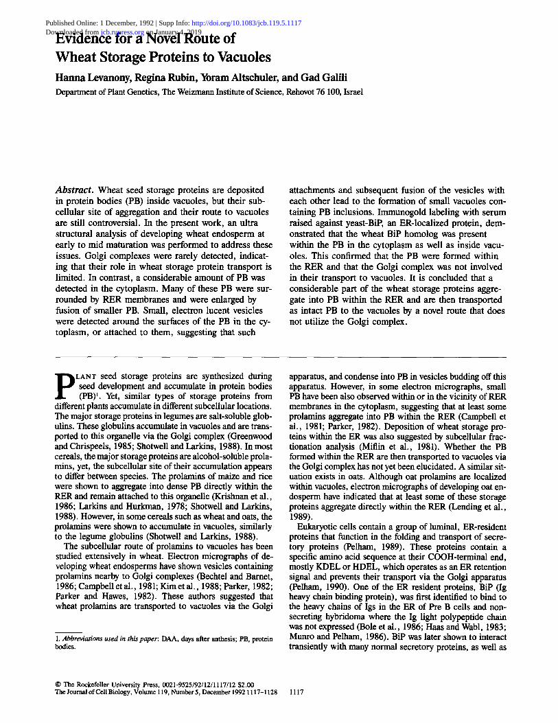
Production of Antibodies against 3,-Gliadins To detect PB in EM sections by immunogold labeling, serum was raised against a wheat prolamin from the type of 7-gli- adins that was produced in E. coli. This serum recognized specifically several gliadin bands on SDS-PAGE which were not recognized by the preimmune serum (Fig. 1 A). When the gliadins were fractionated on acidic PAGE, the serum recognized specifically the 7-gliadins (Fig. 1 B). This "y-gli- adin specific serum was suited for immunological detection of all types of PB in the wheat endosperm cells as immuno- gold labeling of EM sections from wheat endosperm with se- rum raised against different types of prolamins (Kim et al., 1988), or against total wheat prolamins (Levanony, H., and G. Galili, unpublished data) yielded identical results. The preimmune serum did not produce any detectable gold label- ing after being applied to sections of wheat endosperm (see Fig. 3, c and d and Fig. 5 c). In contrast, the immune serum labeled specifically various PB-containing storage proteins, similarly to the immunogold labeling in a previous report (Kim et al., 1988).
Ultrastructure of Wheat Endosperm Cells Endosperms from developing grains of hexaploid (Triticum aestivum) and tetraploid (Triticum dicoccum) wheats were analyzed. Developing grains were harvested at 13 and 16 DAA, representing relatively early to intermediate stages in the synthesis and deposition of the storage proteins. In general, both hexaploid and tetraploid wheats had similar
The Journal of Cell Biology, Volume 119, 1992 1118
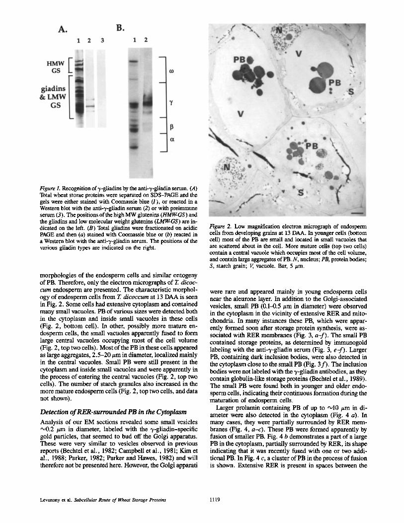
Figure L Recognition of 3,-gliadins by the anti-3,-gliadin serum. (A) Total wheat storae proteins were separated on SDS-PAGE and the gels were either stained with Coomassie blue (1), or reacted in a Western blot with the anti-),-gliadin serum (2) or with preimmune serum (3). The positions of the high MW glutenins (HMIgGS) and the gliadins and low molecular weight glutenins (LMIgGS) are in- dicated on the left. (B) Total gliadins were fractionated on acidic PAGE and then (a) stained with Coomassie blue or (b) reacted in a Western blot with the anti-~/-gliadin serum. The positions of the various gliadin types are indicated on the right.
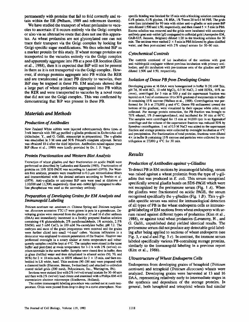
morphologies of the endosperm cells and similar ontogeny of PB. Therefore, only the electron micrographs of T. dicoc- cum endosperm are presented. The characteristic morphol- ogy of endosperm cells from T. dicoccum at 13 DAA is seen in Fig. 2. Some cells had extensive cytoplasm and contained many small vacuoles. PB of various sizes were detected both in the cytoplasm and inside small vacuoles in these cells (Fig. 2, bottom cell). In other, possibly more mature en- dosperm cells, the small vacuoles apparently fused to form large central vacuoles occupying most of the cell volume (Fig. 2, top two cells). Most of the PB in these cells appeared as large aggregates, 2.5-20/zm in diameter, localized mainly in the central vacuoles. Small PB were still present in the cytoplasm and inside small vacuoles and were apparently in the process of entering the central vacuoles (Fig. 2, top two cells). The number of starch granules also increased in the more mature endosperm cells (Fig. 2, top two ceils, and data not shown).
Detection of RER-surrounded PB in the Cytoplasm
Analysis of our EM sections revealed some small vesicles •0.2 #m in diameter, labeled with the ~,-gliadin-specific gold particles, that seemed to bud off the Golgi apparatus. These were very similar to vesicles observed in previous reports (Bechtel et al., 1982; Campbell et al., 1981; Kim et al., 1988; Parker, 1982; Parker and Hawes, 1982) and will therefore not be presented here. However, the Golgi apparati
Figure 2. Low magnification electron micrograph of endosperm cells from developing grains at 13 DAA. In younger cells (bottom cell) most of the PB are small and located in small vacuoles that are scattered about in the cell. More mature cells (top two cells) contain a central vacuole which occupies most of the cell volume, and contain large aggregates of PB. N, nucleus; PB, protein bodies; S, starch grain; V, vacuole. Bar, 5 #m.
were rare and appeared mainly in young endosperm cells near the aleurone layer. In addition to the Golgi-associated vesicles, small PB (0.1-0.5 #m in diameter) were observed in the cytoplasm in the vicinity of extensive RER and mito- chondria. In many instances these PB, which were appar- ently formed soon after storage protein synthesis, were as- sociated with RER membranes (Fig. 3, a-f). The small PB contained storage proteins, as determined by immunogold labeling with the anti-3,-gliadin serum (Fig. 3, e-f). Larger PB, containing dark inclusion bodies, were also detected in the cytoplasm close to the small PB (Fig. 3 f ) . The inclusion bodies were not labeled with the'y-ghadin antibodies, as they contain globulin-like storage proteins (Bechtel et al., 1989). The small PB were found both in younger and older endo- sperm cells, indicating their continuous formation during the maturation of endosperm cells.
Larger prolamin containing PB of up to ,,o10 #m in di- ameter were also detected in the cytoplasm (Fig. 4 a). In many cases, they were partially surrounded by RER mem- branes (Fig. 4, a-c). These PB were formed apparently by fusion of smaller PB. Fig. 4 b demonstrates a part of a large PB in the cytoplasm, partially surrounded by RER, its shape indicating that it was recently fused with one or two addi- tional PB. In Fig. 4 c, a cluster of PB in the process of fusion is shown. Extensive RER is present in spaces between the
Levanony et al. SubceUular Route of Wheat Storage Proteins 1119
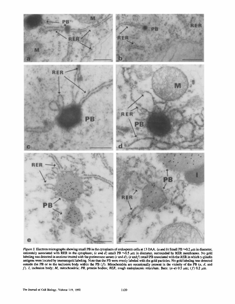
Figure 3. Electron micrographs showing small PB in the cytoplasm of endosperm cells at t3 DAA. (a and b) Small PB ~176 in diameter, intimately associated with PER in the cytoplasm; (c and d) small PB o00.5 ~m in diameter, surrounded by RER membranes. No gold labeling was detected in sections treated with the preimmune serum (c and d); (e and f ) small PB associated with the RER in which ~,-gliadin antigens were located by immunogold labeling. Note that the PB were evenly labeled with the gold particles. No gold labeling was detected outside the PB or in the inclusion body within the PB (f) . Mitochondria are occasionally present in the vicinity of the PB (a, d, and f). I, inclusion body; M, mitochondria; PB, protein bodies; RER, rough endoplasmic reticulum. Bars: (a-e) 0.5/~m; (f) 0.2/~m.
The Journal of Cell Biology, Volume 119, 1992 1120

Figure 4. Electron micrographs of 13 DAA (a) and 16 DAA (b and c) endosperm cells showing large PB in the cytoplasm. (a) Immunogold localization of storage proteins in the intermediate PB. (a and b) Large PB in the cytoplasm, incompletely surrounded by RER membrane. A small ~/-gliadin-containing particle is detected in the vicinity of the PB (a, arrowhead). The PB in b seems as it was recently fused with smaller PB and it also contains an inclusion body. A mitochondrion is detected in the vicinity of the PB (a). (c) A cluster of fusing PB in the cytoplasm with RER membranes in spaces between them. I, inclusion body; PB, protein body; RER, rough endoplasmic reticu- lure. Bars, 0.5 pln.
Levanony et al. Subcellular Route of Wheat Storage Proteins 1121
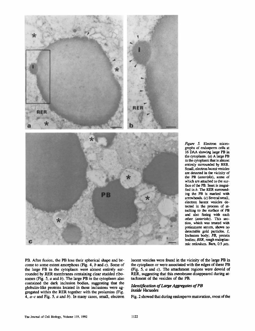
l~gure 5. Electron micro- graphs of endosperm cells at 16 DAA showing large PB in the cytoplasm. (a) A large PB in the cytoplasm that is almost entirely surrounded by RER. Small, electron lucent vesicles are detected in the vicinity of the PB (asterisks), some of which are attached to the sur- face of the PB. Inset is magni- fied in b. The PER surround- ing the PB is marked with arrowheads. (c) Several small, electron lucent vesicles de- tected in the process of at- taching to the surface of PB and also fusing with each other (asterisks). This sec- tion, which was treated with preimmune serum, shows no detectable gold particles. L Inclusion body; PB, protein bodies; RER, rough endoplas- mic reticulum. Bars, 0.5 #m.
PB. After fusion, the PB lose their spherical shape and be- come to some extent amorphous (Fig. 4, b and c). Some of the large PB in the cytoplasm were almost entirely sur- rounded by RER membranes containing clear studded ribo- somes (Fig. 5, a and b). The large PB in the cytoplasm also contained the dark inclusion bodies, suggesting that the globulin-like proteins located in these inclusions were ag- gregated within the RER together with the prolamins (Fig. 4, a-c and Fig. 5, a and b). In many cases, small, electron
lucent vesicles were found in the vicinity of the large PB in the cytoplasm or were associated with the edges of these PB (Fig. 5, a and c). The attachment regions were devoid of RER, suggesting that this membrane disappeared during at- tachment of the vesicles of the PB.
Identification of Large Aggregates of PB inside Vacuoles
Fig. 2 showed that during endosperm maturation, most of the
The Journal of Cell Biology, Volume 119, 1992 1122
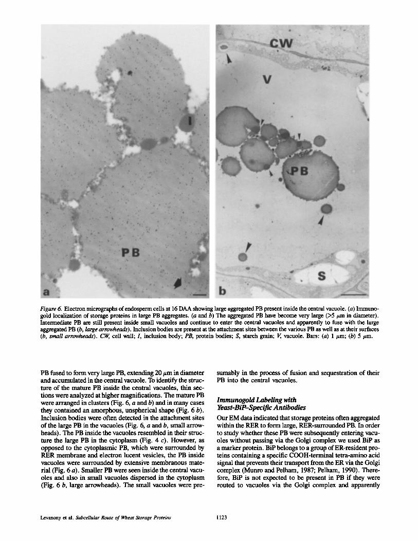
Figure 6. Electron micrographs of endosperm ceils at 16 DAA showing large aggregated PB present inside the central vacuole. (a) Immuno- gold localization of storage proteins in large PB aggregates. (a and b) The aggregated PB have become very large (>5 t~m in diameter). Intermediate PB are still present inside small vacuoles and continue to enter the central vacuoles and apparently to fuse with the large aggregated PB (b, large arrowheads). Inclusion bodies are present at the attachment sites between the various PB as well as at their surfaces (b, small arrowheads). ~ cell wall; I, inclusion body; PB, protein bodies; S, starch grain; V, vacuole. Bars: (a) 1/~m; (b) 5 #m.
PB fused to form very large PB, extending 20/~m in diameter and accumulated in the central vacuole. To identify the struc- ture of the mature PB inside the central vacuoles, thin sec- tions were analyzed at higher magnifications. The mature PB were arranged in clusters (Fig. 6, a and b) and in many cases they contained an amorphous, unspherical shape (Fig. 6 b). Inclusion bodies were often detected in the attachment sites of the large PB in the vacuoles (Fig. 6, a and b, small arrow- heads). The PB inside the vacuoles resembled in their struc- ture the large PB in the cytoplasm (Fig. 4 c). However, as opposed to the cytoplasmic PB, which were surrounded by PER membrane and electron lucent vesicles, the PB inside vacuoles were surrounded by extensive membranous mate- rial (Fig. 6 a). Smaller PB were seen inside the central vacu- oles and also in small vacuoles dispersed in the cytoplasm (Fig. 6 b, large arrowheads). The small vacuoles were pre-
sumably in the process of fusion and sequestration of their PB into the central vacuoles.
Immunogold Labeling with Yeast-BiP--Specific Antibodies
Our EM data indicated that storage proteins often aggregated within the RER to form large, RER-surrounded PB. In order to study whether these PB were subsequently entering vacu- oles without passing via the Golgi complex we used BiP as a marker protein. BiP belongs to a group of ER-resident pro- teins containing a specific COOH-terminal tetra-amino acid signal that prevents their transport from the ER via the Golgi complex (Munro and Pelham, 1987; Pelham, 1990). There- fore, BiP is not expected to be present in PB if they were routed to vacuoles via the Golgi complex and apparently
Levanony et al. Subcellular Route of Wheat Storage Proteins 1123
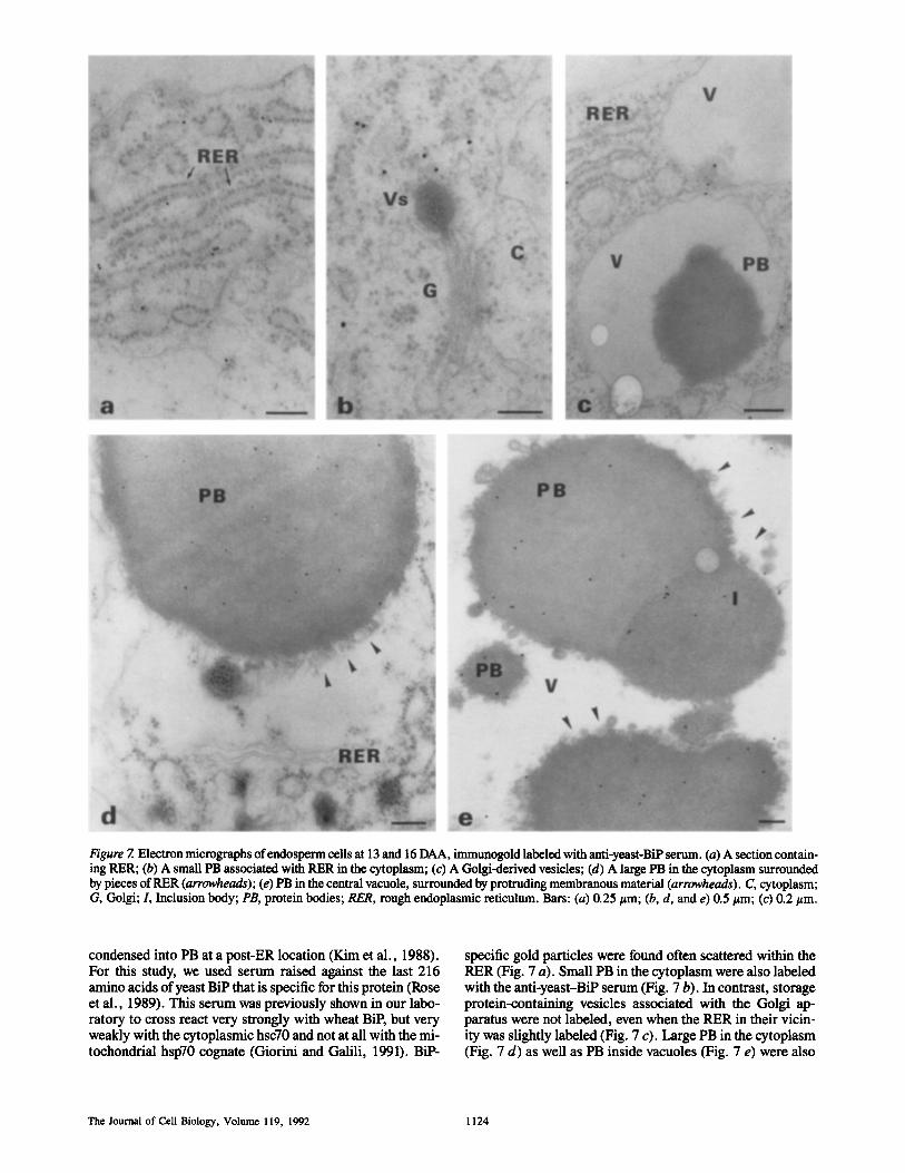
Figure 7. Electron micrographs of endosperm cells at 13 and 16 DAA, immanogold labeled with anti-yeast-BiP serum. (a) A section contain- ing RER; (b) A small PB associated with RER in the cytoplasm; (c) A Golgi-derived vesicles; (d) A large PB in the cytoplasm surrounded by pieces of RER (arrowheads); (e) PB in the central vacuole, surrounded by protruding membranous material (arrowheads). C, cytoplasm; G, Golgi; L Inclusion body; PB, protein bodies; RER, rough endoplasmic reticulum. Bars: (a) 0.25/~m; (b, d, and e) 0.5/~m; (c) 0.2 #m.
condensed into PB at a post-ER location (Kim et al., 1988). For this study, we used serum raised against the last 216 amino acids of yeast BiP that is specific for this protein (Rose et al., 1989). This serum was previously shown in our labo- ratory to cross react very strongly with wheat BiP, but very weakly with the cytoplasmic hsc70 and not at all with the mi- tochondrial hsp70 cognate (Giorini and Galili, 1991). BiP-
specific gold particles were found often scattered within the RER (Fig. 7 a). Small PB in the cytoplasm were also labeled with the anti-ycast-BiP serum (Fig. 7 b). In contrast, storage protein-containing vesicles associated with the Golgi ap- paratus were not labeled, even when the RER in their vicin- ity was slightly labeled (Fig. 7 c). Large PB in the cytoplasm (Fig. 7 d) as well as PB inside vacuoles (Fig. 7 e) were also
The Journal of Cell Biology, Volume 119, 1992 1124
clearly labeled with the yeast-BiP antibodies. BiP was also immunogold localized in the globulin-containing inclusions (Fig. 7 e). Labeling of the PB inside vacuoles with the yeast- BiP antibodies was very specific, as gold particles were hardly observed in the matrix of the vacuoles (Fig. 7 e). Moreover, the gold particles were always present homogene- ously inside the PB and not merely on their surface, eliminat- ing the possibility of an artifact due to attachment of RER membranes to the PB. Gold particles were scarcely present in sections treated with the BiP preimmune serum (data not shown). To quantitate the intensity of BiP-specific gold label- ing in the cytoplasm, PB, and vacuolar sap (as a control), we checked ,,050 randomly chosen fields of 4-cm 2 each, taken from 10 separate micrographs that were obtained from different labeling experiments. The average number of gold particles per field were 8.1 for the PB, 2.9 for the cytoplasm, and 0 for the vacuolar sap.
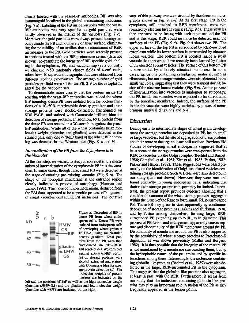
To demonstrate more clearly that the protein inside PB reacting with the yeast-BiP antibodies was indeed the wheat BiP homolog, dense PB were isolated from the bottom frac- tions of a 10-50% metrizamide density gradient and their storage proteins were alcohol-extracted, fractionated on SDS-PAGE, and stained with Coomassie brilliant blue for detection of storage proteins. In addition, total protein from the dense PB was reacted in a Western blot against the yeast- BiP antibodies. While all of the wheat prolamins (high mo- lecular weight glutenins and gliadins) were detected in the stained gels, only one ~78-kD band of the wheat BiP homo- log was detected in the Western blot (Fig. 8, a and b).
Internalization of the PB from the Cytoplasm into the Vacuoles
At the next step, we wished to study in more detail the mech- anism of internalization of the cytoplasmic PB into the vacu- oles. In some cases, though rare, small PB were detected at the stage of entering pre-existing vacuoles (Fig. 9 a). The shape of the tonoplast membrane at the entrance region clearly indicated a process of autophagy (Herman and Lamb, 1992). The more common mechanism, deduced from the EM data, appeared to be a specific process of biogenesis of small vacuoles containing PB inclusions. The putative
Figure 8. Detection of BiP in dense PB from wheat endo- sperm ceils. Dense PB were isolated from endosperm cells of developing wheat grains at 16 DAA, using metrizamide density gradient. Total pro- teins from the PB were then fractionated on SDS-PAGE and reacted in a Western blot against anti-yeast-BiP serum (a) or storage proteins were alcohol extracted and stained with Coomassie blue for stor- age protein detection (b). The molecular weights of protein markers are indicated on the
left and the positions of BiP as well as the high molecular weight glutenins (HMIr~Gs) and the gliadins and low molecular weight glutenins (LMI~GS) are indicated on the right.
steps of this pathway are reconstructed by the electron micro- graphs shown in Fig. 9, b-f. At the first stage, PB in the cytoplasm, still attached to RER membrane, were sur- rounded by electron lucent vesicles (Fig. 9 b). These vesicles then appeared to be fusing with each other around the PB and at this stage, RER could no more be detected near the surfaces of the PB (Fig. 9 c). Fig. 9 d shows two PB. The upper surface of the top PB is surrounded by RER-enriched cytoplasm while its lower surface is surrounded by electron lucent vesicles. The bottom PB is located inside a small vacuole that appears to have recently been formed by fusion of the electron lucent vesicles. The surface of this bottom PB is surrounded by a loosely attached membrane. In some cases, inclusions containing cytoplasmic material, such as ribosomes, but not storage proteins, were also detected in the small vacuoles, suggesting that they were trapped during fu- sion of the electron lucent vesicles (Fig. 9 e). As this process of internalization into vacuoles is analogous to autophagy, the PB inside the vacuoles were expected to be surrounded by the tonoplast membrane. Indeed, the surfaces of the PB inside the vacuoles were highly enriched by pieces of mem- branous material (Figs. 9 f and 6 a).
Discussion
During early to intermediate stages of wheat grain develop- ment the storage proteins are deposited in PB inside small or large vacuoles, but the site of aggregation of these proteins and their route to the organelle are still nuclear. Previous EM studies of developing wheat endosperms suggested that at least some of the storage proteins were transported from the RER to vacuoles via the Golgi complex (Bechtel and Barnet, 1986; Campbell et al., 1981; Kim et ai., 1988; Parker, 1982; Parker and Hawes, 1982). These suggestions were based pri- marily on the identification of Golgi-associated vesicles con- taining storage proteins. Such vesicles were also detected in our study (data not shown). However, they were rare and found primarily in young endosperm cells, indicating that their role in storage protein transport may be limited. In con- trast, the present report provides evidence showing that a considerable amount of the wheat storage proteins aggregate within the lumen of the RER to form small, RER-surrounded PB. These PB may grow in size, apparently by continuous deposition of storage proteins (Larkins and Hurkman, 1978) and by fusion among themselves, forming large, RER- surrounded PB extending up to ~10 tzm in diameter. This process of PB fusion and enlargement apparently causes rup- ture and discontinuity of the RER membrane around the PB. Discontinuity of membrane around the PB is also supported by the sensitivity of wheat storage proteins to Proteinase-K digestion, as was shown previously (Miflin and Burgess, 1982). It is thus possible that the integrity of the mature PB is not maintained by a membrane surrounding them, but by the hydrophobic nature of the prolamins and by specific in- teractions among them. Interestingly, the inclusions contain- ing globulin-like proteins (Bechtel et al., 1989) were also de- tected in the large, RER-surrounded PB in the cytoplasm. This suggests that the globulin-like proteins also aggregate, at least in part, with the RER. Furthermore, it seems from our study that the inclusions containing globulin-like pro- teins may play an important role in fusion of the PB as they frequently appeared in the fusion pOints.
Levanony et al. Subcellular Route of Wheat Storage Proteins 1125

Figure 9. Electron micrographs of endosperm cells at 16 DAA showing progressive stages of the formation of small vacuoles containing PB inclusions. The electron lucent vesicles are indicated by asterisks. (a) A small PB (arrow) enters the vacuole by autophagy; (b) electron lucent vesicles are attached to a RER surrounded PB in the cytoplasm; (r the electron lucent vesicles fuse and encircle the entire surface of the PB; (d) two PB in diffcrant stages of entering the small vacuole. The top PB is surrounded partially by RER-enriched cytoplasm and partially by the electron lucent vesicle. The bottom PB is already present inside a small vacuole. This bottom PB is surrounded by a loosely attached membrane (arrowhead); (e) a small vacuole containing a PB and two additional inclusions containing cytoplasmic mate- rial (arrows). Membranes are also detected in this vacuole (arrowheads); ( f ) the surface of a PB inside the vacuole is highly enriched with membranous material (arrowheads). Sections were immunogold labeled with sera against ~-gliadin (b, d, e) and against yeast-BiP (c and f ) . M, mitochondria; PB, protein bodies; RER, rough endoplasmic reticulum. Bars: (a, r e, and f ) 0.5 #m; (b and d) 0.25 ~ .
The Journal of Cell Biology, Volume 119, 1992 1126

The mechanism by which the RER-surrounded PB in the cytoplasm enter vacuoles is not understood. As these PB are much larger than the Golgi complex, it is unexpected that this organelle is involved in their transport. This question is difficult to address by pulse chase studies because wheat storage proteins are not glycosylated and it is not possible to trace their transport via the Golgi complex by testing for Golgi-specific sugar modifications. One of our approaches to study transport was to reconstruct a route based upon a series of static electron micrographs. Although data derived from such micrographs should be interpreted with caution, the results of these studies suggested that PB in the cytoplasm may enter vacuoles by a specific process that may be analo- gous to autophagy. This proposed process apparently initi- ates by attachment of many small, electron lucent vesicles to the surface of the PB in the cytoplasm followed by fusion of the attached vesicles among themselves, thus forming small vacuoles containing PB inclusions inside them. During this process, the RER membrane surrounding the PB disap- peared. In such a process, it is expected that some cytoplas- mic material may also be trapped inside the newly formed vacuoles. This has indeed been observed (Fig. 9 e). PB that have entered vacuoles by an autophagy-like process are also expected to be surrounded by the tonoplast (Herman and Lamb, 1992). This is not expected if the PB entered by mem- brane fusion. The surfaces of the PB inside the vacuoles and the adjacent regions were highly enriched with loosely at- tached membranous material. It seems likely that during in- ternalization to the vacuoles, the PB became surrounded by the tonoplast membrane, and that this membrane was subse- quently detached and degraded during further fusion of the PB inside the vacuoles. The small vacuoles containing PB in- clusions that are shown in the bottom cell of Fig. 2, may have been formed by such an internalization process. During maturation of the endosperm cells, these small vacuoles may further fuse to form finally a central vacuole to which the PB are sequestered. These PB apparently continue to enlarge by fusions with each other inside the vacuoles (Fig. 2, top two cells). Formation of central vacuoles by fusion of small vacuoles is a common process in plant cells (Boiler and Wiemken, 1986; Marty et al., 1980). However, routing of PB into small vacuoles and their subsequent fusion to form large PB suggests a novel mechanism for storage proteins transport which differs from previous models of PB forma- tion in seeds of dicotyledonous plants. Light microscopy and ultrastructural studies of developing seeds from a variety of dicotyledonous plants suggested that during PB formation, the central vacuole is subdivided into many small vacuoles (Craig et al., 1979; Lott, 1980).
BiP As a Marker Protein for Wheat Storage Protein Transport
The demonstration that BiP was present inside PB in the cytoplasm and within vacuoles provided additional support to the interpretation of our EM data, suggesting that these PB were formed within the lumen of the RER and then trans- ported directly to vacuoles without passing via the Golgi complex. BiP belongs to a family of luminal, ER-resident proteins containing the ER retention signal KDEL or HDEL within the COOH-terminal region which prevents transport via the Golgi (Pelham, 1990). BiP appears to be ubiquitous
in all eukaryotic cells and was recently identified in wheat endosperm cells (Giorini and Galili, 1991). Analyses of BiP cloned from several plant species have confirmed that the plant proteins also contain the HDEL signal (Denecke et al., 1991; Meyer et al., 1991). Although we never detected BiP in the Golgi complex, it is still possible that some BiP will escape retention and transport via the Golgi complex inas- much as the HDEL retention signal is known to be saturable (Pelham, 1990). However, even if some wheat BiP escapes the retention receptor, it is expected to go through a default pathway to the cell membrane and not to vacuoles, because it apparently lacks a vacuolar targeting signal. Indeed, a yeast BiP with the HDEL signal removed was transported in yeast cells to the cell membrane and not to the vacuole (Hardwick et al., 1990). Therefore, if wheat storage proteins would have been transported to the Golgi complex and con- densed into PB in vesicles budding off this organdie, the PB would not be expected to contain significant amounts of BiP. Presence of considerable amounts of BiP inside PB would be expected only if BiP was trapped during aggregation of stor- age proteins within the lumen of the RER.
Our study is not the only one suggesting that the paradigm of transport to vacuoles via the ER and the Golgi complex does not hold true for all proteins. The yeast vacuolar protein a-mannosidase was shown to be transported directly from the cytoplasm to vacuoles with no passage via the endomem- brane system (Chiang and Schekman, 1991; Yoshihida and Anraku, 1990). In addition, Herman and Lamb (1992) have recently demonstrated that tobacco arabinose-rich glycopro- teins are internalized from the periplasmic space into the vacuoles by an autophagy-like process.
Aggregation of the water insoluble prolamins within the RER is not unique to wheat. The prolamins of maize and rice also aggregate into PB within the RER (Krishnan et al., 1986; Larkins and Hurkman, 1978). However, in maize and rice the PB remain attached to the RER, while in wheat they are disconnected from the continuous RER and enter vacu- oles. The difference between these plants may be explained by the fact that in wheat the relatively large size of the PB causes rupture and detachment of the RER, while in maize and rice the PB are smaller and remain surrounded by RER membranes. As subsequent attachment of the wheat PB to the electron lucent vesicles apparently involves the surfaces of the PB themselves and not the RER, these surfaces would be masked by the RER membrane in maize and rice PB (Krishnan et al., 1986; Larkins and Hurkman, 1978). A similar alternative route of prolamins to vacuoles may also exist in other cereals such as oats. The prolamins of oats are also located inside vacuoles, but EM studies have indicated that they aggregate within the RER forming large PB (Lend- ing et al., 1989).
Our study suggests that aggregation of wheat prolamins within the RER and their novel route to vacuoles takes place during early to intermediate stages of grain development (13-16 DAA) and at the early stages it operates in concert with an independent Golgi-mediated route. It is possible that following entrance into the RER, storage proteins initiate a slow process of aggregation and that the aggregation state will dictate the pathway taken. At earlier stages, more pro= tein may escape aggregation within the RER, transport to the Golgi, and condense into PB in vesicles budding off this ap= paratus (Bechtel et al., 1982; Kim et al., 1988). This may
Levanony et al. Subcellular Route of Wheat Storage Proteins 1127
be due to the lower amount of prolamins present within the RER of young grains or due to ER-localized factors that are differentially expressed during grain development. At later stages of grain development a considerable amount of the storage proteins also aggregate into PB within the PER, but it is not clear whether all of these PB subsequently enter vacuoles as it is difficult to detect vacuoles at this stage (Levanony, H., and G. Galili, unpublished data).
This paper is dedicated to the late Mrs. Batia Romano to whom we are greatly obliged for her excellent technical assistance. We thank Dr. N. Rosenberg, Dr. R. Fluhr, Prof. J. Gressel, and Dr. M. Volokita for critical reading of the manuscript and their invaluable suggestions as well as Dr. J. P. Vogel for the yeast-Bip antibodies. G. Galili is an incumbent of the Abraham and Jenny Fialkow Career Development Chair in Biology.
We thank Dr. N. Rosenberg, Dr. R. Fluhr, Prof. J. Gressel, and Dr. M. Volokita for critical reading of the manuscript and their invaluable sugges- tions; Dr. J. P. Vogel for the yeast-BiP antibodies and Mrs. Batia Romano for assistance in thin sectioning. G. G. is an incumbent of the Abraham and Jenny Fialkov career development chair in biology.
Received for publication 2 December 1991 and in revised form 16 August 1992.
References
Bechtel, D. B., and B. D. Barnet. 1986. A freeze fracture study on the storage protein accumulation in unfixed wheat starchy endosperm. Cereal Chem. 63:232-240.
Bechtel, D. B., R. L. Gaines, and Y. Pomeranz. 1982. Early stages in wheat endosperm formation and protein body initiation. Ann. Bat. 50:507-518.
Bechtel, D. B., J. D. Wilson, and P. R. Shewry. 1989. Identification of legumine-like proteins within sections of developing wheat endosperm by immanocytochemical procedures. Cereal Foods World. 34:784 Suppl.
Bole, D. G., L. M. Hendershot, and J. F. Kearney. 1986. Post-translational association of immunoglobulin heavy chain binding protein with nascent heavy chains in nonsecreting and secreting hybridomas. J. Cell Biol. 102: 1558-1566.
Boiler, T., and A. Wiemken. 1986. Dynamics of vacuolar compartmentation. Ann. Rev. Plant Physiol. 37:137-164.
Campbell, W. P., J. W. Lee, T. P. O'Brien, and M. G. Smart. 1981. En- dosperm morphology and protein body formation in developing wheat grains. Aust. J. Plant Physiol. 8:5-19.
Chiang, H.-L., and R. Schekman. 1991. Regulated import and degradation of a cytosolic protein in the yeast vacuole. Nature (Lond.). 350:313-318.
Craig, S., D. J. Goodchild, and A. R. Hardham. 1979. Structural aspects of protein accumulation in developing pea cotyledons I. Qualitative and quan- titative changes in pharenchyma cell vacuoles. Aust. J. Plant Physiol. 6: 81-98.
Denecke, J., M. H. S. Goldman, J. Demolder, J. Seurinck, and L Botterman. 1991. The tobacco luminal binding protein is encoded by a multigene family. Plant Cell. 3:1025-1035.
Giorini, S., and G. Galili. 1991. Characterization of HSP-70 cognate proteins from wheat. Theor. Appl. Genet. 82:615-620.
Greenwood, J. S., and M. J. Chrispeels. 1985. Immunocytochemical localiza- tion of phaseolin and phytohemagglntinin in the endoplasmic reticulum and Golgi complex of developing bean cotyledon. Planta. 164:295-302.
Haas. I. G.. and M. Wabl. 1983. Immunoglobulin beavv chain binding nrotein.
Nature (Lond.). 306:387-389. Hardwick, K. G., M. J. Lewis, J. Semenza, N. Dean, and H. R. B. Pelham.
1990. ERD1, a yeast gene required for the retention of luminal endoplasmic reticulum proteins, affects glycoprotein processing in the Golgi apparatus. EMBO (Fur. biol. Biol. Organ.). J. 9:623-630.
Herman, E. M., and C. J. Lamb. 1992. Arabinogalactan-rich glycoproteins are localized on the cell surface and in intravacuolar multivesicular bodies. Plant Physiol. 98:264-272.
Kim, W. T., V. R. Franceschi, H. B. Krishnan, andT. W. Okita. 1988. Forma- tion of wheat protein bodies: involvement of the Golgi apparatus in gliadin transport. Planta. 176:173-182.
Krishnan, H. B., V. R. Franceschi, and T. W. OkRa. 1986. Immunocytochemi- cal studies on the role of the Golgi complex in protein-body formation in rice seeds. Planta. 169:471--480.
Laemmli, U. K. 1970. Cleavage of structural proteins during the assembly of the head of bacteriophage T4. Nature (Lond.). 227:680-685.
Lafiandra, D., and D. D. Kasarda. 1985. One- and two-dimensional (two-pH) polyacrylamide gel electrophoresis in a stained gel: separation of wheat pro- teins. Cereal Chem. 62:314-319.
Larkins, B. A., and W. J. Hurkman. 1978. Synthesis and deposition of zein in protein bodies of maize endosperm. Plant Physiol. 62:256-263.
Lending, C. R., R. S. Chesnnt, K. L. Shaw, andB. A. Larkins. 1989. Immu- nolocalization of avenin and globulin storage proteins in developing endo- sperm of Arena sativa L. Planta. 178:315-324.
Lott, J. N. A. 1980. Protein bodies. In The Biochemistry of Plants. B. J. Miflin, editor. Academic Press, New York. 589-623.
Marty, F., D. Branton, and R. A. Leigh. 1980. Plant vacuoles. In The Biochem- istry of Plants: A Comprehensive Treatise. N. E. Tolbert, editor. Macmil- lan, New York. 625-658.
Meyer, D. J., N. N. Ewing, and A. B. Bennett. 1991. Identification and charac- terization of a BiP-like endoplasmic reticalum-localized molecular chaperon in tomato. In Keystone Symposium on the Genetic Dissection of Plant Cell Processes. Keystone, CO. Suppl. 15A, A339. 76 pp.
Miflin, B. J., and S. R. Burgess. 1982. Protein bodies from developing seeds of barley, maize, wheat and peas: the effect of protease treatment. J. Exp. Bot. 33:251-260.
Miflin, B. J., S. R. Burgess, and P. R. Shewry. 1981. The development of pro- tein bodies in the storage tissues of seeds. J. Exp. Bot. 32:199-219.
Munro, S., and H. R. B. Pelham. 1986. An hsp70-1ike protein in the ER: iden- tity with the 78 kd glucose-regulated protein and immunoglobulin heavy chain binding protein. Cell. 46:291-300.
Munro, S., and H. R. B. Pelham. 1987. A C-terminal signal prevents secretion of luminal ER proteins. Cell. 48:899-907.
Parker, M. L. 1982. Protein accumulation in the developing endosperm of a high-protein line of triticum dicoccoides. Plant Cell Environ. 5:37--43.
Parker, M. L., and C. R. Hawes. 1982. The Golgi apparatus in developing en- dosperm of wheat (Triticum aestivum L.). Planta. 154:277-283.
Pelham, H. R. B. 1989. Control of proteins exit from the endoplasmic reticu- lure. Annu. Rev. Cell Biol. 5:1-23.
Pelham, H. R. B. 1990. The retentional signal for soluble proteins of the en- doplasmic reticulum. Trends Biochem. Sci. 15:483--486.
Rose, M. D., L. M. Misra, and J. P. Vogel. 1989. Kar2, a karyogamy gene, is the yeast homolog of the mammalian BiP/GRP78 gene. Cell. 57:1211- 1221.
Shotwell, M. A., and B. A. Larkins. 1988. The biochemistry and molecular biology of seed storage proteins. In The Biochemistry of Plants. B. J. Miflin, editor. Academic Press, New York. 297-345.
Towbin, H., T. Staehelin, and J. Gordon. 1979. Electrophoretic transfer of pro- teins from polyacrylamide gels to nitrocellulose sheets: procedure and some applications. Proc. Natl. Acad. Sci. USA. 76:4350--4354.
Wallace, J. C., G. Galili, E. E. Kawata, R. E. Cuellar, M. A. Shotwell, and B. A. Larkins. 1988. Aggregation of lysine-containing zeins into protein bodies in Xenopus oocytes. Science (Wash. DC). 240:662-664.
Yoshihida, T., and Y. Anraku. 1990. A novel pathway of inport of ~-man- nosidase, a marker enzyme of vacuolar membrane, in Saecharomyces cere- visiae. J. Biol. Chem. 265:22418-22425.
The Journal of Cell Biology, Volume 119, 1992 1128