evidence of an asymmetrical endophenotype in...
TRANSCRIPT

Evidence of an Asymmetrical Endophenotype inCongenital Fibrosis of Extraocular MusclesType 3 Resulting from TUBB3 Mutations
Joseph L. Demer,1,2,3,4 Robert A. Clark,1 Max A. Tischfield,5,6
and Elizabeth C. Engle6,7,8,9,10,11
PURPOSE. Orbital magnetic resonance imaging (MRI) was usedto investigate the structural basis of motility abnormalities incongenital fibrosis of the extraocular muscles type 3 (CFEOM3),a disorder resulting from missense mutations in TUBB3, whichencodes neuron-specific �-tubulin isotype III.
METHODS. Ophthalmic examinations in 13 volunteers from fourCFEOM3 pedigrees and normal control subjects, were corre-lated with TUBB3 mutation and MRI findings that demon-strated extraocular muscle (EOM) size, location, contractility,and innervation.
RESULTS. Volunteers included clinically affected and clinicallyunaffected carriers of R262C and D417N TUBB3 amino acidsubstitutions and one unaffected, mutation-negative familymember. Subjects with CFEOM3 frequently had asymmetricalblepharoptosis, limited vertical duction, variable ophthalmo-plegia, exotropia, and paradoxical abduction in infraduction.MRI demonstrated variable, asymmetrical levator palpebraesuperioris and superior rectus EOM atrophy that correlatedwith blepharoptosis, deficient supraduction, and small orbitalmotor nerves. Additional EOMs exhibited variable hypoplasiathat correlated with duction deficit, but the superior obliquemuscle was spared. Ophthalmoplegia occurred only when thesubarachnoid width of CN3 was �1.9 mm. A-pattern exotropiawas frequent, correlating with apparent lateral rectus (LR)muscle misinnervation by CN3. Optic nerve (ON) cross sec-tions were subnormal, but rectus pulley locations were normal.
CONCLUSIONS. CFEOM3 caused by TUBB3 R262C and D417Namino acid substitutions features abnormalities of EOM innerva-tion and function that correlate with subarachnoid CN3 hypopla-sia, occasional abducens nerve hypoplasia, and subclinical ON
hypoplasia that can resemble CFEOM1. Clinical and MRI findingsin CFEOM3 are more variable than those in CFEOM1 and are oftenasymmetrical. Apparent LR innervation by the inferior rectusmotor nerve is an overlapping feature of Duane retraction syn-drome and CFEOM1. These findings suggest that CFEOM3 is anasymmetrical, variably penetrant, congenital cranial dysinnerva-tion disorder leading to secondary EOM atrophy. (Invest Ophthal-mol Vis Sci. 2010;51:4600–4611) DOI:10.1167/iovs.10-5438
Congenital fibrosis of the extraocular muscles (CFEOM) istypically a nonprogressive disorder of ocular dysmotility
with accompanying blepharoptosis. CFEOM is classified, alongwith Duane syndrome, congenital ptosis, congenital facialpalsy, and Moebius syndrome, as a congenital cranial dysinner-vation disorder (CCDD), proposed to be caused by aberrantinnervation of ocular and facial muscles.1 Three distinct phe-notypes, CFEOM1, -2, and -3, are recognized. The classic form,CFEOM1 (MIM 135700; http://www.ncbi.nlm.nih.gov/Omim/,provided in the public domain by the National Center forBiotechnology Information [NCBI], Bethesda, MD), is typifiedby bilateral congenital blepharoptosis and ophthalmoplegia,with the eyes restricted to infraduction.2 Horizontal strabismusmay coexist. CFEOM1 is autosomal dominant, maps to chro-mosome 12,3,4 and is caused by a small number of recurrentheterozygous missense mutations in the kinesin motor proteinencoded by KIF21A.5 Engle et al.2 have suggested thatCFEOM1 is the result of primary maldevelopment of cranialmotor neurons and their axons, which causes secondary hyp-oplasia or atrophy of the EOMs that they innervate and con-tracture of their antagonists.
Persons with CFEOM2 (OMIM 602078) have congenitallybilateral exotropic ophthalmoplegia and blepharoptosis andmay also have infra- or supraduction of the eyes. This form ofCFEOM is a recessive one, identified in consanguineous pedi-grees. It maps to the FEOM2 locus on the long arm of chro-mosome 11, region 136 and is caused by homozygous muta-tions in PHOX2A (ARIX),7 a homeodomain transcription factornecessary for development of oculomotor and trochlear motorneurons in mice and zebrafish.8,9 Affected individuals do nothave detectable oculomotor or trochlear nerves by MRI,10 andthese cranial nuclei most likely fail to form in CFEOM2.
The third CFEOM variant, CFEOM3, encompasses individu-als from pedigrees with CFEOM that can be clinically indistin-guishable from CFEOM1 or CFEOM2. Other pedigree mem-bers, however, have absent or unilateral ptosis, unilateralophthalmoplegia, noninfraducted resting eye position, and/orthe ability to supraduct one or both eyes above central posi-tion. CFEOM3 can be autosomal dominant, and large familiesthat segregate CFEOM3 typically include affected individualswho meet CFEOM1 criteria.11 To avoid overlapping classifica-tions, we defined a CFEOM1 pedigree as one in which allaffected individuals meet CFEOM1 criteria and a CFEOM3 ped-
From the 1Jules Stein Eye Institute, Department of Ophthalmol-ogy, the 2Department of Neurology, and the Interdepartmental Pro-grams of 3Bioengineering and 4Neuroscience, University of California,Los Angeles, Los Angeles, California; the 5Department of MolecularBiology and Genetics, Johns Hopkins University School of Medicine,Baltimore, Maryland; 6Howard Hughes Medical Institute, Chevy Chase,Maryland; the Departments of 7Neurology, 8Ophthalmology, and 9Med-icine (Genetics and Genomics), the F. M. Kirby Neurobiology Center,and 10The Manton Center for Orphan Disease Research, Children’sHospital, Boston, Massachusetts; and the 11Department of Neurology,Harvard Medical School, Boston, Massachusetts.
Supported by National Eye Institute Grants EY13583, EY12498,EY08313, and EY00331. JLD received an award from Research toPrevent Blindness and is Leonard Apt Professor of Ophthalmology. ECEis a Howard Hughes Medical Institute Investigator.
Submitted for publication February 24, 2010; revised March 25,2010; accepted March 26, 2010.
Disclosure: J.L. Demer, None; R.A. Clark, None; M.A. Tischfield,None; E.C. Engle, None
Corresponding author: Joseph L. Demer, Jules Stein Eye Institute,100 Stein Plaza, UCLA, Los Angeles, CA 90095-7002; [email protected].
Eye Movements, Strabismus, Amblyopia, and Neuro-Ophthalmology
Investigative Ophthalmology & Visual Science, September 2010, Vol. 51, No. 94600 Copyright © Association for Research in Vision and Ophthalmology
Downloaded From: http://iovs.arvojournals.org/pdfaccess.ashx?url=/data/journals/iovs/932963/ on 05/19/2018

igree as one in which at least one affected individual failed tomeet CFEOM1 criteria.12 Although in most large pedigrees,CFEOM3 maps to the FEOM3 locus on the long arm of chro-mosome 1611,13 (OMIM 600638), in rare families it maps to theFEOM1 locus,14 and affected members harbor KIF21A muta-tions15 (CFEOM3A; OMIM 607034).
We recently identified the FEOM3 gene as TUBB3 (GenBank10381; http://www.ncbi.nlm.nih.gov/Genbank; provided in thepublic domain by NCBI), encoding neuron-specific �-tubulin iso-type III.16 CFEOM3 can be caused by one of at least eight differentheterozygous missense mutations in TUBB3. These TUBB3 mu-tations alter dynamic instability, and some also alter microtu-bule interactions with kinesins, including KIF21A. Moreover,although some TUBB3 mutations result in relatively isolatedCFEOM, other mutations cause additional central and periph-eral nervous system dysfunction.16
The clinical findings of blepharoptosis and strabismus arenot highly specific, rendering them susceptible to phenocopyfrom a wide variety of causes, as well as to potential variationin presentation. The external phenotypic heterogeneity ofCFEOM3 has particular potential to confound identification ofthe genotype or genotypes of this disorder. Magnetic reso-nance imaging (MRI) now affords the opportunity for detailedstudy of the endophenotype of EOMs and nerves in the orbitsof living subjects,17 and the subarachnoid cranial nerves (CNs)can be imaged as they exit the brain stem.18 We used MRI toinvestigate the internal phenotype, or endophenotype, of awell-defined group of subjects with CFEOM3 caused by muta-tions in TUBB3. The purpose was to characterize orbital inner-vation and the structure and function of EOMs, to distinguishendophenotypes that may be unique to CFEOM3. Our findingssupport the hypothesis that CFEOM3 is a primary motor neu-ropathy that mainly affects the oculomotor nerve (CN3) andarises from the inability of motor axons to correctly target thecranial musculature.16 Moreover, although in some individualsMRI findings may be indistinguishable from CFEOM1, CFEOM3is more often asymmetric and variable in its clinical and MRIpresentation.
METHODS
SubjectsThirteen subjects from four unrelated and previously reported pedi-grees participated in the study.16 The diagnosis of CFEOM3 was estab-lished by clinical criteria and was correlated with TUBB3 mutationstatus. Pedigrees AT, BT, and QX segregate the most common TUBB3mutation that results in a R262C amino acid substitution. We estab-lished that most individuals harboring this mutation have CFEOM3without other nervous system findings.16 Pedigree D segregates themutation resulting in a D417N amino acid substitution. We establishedthat most individuals harboring this mutation are born with CFEOM3and then develop a later-onset axonal sensorimotor peripheral neurop-athy.16 Subjects gave written informed consent to a protocol conform-ing to the Declaration of Helsinki and approved by the InstitutionalReview Boards of the University of California, Los Angeles (UCLA), andChildren’s Hospital Boston. At UCLA, subjects provided ophthalmichistories and underwent complete ophthalmic examination. They alsounderwent measurement of palpebral fissure height and levator func-tion, with video recording of ocular versions and eyelid motility.Affected subjects also underwent an attempt to elicit Bell’s phenome-non of involuntary supraduction on attempted eyelid closure; thismaneuver was omitted in unaffected subjects who exhibited normalvoluntary supraduction. Levator function was taken as the maximumvertical excursion of the upper eyelid associated with vertical duction.
Quantitative MRIImaging was performed at UCLA by using published methods with a1.5-T MRI scanner (General Electric Signa; Milwaukee, WI) supple-
mented with an array of surface coils embedded in a transparent facemask (Medical Advances, Milwaukee, WI). Illuminated fixation targetswere used to minimize eye motion.19,20 Targets were secured asclosely as possible to central position for each eye and, in selectedcases, in secondary and tertiary gazes.
Imaging at and posterior to the orbital apex was performed withstandard head coils. With surface coils, T1-21 or T2-weighted fast spinecho (FSE)22 quasicoronal images of 2 mm thickness in a matrix of256 � 256 were obtained over an 8-cm field of view (FOV; resolutionin the 312-�m plane, with two excitations) for imaging of extraocularmuscles (EOMs), and 6 cm for imaging of intraorbital CNs (resolutionin plane 234 �m). Quasisagittal images parallel to the orbital axis wereobtained with an 8-cm FOV. Imaging of intracranial portions of CNsand the skull base region was performed in 0.8-mm thickness planesparallel to the optic chiasm and major cranial nerves to the orbit byusing the heavily T2-weighted FIESTA (fast imaging with steady stateacquisition) sequence.18 In-plane resolution was 195 �m over a 10-cmFOV (matrix 512 � 512) with 10 excitations.
Digital MRI images were analyzed with the program ImageJ (WayneRasband; National Institutes of Health, Bethesda, MD, http://rsb.info.nih.gov/ij/). The method of quantitative analysis is describedelsewhere.23We computed rectus and SO EOM volumes in normalcontrol subjects, affected subjects with CFEOM3 and unaffected familymembers, and strabismic subjects without CFEOM3 who had under-gone surgical strabismus correction. Volumes were computed from atotal of six contiguous image planes, beginning with the one thatincluded the globe-ON junction and extending five image planes pos-teriorly. Thus, volume computations did not include the region of theorbital apex, which was generally deep in relation to the field ofimaging. Even in the presence of prior strabismus surgery, orbital MRIis considered to reasonably reflect the sizes and positions of the rectusEOM bellies, since surgery is largely confined to the region of theinsertional tendons, and because strabismus surgery does not altermeasured EOM volumes in mid orbit.24 The exception may be theinferior rectus (IR) muscle, which had been freely tenotomized andallowed to retract posteriorly in several cases of CFEOM3. This priorsurgery would cause the IR volume to shift posteriorly, potentiallyeven posterior to the orbital region images. Confounding by thispotential artifact may result in exaggeration of IR volumes. Volumes ofthe IO were computed from complete quasisagittal image sets fromorigin to insertion. Volume measurement was not attempted for thelevator palpebrae superioris (LPS).
RESULTS
Subjects
General characteristics are summarized in Table 1 for the sevenmale and six female subjects. Ten of them met clinical criteriafor CFEOM3 and were TUBB3-mutation positive, whereas sub-jects 11 and 12 did not have CFEOM3 but were TUBB3-mutation positive, and subject 13 did not have CFEOM3 andwas TUBB3-mutation negative. Subject 13 was included as animmediate family member of affected, mutation-positive sub-jects, to provide insight into the effects of family geneticbackground. The mean age of subjects with the CFEOM3phenotype was 45 � 6 years (mean � SEM, range, 17–74),whereas clinically unaffected subjects had an average age of18 � 3.2 years (range 14 – 24). Although not tested formally,the subjects had generally normal intellectual function, al-though several reported having had developmental delay aschildren.
Comparisons are provided here with previously publishedrectus EOM data from 21 normal volunteers, 10 men and 11women, who underwent MRI with the current technique withan 8-cm quasicoronal FOV.24 Six normal volunteers underwentMRI of the CNs in the skull base. Ten control subjects whocontributed rectus and superior oblique (SO) EOM data had an
IOVS, September 2010, Vol. 51, No. 9 Magnetic Resonance Imaging in CFEOM3 4601
Downloaded From: http://iovs.arvojournals.org/pdfaccess.ashx?url=/data/journals/iovs/932963/ on 05/19/2018
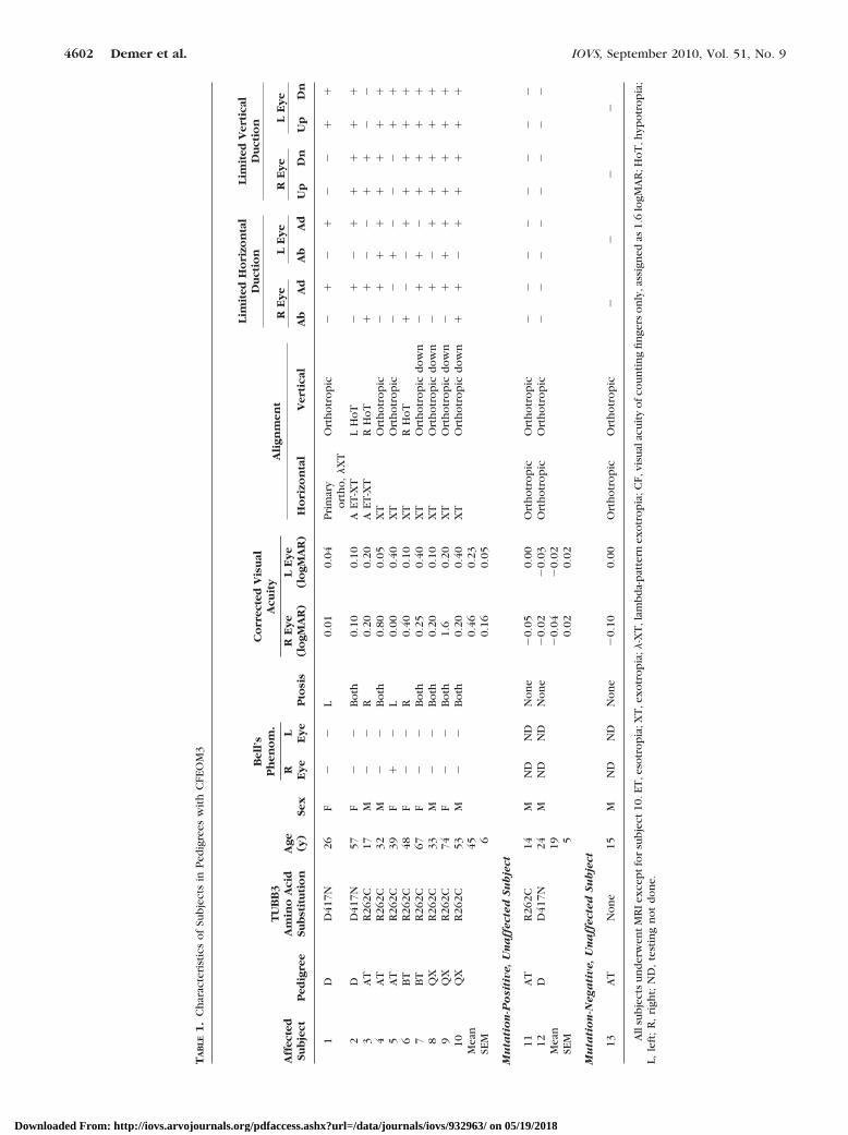
TA
BLE
1.
Ch
arac
teri
stic
so
fSu
bje
cts
inP
edig
rees
wit
hC
FEO
M3
Aff
ecte
dSu
bje
ctP
edig
ree
TU
BB
3A
min
oA
cid
Sub
stit
uti
on
Age (y)
Sex
Bel
l’s
Ph
eno
m.
Pto
sis
Co
rrec
ted
Vis
ual
Acu
ity
Ali
gnm
ent
Lim
ited
Ho
rizo
nta
lD
uct
ion
Lim
ited
Ver
tica
lD
uct
ion
RE
ye
LE
ye
RE
ye
LE
ye
R Ey
eL
Ey
eR
Ey
e(l
ogM
AR
)L
Ey
e(l
ogM
AR
)H
ori
zon
tal
Ver
tica
lA
bA
dA
bA
dU
pD
nU
pD
n
1D
D41
7N26
F�
�L
0.01
0.04
Pri
mar
yo
rth
o,
�X
TO
rth
otr
op
ic�
��
��
��
�
2D
D41
7N57
F�
�B
oth
0.10
0.10
AET
-XT
LH
oT
��
��
��
��
3A
TR
262C
17M
��
R0.
200.
20A
ET-X
TR
Ho
T�
��
��
��
�4
AT
R26
2C32
M�
�B
oth
0.80
0.05
XT
Ort
ho
tro
pic
��
��
��
��
5A
TR
262C
39F
��
L0.
000.
40X
TO
rth
otr
op
ic�
��
��
��
�6
BT
R26
2C48
F�
�R
0.40
0.10
XT
RH
oT
��
��
��
��
7B
TR
262C
67F
��
Bo
th0.
250.
40X
TO
rth
otr
op
icd
ow
n�
��
��
��
�8
QX
R26
2C33
M�
�B
oth
0.20
0.10
XT
Ort
ho
tro
pic
do
wn
��
��
��
��
9Q
XR
262C
74F
��
Bo
th1.
60.
20X
TO
rth
otr
op
icd
ow
n�
��
��
��
�10
QX
R26
2C53
M�
�B
oth
0.20
0.40
XT
Ort
ho
tro
pic
do
wn
��
��
��
��
Mea
n45
0.46
0.23
SEM
60.
160.
05
Mu
tati
on
-Po
siti
ve,
Un
aff
ecte
dSu
bje
ct
11A
TR
262C
14M
ND
ND
No
ne
�0.
050.
00O
rth
otr
op
icO
rth
otr
op
ic�
��
��
��
�12
DD
417N
24M
ND
ND
No
ne
�0.
02�
0.03
Ort
ho
tro
pic
Ort
ho
tro
pic
��
��
��
��
Mea
n19
�0.
04�
0.02
SEM
50.
020.
02
Mu
tati
on
-Neg
ati
ve,
Un
aff
ecte
dSu
bje
ct
13A
TN
on
e15
MN
DN
DN
on
e�
0.10
0.00
Ort
ho
tro
pic
Ort
ho
tro
pic
��
��
All
sub
ject
su
nd
erw
ent
MR
Iex
cep
tfo
rsu
bje
ct10
.ET
,eso
tro
pia
;XT
,ex
otr
op
ia;�
-XT
,lam
bd
a-p
atte
rnex
otr
op
ia;C
F,vi
sual
acu
ity
ofc
ou
nti
ng
fin
gers
on
ly,a
ssig
ned
as1.
6lo
gMA
R;H
oT
,hyp
otr
op
ia;
L,le
ft;
R,
righ
t;N
D,
test
ing
no
td
on
e.
4602 Demer et al. IOVS, September 2010, Vol. 51, No. 9
Downloaded From: http://iovs.arvojournals.org/pdfaccess.ashx?url=/data/journals/iovs/932963/ on 05/19/2018

average age of 20 � 1 years (mean � SEM; range, 14–28). Anadditional 13 control subjects of average age 23 � 1 years(range, 14–33) underwent higher resolution MRI of the opticnerves (ONs) in quasicoronal planes with a 6-cm FOV, and 10subjects underwent MRI of the IO muscles in the quasisagittalplanes. All control subjects had normal ocular and lid motilityand visual acuity in each eye correctable to 0 logarithm of theminimum angle resolvable in arcmin (logMAR, 20/20), or bet-ter.
Clinical Findings in CFEOM3
Overall, subjects with CFEOM3 had slightly subnormal visualacuities. The mean corrected visual acuity of the subjectsexhibiting the CFEOM3 phenotype was 0.29 � 0.08 averagedover the two eyes (Table 1). The mean corrected visual acuityof the two eyes of the three clinically unaffected subjects wasnormal at �0.03 � 0.02 logMAR.
All subjects exhibiting the CFEOM3 phenotype had symmet-rical pupils ranging in diameter from 5 to 8 mm and normalpupillary reactions, without afferent pupillary defect or light-near dissociation. The ophthalmoscopic appearance of the ONhead was normal in all subjects with CFEOM3 except forsubject 3, who had unilateral, and subject 4, who had bilateralON hypoplasia. Unaffected, mutation-negative subject 13 hadunilateral ON hypoplasia.
Subjects with CFEOM3 had unilateral or mild bilateralblepharoptosis. Four had unilateral blepharoptosis, and six hadbilateral blepharoptosis. Seven subjects who exhibited theCFEOM3 phenotype had undergone one to two surgeries eachfor blepharoptosis.
All subjects who exhibited the CFEOM3 phenotype hadundergone 1 to 15 strabismus surgeries. Even after strabismussurgeries, all subjects exhibiting the CFEOM3 phenotype hadlimited supraduction and were exotropic in some gaze posi-tions. Although subjects 1, 4, 5, 7, 8, 9, and 10 had normalvertical binocular alignment, subjects 2, 3, and 6 had hypo-tropia due to asymmetric limitation of supraduction. Affectedsubjects 4, 5, 7, 9, and 10 exhibited abduction of one eye indeorsumversion (binocular downward gaze), and subjects 1 to3 exhibited increased exotropia in deorsumversion, constitut-ing a �- or A-pattern incomitance. Bell’s phenomenon wasabsent in all orbits of affected subjects, with the exception ofsubject 5, who exhibited normal Bell’s phenomenon in herexternally unaffected right eye.
Because of the heterogeneity of the CFEOM3 phenotypewithin pedigrees, informative features are described for eachaffected subject.
Ocular Motility in Subjects Manifesting theCFEOM3 Phenotype
D417N Amino Acid Substitution. Subject 1 from pedi-gree D was a 26-year-old woman who harbored the D417NTUBB3 amino acid substitution and had CFEOM3 and a pro-gressive sensorimotor axonal neuropathy that is common tothis and several other specific TUBB3 mutations.16 She hadundergone surgery for left-side blepharoptosis at 8 months ofage and strabismus surgeries of the left eye at 9 and 11 years.Left eye supraduction and adduction were markedly limited,right eye adduction was mildly limited, and the both eyesabducted markedly during deorsumversion (binocular down-ward gaze, Fig. 1). Levator function was decreased to 4 mm onthe left and was normal at 16 mm on the right.
Subject 2, also from pedigree D, was a 57-year-old womanand the mother of patient 1. She also harbored the D417Namino acid substitution and had both CFEOM3 and later-onsetupper and lower extremity weakness and sensory loss. She wasborn with exotropia, limited supraduction, and blepharoptosisgreater on the right side than on the left. She underwentmultiple eyelid and numerous strabismus surgeries in child-hood and bilateral frontalis suspension surgery for eyelid ele-vation at 27 years of age. The left but not right eye exhibitedhigh myopia and astigmatism, but vision in both eyes wascorrectable with spectacles to 0.1 logMAR. There was fine,horizontal, spontaneous nystagmus in central gaze and conver-gence nystagmus in levoversion. Neither eye supraductedabove horizontal midposition. The left eye adducted markedlyin attempted supraversion and abducted markedly in at-tempted deorsumversion. Levator function was absent on theright and mildly subnormal at 10 mm on the left.
Subject 12 from pedigree D was the 24-year-old son ofsubject 2 and was clinically unaffected, despite harboring theD417N amino acid substitution. Visual acuity in each eye wasnormal after correction of low myopia. Lid position and ocularmotility were normal as was stereopsis when tested by theTitmus method at 40 arcsec. The ONs also appeared normal.The subject had no symptoms of peripheral neuropathy.
R262C Amino Acid Substitution. Subject 3 from pedigreeAT was a 17-year-old boy who harbored the R262C substitutionand had a mild intellectual deficit. He had A-pattern horizontal
FIGURE 1. Subject 1 with the CFEOM3 phenotype, exhibiting left-side blepharoptosis, limited supraduction and adduction, and �-patternexotropia. Ductions were are normal for the right eye. The eyelids were manually elevated for photography in deorsumversion.
IOVS, September 2010, Vol. 51, No. 9 Magnetic Resonance Imaging in CFEOM3 4603
Downloaded From: http://iovs.arvojournals.org/pdfaccess.ashx?url=/data/journals/iovs/932963/ on 05/19/2018
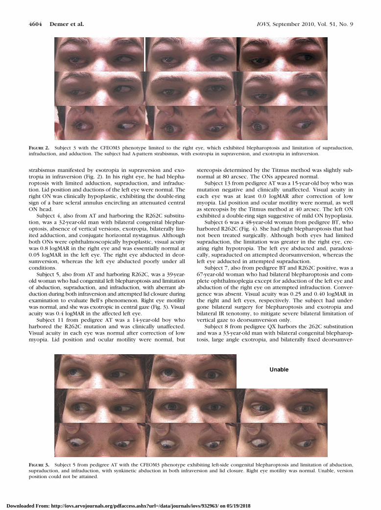
strabismus manifested by esotropia in supraversion and exo-tropia in infraversion (Fig. 2). In his right eye, he had blepha-roptosis with limited adduction, supraduction, and infraduc-tion. Lid position and ductions of the left eye were normal. Theright ON was clinically hypoplastic, exhibiting the double-ringsign of a bare scleral annulus encircling an attenuated centralON head.
Subject 4, also from AT and harboring the R262C substitu-tion, was a 32-year-old man with bilateral congenital blephar-optosis, absence of vertical versions, exotropia, bilaterally lim-ited adduction, and conjugate horizontal nystagmus. Althoughboth ONs were ophthalmoscopically hypoplastic, visual acuitywas 0.8 logMAR in the right eye and was essentially normal at0.05 logMAR in the left eye. The right eye abducted in deor-sumversion, whereas the left eye abducted poorly under allconditions.
Subject 5, also from AT and harboring R262C, was a 39-year-old woman who had congenital left blepharoptosis and limitationof abduction, supraduction, and infraduction, with aberrant ab-duction during both infraversion and attempted lid closure duringexamination to evaluate Bell’s phenomenon. Right eye motilitywas normal, and she was exotropic in central gaze (Fig. 3). Visualacuity was 0.4 logMAR in the affected left eye.
Subject 11 from pedigree AT was a 14-year-old boy whoharbored the R262C mutation and was clinically unaffected.Visual acuity in each eye was normal after correction of lowmyopia. Lid position and ocular motility were normal, but
stereopsis determined by the Titmus method was slightly sub-normal at 80 arcsec. The ONs appeared normal.
Subject 13 from pedigree AT was a 15-year-old boy who wasmutation negative and clinically unaffected. Visual acuity ineach eye was at least 0.0 logMAR after correction of lowmyopia. Lid position and ocular motility were normal, as wellas stereopsis by the Titmus method at 40 arcsec. The left ONexhibited a double-ring sign suggestive of mild ON hypoplasia.
Subject 6 was a 48-year-old woman from pedigree BT, whoharbored R262C (Fig. 4). She had right blepharoptosis that hadnot been treated surgically. Although both eyes had limitedsupraduction, the limitation was greater in the right eye, cre-ating right hypotropia. The left eye abducted and, paradoxi-cally, supraducted on attempted deorsumversion, whereas theleft eye adducted in attempted supraduction.
Subject 7, also from pedigree BT and R262C positive, was a67-year-old woman who had bilateral blepharoptosis and com-plete ophthalmoplegia except for adduction of the left eye andabduction of the right eye on attempted infraduction. Conver-gence was absent. Visual acuity was 0.25 and 0.40 logMAR inthe right and left eyes, respectively. The subject had under-gone bilateral surgery for blepharoptosis and exotropia andbilateral IR tenotomy, to mitigate severe bilateral limitation ofvertical gaze to deorsumversion only.
Subject 8 from pedigree QX harbors the 262C substitutionand was a 33-year-old man with bilateral congenital blepharop-tosis, large angle exotropia, and bilaterally fixed deorsumver-
FIGURE 2. Subject 3 with the CFEOM3 phenotype limited to the right eye, which exhibited blepharoptosis and limitation of supraduction,infraduction, and adduction. The subject had A-pattern strabismus, with esotropia in supraversion, and exotropia in infraversion.
FIGURE 3. Subject 5 from pedigree AT with the CFEOM3 phenotype exhibiting left-side congenital blepharoptosis and limitation of abduction,supraduction, and infraduction, with synkinetic abduction in both infraversion and lid closure. Right eye motility was normal. Unable, versionposition could not be attained.
4604 Demer et al. IOVS, September 2010, Vol. 51, No. 9
Downloaded From: http://iovs.arvojournals.org/pdfaccess.ashx?url=/data/journals/iovs/932963/ on 05/19/2018

sion. He had nearly complete ophthalmoplegia, except forabduction of both eyes and for the transient ability to align theeyes during proximal convergence. Visual acuity was 0.2 and0.1 logMAR for the right and left eyes, respectively. He exhib-ited disconjugate, jerky horizontal nystagmus. Both IR muscleshad been surgically transected before examination.
Subject 9 from pedigree QX harbored the R262C substitu-tion and was a 74-year-old woman with bilateral congenitalblepharoptosis, large angle exotropia, and bilaterally fixed de-orsumversion. She had undergone surgeries for blepharoptosisand strabismus. Visual acuity was counting fingers in the righteye and 0.2 logMAR in the left eye. She had nearly completeophthalmoplegia, except for abduction of the right eye inattempted infraduction. Both eyes incycloducted in attemptedinfraduction, suggesting bilateral SO function. Both IR muscleshad been surgically transected before her examination.
Subject 10 from pedigree QX harbored the R262C substitu-tion and was a 53-year-old man with bilateral congenitalblepharoptosis, large angle exotropia, and bilaterally fixed de-orsumversion. He had undergone surgeries for blepharoptosisand strabismus. Visual acuity was 0.2 in the right eye and 0.4logMAR in the left eye. He had nearly complete ophthalmople-gia, except for abduction of the left eye in attempted infraduc-tion, and limited adduction with slow adducting saccades bi-laterally. Both eyes incycloducted in attempted infraduction,suggesting bilateral SO function. Claustrophobia precludedMRI in this subject.
MRI Findings in CFEOM3
All except subject 10 underwent MRI. The three affectedsubjects (8, 9, and 10) in pedigree QX harbored the R262Csubstitution and exhibited the most severe ocular findings,with exotropia and infraduction and almost total external oph-thalmoplegia, except for residual abduction and limited incy-cloduction on attempted infraduction. Subject 8 couldintermittently achieve binocular alignment by proximal con-vergence. MRI findings in severely affected subjects 8 and 9revealed profound bilateral hypoplasia of the SR, LPS, and MRmuscles (Figs. 5 and 6). Both subjects also exhibited highlyaspheric globes (Fig. 5), a feature that had been observed inmembers of a large CFEOM3 pedigree25 now known to harborthe R262C substitution.16
Other subjects exhibited a less severe, asymmetrical phe-notype. For example, EOMs in the right orbit of subject 1 frompedigree D appeared normal on quasicoronal (Fig. 7) andquasisagittal (Fig. 8) MRIs, yet in the left obit, there was markedhypoplasia of the LPS and the deep portions of the SR, MR, and
IO muscles. Thus, MRI findings in the left but not right orbit ofsubject 1 were compatible with the CFEOM1 phenotype.
Quantitative Features of EOMs
EOM volumes in affected subjects with CFEOM3 were subnor-mal for the SR, MR, and IR (Table 2). This effect was significantat the 0.0002 level for the SR and at the 0.02 level for the MRand IR. IO volume was slightly but not significantly subnormal,and LR volume was slightly but not significantly greater thannormal. Subjects with CFEOM3 who had evidence of surgicalIR disinsertion had displacement of the IO belly without sig-nificant change in IO volume. Such an IO path change wouldnot have been expected to change apparent IO volume. Onaverage, externally unaffected members of families withCFEOM3 had normal EOM volumes. However, the total num-ber of unaffected family members who were studied was small,and externally unaffected subject 12, who carried the D417Nsubstitution, exhibited in his left orbit volume reductions ofthe SR (220 mm3), MR (278 mm3), and IR (246 mm3) that werebelow the 2.5 percentile for normal EOM volumes. Subject 12had no other subnormal EOM volumes, and subjects 11 and 13did not have any subnormal EOM volumes. Subject 12 was thusregarded as carrying the endophenotype of CFEOM3.
Extraocular Muscle Dysplasia
In externally affected subjects 4, 5, 7, 8, and 9, the LR exhibiteda longitudinal fissure separating it into distinct superior andinferior zones. Such fissuring was also evident in externallyunaffected subjects 11 and 12, who carried the R262C andD417N substitutions, respectively. In addition, in the left orbitof subject 12, CN3 was in continuity with the inferior zone ofthe LR, whereas CN6 entered the superior zone. All otherEOMs were free of dysplasia in the remaining subjects withCFEOM3 who underwent MRI.
Globe Position and EOM Paths
Coordinates of the LR, MR, and SR pulleys in subjects exhibit-ing the CFEOM3 phenotype were not significantly differentfrom normal (P � 0.05), and the lateral coordinate of the IRpulley was minimally abnormal, as a likely result of IR disinser-tion surgeries that had been performed in nearly all affectedorbits.
Imaging of Intraorbital Motor Nerves
Orbits affected by CFEOM3 exhibited abnormal motor inner-vation, in approximate proportion to the degree of clinical
FIGURE 4. Subject 6 from pedigree BT with the CFEOM3 phenotype who had right blepharoptosis and right hypotropia, A-pattern strabismus, andparadoxical supraduction and abduction of the left eye on attempted deorsumversion. Both eyes had limited supraduction. Unable, version positioncould not be attained.
IOVS, September 2010, Vol. 51, No. 9 Magnetic Resonance Imaging in CFEOM3 4605
Downloaded From: http://iovs.arvojournals.org/pdfaccess.ashx?url=/data/journals/iovs/932963/ on 05/19/2018

motility restrictions. It was possible to visualize the inferior butnot the superior division of CN3 in the subjects with CFEOM3who were imaged. The superior division was seen, however,only in a subset of normal subjects. In some subjects withCFEOM3, the inferior division of CN3 was too small to trace itsbranches.
Image resolution was insufficient for quantitative analysis ofintraorbital motor nerve size.24 When the nerve was of suffi-cient size to trace, however, MRI demonstrated misinnervationof EOMs in some subjects with CFEOM3. The inferior divisionof CN3 failed to innervate the MR in subjects 2, 3, 6, and 7 andwas in intimate continuity with the inferior zone of the LR aswell its normal target EOMs in subjects 2, 6, 7, and 9 (Fig. 7).In subjects 1, 2, 4, 8, 9, and 10, CN6 entered the superior zoneof the LR, whereas a branch of CN3 was in continuity with theinferior zone (Fig. 7). Contraction of the LR in infraductionwould explain exotropia in that gaze position, with relaxationin supraduction accounting for the associated esotropia in thatgaze position.
Functional imaging in subject 1 was also consistent with LRmisinnervation. In subjects 1 and 2 from pedigree D, one LRexhibited contractile thickening in attempted infraduction(Fig. 9). Although subjects 4, 5, 7, 9, and 10 also exhibitedabduction on attempted infraduction, MRI could not be per-formed in this situation because infraduction to a target couldnot be maintained in the scanner. Misinnervation of the LR bya CN3 branch normally destined for the IR appears to accountfor the A- and �-pattern strabismus in pedigrees D and AT andfor the anomalous abduction in attempted infraduction in ped-igrees BT and QX.
Imaging of Intracranial Motor Nerves
Heavily T2-weighted imaging of the skull base region has suf-ficient resolution to demonstrate easily and consistently thecourse of CN3 of normal subjects.26 CN3s were strikinglyhypoplastic ipsilateral to all affected orbits, except for thenearly normal left CN3 of subject 5. CN3 was of normal size insubject 1’s unaffected right orbit and was of normal size bilat-erally in the clinically unaffected family members, regardless ofmutation status (Table 3). No orbit was affected by the clinicalCFEOM3 phenotype when the subarachnoid oculomotor nerveipsilateral to that orbit had a width of at least 1.9 mm. For
FIGURE 6. Quasi-sagittal T2 FSE MRI in subject 9 with a severeCFEOM3 phenotype, exhibiting severe bilateral hypoplasia of the LPSand SR muscles, as well as bilateral absence of the IO muscle. Anepithelial inclusion cyst and artificial IOL were present in the surgicallytreated left orbit.
FIGURE 5. Quasicoronal T2 FSE MRI in subject 9 with a severeCFEOM3 phenotype. Image planes are 2 mm thick with a 4-mm gapbetween them, arranged from posterior at top to anterior at bottom.There was marked bilateral hypoplasia of the LPS, SR, and MR. The IRwas mildly hypoplastic bilaterally. The SO muscle was preserved. Anepithelial inclusion cyst present near the IR insertion was a probableeffect of prior IR surgery. The globe cross section was bilaterallyirregular and nonspherical. SOT, reflected SO tendon.
4606 Demer et al. IOVS, September 2010, Vol. 51, No. 9
Downloaded From: http://iovs.arvojournals.org/pdfaccess.ashx?url=/data/journals/iovs/932963/ on 05/19/2018

comparison, normal CN3 width measured using this techniqueis 2.01 � 0.36 mm.26 Asymmetry of subarachnoid CN3 size isillustrated in Figure 10 for subject 1. The left CN3 was affectedwith a width of 1.05 mm, whereas on the right side, CN3 wasnormal, with a width of 2.01 mm.
In normal subjects, the heavily T2-weighted imaging tech-nique also demonstrates the course of CN6 after it exits thepons.27,28 The CN6 was unilaterally or bilaterally hypoplastic insubjects 7, 8, and 9 with the CFEOM3 phenotype; abnormali-ties of CN6 were not evident in the remaining subjects.
Optic Nerve
ON cross sections normally decrease anteriorly to posteriorly,because of the reduction of connective tissues surroundingaxon bundles.29 Thus, ON cross section was analyzed asclosely as possible posterior to the globe-ON junction. Subjectswith the CFEOM3 phenotype exhibited mean ON cross sec-tions of 9.19 � 0.60 mm2, which was a 35% reduction andsignificantly smaller than the mean of 14.1 � 0.77 mm2 for 17control ONs (P � 0.00005). All ophthalmoscopically hypoplas-tic ONs exhibited reduced MRI cross sections, yet not all ONswith reduced MRI cross sections were ophthalmoscopicallyhypoplastic. The ophthalmoscopic finding of ON hypoplasiawas concordant with MRI in subject 4, whose right ON had across section of 4.3 and left ON had cross section of 4.8 mm2.The unilateral ophthalmoscopic finding of the double-ring signindicative of right ON hypoplasia in subject 3 also correlatedwith reduction in MRI cross section to 7.5 mm2, comparedwith 13.1 mm2 on the ophthalmoscopically left side. Subject 3also had a dysplastic corpus callosum on sagittal MRI. Theophthalmoscopically hypoplastic left ON of CFEOM3-unaf-fected, mutation-negative subject 13 had a subnormal crosssection of 8.0 mm2, compared with 10.0 for his ophthalmo-scopically normal right ON. However, ONs were ophthalmo-scopically normal in on the right-affected subject 8, whoseright ON cross section was 7.0 mm2, and on the left in affectedsubject 1, whose left ON cross section was 6.7 mm2.
Facial Nerve
The facial nerve was bilaterally normal in all subjects imaged.Consistent with this, no subject exhibited clinical signs of facial
FIGURE 8. Quasisagittal T1 MRI in less affected subject 1, in whomonly the left eye was phenotypically involved. The subject exhibitedhypoplasia of the involved left LPS and SR muscles, as well as severehypoplasia of the left IO muscle in this image plane. The left IR musclehad been surgically transected, displacing the IO nasally.
FIGURE 7. Quasicoronal T1 MRI in phenotypically less affected subject1, with asymmetric hypoplasia of the left LPS and milder hypoplasia ofthe deep portion of the left MR and SR muscles. The inferior divisionof the left oculomotor nerve (CN3) was in contact with the inferiorportion of the left LR muscle (top right). The motor nerve branch tothe right but not left MR was visualized. CN6, abducens nerve; N to IO,motor nerve to inferior oblique muscle.
IOVS, September 2010, Vol. 51, No. 9 Magnetic Resonance Imaging in CFEOM3 4607
Downloaded From: http://iovs.arvojournals.org/pdfaccess.ashx?url=/data/journals/iovs/932963/ on 05/19/2018
weakness, and we have not found facial weakness to cosegre-gate with either the R262C or D417N substitutions.16
DISCUSSION
Employing high-resolution MRI, we demonstrated variable andasymmetric affection of CNs and EOMs correlating closely withthe external findings of CFEOM3. This correlation confirms andextends imaging findings in other CCDDs that manifest ocularmotility disorders. The most severely affected phenotypes inCFEOM3 may be indistinguishable from CFEOM1, both exter-nally and in the internal involvement of EOMs and associatednerves by orbital MRI. These phenotypes include severe bilat-eral hypoplasia of the SR, LPS, MR, and occasionally the IO,with anatomic contiguity suggestive of misinnervation of theLR by CN3 and marked bilateral hypoplasia of the subarach-noid CN3. Unlike individuals with CFEOM1, some withCFEOM3 who are less severely affected may exhibit only uni-lateral, relatively mild hypoplasia of a few EOMs, with lesssevere hypoplasia of the subarachnoid portion of CN3.
External Ocular Phenotype of CFEOM3
Affected subjects had a range of presentations, typically withexotropia in at least one gaze position and limited supraduc-tion in at least one eye. Most, but not all, subjects also had
either vertical gaze restricted to deorsumversion or unilateralhypotropia. None had pupillary abnormalities. Corrected visualacuities averaged approximately 0.3 logMAR (20/40 Snellen),despite strabismus and occasional clinically evident ON hyp-oplasia. The mild nystagmus present in some subjects wassimilar to that observed in association in early-onset strabismus.No affected subject had vision binocularly impaired to thedegree that nystagmus might be attributable to sensory loss. Ifpresented as isolated cases, subject 1 from pedigree D andsubject 5 from pedigree AT might be considered to haveunilateral partial CN3 palsies. At the other end of the spectrum,subject 7 from pedigree BT and subjects 8, 9, and 10 frompedigree QX exhibited profound bilateral hypotropia andblepharoptosis with A- or �-pattern exotropia similar to thepresentation of a subset of subjects with CFEOM1. Bell’s phe-nomenon was uniformly absent in affected eyes. Thus, theexternal phenotype of CFEOM3 can vary widely among af-fected individuals within CFEOM3 families. However, severecases of CFEOM3 always present with exotropia and verticalgaze limited to deorsumversion.
Structural Correlations in CFEOM3
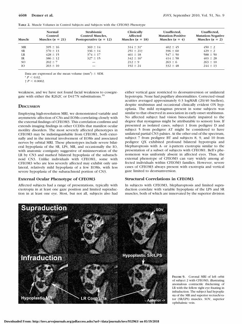
In subjects with CFEOM3, blepharoptosis and limited supra-duction correlate with variable hypoplasia of the LPS and SRmuscles, both of which are innervated by the superior division
FIGURE 9. Coronal MRI of left orbitof subject 2 with CFEOM3, illustratinganomalous contractile thickening ofLR with the fellow right eye fixating ininfraduction. The subject had hypopla-sia of the MR and superior rectus/leva-tor (SR/LPS) muscles. SOV, superiorophthalmic vein.
TABLE 2. Muscle Volumes in Control Subjects and Subjects with the CFEOM3 Phenotype
Muscle
NormalControl
Muscles (n � 21)
StrabismicControl Muscles,
Postoperative (n � 12)
ClinicallyAffected
Muscles (n � 18)
Unaffected,Mutation-PositiveMuscles (n � 4)
Unaffected,Mutation-NegativeMuscles (n � 2)
MR 395 � 16 369 � 14 314 � 31* 402 � 45 450 � 2SR 370 � 13 336 � 14 251 � 21† 396 � 60 429 � 2LR 428 � 15 374 � 17 461 � 18 547 � 50 588 � 50IR 386 � 12 327 � 15 342 � 16* 414 � 58 403 � 28SO 202 � 7 — 212 � 9 263 � 6 263 � 10IO 263 � 10 — 192 � 24 332 � 48 244 � 13
Data are expressed as the mean volume (mm3) � SEM.* P � 0.02.† P � 0.0002.
4608 Demer et al. IOVS, September 2010, Vol. 51, No. 9
Downloaded From: http://iovs.arvojournals.org/pdfaccess.ashx?url=/data/journals/iovs/932963/ on 05/19/2018

of CN3. Other EOMs exhibiting clinically deficient function,such as the MR and IR, also exhibit hypoplasia that correlatesin severity with the corresponding functional deficit. This EOMhypoplasia is also found in typical cases of CFEOM124 andoccasionally in cases of DURS2,28 which are CCDD disordersdue to mutation in KIF21A5 and CHN1,30 respectively. Curi-ously, EOM hypoplasia is not a feature of Duane radial raysyndrome,27 another form of Duane syndrome caused by mu-tation in the zinc finger transcription factor SALL4.31
While his visual function and ocular motility were entirelynormal, externally unaffected subject 12 with the D417N sub-stitution exhibited the endophenotype of significant unilateralhypoplasia of multiple EOMs innervated by CN3. Subjects 1and 2 with this same substitution exhibited unilateral blepha-roptosis, bilateral ocular motility abnormalities, and A- or �-pat-tern strabismus. These different presentations highlight thevariability in CFEOM3 phenotype resulting from the same mu-tation, even within the same pedigree.
Aspheric globes were observed in subjects 8 and 9, neitherof whom was highly myopic. Little is known about globeasphericity in the absence of high myopia.32 Hemiretinal de-privation33 and regional defocus34 can also create asphericglobes by induction of local eye shape changes in primates.Conceivably, both of these factors could have been present insubjects 8 and 9. It is also possible that abnormal EOM forcesin CFEOM altered globe shape or that unknown factors con-tribute.
Oculomotor Nerve Hypoplasia
Although the number of subjects was small, evidence suggestsa threshold effect of subarachnoid CN3 width in CFEOM3 onmanifestation of EOM hypoplasia. The external phenotype ofblepharoptosis and impaired motility is most commonly evi-dent when CN3 width is less than 1.0 mm. Subarachnoid CN3width in the internally affected left orbit of subject 12, who hadno external phenotype was 2.0 mm, similar to the width ofCN3 in the unaffected right orbit and to that in the unaffectedleft orbit of mutation-negative subject 13. The current, variableMRI findings of small subarachnoid CN3 supports previousautopsy findings showing a decrease in axon number andoverall size in the proximal common trunk of CN3 inCFEOM1.2 Since the CN3 cross-sectional area and its maximum
possible number of axons varies with the square of the CN3radius, it may be estimated that the CFEOM phenotype is likelyto be manifested when the total number of axons in CN3 isreduced to less than approximately 25% of normal. Of course,this reduction need not be homogeneous and, for example,may be more severe in the superior than in the inferior divisionof CN3. The separate divisions were not distinguishable in thisstudy by overall measurements of subarachnoid CN3 width.Like CFEOM1, the SR and LPS EOMs innervated by the superiordivision of CN3 were most severely affected by CFEOM3. Theoblique EOMs were more likely to be spared in CFEOM3, butmight be involved.
LR Muscle
The present findings confirm widespread LR misinnervation inCFEOM3. Particularly common was A-pattern exotropia, in-creasing in deorsumversion. MRI confirmed that this phenom-enon is due to LR contraction in infraduction, presumablybecause a branch of CN3 normally destined to innervate the IRmuscle also innervates the LR. This finding is typical of bothCFEOM1 resulting from KIF21A mutations,24 and chromosome2-linked Duane’s syndrome (DURS2),28 which we recentlydemonstrated to result from gain-of-function missense muta-tions in the signaling protein �-2-chimerin, which is responsi-ble for ocular motor axon pathfinding.30 Similarly, CFEOM3caused by TUBB3 mutations results in axon guidance defectsin mouse.16 Overall, these findings further support a primarydefect in cranial motor neuron axon guidance in CFEOM3 andother closely related strabismus disorders.
In several CCDDs, particularly CFEOM124 and DURS2,28 thedeep LR muscle is commonly dysplastic, typically split longi-tudinally into superior and inferior portions or otherwise struc-turally disorganized. Dysplasia of the LR was observed in sub-jects 2, 4, 7, 8, 9, and 12, but this finding was less prominentthan in the other CCDDs. We proposed in earlier work thatnormal CN6 innervation is essential for normal structure of thedeep LR.28 We proposed that variability in severity and patternof ophthalmoplegia in DURS2 depends on the degree to whichresidual axons of the hypoplastic inferior division of CN3 reachtheir normal target EOMs or are misdirected to innervate the
FIGURE 10. Heavily T2-weighted MRI of the subarachnoid oculomotornerve (CN3) in subject 1 with a unilateral left CFEOM3 phenotype. Theright CN3 was normal at 2.0 mm wide, whereas the left CN3 wassubnormal at 1.0 mm wide.
TABLE 3. Duction Abnormalities and Oculomotor Nerve Width inMembers of Families Affected by CFEOM3
Subject
DuctionAbnormality
SubarachnoidOculomotor
NerveWidth (mm)
Right Eye Left Eye Right Left
1 � � 2.01 1.052 � � 0.87 0.703 � � 1.18 2.904 � � 1.09 1.045 � � 1.90 1.796 � � 0.82 0.977 � � 0.35 0.698 � � 0.62 0.929 � � 0.42 0.73
10 � � N.D. ND11 � � 2.14 2.4612 � � 2.01 2.0113 � � 2.27 1.98
Normal mean 2.01Normal SD 0.36
IOVS, September 2010, Vol. 51, No. 9 Magnetic Resonance Imaging in CFEOM3 4609
Downloaded From: http://iovs.arvojournals.org/pdfaccess.ashx?url=/data/journals/iovs/932963/ on 05/19/2018

LR.28 It seems reasonable to suppose that CFEOM3 is due toone or several cranial nerve targeting abnormalities that arerelatively specific for CN3.
SO Muscle
The SO is hypoplastic in acquired and congenital SO palsy,17,35
commonly hypoplastic in CFEOM1,24 occasionally hypoplasticin DURS2,28 but not hypoplastic in DRRS.27 Mean SO volume ispreserved in subjects exhibiting the CFEOM3 phenotype andin externally unaffected carriers of the R262C and D417Nsubstitutions (subjects 11 and 12). No subject in the presentstudy had SO volume less than the 5th percentile of normal.
Optic Nerve Involvement in CFEOM3
We have found quantitative MRI useful for ON analysis.29 Sizeof the ON is normal in DRRS.27 However, subjects withCFEOM1 consistently exhibit a subclinical reduction from nor-mal of approximately 30% to 40% in ON cross section,24 andsubjects with dominant Duane syndrome linked to chromo-some 2 (DURS2) average a 25% reduction in ON cross sec-tion.28 Two of the subjects with CFEOM3 and unaffected,mutation-negative subject 13 exhibited clinically evident ONhypoplasia on ophthalmoscopy, but typical subjects affectedwith CFEOM3 manifest only subclinical ON hypoplasia. Thepresent finding of a significant but usually subclinical 35%reduction in ON cross section in CFEOM3 is similar. Notably,ON hypoplasia can cosegregate with polymicrogyria resultingfrom homozygous truncating mutations in TUBA8, suggestingthat this phenotype is commonly associated with tubulin mu-tations in general. No subject with CFEOM3 has a gross visualfield deficit or afferent pupillary defect. The only marked visualloss in the subjects with CFEOM3 is due to amblyopia associ-ated with anisometropia and strabismus.
Absence of Widespread Pulley Abnormalities
Pulley disorders can cause strabismus.17,36 Markedly abnormalrectus EOM paths due to misplaced pulleys are associated withcraniosynostosis syndromes that are caused by mutations inFGFR37 that spare orbital nerves and EOM volumes. InCFEOM1,24 DRRS,27 and DURS2,28 EOM paths are normal oronly minimally abnormal. Overall, rectus pulley positions inthe coronal plane remain remarkably normal in CFEOM3, de-spite severe congenital dysinnervation. This finding furtherbuttresses the accumulating genetic evidence that normal EOMinnervation is not requisite for establishment and maintenanceof normal rectus pulley positions.24,27,28 It provides additionalevidence favoring the idea that pulley abnormalities are pri-mary in cases of incomitant strabismus with which they areassociated38–40 and that pulley heterotopy is unlikely to besecondary to acquired EOM denervation.39
Fundamental Implications
We previously reported that subjects harboring the TUBB3mutations R262C and D417N can have thinning of the anteriorcommissure and corpus callosum and, as found in subjects 1and 2, D417N can result in a peripheral axonal neuropathy.16
Subject 3 exhibited localized corpus callosum thinning anddysplasia, as well as in intellectual deficit. TUBB3 mutationsnot represented in this study can be associated with moresevere structural brain abnormalities and fairly consistent andsevere external ophthalmoplegia.16 Thus, it is possible that theocular motor endophenotype associated with other mutationsdiffers from the present cases of CFEOM3, but we anticipatethat it would be similar to the more severely affected subjectsincluded in this study.
Overall, these findings suggest that CFEOM3 is an asymmet-ric, variably penetrant, congenital cranial dysinnervation disor-der characterized mainly by oculomotor nerve hypoplasia thataffects the superior more than the inferior division and leads tosecondary atrophy of EOMs, with associated occasional CN6and mild ON hypoplasia. The CFEOM3 ocular phenotype pre-senting asymmetrically may closely resemble incomitant stra-bismus due to a variety of etiologies, including trauma17 andanomalous EOM bands.41 High-resolution imaging would beuseful in recognition of the MRI features of CFEOM3 in casespresenting outside of well-established dominant pedigrees andmay reveal clinically unsuspected structural abnormalities ofEOMs and their innervation.
References
1. Gutowski NJ, Bosley TM, Engle EC. The congenital cranial dysin-nervation disorders (CCDDs). 110th ENMCC International Work-shop, Naarden, The Netherlands, October, 25–27, 2002. Neuro-musc Dis. 2003;13:573–578.
2. Engle EC, Goumnerov BC, McKeown CA, et al. Oculomotor nerveand muscle abnormalities in congenital fibrosis of the extraocularmuscles. Ann Neurol. 1997;41:314–325.
3. Engle EC, Kunkel LM, Specht LA, Beggs AH. Mapping a gene forcongenital fibrosis of the extraocular muscles to the centromericregion of chromosome 12. Nat Genet. 1994;7:69–73.
4. Engle EC, Marondel I, Houtman WA, et al. Congenital fibrosis ofthe extraocular muscles (autosomal dominant congenital externalophthalmoplegia): genetic homogeneity, linkage refinement, andphysical mapping on chromosome 12. Am J Hum Genet. 1995;58:1086–1094.
5. Yamada K, Andrews C, Chan W-M, et al. Heterozygous mutationsof the kinesin KIF21A in congenital fibrosis of the extraocularmuscles type 1 (CFEOM1). Nat Genet. 2003;35:318–321.
6. Wang SM, Zwann J, Mullaney PB, et al. Congenital fibrosis of theextraocular muscles type 2, an inherited exotropic strabismusfixus, maps to distal 11q13. Am J Hum Genet. 1998;63:517–525.
7. Nakano M, Yamada K, Fain J, et al. Homozygous mutations inARIX(PHOX2A) result in congenital fibrosis of the extraocularmuscles type 2. Nat Genet. 2001;29:315–320.
8. Pattyn A, Morin X, Cremer H, Goridis C, Brunet JF. Expression andinteractions of the two closely related homeobox genes Phox2Aand Phox2b during neurogenesis. Development. 1997;124:4065–4075.
9. Guo S, Brush J, Teraoka H, et al. Development of a noradrenergicneurons in the zebrafish hindbrain requires BMP, FGF8, and thehomeodomain protein soulless/Phox2a. Neuron. 1999;24:555–566.
10. Bosley TM, Oystreck DT, Robertson RL, al Awad A, Abu-Amero K,Engle EC. Neurological features of congenital fibrosis of the ex-traocular muscles type 2 with mutations in PHOX2A. Brain. 2006;129:2363–2374.
11. Doherty EJ, Macy ME, Wang SM, Dykman CP, Melanson MT, EngleEC. CFEOM3: A new extraocular congenital fibrosis syndrome thatmaps to 16q24.2-q24.3. Invest Ophthalmol Vis Sci. 1999;40:1687–1694.
12. Engle EC, McIntosh N, Yamada K, et al. CFEOM1, the classicfamilial form of congenital fibrosis of the extraocular muscles, isgenetically heterogeneous but does not result from mutations inARIX. BMC Genet. 2002;3:3.
13. Mackey DA, Chan WM, Chan C, et al. Congenital fibrosis of thevertically acting extraocular muscles maps to the FEOM3 locus.Hum Genet. 2002;110:510–512.
14. Sener EC, Lee BA, Turgut B, Akarsu AN, Engle EC. A clinicallyvariant fibrosis syndrome in a Turkish family maps to the CFEOM1locus on chromosome 12. Arch Ophthalmol. 2000;118:1090–1097.
15. Yamada K, Chan W-W, Andrews C, et al. Identification of KIF21Amutations as a rare cause of congenital fibrosis of the extraocularmuscles type 3 (CFEOM3). Invest Ophthalmol Vis Sci. 2004;45:2218–2223.
4610 Demer et al. IOVS, September 2010, Vol. 51, No. 9
Downloaded From: http://iovs.arvojournals.org/pdfaccess.ashx?url=/data/journals/iovs/932963/ on 05/19/2018

16. Tischfield MA, Baris HN, Gupta ML, et al. Human TUBB3 mutationsperturb microtubule dynamics, kinesin interactions, and neuronalcircuitry. Cell. 2010;140:74–87.
17. Demer JL. A 12 year, prospective study of extraocular muscleimaging in complex strabismus. J AAPOS. 2003;6:337–347.
18. Seitz J, Held P, Strotzer M, et al. MR imaging of cranial nervelesions using six different high-resolution T1 and T2(*)-weighted3D and 2D sequences. Acta Radiol. 2002;43:349–353.
19. Demer JL, Miller JM. Orbital imaging in strabismus surgery. In: Rosen-baum AL, Santiago AP, eds. Clinical Strabismus Management: Prin-ciples and Techniques. Philadelphia: WB Saunders; 1999:84–98.
20. Clark RA, Miller JM, Demer JL. Three-dimensional location ofhuman rectus pulleys by path inflections in secondary gaze posi-tions. Invest Ophthalmol Vis Sci. 2000;41:3787–3797.
21. Demer JL, Ortube MC, Engle EC, Thacker N. High resolutionmagnetic resonance imaging demonstrates abnormalities of motornerves and extraocular muscles in patients with neuropathic stra-bismus. J AAPOS. 2006;10:135–142.
22. Demer JL, Narasimhan A. T2-weighted magnetic resonance imag-ing (MRI) of extraoccular muscles (EOMs). Abstracts of 35th An-nual Meeting of the American Association for Pediatric Ophthal-mology and Strabismus. 2009:Abstract 56.
23. Demer JL, Oh SY, Clark RA, Poukens V. Evidence for a pulley of theinferior oblique muscle. Invest Ophthalmol Vis Sci. 2003;44:3856–3865.
24. Demer JL, Clark RA, Engle EC. Magnetic resonance imaging evi-dence for widespread orbital dysinnervation in congenital fibrosisof extraocular muscles due to mutations in KIF21A. Invest Oph-thalmol Vis Sci. 2005;46:530–539.
25. Gillies WE, Harris AJ, Brooks AM, Rivers MR, Wolfe RJ. Congenitalfibrosis of the vertically acting extraocular muscles: a new group ofdominantly inherited ocular fibrosis with radiologic findings. Oph-thalmology. 1995;102:607–612.
26. Lim KH, Engle EC, Demer JL. Abnormalities of the oculomotornerve in congenital fibrosis of the extraocular muscles and con-genital oculomotor palsy. Invest Ophthalmol Vis Sci. 2007;48:1601–1606.
27. Demer JL, Clark RA, Lim K-H, Engle EC. Magnetic resonanceimaging of innervational and extraocular muscle abnormalities inDuane-radial ray syndrome. Invest Ophthalmol Vis Sci. 2007;48:5505–5511.
28. Demer JL, Clark RA, Lim KH, Engle EC. Magnetic resonance imag-ing evidence for widespread orbital dysinnervation in dominantDuane’s retraction syndrome linked to the DURS2 locus. InvestOphthalmol Vis Sci. 2007;48:194–202.
29. Karim S, Clark RA, Poukens V, Demer JL. Quantitative magneticresonance imaging and histology demonstrates systematic varia-tion in human intraorbital optic nerve size. Invest Ophthalmol VisSci. 2004;45:1047–1051.
30. Miyake N, Chilton J, Psatha M, et al. Human CHN1 mutationshyperactivate alpha2-chimaerin and cause Duane’s retraction syn-drome. Science. 2008;321:839–843.
31. Al-Baradie R, Yamada K, St Hilaire C, et al. Duane radial raysyndrome (Okihiro syndrome) maps to 20q13 and results frommutations in SALL4, a new member of the SAL family. Am J HumGenet. 2002;71:1195–1199.
32. Atchison DA, Jones CE, Schmid KL, et al. Eye shape in emmetropiaand myopia. Invest Ophthalmol Vis Sci. 2004;45:3380–3386.
33. Smith EL, Huang J, Hung LF, et al. Hemiretinal form deprivation:evidence for local control of eye growth and refractive develop-ment in infant monkeys. Invest Ophthalmol Vis Sci. 2009;50:5057–5069.
34. Smith EL, Hung LF, Huang J, Blasdel TL, Humbird TL, BockhorstKH. Optical defocus influences refractive development in mon-keys via local, regionally selective mechanisms. Invest OphthalmolVis Sci. Published online March 10, 2010.
35. Demer JL, Miller MJ, Koo EY, Rosenbaum AL, Bateman JB. Trueversus masquerading superior oblique palsies: muscle mechanismsrevealed by magnetic resonance imaging. In: Lennerstrand G, ed.Update on Strabismus and Pediatric Ophthalmology. Boca Ra-ton, FL: CRC Press; 1995:303–306.
36. Oh SY, Clark RA, Velez F, Rosenbaum AL, Demer JL. Incomitantstrabismus associated with instability of rectus pulleys. InvestOphthalmol Vis Sci. 2002;43:2169–2178.
37. Muller U, Steinberger D, Kunze S. Molecular genetics of cranio-synostotic syndromes. Graefes Arch Clin Exp Ophthalmol. 1997;235:545–550.
38. Clark RA, Miller JM, Rosenbaum AL, Demer JL. Heterotopic musclepulleys or oblique muscle dysfunction? J AAPOS. 1998;2:17–25.
39. Clark RA, Miller JM, Demer JL. Displacement of the medial rectuspulley in superior oblique palsy. Invest Ophthalmol Vis Sci. 1998;39:207–212.
40. Demer JL, Clark RA, Miller JM. Heterotopy of extraocular musclepulleys causes incomitant strabismus. In: Lennerstrand G, ed. Ad-vances in Strabismology. Buren, The Netherlands: Aeolus Press;1999:91–94.
41. Khitri MR, Demer JL. Magnetic resonance imaging of tissues compat-ible with supernumerary extraocular muscles. Am J Ophthalmol. Inpress.
IOVS, September 2010, Vol. 51, No. 9 Magnetic Resonance Imaging in CFEOM3 4611
Downloaded From: http://iovs.arvojournals.org/pdfaccess.ashx?url=/data/journals/iovs/932963/ on 05/19/2018