evidence of an epithelial stem/progenitor cell …evidence of an epithelial stem/progenitor cell...
TRANSCRIPT

Evidence of an epithelial stem/progenitor cellhierarchy in the adult mouse lungJonathan L. McQualtera,b,1, Karen Yuena, Brenda Williamsa, and Ivan Bertoncelloa,b
aAustralian Stem Cell Centre, Clayton, Victoria 3800, Australia; and bDepartment of Anatomy and Developmental Biology, Monash University, Clayton,Victoria 3800, Australia
Edited by Darwin J. Prockop, Texas A&M Health Science Center, Temple, TX, and approved December 8, 2009 (received for review August 13, 2009)
The role of lung epithelial stem cells in maintenance and repair ofthe adult lung is ill-defined, and their identity remains contentiousbecause of the lack of definitive markers for their prospectiveisolation and the absence of clonogenic assays able to measuretheir stem/progenitor cell potential. In this study, we show thatreplication of epithelial–mesenchymal interactions in a previouslyundescribed matrigel-based clonogenic assay enables the identifi-cation of lung epithelial stem/progenitor cells by their colony-forming potential in vitro. We describe a population of EpCAMhi
CD49fpos CD104pos CD24low epithelial cfus that generate coloniescomprising airway, alveolar, or mixed lung epithelial cell lineageswhen cocultured with EpCAMneg Sca-1pos lung mesenchymal cells.We show that soluble fibroblast growth factor-10 and hepatocytegrowth factor partially replace the requirement for mesenchymalsupport of epithelial colony formation, allowing clonal passagingand demonstration of their capacity for self-renewal. These datasupport a model in which the adult mouse lung contains a minorpopulation of multipotent epithelial stem/progenitor cells withthe capacity for self-renewal and whose descendants give rise toairway and alveolar epithelial cell lineages in vitro.
colony-forming assay | lung epithelium | lineage specificity |differentiation | EpCAM
During lung development in the mouse, progenitors of theanterior foregut endoderm undergo directed differentiation
to establish distinct respiratory epithelial cell compartments. Thetracheobronchial airways are composed predominantly of cili-ated, Clara, and basal cells as well as a lesser number of gobletand neuroendocrine cells. Submucosal glands are restricted tothe highest reaches of the cartilaginous tracheal airway. In thedistal lung, the bronchiolar epithelium is composed largely ofClara and ciliated cells with intermittent clusters of neuro-endocrine cells, whereas type I and type II alveolar epithelialcells (AEC I/II) make up the gas exchange surface of the alveoli.However, in comparison to our understanding of the mecha-nisms regulating epithelial cell proliferation and differentiationduring lung development (1), our knowledge of the organizationand regulation of endogenous lung stem and progenitor cellsinvolved in maintenance, remodeling, regeneration, and repairof the postnatal lung is relatively poor.For themost part, cell lineage tracing studies and the analysis of
mouse lung injury models suggest that the adult lung epithelium ismaintained by divergent progenitor cells residing in discretemicroenvironmental niches along the proximal-distal axis of therespiratory tree (2), consistent with the existence of a “non-classical” stem cell hierarchy in which relatively quiescent differ-entiated progenitor cells function as facultative stem cells (3).However, Rawlins et al. (4) recently provided compelling evidenceof the existence of multipotent stem/progenitor cells in the fetallung distal tip that are able to self-renew and contribute to all lungepithelial cell lineages during development. Others have alsoposited that the processes of adult lung regeneration and repairare regulated by highly conserved mechanisms that recapitulateontogeny (5). These processes are key to understanding normaland pathophysiological lung stem/progenitor cell behavior, which,
in turn, leads to the identification of cellular and molecular ther-apeutical targets that could be exploited for the attenuation orreversal of intractable lung diseases. However, their elucidationhas been confounded by a lack of specific markers and functionalassays for the prospective isolation and characterization of adultlung epithelial stem and progenitor cells and the measurement oftheir proliferative and differentiative potential.In this study, we describe a previously undescribed and robust
organotypic epithelial colony-forming assay which has enabledus to identify and characterize a minor population of renewingEpCAMhi CD49fpos CD104pos CD24low epithelial cfus in theadult mouse lung which give rise to more committed clonogenicairway and alveolar epithelial progenitor cells in vitro.
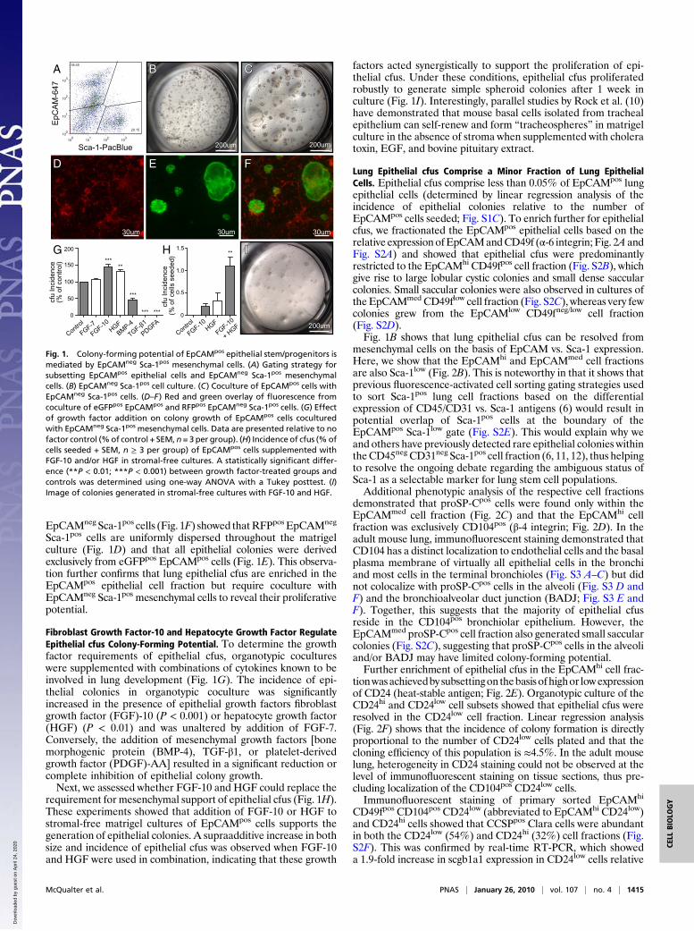
ResultsMesenchymal Progenitor Cells Regulate the Growth of Lung EpithelialStem/Progenitor Cells in Vitro. To separate lung epithelial cellsfrom mesenchymal cells (6), we sorted nonhematopoietic(CD45neg) and nonendothelial (CD31neg) cells from dis-aggregated adult lung cell suspensions on the basis of the dif-ferential expression of a panepithelial cell marker (EpCAM;CD326) and the Sca-1 antigen. We show that EpCAMpos epi-thelial cells (40.6 ± 2.0%, mean ± SEM, n = 7) are easilyresolved from EpCAMneg Sca-1pos mesenchymal cells (Fig. 1A),which generate mesenchymal cell colonies in matrigel (Fig. 1B)defined by Oil-Red O (lipofibroblasts) and α-smooth muscleactin (SMA; myofibroblasts) expression (Fig. S1A).In contrast, EpCAMpos cells did not exhibit clonogenic growth
when cultured alone inmatrigel but did generate complex epithelialcell colonies (Fig. 1C) when cocultured with EpCAMneg Sca-1pos
mesenchymal cells. Significantly, when EpCAMneg Sca-1pos mes-enchymal cells were grown in the lower chamber of the matrigeltranswell culture separated from EpCAMpos epithelial cells, epi-thelial colony formation was not supported, demonstrating thatthe recapitulation of physiological epithelial mesenchymal celland/or paracrine interactions is critical for lung epithelial colonyformation in vitro. Counterstaining of cocultures for α-SMAshowed that αSMApos mesenchymal cells were tightly wrappedaround epithelial colonies (Fig. S1B), also suggesting that, as inlungmorphogenesis during development, adult lung epithelial cellfate specification is orchestrated by smooth muscle progenitors inthe lung mesenchyme (7–9).
Epithelial cfus Are Enriched in the EpCAMpos Cell Fraction. Cellmixing experiments using eGFPpos EpCAMpos cells and RFPpos
Author contributions: J.L.M. and I.B. designed research; J.L.M., K.Y., and B.W. performedresearch; J.L.M. and I.B. contributed new reagents/analytic tools; J.L.M. and I.B. analyzeddata; and J.L.M. and I.B. wrote the paper.
The authors declare no conflict of interest.
This article is a PNAS Direct Submission.1To whom correspondence should be addressed at: Australian Stem Cell Centre, ThirdFloor, Building 75 (STRIP), Monash University, Wellington Road, Clayton, Victoria 3800,Australia. E-mail: [email protected].
This article contains supporting information online at www.pnas.org/cgi/content/full/0909207107/DCSupplemental.
1414–1419 | PNAS | January 26, 2010 | vol. 107 | no. 4 www.pnas.org/cgi/doi/10.1073/pnas.0909207107
Dow
nloa
ded
by g
uest
on
Apr
il 24
, 202
0
EpCAMneg Sca-1pos cells (Fig. 1F) showed thatRFPpos EpCAMneg
Sca-1pos cells are uniformly dispersed throughout the matrigelculture (Fig. 1D) and that all epithelial colonies were derivedexclusively from eGFPpos EpCAMpos cells (Fig. 1E). This observa-tion further confirms that lung epithelial cfus are enriched in theEpCAMpos epithelial cell fraction but require coculture withEpCAMneg Sca-1pos mesenchymal cells to reveal their proliferativepotential.
Fibroblast Growth Factor-10 and Hepatocyte Growth Factor RegulateEpithelial cfus Colony-Forming Potential. To determine the growthfactor requirements of epithelial cfus, organotypic cocultureswere supplemented with combinations of cytokines known to beinvolved in lung development (Fig. 1G). The incidence of epi-thelial colonies in organotypic coculture was significantlyincreased in the presence of epithelial growth factors fibroblastgrowth factor (FGF)-10 (P < 0.001) or hepatocyte growth factor(HGF) (P < 0.01) and was unaltered by addition of FGF-7.Conversely, the addition of mesenchymal growth factors [bonemorphogenic protein (BMP-4), TGF-β1, or platelet-derivedgrowth factor (PDGF)-AA] resulted in a significant reduction orcomplete inhibition of epithelial colony growth.Next, we assessed whether FGF-10 and HGF could replace the
requirement for mesenchymal support of epithelial cfus (Fig. 1H).These experiments showed that addition of FGF-10 or HGF tostromal-free matrigel cultures of EpCAMpos cells supports thegeneration of epithelial colonies. A supraadditive increase in bothsize and incidence of epithelial cfus was observed when FGF-10and HGF were used in combination, indicating that these growth
factors acted synergistically to support the proliferation of epi-thelial cfus. Under these conditions, epithelial cfus proliferatedrobustly to generate simple spheroid colonies after 1 week inculture (Fig. 1I). Interestingly, parallel studies by Rock et al. (10)have demonstrated that mouse basal cells isolated from trachealepithelium can self-renew and form “tracheospheres” in matrigelculture in the absence of stroma when supplemented with choleratoxin, EGF, and bovine pituitary extract.
Lung Epithelial cfus Comprise a Minor Fraction of Lung EpithelialCells. Epithelial cfus comprise less than 0.05% of EpCAMpos lungepithelial cells (determined by linear regression analysis of theincidence of epithelial colonies relative to the number ofEpCAMpos cells seeded; Fig. S1C). To enrich further for epithelialcfus, we fractionated the EpCAMpos epithelial cells based on therelative expression ofEpCAMandCD49f (α-6 integrin; Fig. 2A andFig. S2A) and showed that epithelial cfus were predominantlyrestricted to the EpCAMhi CD49fpos cell fraction (Fig. S2B), whichgive rise to large lobular cystic colonies and small dense saccularcolonies. Small saccular colonies were also observed in cultures ofthe EpCAMmed CD49flow cell fraction (Fig. S2C), whereas very fewcolonies grew from the EpCAMlow CD49fneg/low cell fraction(Fig. S2D).Fig. 1B shows that lung epithelial cfus can be resolved from
mesenchymal cells on the basis of EpCAM vs. Sca-1 expression.Here, we show that the EpCAMhi and EpCAMmed cell fractionsare also Sca-1low (Fig. 2B). This is noteworthy in that it shows thatprevious fluorescence-activated cell sorting gating strategies usedto sort Sca-1pos lung cell fractions based on the differentialexpression of CD45/CD31 vs. Sca-1 antigens (6) would result inpotential overlap of Sca-1pos cells at the boundary of theEpCAMpos Sca-1low gate (Fig. S2E). This would explain why weand others have previously detected rare epithelial colonies withintheCD45neg CD31neg Sca-1pos cell fraction (6, 11, 12), thus helpingto resolve the ongoing debate regarding the ambiguous status ofSca-1 as a selectable marker for lung stem cell populations.Additional phenotypic analysis of the respective cell fractions
demonstrated that proSP-Cpos cells were found only within theEpCAMmed cell fraction (Fig. 2C) and that the EpCAMhi cellfraction was exclusively CD104pos (β-4 integrin; Fig. 2D). In theadult mouse lung, immunofluorescent staining demonstrated thatCD104 has a distinct localization to endothelial cells and the basalplasma membrane of virtually all epithelial cells in the bronchiand most cells in the terminal bronchioles (Fig. S3 A–C) but didnot colocalize with proSP-Cpos cells in the alveoli (Fig. S3 D andF) and the bronchioalveolar duct junction (BADJ; Fig. S3 E andF). Together, this suggests that the majority of epithelial cfusreside in the CD104pos bronchiolar epithelium. However, theEpCAMmed proSP-Cpos cell fraction also generated small saccularcolonies (Fig. S2C), suggesting that proSP-Cpos cells in the alveoliand/or BADJ may have limited colony-forming potential.Further enrichment of epithelial cfus in the EpCAMhi cell frac-
tionwasachievedby subsettingon thebasis ofhighor lowexpressionof CD24 (heat-stable antigen; Fig. 2E). Organotypic culture of theCD24hi and CD24low cell subsets showed that epithelial cfus wereresolved in the CD24low cell fraction. Linear regression analysis(Fig. 2F) shows that the incidence of colony formation is directlyproportional to the number of CD24low cells plated and that thecloning efficiency of this population is ≈4.5%. In the adult mouselung, heterogeneity in CD24 staining could not be observed at thelevel of immunofluorescent staining on tissue sections, thus pre-cluding localization of the CD104pos CD24low cells.Immunofluorescent staining of primary sorted EpCAMhi
CD49fpos CD104pos CD24low (abbreviated to EpCAMhi CD24low)and CD24hi cells showed that CCSPpos Clara cells were abundantin both the CD24low (54%) and CD24hi (32%) cell fractions (Fig.S2F). This was confirmed by real-time RT-PCR, which showeda 1.9-fold increase in scgb1a1 expression in CD24low cells relative
EpC
AM
-647
Sca-1-PacBlue
A BB CC
200um 200um
cfu
Inci
denc
e (%
of c
ells
see
ded)
Control
G
0.5
1.0
1.5
0
FGF-10HGF
FGF-10
+ HGF
IIH
cfu
Inci
denc
e(%
of c
ontr
ol)
Control
FGF-7
FGF-10HGF
BMP-4
TGF-β1
PDGFA
50
100
150
200
0
DD EE FF
30um30um30um
200um
*****
***
*** ***
**
Fig. 1. Colony-forming potential of EpCAMpos epithelial stem/progenitors ismediated by EpCAMneg Sca-1pos mesenchymal cells. (A) Gating strategy forsubsetting EpCAMpos epithelial cells and EpCAMneg Sca-1pos mesenchymalcells. (B) EpCAMneg Sca-1pos cell culture. (C) Coculture of EpCAMpos cells withEpCAMneg Sca-1pos cells. (D–F) Red and green overlay of fluorescence fromcoculture of eGFPpos EpCAMpos and RFPpos EpCAMneg Sca-1pos cells. (G) Effectof growth factor addition on colony growth of EpCAMpos cells coculturedwith EpCAMneg Sca-1pos mesenchymal cells. Data are presented relative to nofactor control (%of control + SEM, n= 3 per group). (H) Incidence of cfus (%ofcells seeded + SEM, n ≥ 3 per group) of EpCAMpos cells supplemented withFGF-10 and/or HGF in stromal-free cultures. A statistically significant differ-ence (**P < 0.01; ***P < 0.001) between growth factor-treated groups andcontrols was determined using one-way ANOVA with a Tukey posttest. (I)Image of colonies generated in stromal-free cultures with FGF-10 and HGF.
McQualter et al. PNAS | January 26, 2010 | vol. 107 | no. 4 | 1415
CELL
BIOLO
GY
Dow
nloa
ded
by g
uest
on
Apr
il 24
, 202
0

to CD24hi cells (CD24low: ΔCt = 4.201 ± 0.051, CD24hi: ΔCt =5.117± 0.067, whereΔCt is the difference in the cycle threshold ofthe sample relative to an 18sRNAcontrol). In addition, expressionof Foxj1 (ciliated cells) was detected in both fractions, albeitenriched 22.4-fold in CD24hi cells (CD24low: ΔCt = 17.332 ±0.088, CD24hi: ΔCt = 12.848 ± 0.088). Therefore, although boththe CD24hi and CD24low cell fractions comprise Clara and ciliatedbronchiolar epithelial cells, only CD24low cells demonstrate cfupotential, suggesting that only a restricted subpopulation ofEpCAMhi CD49fpos CD104pos CD24low bronchiolar epithelialcells have the capacity to serve as stem/progenitor cells. This is atvariance with previous reports suggesting that the majority ofClara cells in the bronchioles can self-renew and generate differ-entiated progeny (13). The relation of our EpCAMhi CD24low
population to the Clara or variant Clara cell is unclear (3) and willrequire identification of previously undescribedClara cell markersfor further subsetting.
Epithelial Colonies Are Clonally Derived. Previous studies haveshown that heterogeneous embryonic lung cells comprising bothepithelial and mesenchymal elements are able to undergoorganotypic rearrangement to generate complex epithelialstructures in Matrigel without the requirement for proliferation(14). To demonstrate the clonal proliferation of epithelial cfus,we performed mixing experiments using equal numbers ofRFPpos and GFPpos EpCAMhi CD24low cells in coculture withWT EpCAMneg Sca-1pos mesenchymal cells (Fig. 3A). All epi-thelial colonies (n = 259) were exclusively monochromatic(RFPpos or eGFPpos), whereas surrounding mesenchymal cellswere nonfluorescent (Fig. 3 B–D), confirming that epithelialcolonies are clonally derived from the proliferation of singleEpCAMhi CD24low cfus rather than by aggregation, which wouldgive rise to colonies comprising both RFPpos and eGFPpos cells.
Lung Epithelial cfus Possess the Capacity for in Vitro Self-Renewal. Todetermine whether lung epithelial cfus exhibit the capacity forself-renewal, primary EpCAMhi CD24low cell-derived coloniesgrown in stromal-free cultures supplemented with FGF-10 andHGF were dispersed by enzymatic digestion and seriallyrecloned to measure their secondary and subsequent epithelialcolony-forming potential. Initially, bulk passaging of whole cul-tures (Fig. 3E) demonstrated that a subset of EpCAMhi
CD104pos epithelial cfus retain their colony-forming potentialafter serial passaging (Fig. 3F). Importantly, when epithelialcolonies are dissociated and replated, there is a progressiveincrease in cfu number (Fig. 3G), suggesting that self-renewal
rather than cell survival is occurring. The reanalysis of theseserially propagated cells after three passages also demonstratedthat the majority of propagated cells maintained their EpCAMhi
CD104pos phenotype, although the remainder exhibited areduction in EpCAM and CD104 expression (Fig. 3H), corre-lated with preservation and loss of colony-forming potential,respectively (Fig. 3I). Significantly, if transformation had occur-red within the colonies or during serial passaging, it would beexpected that colony-forming potential would be uncoupledfrom the level of EpCAM expression. More importantly, serialpassaging of single colonies (Fig. 3J) demonstrated the robustself-renewal of single cfus over three generations (Fig. 3K).
Lung Epithelial cfus Comprise Lineage-Committed and MultilineageSubtypes. Morphological characterization of epithelial coloniesgrown in organotypic cultures demonstrates the generation ofthree distinct subtypes of epithelial colonies from the EpCAMhi
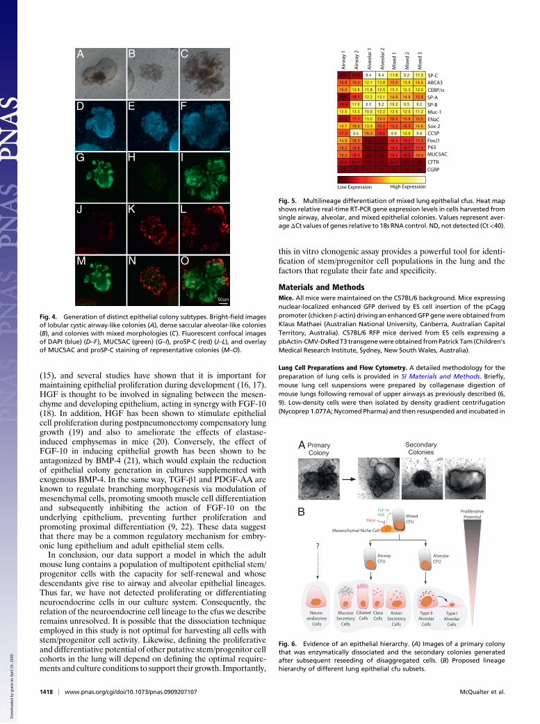
CD24low cell fraction, including large airway-like lobular cysticcolonies with a clearly defined lumen (46% of colonies, n = 150of 326; Fig. 4A), small dense saccular colonies (35% of colonies,n = 114 of 326; Fig. 4B), and colonies of mixed phenotype withdistinct budding (19% of colonies, n = 62 of 326; Fig. 4C).Immunofluorescent labeling of epithelial colonies in fixed whole-mount organotypic cultures confirmed the presence of threedistinct colony subtypes. In cystic colonies, cells expressed thepolymeric mucin MUC5AC, which was secreted into the lumenbut did not express the AEC II marker, proSP-C (Figs. 4 D, G, J,andM). Fluid could be seen circulating within the lumen of cysticcolonies along with beating cilia in cell patches on the innersurface of the lumen. Given that MUC5AC is produced specif-ically by airway mucous-secreting epithelial cells, these datasuggest that cystic colonies comprise cells of the airway lineage.In contrast, the majority of cells in the smaller saccular coloniesexpressed proSP-C but did not stain for MUC5AC (Fig. 4 E, H,K, and N), suggesting that these colonies comprised alveolarAEC II epithelial cells and their progeny. Mixed colonies showedimmunoreactivity for MUC5AC and proSP-C (Fig. 4 F, I, L, andO), suggesting that both airway and alveolar epithelial lung celllineages can be derived from a multipotent lung epithelial cfu.Interestingly, proSP-C staining in mixed colonies was onlyobserved on cells at the peripheral tips of the colonies.Real-time RT-PCR gene expression analysis of individual
colonies (Fig. 5) established that genes encoding airway lineagemarkers, including FoxJ1 (Foxj1; ciliated cells), P63 (Trp63;basal cells), MUC5AC (Muc5ac; goblet cells) and CFTR (Cftr;anion secretory cells) were exclusively detected in cystic and
EpC
AM
-PE
CD49f-488
A%
of M
ax
CD104-APC CD24-488
D E
No.
of c
olon
ies
No. of cells seeded
5050
100100
150150
200200
250250
10001000 20002000 30003000 400040000000
% o
f Max
CD24lo
CD24hiCD24
hi
CD24lo
F
C
proSP-C-488
% o
f Max
B
Sca-1-PacBlue%
of M
ax
Fig. 2. Lung epithelial cfus are enriched in the EpCAMhi
CD49fpos CD104pos CD24low cell fraction. (A) Fractionation ofCD45neg CD31neg lung cells based on EpCAM and CD49fexpression. Sca-1 (B), proSP-C (C), CD104 (D), and CD24 (E)expression on EpCAMhi (blue), EpCAMmed (green), andEpCAMlow (red) cell fractions and isotype controls (gray). (F)Cloning efficiency of EpCAMhi CD24low (blue) and CD24hi (red)cells with linear regression analysis (r2 = 0.9481, 1/slope =22.45, n = 7).
1416 | www.pnas.org/cgi/doi/10.1073/pnas.0909207107 McQualter et al.
Dow
nloa
ded
by g
uest
on
Apr
il 24
, 202
0

mixed cfus. The alveolar markers, ABCA3 (Abca3), CEBP/alpha(Cebpa) and SP-C (Sftpc), as well as SP-A (Sftpa1) were highlyexpressed in saccular and mixed colonies, but were not detected(SP-C and SP-A) or expressed at lower levels (ABCA3 andCEBP/alpha) in cystic colonies. The Clara cell marker, CCSP(Scgb1a1) was enriched in cystic and mixed colonies, but alsodetected in saccular colonies, and SP-B (Sftpb), MUC-1 (Muc1),ENaC (Scnn1g) and Sox-2 (Sox2) were expressed by all epithelialcolony subtypes. CGRP (Calca) was not detected in any of theepithelial colonies examined suggesting that neuroendocrinecells are part of a separate lineage. Taken together, the differ-ential expression of airway versus alveolar lineage markers sup-ports the concept that different epithelial colony subtypes arederived from either lineage-committed epithelial cfus or multi-potent lung epithelial cfus.
Evidence of an Adult Lung Epithelial Stem/Progenitor Cell Hierarchy.In support of the hypothesis that an epithelial stem/progenitor cellhierarchy exists in the adult mouse lung, we have shown that dis-sociation and reseeding of primary mixed colonies in coculturewithEpCAMneg Sca-1pos stromal cells resulted in the generation ofsecondary mixed, airway and alveolar colonies (Fig. 6A). In con-trast, cystic airway and saccular alveolar colonies failed to generatesecondary colonies, suggesting that these progenitor cells haveonly limited proliferative potential. However, it is not possible atthis stage to categorically exclude the possibility that mixed colo-nies may be derived from a multipotent regenerative cfu, consist-ing of juxtaposed cells rather than a single multipotent stem cell.Ultimately, single cell deposition would be required to resolve thisissue. The complex growth requirements of these cells and theirlow incidence have made this goal elusive to date.Taken together, these findings suggest that the adultmouse lung
contains a population of multipotent epithelial stem/progenitorcells with the capacity for self-renewal, whose fate and specificity isregulated by cell-cell and/or paracrine interactions with the sur-roundingmesenchyme.Wepropose amodel in which descendantsof mixed cfus give rise to lineage-committed progenitors withlimited proliferative potential (Fig. 6B) which in turn give rise toairway or alveolar epithelial lineages in the adult lung. Althoughwe have not directly investigated the differentiation of AEC I inthis system, it is generally believed that these cells are descendantsof AEC II cells, as indicated in the hierarchy schematic (Fig. 6B).
DiscussionIn this study, we have identified epithelial stem/progenitor cellsubsets in the adult mouse lung that give rise to clonally derivedairway alveolar or mixed lung epithelial colonies in vitro, pro-viding evidence that the adult lung epithelium may be organizedin a hierarchical manner such that lung epithelial stem cells giverise to differentiated lung epithelium via a series of lineage-committed progenitors. We have shown that both mixed andlineage-committed cfus can be harvested from the adult mouselung, with the latter beingmore abundant, suggesting that lineage-committed cfus may be the primary reserve for maintenance ofdistinct lineages in vivo.To date, there is no convincing evidence to suggest that a
multipotent stem cell in the bronchiolar airways is capable ofmigrating to alveoli to replenish alveolar epithelium in vivo.Indeed, there is a strong belief that alveolar and bronchiolarepithelial cells are maintained by distinct lineages, questioningthe biological relevance of multipotent lung epithelial stem cells.However, the existence of a quiescent multipotent lung epithelialstem cell pool should not be discounted. Rawlins et al. (4)recently provided compelling evidence using a lineage-tracingstrategy, based on the restricted expression of Id2 (inhibitor ofdifferentiation 2), that multipotent progenitors in the fetal lungdistal tip are able to self-renew and contribute to both bron-chiolar and alveolar lineages during development of the lung (4).Importantly, it should also be noted that although we demon-strate that multipotent lung epithelial cfus can generate mixed-lineage colonies in vitro, this does not necessarily mean that theyexhibit this potential in vivo. The capacity for multilineage dif-ferentiation of EpCAMhi CD104pos CD24low cells in the adultlung may be indicative of an enduring developmental potentialcharacterizing cells with the capacity to replenish regional fac-ultative stem cell pools whose fate specification is regulated bythe niche in which they reside.We propose that EpCAMneg Sca-1pos mesenchymal elements
of lung epithelial stem/progenitor cell niches provide instruc-tional cues for proliferation and differentiation of EpCAMhi
CD24low lung epithelial cfus by the local release of growth fac-tors, particularly FGF-10 and HGF. In the developing lung,FGF-10 is expressed in mesenchymal progenitors adjacent toepithelial buds, where it regulates branching morphogenesis
H D
cfu
Inci
denc
e (%
of c
ells
see
ded)
F
2
4
6
0
EpC
AM
-PE
CD104-APC
IgG
2a-P
E
IgG2a-APC
Tota
l Col
onie
s
G I
Tota
l Col
onie
s
No. of cells seeded
5050
100100
150150
10001000 20002000 30003000 40004000 500050000000
5050
100100
150150
00
Singlecells
Epithelialcysts
Singlecells
Epithelialcysts
E
J
P0 P0 P1 P1 P2 P2 P3 P3 P4 P4 P5 P5
P0 P0 P1 P1 P2 P2
8
K
00 G1 G2 G3
1010
2020
3030
4040
5050
Tota
l Col
onie
s
Enzymatic digestionof whole culture
Enzymatic digestionof single cysts
Bulk passagecolony-forming assay
Clonal passagecolony-forming assay
Aggregation
Clonal
Proliferation
Monochromatic
Mixed Colour
A B
D
C
Tota
l Col
onie
s
RedGreen
Mixed
100100
5050
150150
00
30um
30um
Fig. 3. Lung epithelial cfus are capable of clonal proliferation and self-renewal in vitro. (A) Schematic representation of potential outcomes ofmixing experiment. (B) Total colonies that were red, green, or mixed color.(C) Phase contrast image of colonies. (D) Overlay of fluorescence fromcocultures using RFPpos and eGFPpos EpCAMhi CD24low cells with WTEpCAMneg Sca-1pos cells. (E) Schematic representation of stromal-free bulkserial passaging. (F) Incidence of cfus (% of cells seeded + SEM, n ≥ 2) ofEpCAMhi CD24low cells after bulk serial passage in stromal-free cultures withFGF-10 and HGF. (G) Increase in total number of epithelial cfus generatedafter serial passaging. (H) EpCAM vs. CD104 subsetting of passaged cells (P3).(I) Cloning efficiency of EpCAMhi CD104hi (blue) and EpCAMlow CD104low
(red) passaged cells (P3) replated in stromal-free culture with FGF-10 andHGF. (J) Schematic representation of stromal-free clonal serial passaging. (K)Incidence of cfus (mean number of colonies + SEM, n ≥ 3 colonies for eachgeneration) from serial recloning of single colonies.
McQualter et al. PNAS | January 26, 2010 | vol. 107 | no. 4 | 1417
CELL
BIOLO
GY
Dow
nloa
ded
by g
uest
on
Apr
il 24
, 202
0
(15), and several studies have shown that it is important formaintaining epithelial proliferation during development (16, 17).HGF is thought to be involved in signaling between the mesen-chyme and developing epithelium, acting in synergy with FGF-10(18). In addition, HGF has been shown to stimulate epithelialcell proliferation during postpneumonectomy compensatory lunggrowth (19) and also to ameliorate the effects of elastase-induced emphysemas in mice (20). Conversely, the effect ofFGF-10 in inducing epithelial growth has been shown to beantagonized by BMP-4 (21), which would explain the reductionof epithelial colony generation in cultures supplemented withexogenous BMP-4. In the same way, TGF-β1 and PDGF-AA areknown to regulate branching morphogenesis via modulation ofmesenchymal cells, promoting smooth muscle cell differentiationand subsequently inhibiting the action of FGF-10 on theunderlying epithelium, preventing further proliferation andpromoting proximal differentiation (9, 22). These data suggestthat there may be a common regulatory mechanism for embry-onic lung epithelium and adult epithelial stem cells.In conclusion, our data support a model in which the adult
mouse lung contains a population of multipotent epithelial stem/progenitor cells with the capacity for self-renewal and whosedescendants give rise to airway and alveolar epithelial lineages.Thus far, we have not detected proliferating or differentiatingneuroendocrine cells in our culture system. Consequently, therelation of the neuroendocrine cell lineage to the cfus we describeremains unresolved. It is possible that the dissociation techniqueemployed in this study is not optimal for harvesting all cells withstem/progenitor cell activity. Likewise, defining the proliferativeand differentiative potential of other putative stem/progenitor cellcohorts in the lung will depend on defining the optimal require-ments and culture conditions to support their growth. Importantly,
this in vitro clonogenic assay provides a powerful tool for identi-fication of stem/progenitor cell populations in the lung and thefactors that regulate their fate and specificity.
Materials and MethodsMice. All mice were maintained on the C57BL/6 background. Mice expressingnuclear-localized enhanced GFP derived by ES cell insertion of the pCaggpromoter (chicken β-actin) driving an enhancedGFP genewere obtained fromKlaus Mathaei (Australian National University, Canberra, Australian CapitalTerritory, Australia). C57BL/6 RFP mice derived from ES cells expressing apbActin-CMV-DsRedT3 transgenewereobtained fromPatrick Tam (Children’sMedical Research Institute, Sydney, New South Wales, Australia).
Lung Cell Preparations and Flow Cytometry. A detailed methodology for thepreparation of lung cells is provided in SI Materials and Methods. Briefly,mouse lung cell suspensions were prepared by collagenase digestion ofmouse lungs following removal of upper airways as previously described (6,9). Low-density cells were then isolated by density gradient centrifugation(Nycoprep 1.077A; Nycomed Pharma) and then resuspended and incubated in
A B C
D E F
G H I
J K L
M N O
50um
Fig. 4. Generation of distinct epithelial colony subtypes. Bright-field imagesof lobular cystic airway-like colonies (A), dense saccular alveolar-like colonies(B), and colonies with mixed morphologies (C). Fluorescent confocal imagesof DAPI (blue) (D–F), MUC5AC (green) (G–I), proSP-C (red) (J–L), and overlayof MUC5AC and proSP-C staining of representative colonies (M–O).
Mix
ed 3
Air
way
1
Air
way
2
Alv
eola
r 1
Alv
eola
r 2
Mix
ed 1
Mix
ed 2
SP-C
SP-A
SP-B
FoxJ1
CEBP/αABCA3
Sox-2
P63MUC5AC
ENaC
Muc-1
CFTR
CCSP
CGRP
High ExpressionLow Expression
N D 23 .6 8 .4 9 .4 11 .6 9 .2 11 .3
16 .8 16 .4 12 .1 13 .8 15 .0 13 .9 14 .4
15 .0 13 .8 11 .8 12 .5 13 .3 12 .3 12 .0
N D 18 .1 12 .2 13 .1 14 .9 14 .6 15 .6
19 .9 11 .0 8 .0 9 .2 10 .2 9 .5 9 .2
12 .5 13 .3 10 .5 12 .2 12 .5 12 .0 11 .2
21 .2 17 .1 13 .0 15 .3 16 .3 15 .8 15 .5
14 .1 15 .4 13 .9 16 .8 16 .5 16 .5 14 .6
17 .3 9 .5 15 .3 18 .6 9 .9 12 .0 9 .8
14 .8 16 .3 N D N D 18.4 19 .2 17 .3
16 .2 16 .9 N D N D 19.0 19 .7 17 .5
16 .3 18 .0 N D N D 19.4 19 .5 16 .4
20 .3 23 .7 N D N D 23.2 N D N D
N D N D N D N D N D N D N D
Fig. 5. Multilineage differentiation of mixed lung epithelial cfus. Heat mapshows relative real-time RT-PCR gene expression levels in cells harvested fromsingle airway, alveolar, and mixed epithelial colonies. Values represent aver-ageΔCt values of genes relative to 18s RNA control. ND, not detected (Ct<40).
Primary Primary ColonyColony
SecondarySecondaryColoniesColonies
A
B
MucousSecretory
Cells
CiliatedCells
ClaraCells
MixedCFU
AnionSecretory
Cells
AirwayCFU
AlveolarCFU
Type IIAlveolar
Cells
Type IAlveolar
Cells
ProliferativePotential
Mesenchymal Niche Cell
Neuro-endocrine
Cells
?
FGF-10HGF
TGF-β
Fig. 6. Evidence of an epithelial hierarchy. (A) Images of a primary colonythat was enzymatically dissociated and the secondary colonies generatedafter subsequent reseeding of disaggregated cells. (B) Proposed lineagehierarchy of different lung epithelial cfu subsets.
1418 | www.pnas.org/cgi/doi/10.1073/pnas.0909207107 McQualter et al.
Dow
nloa
ded
by g
uest
on
Apr
il 24
, 202
0

2% vol/vol FCS or newborn calf serum (PBS-2% vol/vol Se) (5 × 10⁷ cells/mL, 20min on ice) in an optimally pretitered mixture of antibodies, including anti-CD45, anti-CD31, anti-Sca-1, anti-EpCAM, anti-CD49f, anti-CD24, anti-CD104,and relevant isotype controls (Biolegend). Labeled cells were washed in PBS-2% Se, resuspended at 5–10 × 106 cells/mL, and held on ice forflow cytometricanalysis and sorting. Viability was determined by propidium iodide (1 μg/mL)staining, and doublets excluded by forward scatter (height) vs. forwardscatter (area) gating. For proSP-C analysis, cells were fixed using a Fix ‘n’ Permkit (Invitrogen). Sorting was performed using a BD Influx cell sorter (BectonDickinson). Analysis was done using a BD LSRII bench top analyzer (BectonDickinson) and data were analyzed using FlowJo (Tree Star).
Real-Time RT-PCR. RNA from individual colonies or primary sorted cells wasprepared using the RNAqueous MicroElute RNA isolation kit (Applied Bio-systems), and cDNA was prepared using the high-capacity RNA-to-cDNA kit(Applied Biosystems). For real-time PCR, isolated cDNA was subjected to 40cycles of amplification using Applied Biosystems TaqMan gene expressionassays (Table S1) and 18s RNA endogenous control as per the manufacturer’sinstructions. Reactions resulting in a Ct of less than 40 indicated the presenceof target cDNA in the sample, anddatawere expressed as thenumber of cyclesthat the reaction sample differed (ΔCt) from an 18s RNA control. Relativeexpression of the target gene was expressed as raw ΔCt values relative to theendogenous control.
Cell Culture. Sorted cells resuspended in 90 μL of Matrigel (BD Biosciences)prediluted 1:1 (vol/vol) with media were added to a 24-well transwell filterinsert (Millicell-CM;Millipore) in a 24-well tissue culture plate containing 400 μLof media. For cocultures, epithelial cells were mixed with EpCAMneg Sca-1pos
cells (2 × 10⁶ cells/mL) in Matrigel. DMEM/F12 plus L-glutamine plus 2.438 g/Lsodium bicarbonate (Invitrogen) was supplemented with 10% newborn calfserum, penicillin/streptomycin, insulin, transferrin, and selenium for allcultures. Where specified, FGF-7 (100 ng/mL; Millipore), FGF-10 (50 ng/mL;R&D Systems), HGF (30 ng/mL; R&D Systems), BMP-4 (100 ng/mL; R&D Sys-
tems), TGF-β1 (10 ng/mL; Peprotech), and PDGF-AA (50 ng/mL; Millipore)were added to the media. Cultures were incubated at 37°C in a humidifiedincubator (5% vol/vol O2, 10% vol/vol CO2, 85% vol/vol N2) and refed threetimes weekly. All images are representative of cultures grown for 14–16 daysunless otherwise specified. For bulk passaging, whole cultures were dis-sociated in 1 mg/mL Collagenase Type I plus 3 mg/mL Dispase (Roche) in PBSto generate a single-cell suspension. For clonal passaging, single colonieswere picked and dissociated in the Collagenase/Dispase solution. Single cellswere reseeded in stromal-free cultures supplemented with FGF-10 and HGF.
Immunohistochemistry. Whole-mount cultures were fixed with 4% vol/volparaformaldehyde and removed from inserts, washed in PBS, subjected toantigen retrieval by boiling in 10 mM citrate buffer for 20 min, and incu-bated in blocking buffer (1 h; 5% wt/vol BSA, 1% skim milk, 0.05% Triton X-100 in PBS). Cultures were then incubated overnight with antibodies againstproSP-C (goat anti-proSP-C, clone C-19; Santa Cruz Biotechnology, or rabbitanti-proSP-C; Millipore), CCSP (goat anti-CCSP; Santa Cruz Biotechnology),MUC-1 (rabbit anti-MUC-1; Abcam), and MUC5AC (biotin-labeled mouseanti-MUC5AC) and were then washed in PBS (0.05% Tween 20) and incu-bated with Alexafluor-488, -568, or -647–conjugated anti-goat, anti-rabbit,or streptavidin secondary reagents (Invitrogen) for 3 h and then washed.Nuclei were stained with DAPI, and sections were mounted in Vectashield(Vecta Laboratories). Images were acquired using a Leica SP confocalmicroscope and colored and overlaid using Adobe Photoshop (Adobe Sys-tems). Bright-field images and whole-mount fluorescent images of cultureswere taken using an Olympus SZX16 stereo dissecting microscope.
ACKNOWLEDGMENTS. We thank Kate Rutherford and Daniela Cardozo forassistance with experimental animals and Andrew Fryga, Darren Ellemor,and Kathryn Flanagan for assistance with flow cytometry. This study wassupported by the Australian Stem Cell Centre, National Health and MedicalResearch Council of Australia (Grant 400323), and National Health andMedical Research Council Peter Doherty Fellowship 384367 (to J.M.).
1. Weiss DJ, Kolls JK, Ortiz LA, Panoskaltsis-Mortari A, Prockop DJ (2008) Stem cells andcell therapies in lung biology and lung diseases. Proc Am Thorac Soc 5:637–667.
2. Rawlins EL, Hogan BL (2006) Epithelial stem cells of the lung: Privileged few oropportunities for many? Development 133:2455–2465.
3. Stripp BR (2008) Hierarchical organization of lung progenitor cells: Is there an adultlung tissue stem cell? Proc Am Thorac Soc 5:695–698.
4. Rawlins EL, ClarkCP, XueY,HoganBL (2009) The Id2+distal tip lungepitheliumcontainsindividual multipotent embryonic progenitor cells. Development 136:3741–3745.
5. Shi W, Xu J, Warburton D (2009) Development, repair and fibrosis: What is commonand why it matters. Respirology 14:656–665.
6. McQualter JL, et al. (2009) Endogenous fibroblastic progenitor cells in the adult mouselung are highly enriched in the sca-1 positive cell fraction. Stem Cells 27:623–633.
7. Shannon JM, Nielsen LD, Gebb SA, Randell SH (1998) Mesenchyme specifies epithelialdifferentiation in reciprocal recombinants of embryonic lung and trachea. Dev Dyn212:482–494.
8. Taderera JV (1967) Control of lung differentiation in vitro. Dev Biol 16:489–512.9. Lindahl P, et al. (1997) Alveogenesis failure in PDGF-A-deficient mice is coupled to lack
of distal spreading of alveolar smooth muscle cell progenitors during lungdevelopment. Development 124:3943–3953.
10. Rock JR, et al. (2009) Basal cells as stem cells of the mouse trachea and human airwayepithelium. Proc Natl Acad Sci USA 106:12771–12775.
11. Kim CF, et al. (2005) Identification of bronchioalveolar stem cells in normal lung andlung cancer. Cell 121:823–835.
12. Summer R, Fitzsimmons K, Dwyer D, Murphy J, Fine A (2007) Isolation of an adultmouse lung mesenchymal progenitor cell population. Am J Respir Cell Mol Biol 37:152–159.
13. Rawlins EL, et al. (2009) The role of Scgb1a1+ Clara cells in the long-termmaintenanceand repair of lung airway, but not alveolar, epithelium. Cell Stem Cell 4:525–534.
14. Mondrinos MJ, et al. (2006) Engineering three-dimensional pulmonary tissueconstructs. Tissue Eng 12:717–728.
15. Bellusci S, Grindley J, Emoto H, Itoh N, Hogan BL (1997) Fibroblast growth factor 10(FGF10) and branching morphogenesis in the embryonic mouse lung. Development124:4867–4878.
16. Ramasamy SK, et al. (2007) Fgf10 dosage is critical for the amplification of epithelialcell progenitors and for the formation of multiple mesenchymal lineages during lungdevelopment. Dev Biol 307:237–247.
17. Nyeng P, Norgaard GA, Kobberup S, Jensen J (2008) FGF10 maintains distal lung budepithelium and excessive signaling leads to progenitor state arrest, distalization, andgoblet cell metaplasia. BMC Dev Biol 8:2.
18. Ohmichi H, Koshimizu U, Matsumoto K, Nakamura T (1998) Hepatocyte growth factor(HGF) acts as a mesenchyme-derived morphogenic factor during fetal lung devel-opment. Development 125:1315–1324.
19. Sakamaki Y, et al. (2002) Hepatocyte growth factor stimulates proliferation ofrespiratory epithelial cells during postpneumonectomy compensatory lung growth inmice. Am J Respir Cell Mol Biol 26:525–533.
20. Hegab AE, et al. (2008) Intranasal HGF administration ameliorates the physiologic andmorphologic changes in lung emphysema. Mol Ther 16:1417–1426.
21. Weaver M, Dunn NR, Hogan BL (2000) Bmp4 and Fgf10 play opposing roles duringlung bud morphogenesis. Development 127:2695–2704.
22. Boström H, et al. (1996) PDGF-A signaling is a critical event in lung alveolarmyofibroblast development and alveogenesis. Cell 85:863–873.
McQualter et al. PNAS | January 26, 2010 | vol. 107 | no. 4 | 1419
CELL
BIOLO
GY
Dow
nloa
ded
by g
uest
on
Apr
il 24
, 202
0