evolution of cancer stem-like cells in endocrine-...
TRANSCRIPT

Microenvironment and Immunology
Evolution of Cancer Stem-like Cells in Endocrine-Resistant Metastatic Breast Cancers Is Mediatedby Stromal MicrovesiclesPasquale Sansone1, Marjan Berishaj1, Vinagolu K. Rajasekhar1, Claudio Ceccarelli2,Qing Chang1, Antonio Strillacci1,3, Claudia Savini1,2,4, Lauren Shapiro5, Robert L. Bowman6,Chiara Mastroleo1, Sabrina De Carolis2,4, Laura Daly1, Alberto Benito-Martin7,Fabiana Perna8, Nicola Fabbri9, John H. Healey9, Enzo Spisni3, Monica Cricca2,David Lyden7,10,11, Massimiliano Bonaf�e2,4, and Jacqueline Bromberg1,12
Abstract
The hypothesis that microvesicle-mediated miRNA transferconverts noncancer stem cells into cancer stem cells (CSC) leadingto therapy resistance remains poorly investigated. Here we pro-vide direct evidence supporting this hypothesis, by demonstratinghow microvesicles derived from cancer-associated fibroblasts(CAF) transfer miR-221 to promote hormonal therapy resistance(HTR) in models of luminal breast cancer. We determined thatCAF-derived microvesicles horizontally transferred miR-221 totumor cells and, in combinationwith hormone therapy, activatedan ERlo/Notchhi feed-forward loop responsible for the generationof CD133hi CSCs. Importantly, microvesicles from patients withHTR metastatic disease expressed high levels of miR-221. Wefurther determined that the IL6–pStat3 pathway promoted thebiogenesis of onco-miR-221hi CAF microvesicles and establishedstromal CSC niches in experimental and patient-derived breastcancer models. Coinjection of patient-derived CAFs from bone
metastases led to de novo HTR tumors, which was reversed withIL6R blockade. Finally, we generated patient-derived xenograft(PDX) models from patient-derived HTR bone metastases andanalyzed tumor cells, stroma, and microvesicles. Murine andhuman CAFs were enriched in HTR tumors expressing highlevels of CD133hi cells. Depletion of murine CAFs from PDXrestored sensitivity to HT, with a concurrent reduction ofCD133hi CSCs. Conversely, in models of CD133neg, HT-sen-sitive cancer cells, both murine and human CAFs promotedde novo HT resistance via the generation of CD133hi
CSCs that expressed low levels of estrogen receptor alpha.Overall, our results illuminate how microvesicle-mediatedhorizontal transfer of genetic material from host stromal cellsto cancer cells triggers the evolution of therapy-resistantmetastases, with potentially broad implications for theircontrol. Cancer Res; 77(8); 1927–41. �2017 AACR.
IntroductionTumor heterogeneity and resistance to therapy may occur from
microvesicle-mediated transfer of genetic material between cells(1–3). Thus, the characterization of this phenomenon could haveimportant clinical ramificationsmost notably in the developmentof new therapeutically relevant compounds.
Although adjuvant hormonal therapy (HT) improves dis-ease-free survival in luminal breast cancer patients, HT-resistant(HTR) metastatic disease commonly develops in the bones ofthese patients. This observation suggests that the bone micro-environment may foster estrogen receptor (ER)-independentgrowth of luminal breast cancer leading to HTR metastases.
The interaction of stromal cells (cancer-associated fibroblasts;CAF) with tumor cells has been shown to mediate and modulateestrogen receptor–dependent (e.g., fibronectin, collagen) andindependent proliferation (e.g., laminin) of luminal breast cancercells, suggesting that stroma–tumor communication may play apivotal role in the ER-independent self-renewal of breast cancers(4). In the metastatic microenvironment, we hypothesize thatchronic inflammation incurred by anti-estrogen therapy and theeffects of disseminated tumor cells on the local microenviron-ment will lead to the activation of resident stromal cells orcirculating mesenchymal stem cells to become CAFs. Once
1Department of Medicine, Memorial Sloan Kettering Cancer Center, New York,New York. 2Department of Experimental, Diagnostic and Specialty Medicine,AlmaMater Studiorum, Universit�a di Bologna, Bologna, Italy. 3Department ofBiological, Geological and Environmental Sciences, Universit�a di Bologna, Bolo-gna, Italy. 4Center for Applied Biomedical Research Laboratory, PoliclinicoUniversitario S. Orsola-Malpighi AlmaMater Studiorum, Universit�a di Bologna,Bologna, Italy. 5Department of Radiation Oncology, Kaiser Permanente, Oak-land, California. 6Cancer Biology and Genetics Program, Memorial Sloan Ketter-ing Cancer Center, New York, New York. 7Department of Pediatrics, Cell andDevelopmental Biology, Children's Cancer and Blood Foundation Laboratories,Weill Cornell Medicine, New York, New York. 8Molecular Pharmacology andChemistry Program, Memorial Sloan Kettering Cancer Center, New York, NewYork. 9Orthopedics Service, Memorial Sloan Kettering Cancer Center, New York,New York. 10Drukier Institute for Children's Health, Meyer Cancer Center, WeillCornell Medicine, New York, New York. 11Department of Pediatrics, MemorialSloan Kettering Cancer Center, New York, New York. 12Department of Medicine,Weill Cornell Medicine, New York, New York.
Note: Supplementary data for this article are available at Cancer ResearchOnline (http://cancerres.aacrjournals.org/).
D. Lyden, M. Bonaf�e, and J. Bromberg contributed equally to this article.
CorrespondingAuthor: Jacqueline Bromberg, Memorial Sloan Kettering CancerCenter, 1275 York Avenue, Box 397, New York, NY 10065. Phone: 646-888-3112;Fax: 646-888-3200; E-mail: [email protected]
doi: 10.1158/0008-5472.CAN-16-2129
�2017 American Association for Cancer Research.
CancerResearch
www.aacrjournals.org 1927
on June 24, 2018. © 2017 American Association for Cancer Research. cancerres.aacrjournals.org Downloaded from
Published OnlineFirst February 15, 2017; DOI: 10.1158/0008-5472.CAN-16-2129
on June 24, 2018. © 2017 American Association for Cancer Research. cancerres.aacrjournals.org Downloaded from
Published OnlineFirst February 15, 2017; DOI: 10.1158/0008-5472.CAN-16-2129
on June 24, 2018. © 2017 American Association for Cancer Research. cancerres.aacrjournals.org Downloaded from
Published OnlineFirst February 15, 2017; DOI: 10.1158/0008-5472.CAN-16-2129
activated, the CAFs may sustain a feed-forward circuit of self-renewal, proliferation, and differentiation of CSCs, resulting inmetastasis.
As tumors become more metastatic and resistant to targetedtherapies, the number and types of CSCs increases, suggesting thatCSCs evolve fromnon-CSC cells in a given tumor niche (5, 6). Therole of stroma microvesicles (MV) in the generation of therapy-resistant cancer and the regulation of self-renewal remains poorlyinvestigated.
Here, we investigated the hypothesis that HT- and CAF-derivedmicrovesicles converge to promote HT resistance and ER-inde-pendent self-renewal in luminal breast cancer. By employingpatient-derived xenografts from breast cancer bone metastasesand experimental models of luminal breast cancer, we uncovereda unique process of CAF-mediated resistance to HT. Our datademonstrate the formation of therapy-resistant stromal-tumorniches via an IL6/Stat3-driven expansion of CAFs, CAF-MV–mediated oncomiR-221 transfer to cancer cells leading to theexpansion of Notch3hi/ERlo/CD133hi CSCs. These data reinforcethe concept of targeting the stromal niche to prevent both HTresistance and metastatic progression (7–9).
Materials and MethodsMicrovesicle isolation and in vivo education experiment
Plasma (10 mL) was collected and processed within 4 hoursfrom patients with metastatic disease (Supplementary TableS1) and in healthy controls who were consented to an MSKCCbiospecimen protocol #12-137. The plasma and conditionedmedia (CM) from cancer and CAF cultures was collected from107 cells grown in 5 � 10 cm dishes and centrifuged for 20minutes at 3,000 � g at 4�C. The supernatant was subsequentlycentrifuged for 30 minutes at 12,500 � g at 4�C. The superna-tant was transferred and centrifuged at 100,000 � g for 90minutes at 4�C. The supernatant was discarded while the pellet,containing microvesicles, was resuspended in 25 mL of PBS andloaded onto a 5 mL 30% sucrose cushion to deplete micro-vesicles from extracellular proteins (300 g/L sucrose, 24 g/L Trisbase, pH 7.4). Samples were centrifuged at 100,000 � g for 90minutes 4�C. Cushion (3.5 mL), containing microvesicles, wasdiluted with 10 mL of PBS and centrifuged at 100,000 � g for90 minutes at 4�C. The supernatant was discarded and thepellet resuspended in 25 mL of PBS. Microvesicles were treatedwith 0.1 mg/mL of DNase I solution (Epicentre) to eliminatecontaminating DNA bound to the microvesicles surface orpresent in solution. Nanosight (Lyden laboratory, CornellMedical Center) and electron microscopy (MSKCC ElectronMicroscopy Core) were used to characterize the physical struc-tures of these microvesicles (size and distribution). Confocalmicroscopy (MSKCC Microscopy Core) of cancer cells educatedwith prelabeled (PKH67-Green Fluorescent Cell Linker Kit,Sigma) CAF-MVs was performed to ensure transfer and uptake.The in vivo role of CAF-MVs in the promotion of HT-resistantluminal breast cancer was determined by injecting CAF-MVs(Mu-CAFs, isolated from HT-resistant xenografts and culturedin vitro) and control microvesicles (from MCF7 cells) into thearterial circulation (retro-orbital injection, 3 � 109 particles/mouse/weekly) of tumor-bearing mice (MCF7 cells). Oncemammary fat pad (MFP) xenografts were established (after4 months), mice were treated with HT (fulvestrant; 100mg/injection/once a week for 3 months).
Primary cultures and patient-derived xenografts of endocrine-resistant luminal breast cancer bone metastases
Patient-derived xenografts (PDX) were established from n ¼ 2of 5 HT-resistant and 1 de novo stage IV breast cancer bonemetastatic tissue isolated at MSKCC (Supplementary Table S2).Patients who developedmetastatic breast cancer in the bone wereenrolled in the study (IRB protocol #97-094). Following surgery,tissue was processed by the pathologist, 60% of the specimen wasused for confirmation of diagnosis and molecular analyses (IHC,IMPACT analyses), while 40% was used for further analysis.Tissues were placed in sterile Epicult (Voden Medical), mincedwith sterile razor blades and incubated at 37�C for 8 to 12 hours inthe presence of Collagenase/Hyaluronidase enzyme mix (1,000Units, Voden Medical). To grow tumor cells devoid of its cognatestroma, we performed serial centrifugations to separate epithelialcells as mammosphere cultures (MS). Secondary and tertiary MSpotential (II or III-MS) was performed as follows: 7-day primaryMS started to form after 4–6 days, then they were disaggregated in1� Trypsin-EDTA (StemCell Technologies), washed in completeMEGM, filtered through a 40-mmnylonmesh, and seeded to formsecond generation MS. Number of MS was assessed by countingthe total number of spheres (size > 100 mm) from cells seeded inlow-attachment plates (from 100 to 1,000). To establish pri-mary CAF cultures from patient-derived tissue, MS-depletedsupernatant was centrifuged at 450 � g for 10 minutes; thispellet was enriched with stromal cells was plated onto 10-cmplates supplemented with DMEM 10% serum media. CAFprimary cultures were expanded in vitro for n ¼ 10 passages.III-MS primary cultures were used to establish PDXs: 50–100MS (size �100 mm) were injected in the MFP of NOD/SCIDmice and tumor growth was determined over a period of 5months. At the endpoint of the experiment, xenograft tissueswere collected and primary PDX cultures were established.Multiple passaged PDX were generated following repeatedorthotopic injection of PDX-derived EpCAMpos cancer cells(recognizes only human cells) in the MFPs of NOD/SCID mice(from 1st to 4th generation). Luminal breast cancer cellsexpressing a vector for GFP/Luciferase were generated and usedfor all the in vivo experiments. Tumor growth was determinedusing in vivo bioluminescence technology (BLI: Xenogen, IVISSystem). Luminal cancer xenografts from the coinjection ofhuman CAFs and MCF7 cells were also generated to determinethe effect of the stroma on the generation of de novo–resistantendocrine tumors
Xenograft assays and preclinical trialsAll cancer cell lines were engineered to express a GFP-positive
luciferase expression vector. Prior to in vivo inoculation, cancercells were FACS purified (for GFP) and injected bilaterally in theMFPs of 5- to 7-week-old NOD/SCID) mice (obtained from NCI,Frederick, MD). For each in vivo experiment, cancer cells weremixed with an equal volume of Matrigel (BD Biosciences) in atotal volumeof 50mL. Bioluminescencewas used tomonitor bothtumour growth (weekly) and metastatic burden (at necropsy).Luminal cancer xenografts from the coinjection of human CAFs(HTR bone metastases) and MCF7 cells (103 cells) were alsogenerated to determine the effect of the stroma on the generationof de novo–resistant endocrine tumors. In addition, human bonemarrow stromal cells HS27a, HS27shC, and HS27shIL6 (100cells/injection) were coinjected with MCF7 cells (103 cells/injec-tion) into the MFP. For immunostaining assays, organs were
Sansone et al.
Cancer Res; 77(8) April 15, 2017 Cancer Research1928
on June 24, 2018. © 2017 American Association for Cancer Research. cancerres.aacrjournals.org Downloaded from
Published OnlineFirst February 15, 2017; DOI: 10.1158/0008-5472.CAN-16-2129
collected and fixed overnight in 4% paraformaldehyde, washed,embedded in paraffin, and sectioned (Histo-Serve Core). Hema-toxylin and eosin (H&E) staining was performed by standardmethods. For the detection of metastases at secondary sites, weperformed in vivo BLI as well as immunofluorescence/IHC stain-ing for GFP and ER. All the surgical procedures and animal carefollowed the institutional guidelines and an approved protocolfrom our IACUC at MSKCC. For the preclinical studies, injectablefulvestrant (Faslodex, AstraZeneca) was given intramuscularly inthe tibialis posterior/popliteal muscles (100 mg/injection/once aweek) for 2 months. Tocilizumab (Actemra, Roche Pharmaceu-ticals) was diluted in PBS at a final concentration of 20 mg/mL. Adosage of 100 mg/g/mouse was administered intraperitoneallyevery week (this is �5-fold higher than the physiologic range,patients receive 8 mg/kg i.v.). Control mice received isotypecontrol (placebo) or PBS injection.
Cell lines and FACSHuman cancer cell lines (Namalwa, lymphoma; HeLa, cer-
vical carcinoma), human breast cancer cell lines (MCF7,ZR751, T47D, and BT474), human bone marrow stromal celllines (HS5, HS27a), and human normal fibroblasts (MRC5,HMF) were purchased from the ATCC and authenticated byshort tandem repeat (STR) DNA profiling (Genomic CoreMSKCC). Murine CAFs (Mu-CAF) were isolated from HTRxenografts and PDXs by FACS purification (GFP�/EpCAM�).Cells were mycoplasma free and maintained in minimumessential medium and RPMI (ATCC and Media Core) supple-mented with 5% FBS (Media Core), 2 mmol/L glutamine, 100U mL�1 penicillin, and 0.1 mg mL�1 streptomycin (MediaCore). Cancer cells from xenografts were isolated from primaryand metastatic tissues by enzymatic digestion (Collagenase/Hyaluronidase, Sigma-Aldrich), sorted (GFPþ/DAPI�), andcultured in vitro. The following reagents: 4-hydroxytamoxifenand fulvestrant were purchased from Sigma (Sigma-Aldrich).For FACS/flow analyses, tumors were digested in sterile Epicultmedia (StemCell Technology), minced with sterile razorblades, and incubated for 3 hours in the presence of collage-nase/hyaluronidase (1,000 Units/sample). Cells were washedwith sterile filtered PBS supplemented with 1% BSA (PBS-BSA 1%) and filtered through a 40 mm nylon mesh (BD Bio-sciences). For the detection of CD44 and CD133, EpCAMantigens, cells were stained in a volume of 100 mL (PBS-BSA1%) with each antibody CD44-APC (100 ng/106 –108
cells Clone IM7, eBiosciences), CD133/1-PE (100 ng/106–108 cells, clone AC133, Miltenyi Biotech) and EpCAM-FITC(250 ng/106–108 cells, Clone VU-1D9, StemCell Technolo-gies). Cells were labeled on ice for 30 minutes and analyzed(BDFACS Aria III, Flow Core). Samples were analyzed for cellpopulation distribution and sorted for GFP/viability (GFPþ/DAPI�) and CD133/CD44 expression. For flow plot analyses,samples were run using FlowJo 7.5 software (Tree Star). shRNAsfor Notch3 and IL6 were previously described (10, 11).
Microarray and miRNA analysesNormalized gene expression values were downloaded from the
GEO under accession number GSE17705 and probes were aggre-gated to median gene level expression. A CAF gene set fromAllinen and colleagues (12) was used in a single sample gene setenrichment analysis (ssGSEA). ssGSEA scores were z-scored and a"CAF score" was assigned to each patient. Patients were split at the
median into CAF-high and CAF-low groups. PROM1-high andPROM1-low groups were split based on PROM1 (CD133) medi-an expression. Statistical significance for differences in PROM1expression and ESR1 expression were assessed with a Student ttest. The heatmap for CAF signature gene and PROM1was plottedin R with the heatmap.2 function. For real-time PCR (qPCR), weextracted RNA using TRIzol (Invitrogen). RNA concentration wasdetermined with a NanoDrop 2000. For microarray analysis ofpublished datasets, normalized gene expression data were down-loaded from theGeneExpressionOmnibus (GEO). Each genewasmean centered and scaled by SD. All analyses were conducted inR. Normalized gene expression data was downloaded from theNCBI for dataset GSE69280 (5). For qPCR, 1 mg of total RNA wasreverse transcribed to cDNA using iScript Select cDNA SynthesisKit (Bio-Rad) following the manufacturer's protocol. Reversetranscription PCR (RT-PCR) analysis was performed using thefollowing primers: ERa; forward 50-TGAAAGTGGGATACGAAAA-GAC-30, reverse 50-CAGGATCTCTAGCCAGGCACAT-30; b2m for-ward 50-ACCCCCACTGAAAAAGATGA-30, reverse 50-ATCTTCA-AACCTCCATGA-30. DNA was isolated using phenol/chloroformtechnique from PDX-derived EpCAM-positive/negative cells. Thepresence of murine and human cells was determined on 2 ng ofDNA by PCR for GAPDH (murine: forward 50-AGCAGCCG-CATCTTCTTGTGCAGTG-30, reverse 50- GGCCTTGACTGTGCC-GTTGAATTT-30; Human: forward 50-CTCTGCTCCTCCTGTTC-GAC-30, reverse 50- ACGACCAAATCCGTTGACTC-30). miRNAexpression was analyzed as described previously (13). Briefly,miRNA were reverse transcribed using stem-loop RT-PCR tech-nology (14) and amplified by real-time PCR using SYBR SelectMaster Mix (Applied Biosystems) and ViiA 7 Real-Time PCRSystem (Applied Biosystems) according to the manufacturer'sinstructions. The melting curve data were collected to check PCRspecificity. miRNA expression was normalized against RNA U6levels: (RT-miR-221) 50-GTCGTATCCAGTGCAGGGTCCGAG-GTATTCGCACTGGATACGACGAAACCC-30; (RT-miR-222) 50-GTCGTATCCAGTGCAGGGTCCGAGGTATTCGCACTGGATAC-GACACCCAGT-30; (RT-miR-101) 50-GTCGTATCCAGTGCA-GGGTCCGAGGTATTCGCACTGGATACGACTTCAGTT-30; (for-ward-miR-221) 50-AGCTACATTGTCTGCTGGGTTTC-30; (for-ward miR-222) 50-AGCTACATCTGGCTACTGGGT-30; (forwardmiR-101) 50-GCCGCTACAGTACTGTGA-30; (forward U6) 50-CTTCGGCAGCACATATACT-30; (reverse U6) 50-AAAATATG-GAACGCTTCACG-30 (reverse all miRs) 50-TGCAGGGTCC-GAGGTAT-30. All primers were purchased from Eurofins MWGOperon. miRNA expression was analyzed as described else-where (13).
Protein and in vitro studiesFor immunoblotting assays, cells were lysed in buffer (50
mmol/L Tris at pH 7.5, 150 mmol/L NaCl, 5 mg/mL aprotinin,pepstatin, 1% NP-40, 1 mmol/L EDTA, 0.25% deoxycholate,and protease inhibitor cocktail tablet, Sigma). Proteins wereseparated by SDS-PAGE, transferred to polyvinylidene difluor-ide membranes and blotted with specific antibodies (Supple-mentary Table S3). For functional interference studies, anti-miR-221 and control RNA oligonucleotide were purchasedfrom Applied Biosystems. MCF7 cells were seeded in a 6-wellplate (8 � 105 cells/well) at 60% confluence. After 24 hours,cells were transfected using Lipofectamine 2000 transfectionreagent (Invitrogen) according to the manufacturer's instruc-tions (RNA final concentration, 200 nmol/L). After 6 hours of
Stroma Microvesicles Regulate Endocrine Therapy Resistance
www.aacrjournals.org Cancer Res; 77(8) April 15, 2017 1929
on June 24, 2018. © 2017 American Association for Cancer Research. cancerres.aacrjournals.org Downloaded from
Published OnlineFirst February 15, 2017; DOI: 10.1158/0008-5472.CAN-16-2129

incubation at 37�C, transfection medium was replaced with2 mL of complete medium containing 10% FCS supplementedwith/without CAF-MVs. For determination of cell viability, weseeded 2,500 cells per well in 96-well plates and treated themwith fulvestrant (10 mmol/L). Viable cells were determined7–14 days after treatment using Trypan blue and cell countingwas done using bright-field microscopy or DAPI staining byflow cytometry (Dako Cytomation). Crystal violet assay wasperformed to obtain information of the relative cell densityat the endpoint of proliferation potential experiments. IL6ELISA assays were performed using the conditioned mediumcollected from 5-day cultures of CAF-derived cells seeded at2 � 106 cells/plate. Proliferation assay was carried out usingCalceinAM technology (Invitrogen): cells were seeded in96-well plates treated with the prefluorescent compound for20 minutes and fluorescence was read using a plate reader(SpectraMax plate platform). To determine the selectivegrowth potential of cancer cells over stroma cells, we analyzedthe proliferation potential of luciferase-positive cancer cells byin vitro BLI: cells were seeded in 96-well plates in presence/absence of distinct CAFs/normal fibroblast (1:10 ratio of CAFsto tumor cells) and treated with fulvestrant (10 mmol/L/weeklyfor 3 weeks). Luciferase activity was measured weekly. Cyto-kine Arrays were performed on 10 mg of CM-derived proteinsaccording to manufacturer's protocol (Antibody Array 3, Ray-Biotech. Inc.).
Immunostaining analysisSerial sections of formalin-fixed paraffin-embedded samples
were immunostained using monoclonal anti-CD133 diluted1:70 (clone W6B3C1, Miltenyi Biotec), anti-ERa RTU (cloneSP1, Ventana), anti-Pankeratin RTU (clone AE1/AE3/PCK26,Ventana), and polyclonal anti-Notch-3 diluted 1:400 (M-134,Santa Cruz Biotechnology). CD133 and Notch3 immunostain-ing was performed as follows: sections were dewaxed, rehy-drated, and subjected to antigen retrieval treatment. Antigenswere unmasked with a Tris-EDTA pH 9.0 buffer at 98�C for20 minutes in a waterbath. Endogenous peroxidase activitywas inhibited using a 0.5% H2O2 solution in methanol for20 minutes at room temperature. Sections were processedusing a non-biotin–amplified method (Novolink, Novocastra)according to the manufacturer's protocols. When mouse tissuewas used, a short treatment (30 minutes at room temperature)with MOM blocking solution (Vector Laboratories Inc.) wasconducted prior to primary antibody overnight incubation at4�C. The reaction was visualized using the UltraView DABDetection System. The immunologic reaction was developedusing a 3,30-diaminobenzidine (DAB)/H2O2 PBS pH 7.2–7.4solution for 10 minutes. Sections were then washed in distilledwater and counterstained in Harris hematoxylin. Anti-ERa (ER)and Pankeratin (CK) immunostaining was performed on anautomated immunostainer (Benchmark Ultra, Ventana) usingthe UltraView DAB Detection kit according to the manufac-turer's protocol. Antigen retrieval was performed onboard withUltraCC1 buffer (pH 8.2–8.5) at 95�C for 52 minutes (ER) or20 minutes (CK). Primary antibodies were incubated 28 min-utes at 37�C (ER) or 8 minutes at room temperature (CK). ForCD133 and Notch3 evaluation, each section was examined at400�. In each microscopic field, the neoplastic cells wereclassified according to both positive percentage and stainingintensity: [percentage ¼ 0 if <1%, 1 if >1% < 25%, 2 if >25% <
50%, 3 if >50% <75%, 4 if >75%; intensity ¼ 1 (weak), 2(moderate), and 3 (strong)]. A final classification was obtainedby multiplying the two mean values (percentage and intensity,IRS score). As for ER evaluation, the neoplastic population wasscanned using Image Cytometry and reported as percentage ofpositive cells (%) (IMAGE-Pro Plus V5.0.1, Media CyberneticsInc.). A detailed histologic examination of xenograft tissues wasperformed at the collaborating institution (University of Bolo-gna, Bologna, Italy). Xenograft tissue was stained with hema-toxylin and eosin and examined by three independent pathol-ogists (C. Ceccarelli, Donatella Santini, and M. Bonafe, fromthe University Hospital of Bologna, Bologna, Italy). For eachmicroscope field (200�), the area occupied from cancer cells,stromal cells, and necrotic components was evaluated andrepresented as percentage.
Characterization of CAFsSerial sections (5 mm) of paraformaldehyde-fixed paraffin-
embedded samples underwent antigen retrieval using LeicaBond ER2 Buffer (Leica Biosystems) for 20 minutes at 100�Cbefore staining with 1 mg/mL Desmin rabbit polyclonal anti-body (Abcam catalog no. ab8592) and 1 mg/mL pStat3 (cloneD3A7, Cell Signaling Technology) for 1 hour using LeicaProtocol F (Molecular Imaging Core facility, MSKCC, NewYork, NY). Quantification of Desmin/pStat3 staining was per-formed using ImageJ/FIJI (NIH, Bethesda, MD). At least 19fields at 400� were randomly selected and evaluated. Theresults were expressed as percentage of immunostained cells/over total area of tissue. To discriminate between cancer andstromal cells, fortified H&E staining was also performed (His-toServ Inc). A color deconvolution algorithm was then used,with RGB vectors for the stromal component and counterstain/background stain created from regions of interest drawn fromexample images (Molecular Imaging Core facility, MSKCC,New York, NY). Appropriate thresholds were then set for eachcell type of interest and area measurements were taken for allimages. To rule out possible non-CAFs/noncancer cells com-ponent, specific staining for CAFs (desmin-murine CAFs) andcancer cells (human pankeratin) was also performed in serialsection slides. Stroma–tumor niches were evaluated as area oftissue slide with the copresence of pankeratin-positive cells andstroma cells.
ALDEFLUOR assayALDEFLUOR analysis was performed using the ALDEFLUOR
Kit (StemCell Technologies) according to the manufacturer'sprotocol. Cancer cells from PDX primary cultures were washedwith 5mL10%PBS supplementedwithAccumax (InnovativeCellTechnologies), and single-cell suspensions were first stained withanti-CD133-PE–conjugated antibody for 20 minutes, washedtwice with PBS-BSA (5%), and then incubated with ALDEFLUORreagent.
CM preparation and phenotypic assaysCM was isolated from CAFs and cancer cell lines (108 cells),
concentrated using Amicon Ultra-15 centrifuge tubes (Millipore),and protein levels were measured by the Lowry technique; 10 mgof total extracellular protein was loaded for zymographic/protein(MMP-2, MMP-9) and in vitro studies (invasion capacity). Cellgrowth of cocultured cancer cells with CAFs was determined with
Sansone et al.
Cancer Res; 77(8) April 15, 2017 Cancer Research1930
on June 24, 2018. © 2017 American Association for Cancer Research. cancerres.aacrjournals.org Downloaded from
Published OnlineFirst February 15, 2017; DOI: 10.1158/0008-5472.CAN-16-2129
and without anti-Jagged1/Notch3 blocking antibody (AF1277,R&D Systems 500 ng every 72 hours). Briefly, luciferase-positivebreast cancer cells (MCF7) grownwithCAFs (1:50)were seeded in96-well plate and treated with fulvestrant (10 mmol/L/weekly) inthe presence ofmouse anti-Jagged1blocking antibody. BLI signalswere measured every 48 hours and growth curves were generatedaccordingly.
Luciferase assaysCells were plated in 6-well plates at a density of 2� 105 cells per
well. Cells were transfected with 0.3 mg of promoter luciferase(CD133; ref. 15) and the activated form ofNotch3 (pNICD3 2 mg;ref. 10). To normalize transfection efficiency, cells were alsocotransfected with 0.1 mg of the pRL-CMV (Renilla luciferase,Promega). Forty-eight hours after transfection, luciferase activitywas measured using the Dual-Luciferase Assay Kit (Promega).Three independent experiments were performed, and the calcu-lated means and SDs are presented.
TaqMan gene expression profile and RT-PCRqPCR was performed on 100 ng of cDNA using TaqMan
precustom probes (Applied Biosystems, ERaHs00174860 62 pb,GATA3 Hs00231122 80 bp, FOXA1 Hs0418755 59 bp, GREB1Hs00536409 67 bp, EGR3 Hs00231780 91 bp, CCL5Hs00174575 63 bp, PGR Hs01556702 77 bp) and SYBR Greentechnique (a-sma forward 50- CAGGGCTGTTTTCCCATCCAT-30,reverse 50-GCCATGTTCTATCGGGTACTTC-30; SDF-1a forward50-CCATGAACGCCAAGGTCGTG-30, reverse 50- CCAGGTACT-CCTGAATCCAC-30; Vimentin forward 50-TGGCACGTCTT-GACCTTGAAA-30, reverse 50- GGTCATCGTGATGCTGAGAA -30;Slug 50-AGATGCATATTC GGACCCACA-30, reverse 50- CCT-CATGTTTGTGCAGGAGA-30; CD44 forward 50-CAGCAACCC-TACTGATGATGACG-30, reverse 50- GCCAAGAGGGATGCCAA-GATGA -30). ViiATM 7 Real-Time PCR System was used (AppliedBiosystems) in accordance with the manufacturer's instructions.For analysis, DCt method was applied and fold change wascalculated (2�DDCt). All valueswere normalized toGAPDHexpres-sion (TaqMan, Hs02758991). RT-PCR for Notch3 (forward 50-TCAGGCTCTCACCCTTGG-30, reverse 50-AGTCACTGGCAC-GGTTGTAG-30), Jagged1 (forward 50-TCGCTGTATCTGTCCAC-CTG-30, reverse 50-AGTCACTGGCACGGTTGTAG-30) and b2m asinternal control 50-ACCCCCACTGAAAAAGATGA-30, reverse 50-ATCTTCAAACCTCCATGA-30 was performed in MCF7 cells con-trol and shNotch3 and mCAFs/fibroblast cell lines.
Statistical analysisStatistical analysis was performed by SPSS (SPSS Inc.). Con-
tinuous variables were analyzed by unequal variance t test,paired t test (samples, n ¼ 2), general linear model (GLM)ANOVA, or GLM for repeated measures (samples, n > 2).Mann–Whitney and Wilcoxon tests were used to analyze ordi-nal variables. P values were adjusted for multiple comparisonsaccording to Bonferroni correction. Association among quan-titative variables was quantified by Pearson correlation coeffi-cient. Categorical variables were analyzed by Monte Carlo c2
test. All the tests were two-sided. P < 0.05 was consideredsignificant. Elda software was used to measure the statistics oflimiting dilution experiments (bioinf.wehi.edu.au/<http://bioinf.wehi.edu.au/>software/elda/).
ResultsMicrovesicles from CAF-mediated HT resistance
The presence of CAFs have been assessed as prognosticators inbreast cancer and an "active stromal signature" in normal fibro-blasts exhibits a tumor-promoting phenotype (16). Many stro-mal-secreted factors including IL6, SFD-1a, and HFG participatein the communication between CAFs and tumor cells within thetumor microenvironment.
Stromal microvesicles have also been implicated in tumorprogression in glioblastomas and ovarian cancers (17, 18). How-ever, the molecular and pathologic relevance of CAF-derivedmicrovesicles in luminal breast cancer remains unclear.
To study tumor progression in luminal breast cancer, weestablished long-term xenografts of highly tumorigenic MCF7and ZR751 cells (5). Following tumor establishment (1 cm), allmice receivedHT (fulvestrant a selective estrogen receptor degrad-er commonly given to patients with ERþ metastatic disease, 10mmol/L) for 3 months. Although the majority of xenograftsdisplayed sensitivity to HT (HTS, stable disease or remission),approximately 10% of the xenografts (data not shown) grew inthe presence of therapy (Fig. 1A, HTR resistance to HT). Interest-ingly, the histologic analysis of these tissues revealed the enrich-ment of CAFs in the HTR xenografts (Fig. 1B; Supplementary Fig.S1A). Furthermore, we could isolate and in vitro passage CAFsfrom HTR-derived xenograft tissues. These CAF cell lines dis-played the upregulation of CAF markers by Western blot analysisand the capability of growth for multi passages (more than 20passages; Fig. 1B and data not shown). Although we were able toisolate CAFs from HTS lesions in a small fraction of xenografts(5%, n¼ 3 out of 60), we could not propagate them in culture formore than 2 passages (2weeks). Therefore, noCAF cell lines (0%)were established from HTS xenografts.
To further characterize these tumor-associated stromal cells, weculturedHTR tumors and isolated stromal cells by FACS (negativeselection with EpCAM, which recognizes epithelial cells) anddetermined that EpCAMneg cells were morphologically spindleshaped, were murine in origin (expressed murine genomic DNA,data not shown) and expressed markers of activated CAFs includ-ing Fap, vimentin, fibronectin, and activated Stat3 (Fig. 1B,phospho tyrosine 705 Stat3 pStat3).
Next, we asked whether these CAFs could promote de novoHTR disease. We cocultured murine CAFs and human-HS27a"CAF" like cells (bone marrow–derived immortalized mesen-chymal cells) with HT (Luciferasepos) na€�ve cancer cells in thepresence/absence of HT (fulvestrant, 10 mmol/L/weekly) andcancer cell growth was analyzed by in vitro bioluminescenceafter 2 weeks (Fig. 1C). We found that CAFs promoted tumorcell growth following HT, whereas no difference was found inthe absence of therapy (Fig. 1D; Supplementary Fig. S1B). Incontrast to HS27a cells and murine CAFs, normal fibroblasts(mammary and lung) did not confer resistance to HT incocultures (Supplementary Fig. S1C).
In addition to growth factors, stromal cells have been shown tosecrete microvesicles, which can horizontally transfer numerousprosurvival factors and confer resistance to radiotherapy (19).Here we determined that the number of microvesicles producedby murine CAFs and HS27a cells was much greater than MCF7tumor cells (Fig. 1E). To determine whether these microvesiclescould confer a protumorigenic advantage, we set up an in vivomodel (Fig. 1F).MFP xenografts fromHT-na€�ve cells (MCF7)were
Stroma Microvesicles Regulate Endocrine Therapy Resistance
www.aacrjournals.org Cancer Res; 77(8) April 15, 2017 1931
on June 24, 2018. © 2017 American Association for Cancer Research. cancerres.aacrjournals.org Downloaded from
Published OnlineFirst February 15, 2017; DOI: 10.1158/0008-5472.CAN-16-2129

established; CAF-MVs (3 � 109) and control microvesicles(tumor-derived microvesicles, MCF7) were injected retro-orbi-tally weekly for 7 months. Once tumors were established (after 4months) all mice received HT (fulvestrant weekly). Althoughthere was no difference in tumor growth beforeHT, those animalstreated with CAF-MVs had tumors resistant to HT (HTR) whileMCF7-MVs provided no benefit as tumors regressed with HT (Fig.1G). Overall, these data demonstrated that circulating stromalmicrovesicles can induce resistance to HT in vivo.
CAF-derived microvesicle transfer of oncomiR-221/222 tocancer cells promotes de novo HT resistance
Distinct genes and pathways have been associated withresistance to HT including the activation of mutations in theESR1 gene (20), increased Her2 expression (21), decreased ER
levels, and ER transcriptional signatures (22, 23), increasedexpression of oncomiRs including the ER repressor miR-221/222 (24) and, more recently, increased Notch signaling in CSCs(5, 25).
As a reduction in ER expression is associated with resistance toHT (22), and CD133hi CSCs have lower ER levels (mRNA andprotein) as compared with CD133lo/CD44lo cells, we reasonedthat the suppression of ER signaling could be a mechanism ofstroma-mediated expansion of therapy-resistant CSCs (CD133hi/Notch3hi).
To test our hypothesis, we first demonstrated that the CMfrom CAFs (murine and human), but not normal fibroblasts,led to a decrease in ER protein expression and ER-dependenttranscripts (e.g., GATA3, FOXA1, GREB1, EGR3, CCL5, PGR)in MCF7 cells (Fig. 2A; Supplementary Fig. S2A). As the
Figure 1.
Microvesicles fromCAFsmediatedHTR.A,Generation of HTR (purple) andHTS (green) xenografts in luminal breast cancer. Highly tumorigenic luminal breast cancercells (MCF7 and ZR751 luciferase positive) were injected in the MFP; xenograft-bearing mice were treated with fulvestrant starting at 4 months for 3 months(HT, fulvestrant intramuscular injection 100mg/mouse/weekly). Some tumors grew in the presence of HT (HTR),while themajority of themwas sensitive toHT (HTS).Data reported as error bars mean bioluminescence (BLI value � SEM) at the endpoint of the experiment (7 months, n ¼ 10 mice/group). B, HTR cultureswere enriched with murine CAFs (Mu-CAFs, red). Representative fortified H&E staining of HTR xenografts (from A, MCF7, scale bar, 50 mm; yellow, stromadesmoplastic reaction), bright-field images of primary cultures from HTS and HTR xenografts andWestern blot analysis of FACS-purified cancer cells (Hu-HTR) andmurine cells (Mu-CAFs). C, Schematic of the experiment. HT-na€�ve cancer cells (GFPþ/luciferaseþ) were cocultured with FACS purified Mu-CAFs (purple),humanBMSC/CAFHS27a cells (red), or in the absence of stromal cells (green). Cellswere treatedwith orwithout HT (fulvestrant, 10 mmol/L/weekly).D, Proliferationpotential (after 2 weeks, BLI) was determined �HT (fulvestrant, 10 mmol/L). Data are reported as error bars, mean � SD of n ¼ 3 independent experiments.� , P < 0.05 (Student t test). E, Electron microscopy images and quantification (number by nanosight) of CAF-derived microvesicles (murine and human, HS27a).Scale bar, 200 nm; data reported as error bars, mean � SD of number of particles/mL for 106 cells. � , P < 0.05 (Student t test) of n ¼ 3 independent experiments.F, Schematic of the experiment. Mice were injected in the MFP with HTS cells (MCF7) and subsequently injected weekly with either Mu-CAFs microvesicles orcontrol microvesicles (MCF7 derived; see Materials and Methods). After 4 months, HT was administered for 3 months (fulvestrant intramuscular injection100 mg/mouse/weekly). G, Dot plot of tumor growth of HTS cells (luciferase/p/s) grown in the MFP of mice educated with CAF MVs or CTRL MVs (MCF7) in thepresence/absence of HT (fulvestrant, see F; microvesicles, retro-orbital injection, 3 � 109 particles/mouse/weekly); error bars, mean � SEM of the last pointof the growth curve before (4 months) and after HT (7 months). � , P < 0.05 values refer two-way ANOVA (G). NS, not significant.
Sansone et al.
Cancer Res; 77(8) April 15, 2017 Cancer Research1932
on June 24, 2018. © 2017 American Association for Cancer Research. cancerres.aacrjournals.org Downloaded from
Published OnlineFirst February 15, 2017; DOI: 10.1158/0008-5472.CAN-16-2129

suppression of ER protein occurred with CM from both murineand human CAFs, we hypothesized that rather than solublefactors (which can be typically species-specific; refs. 26, 27),CAF-derived miRNAs might be mediating the downregulation ofER expression. Microvesicles have been suggested to bemediatorsof nucleic acid transfer including miRNAs (27). Among differentmiRNAs, the forced overexpression of oncomiR-221/222 in lumi-nal breast cancer cells has been found to reduce ER expression andpromote HT resistance (24). In addition, increased plasma levelsofmiR-221were found in ER-negative breast cancer patients (28).The administration of 108 microvesicles from mCAFs to ERþ
cancer cells (MCF7) reduced ER levels after 48 hours (Fig. 2B).We showed that oncomiR-221/222 sequences are conservedbetween human and mouse species, suggesting possible func-tional cross-species interactions (Fig. 2C). Importantly, miR-221expression was found in circulating microvesicles from patientswith HTR metastatic disease (independent of tumor burden) ascompared with healthy controls (Fig. 2D, n¼ 11; SupplementaryTable S1).
We determined that CAF-derived microvesicles wereenriched for miR-221 compared with normal fibroblasts anddistinct cancer cells lines (breast, cervical, and lymphoma),
Figure 2.
Microvesicle-mediated OncomiR-221 transfer from CAFs to cancer cells promotes an ERlo phenotype, leading to HTR resistance A, Western blot analysis ofERa protein in MCF7 cells following treatment with the CM of human normal mammary fibroblasts (HMF), mCAFs (red font), and HS27a cells (blue font, 48 hours).B, Western blot of ERa protein in MCF7 cells treated with and without mCAF-derived microvesicles (108 particles, 48 hours). C, Image showing theOncomiR-221 sequence conservation in Mus Musculus and Homo Sapiens. D, MV-miR221 expression as determined by qPCR as fold increase from patients withHTR disease and healthy controls (reference MCF7-MVs was used and normalized to total RNA expression, n ¼ 3 replicates; Supplementary Table S1).Patients with high volume disease (>10% of organ involvement) are denoted in red and low volume disease (<1% of organ involvement) is denoted in blue. E,Bar graphs representing oncomiR-221 expression (qPCR) in microvesicles from indicated sources (108 cells). Data are reported as fold increase (221/U6expression) � SD of n ¼ 3 replicates (MV-MCF7 is used as reference). F, RT-PCR analysis of miR-221 and 101 in MCF7, mCAFs, and mCAF-derived microvesicles(RNA was isolated from 1015 particles) and bar graph showing oncomiR-221 expression (qPCR, Fold, normalized to U6 values) in mCAFs and HS27a cells andtheir respective microvesicles. The expression of oncomiR-221 in MCF7 was used as a reference control. G, Bar graph of oncomiR-221 (qPCR, Fold) in MCF7cells following chronic mCAF-MV education (one month, 108 particles weekly). Microvesicles were isolated from MRC5 cells (normal lung fibroblasts) and usedas control microvesicles (CT-MVs). H, Western blot analysis of ERa protein in MCF7 cells treated with mCAF-MVs (108 particles, 48 hours) previouslytransfected (24hours beforemicrovesicle education)with the anti-miR-221 or control (CT; seeMaterials andMethods); I,Bar graph representing cell death (byTrypanblue, %) of MCF7 cells transfected with anti-miR-221 and controls, treated with fulvestrant (10 mmol/L, 7 days) in the presence/absence of mCAF and MRC5microvesicles (108 particles every 48 hours). miR-221–negative microvesicles were used as control microvesicles and were obtained from normal lung fibroblasts(MRC5). Representative images of Crystal violet staining of MCF7 cells at the endpoint of the experiment described in H. Data are reported as mean � SD of threeindependent experiments (n ¼ 3). NS, not significant. P values, Student t test (D–G, I).
Stroma Microvesicles Regulate Endocrine Therapy Resistance
www.aacrjournals.org Cancer Res; 77(8) April 15, 2017 1933
on June 24, 2018. © 2017 American Association for Cancer Research. cancerres.aacrjournals.org Downloaded from
Published OnlineFirst February 15, 2017; DOI: 10.1158/0008-5472.CAN-16-2129
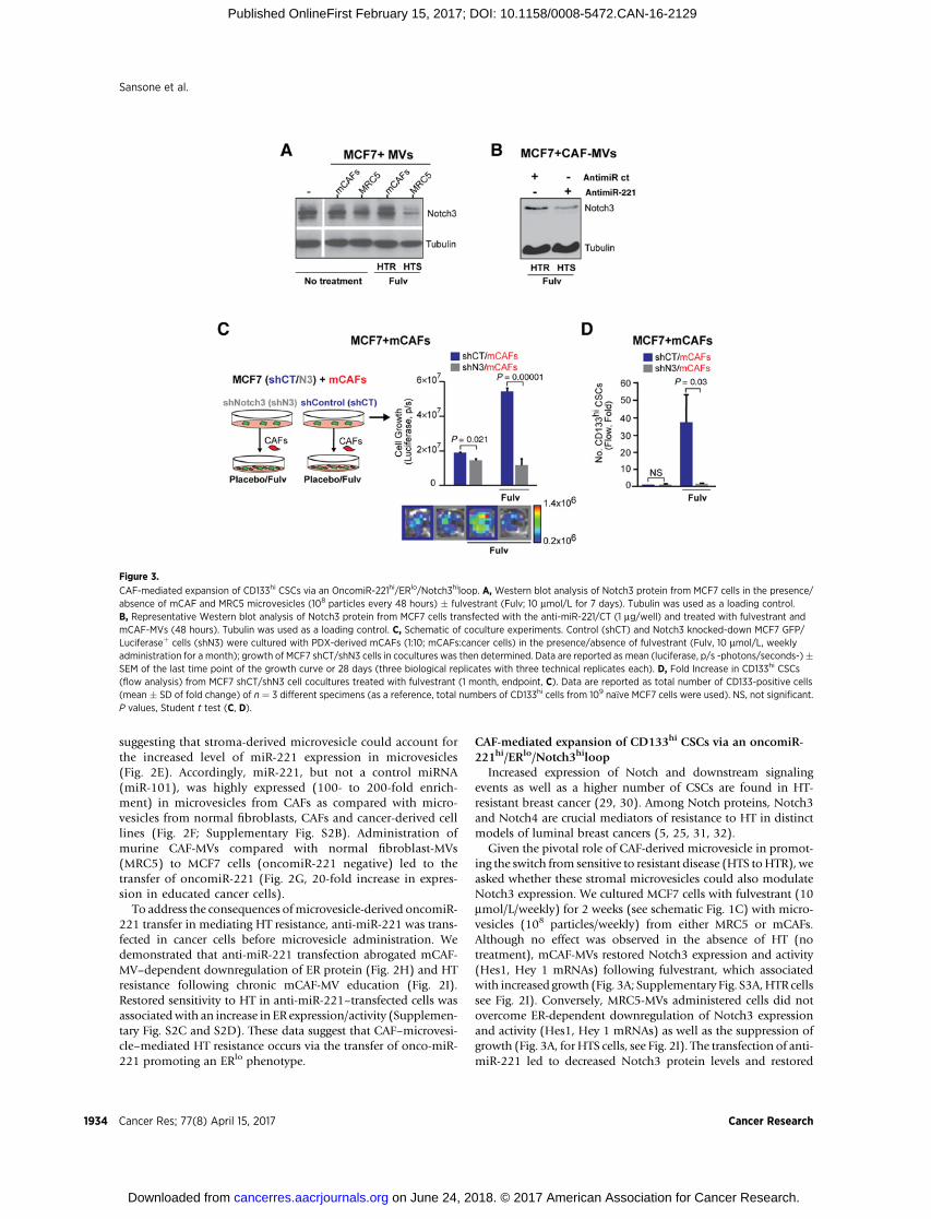
suggesting that stroma-derived microvesicle could account forthe increased level of miR-221 expression in microvesicles(Fig. 2E). Accordingly, miR-221, but not a control miRNA(miR-101), was highly expressed (100- to 200-fold enrich-ment) in microvesicles from CAFs as compared with micro-vesicles from normal fibroblasts, CAFs and cancer-derived celllines (Fig. 2F; Supplementary Fig. S2B). Administration ofmurine CAF-MVs compared with normal fibroblast-MVs(MRC5) to MCF7 cells (oncomiR-221 negative) led to thetransfer of oncomiR-221 (Fig. 2G, 20-fold increase in expres-sion in educated cancer cells).
To address the consequences ofmicrovesicle-derived oncomiR-221 transfer in mediating HT resistance, anti-miR-221 was trans-fected in cancer cells before microvesicle administration. Wedemonstrated that anti-miR-221 transfection abrogated mCAF-MV–dependent downregulation of ER protein (Fig. 2H) and HTresistance following chronic mCAF-MV education (Fig. 2I).Restored sensitivity to HT in anti-miR-221–transfected cells wasassociatedwith an increase in ER expression/activity (Supplemen-tary Fig. S2C and S2D). These data suggest that CAF–microvesi-cle–mediated HT resistance occurs via the transfer of onco-miR-221 promoting an ERlo phenotype.
CAF-mediated expansion of CD133hi CSCs via an oncomiR-221hi/ERlo/Notch3hiloop
Increased expression of Notch and downstream signalingevents as well as a higher number of CSCs are found in HT-resistant breast cancer (29, 30). Among Notch proteins, Notch3and Notch4 are crucial mediators of resistance to HT in distinctmodels of luminal breast cancers (5, 25, 31, 32).
Given the pivotal role of CAF-derived microvesicle in promot-ing the switch from sensitive to resistant disease (HTS toHTR), weasked whether these stromal microvesicles could also modulateNotch3 expression. We cultured MCF7 cells with fulvestrant (10mmol/L/weekly) for 2 weeks (see schematic Fig. 1C) with micro-vesicles (108 particles/weekly) from either MRC5 or mCAFs.Although no effect was observed in the absence of HT (notreatment), mCAF-MVs restored Notch3 expression and activity(Hes1, Hey 1 mRNAs) following fulvestrant, which associatedwith increased growth (Fig. 3A; Supplementary Fig. S3A,HTR cellssee Fig. 2I). Conversely, MRC5-MVs administered cells did notovercome ER-dependent downregulation of Notch3 expressionand activity (Hes1, Hey 1 mRNAs) as well as the suppression ofgrowth (Fig. 3A, for HTS cells, see Fig. 2I). The transfection of anti-miR-221 led to decreased Notch3 protein levels and restored
Figure 3.
CAF-mediated expansion of CD133hi CSCs via an OncomiR-221hi/ERlo/Notch3hiloop. A, Western blot analysis of Notch3 protein from MCF7 cells in the presence/absence of mCAF and MRC5 microvesicles (108 particles every 48 hours) � fulvestrant (Fulv; 10 mmol/L for 7 days). Tubulin was used as a loading control.B, Representative Western blot analysis of Notch3 protein from MCF7 cells transfected with the anti-miR-221/CT (1 mg/well) and treated with fulvestrant andmCAF-MVs (48 hours). Tubulin was used as a loading control. C, Schematic of coculture experiments. Control (shCT) and Notch3 knocked-down MCF7 GFP/Luciferaseþ cells (shN3) were cultured with PDX-derived mCAFs (1:10; mCAFs:cancer cells) in the presence/absence of fulvestrant (Fulv, 10 mmol/L, weeklyadministration for a month); growth of MCF7 shCT/shN3 cells in cocultures was then determined. Data are reported as mean (luciferase, p/s -photons/seconds-)�SEM of the last time point of the growth curve or 28 days (three biological replicates with three technical replicates each). D, Fold Increase in CD133hi CSCs(flow analysis) from MCF7 shCT/shN3 cell cocultures treated with fulvestrant (1 month, endpoint, C). Data are reported as total number of CD133-positive cells(mean � SD of fold change) of n ¼ 3 different specimens (as a reference, total numbers of CD133hi cells from 109 na€�ve MCF7 cells were used). NS, not significant.P values, Student t test (C, D).
Sansone et al.
Cancer Res; 77(8) April 15, 2017 Cancer Research1934
on June 24, 2018. © 2017 American Association for Cancer Research. cancerres.aacrjournals.org Downloaded from
Published OnlineFirst February 15, 2017; DOI: 10.1158/0008-5472.CAN-16-2129

sensitivity to HT (fulvestrant) of CAF-MV–treated cancer cells(Figs. 2I and 3B). These data demonstrate that miR-221 inCAF-MVs can block HT-mediated downregulation of Notch3expression.
We recently described the enrichment of CD133hi/ERlo/Notch3hi CSCs in HT-resistant tumors, which also expressed highlevels of Notch-regulated genes such as Hey1 and Hes1(GSE69280; ref. 5).
Figure 4.
IL6/Stat3 signaling from CAFs promotes the expansion of CD133hi CSCs. A, Cytokine array expression of the CM from MCF7 and HS27a cells (10 mg total protein).Highlighted are overexpressed cytokines and chemokines (IL6, IL6sR, IL8, MIP-1d, and CCL5). B, Dot plot showing phospho-tyrosine 705 Stat3 (pStat3) IHCquantification in HTR-derived primary tumor tissues (see Fig. 1B) in both the stroma and tumor compartments. Quantification of pStat3was performed using ImageJ/FIJI (NIH) of n¼ 19 fields from n¼ 5 different tumors. The results were expressed as ratio of pStat3 IHC value/total tissue area. A representative IHC image is shown.Scale bar, 25 mm. C, Bar graph of the proliferation capacity (Calcein AM, fluorescence) of xenograft-derived mCAFs isolated from HTR xenografts (Fig. 1) andcultured in the presence of vehicle (placebo) or signaling pathway inhibitors including ER (fulvestrant, 10 mmol/L) or PI3K (BYL, 100 nmol/L), HER (lapatinib,100 nmol/L) or JAK/pStat3 (AZD1480, 500 nmol/L). Data are reported as mean (fluorescence) � SEM of the last time point of the growth curve (14 days; threebiological replicates with three technical replicates each). D, Cytokine array expression from the CM (10 mg) of HS27shIL6 versus HS27a cells. Highlighted areoverexpressed cytokines and chemokines (IL6, IL6sR, IL8, MIP-1d, and CCL5). E,Western blot analysis of pStat3, Stat3, CD44, vimentin, caveolin1, and tubulin proteinlevels in HS27a cells CT and shIL6. F, Bar graph of OncomiR-221 expression in microvesicles derived from 108 HS27a and HS27shIL6 cells (qPCR, fold as reference,MCF7 microvesicles were used, normalized to U6 expression). Bar graph of protein levels (mg) in 108 microvesicles isolated from HS27a and HS27shIL6 cells is alsoshown. G and H, Tumor growth and metastatic burden (luciferase, BLI) in MCF7/HS27a and MCF7/HS27shIL6 xenografts. 103 cancer cells were inoculated inboth 4th inguinal MFPwith 102HS27a cells (CT or shIL6). Tumor growthwas examined over 20weeks. Data are reported asmean BLI value� SEM (log scale) for eachtime point (n ¼ 4/5 mice/group). Metastatic burden is mean BLI value � SEM of signal from metastatic tissues including lymph nodes, lungs, and bones. Arepresentative image of primary tumors from MCF7/HS27a versus MCF7/HS27shIL6 is shown. I, Bar graph representing immunohistochemical quantification (IRSscore) of ERa, CD133, and Notch3 in tumor-derived tissues from MCF7/HS27a or MCF7/HS27shIL6 xenografts (G). Data are reported as mean� SD of n¼ 10 tissuesections for each group. J, Bar graph showing percentage (H&E, %) of stroma–tumor niches versus tumor compartment in primary tumor tissues derived fromMCF7/HS27a and MCF7/HS27shIL6 xenografts (G). K, Representative Pankeratin staining of tissues slides from G depicting stroma–tumor niches (cancer cellssurrounded by stromal cells). Data are reported asmean� SD of n¼ 10 tissue slides for each group. P values, t test (B, F,H, I),Wilcoxon two-sample test (J), multiplecomparisons corrected post hoc t test after GLM ANOVA (C), repeated measures GLM ANOVA (G).
Stroma Microvesicles Regulate Endocrine Therapy Resistance
www.aacrjournals.org Cancer Res; 77(8) April 15, 2017 1935
on June 24, 2018. © 2017 American Association for Cancer Research. cancerres.aacrjournals.org Downloaded from
Published OnlineFirst February 15, 2017; DOI: 10.1158/0008-5472.CAN-16-2129

Given the role of CAF or stromal microvesicles in promoting amiR-221hi/ERlo/Notch3hi phenotype, we tested the hypothesisthatCAFs couldpromote the in vivo expansionofCD133hi cells viaNotch3 upregulation. The selective reduction of Notch3 expres-sion in cancer cells (shNotch3) and activity (using an anti-Jagged1blocking antibody) abrogated CAF-mediated HT resistance andthe expansion of CD133hi cancer cells (Fig. 3C and D; Supple-mentary Fig. S3B). In agreement with the knockdown experiment,overexpression of the activated form of Notch3 (pNICD3) inMCF7 cells led to an increase in CD133 promoter luciferaseactivity (pCD133) with HT (fulvestrant) in association with areduction in ER protein levels (Supplementary Fig. S3C and S3D).These data suggest that higher Notch3 activation could promote afeed-forward ERlo/CD133hi loop necessary for the generation ofCD133hi CSCs (Supplementary Fig. S3C and S3D). Overall, ourdata describe a novel mechanism of HT resistance: CAF-MV–mediated transfer of the onco-miR-221 leading to reduced ERexpression and Notch3 upregulation.
IL6/Stat3 activity is required for CAF–CSC niche formationAs the biogenesis of oncomiR-221/222hi microvesicles
occurs preferentially in CAFs (not normal fibroblasts), we
hypothesized that the abrogation of a CAF phenotype wouldinterfere with the generation of HT-resistant CSCs. To investi-gate this hypothesis, we examined possible candidates respon-sible for CAF growth. Compared with breast cancer cells, theCM of CAFs (HS27a cells) expressed higher levels of chemo-kines (e.g., IL8, MIP-1d, CCL5) and cytokines, including IL6 anactivator of Stat3 (Fig. 4A). These findings were further sup-ported by evidence of high pStat3 levels in murine CAFs fromHTR-derived xenografts (Fig. 4B). Differently from other sig-naling pathways (HER, PI3K, ER), pStat3 activity was requiredfor CAF proliferation as well as the generation of oncomiR-221hi microvesicles (Fig. 4C; Supplementary Fig. S4A). Inconcordance with these data, reduced IL6 expression in HS27acells (using an IL6-shRNA) lowered secreted IL8/IL6R/CCL5levels as well as the expression of CAF markers includingpStat3, vimentin, and CD44 (Fig. 4D and E). In addition,compared with HS27shCT cells, HS27shIL6 cells had reducedproliferative and invasive potential (Supplementary Fig. S4Band S4C) as well as lowered MMP2/9 expression and activity,indicating a loss of characteristic CAF features (SupplementaryFig. S4D). Moreover, microvesicles from HS27shIL6 cells hadessentially no expression of oncomiR-221 compared with
Figure 5.
Autocrine CAF-derived IL6 triggers endocrine-resistant disease.A,Bar graph showing IL6 levelsmeasured by ELISA from the CM (10mg total protein) of primary CAFcultures (BM-CAFs) isolated from patient bone–derived breast cancer metastases (Supplementary Table S2). IL6 levels from MCF7 and HS27a are also shown.B, Growth (cell number) of BM-CAFs (specimen 5, Supplementary Table S2) treated with an anti IL6-IL6R antibody tocilizumab (500 ng/mL every 48 hours)versus IgG control. C, Bar graph of IL6 protein (ELISA) from the CM (2 mg total protein) of BM-CAFs treated with tocilizumab or IgG for a week (500 ng/mL every 48hours).D, Bar graph of oncomiR-221 expression by qPCR (Fold, normalized on U6 level) in microvesicles isolated from BM-CAF cultures. Microvesicles isolated fromMCF7 cells were used as a reference control. E, Tumor burden (luciferase) of MCF7 xenografts alone (black) and coinjected with BM-CAFs (specimen 6,Supplementary Table S2) treated with fulvestrant and tocilizumab. Briefly, MCF7 xenografts were established in the MFP of NOD/SCID mice alone (103 cells) or incombination with CAFs (102 cells). When tumors reached �1 cm (3 months), mice were randomized (n ¼ 4/group) to receive either fulvestrant (1 mg/weekly) orfulvestrant and tocilizumab (100 mg/g/mouse) for 4 months. Data are reported as mean BLI value � SEM at the endpoint of the experiment (7 months).Data are reported as mean � SD of three independent experiments (n ¼ 3). P values, Student t test (C, D, E), multiple comparisons corrected post hoc t test afterrepeated measures GLM ANOVA (B).
Sansone et al.
Cancer Res; 77(8) April 15, 2017 Cancer Research1936
on June 24, 2018. © 2017 American Association for Cancer Research. cancerres.aacrjournals.org Downloaded from
Published OnlineFirst February 15, 2017; DOI: 10.1158/0008-5472.CAN-16-2129

HS27shCT cells with no change in microvesicle production(Fig. 4F, protein content as a surrogate marker of microvesicleyield). These data suggest that IL6/pStat3 signaling is crucial forthe proliferation of CAFs and the production of oncomiR-221þ
microvesicles.To address the phenotypic consequences of decreasing IL6
signaling in CAFs, we coinjected MCF7 cells with HS27shCT andHS27shIL6 CAFs into the MFP of mice. Compared with controls(MCF7/HS27a), the coinjection of MCF7/HS27shIL6 cellsresulted in impaired tumor growth, lower metastatic burden, anddecreased expression of CD133hi/Notch3hi/ERalo CSCs (Fig. 4G–I; Supplementary Fig. S4E). In agreement with the loss ofCD133hi/Notch3hi CSCs, MCF7/HS27shIL6–derived tumors hadfewer stroma–tumor niches (Fig. 4J and K) and decreased pStat3expression (Supplementary Fig. S4F). Overall, our data suggestthat IL6-mediated generation of stromal niches is required for theexpansion of CD133hi CSCs.
Anti-IL6 therapy abrogates CAF-mediated de novoresistance to HT
To extend our results to clinical specimens, we establishedprimary cultures of stromal cells from patient-derived bonemetastases (Supplementary Table S2, BM-CAFs). IL6 is a well-known pleiotropic cytokine, secreted at high levels from the bonemarrow microenvironment and CAFs (33, 34). We isolated andculturedCAFs frombonemetastases (Supplementary Fig. S5Aanddata not shown) and detected high levels of IL6 protein from theCM of these primary cultures (Fig. 5A). These levels are similar tothose found from the CM of HS27a cells. Consistent with theHS27model, the abrogation of IL6 signaling, using the anti-IL6R-IL6 antibody (tocilizumab), abrogated the growth in 70% of CAFprimary cultures (Fig. 5B), reduced IL6 secretion and the expres-sion of CAF markers (Fig. 5C; Supplementary Fig. S5B), thisfinding suggests that autocrine IL6 maintains the CAF phenotypeof these cells.
AlthoughmiR-221/222 expression is very low in luminal breastcancer tissues and cells (Fig. 2), high levels of miR-221 was foundin circulatingmicrovesicles fromHTR patients (Fig. 2D). Next, wereasoned that, in agreement with other investigators, CAFs wouldbe the major source of miR-221 (35). We then isolated micro-vesicles from CAF primary cultures derived from bone metastasesand found increased levels of oncomiR-221 as compared withtumor microvesicles (Fig. 5D); these results were similar to thosefound in HS27a cells and mCAFs (Fig. 4). We subsequentlydemonstrated that the coinjection of BM-CAFs with MCF7 cellspromoted HTR tumor growth (treated with fulvestrant) in an IL6-dependent manner, as combination fulvestrant/tocilizumababrogated tumor growth (Fig. 5E).
CAF-mediated expansion of CD133hi CSCsWe and others have recently demonstrated the enrichment of
CSCs expressing CD133 or ALDHhi activity in experimentalluminal breast cancers following HT (5, 25). In addition, expres-sion of Prominin1 (CD133) was identified in tumors frompatients who progressed on adjuvant HT (23). In agreement withother investigators, CD133hi and CD44hi cells are functionallyCSCs as they were capable of engrafting with low cell numbers(<1,000, Supplementary Fig. S6A). We previously demonstratedthat differently from CD44hi cells, CD133hi/CD44lo cellsexpressed embryonic stem cell signatures and increased theexpression of ABCG2, a CSC gene associated with therapy resis-
tance (GSE69280; refs. 5, 36). Here we determined that CD133hi/CD44lo cells expressed normal stem cell signatures by GSEA(Supplementary Fig. S6B, GSE69280; ref. 5). In addition, com-pared with CD133lo/CD44lo and CD44hi/CD133lo cells, theinjection of CD133hi/CD44lo cells gave rise to slow-growingtumors (Supplementary Fig. S6C) with an increased capacity todisseminate to the bonemarrow (Supplementary Fig. S6D). Thesedata suggest that the CD133hi phenotype is a distinct CSC pop-ulation (37).
Next, we investigated the hypothesis that extrinsic (stromalheterogeneity) factors could regulate the evolution of therapy-resistant niches leading to metastatic progression (38, 39). Wedemonstrated high PROM1 (encoding CD133) expression bymicroarray (GSE17705) was associated with increased levels ofa mammary CAF gene signature (12) in the setting of human HT-resistant primary tumors (Fig. 6A,P<1.82�10�4; SupplementaryFig. S7A).
To examine the importance of CD133hi cells in clinicallyrelevant models of metastatic breast cancer, we generated PDXsfrom HT-resistant (HTR) luminal breast cancer bone metastases(Fig. 6B; Supplementary Table S2). Cultured tumor cells wereserially transplanted into theMFP of immunocompromisedmicein the presence of HT (fulvestrant: a selective estrogen receptordegrader). After three sequential passages, the enhanced tumor-igenic capacity (% tumor take) of PDXs was associated withincreased Notch3 expression (Supplementary Fig. S7B), enrich-ment ofCD133hi (�40-fold) cancer cells andmurine stromal cells(�30 fold), which had infiltrated the tumor (Fig. 6B and C;Supplementary Fig. S7C and S7D). We further determined thatthe PDX-derived CD133hi cells had lowALDH activity, suggestingdifferent and unique stem cell populations arising through astroma-mediated metastatic transition within ERþ breast cancer(Supplementary Fig. S7E).
Given the coenrichment of CD133hi cells with CAFs, wehypothesized that CAFs could directly promote the expansion ofHT-resistant CD133hi cells. To address this hypothesis, we deplet-ed HT-resistant PDX primary cultures from CAFs (by FACS forEpCAMpos) andmaintained these cancer cells in culture for severalmonths. When long-term depleted for murine CAFs, EpCAMpos
cells from PDX primary cultures were growth inhibited by HT ascompared with tumor cells freshly isolated from cocultures (sort-ing for EpCAMpos, Fig. 6D and E). Moreover, acquired sensitivityto HT resulted in decreased numbers of self-renewing CD133hi
cells (Fig. 6E and F).To determine whether CAFs could confer de novo HT resis-
tance and promote the biogenesis of CD133hi cells, we culturedHT-sensitive MCF7 cells with murine CAFs, isolated fromxenografts, with/without HT (fulvestrant) for 4 weeks (Fig.6G). When cultured with CAFs, MCF7 cancer cells becameresistant to HT (fulvestrant), as compared with MCF7 cellsalone (Fig. 6G). Concomitant with HTR cancer cell growth, byFACS we observed a marked (30-fold) increase in CD133hi
CSCs as compared with MCF7 cells alone (Fig. 6H). Furthercharacterization of the CD133hi cells revealed that ERa mRNAlevels were lower in these cells than in CD133lo/CD44lo cells(Fig. 6I). In agreement with these data, higher CD133/PROM1expression was associated with decreased ESR1 mRNA expres-sion and activity in HT-resistant primary tumors (Fig. 6J, P <0.0001). Overall, these findings demonstrate that CAFs, in thepresence of HT, can promote the de novo formation of CD133hi
CSCs and HTR disease.
Stroma Microvesicles Regulate Endocrine Therapy Resistance
www.aacrjournals.org Cancer Res; 77(8) April 15, 2017 1937
on June 24, 2018. © 2017 American Association for Cancer Research. cancerres.aacrjournals.org Downloaded from
Published OnlineFirst February 15, 2017; DOI: 10.1158/0008-5472.CAN-16-2129
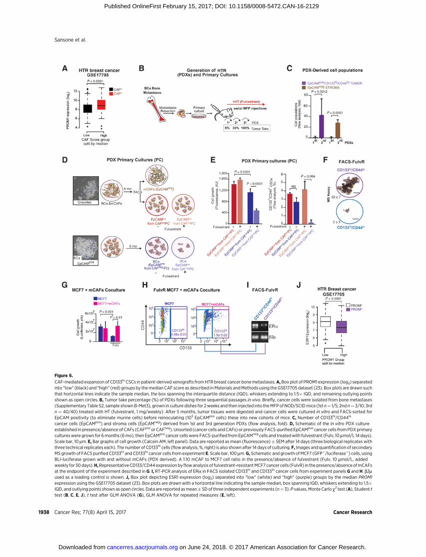
Figure 6.
CAF-mediated expansion of CD133hi CSCs in patient-derived xenografts fromHTR breast cancer bonemetastases.A,Box plot of PROM1 expression (log2) separatedinto "low" (black) and "high" (red) groups by themedian CAF score as described inMaterials andMethods using the GSE17705 dataset (23). Box plots are drawn suchthat horizontal lines indicate the sample median, the box spanning the interquartile distance (IQD), whiskers extending to 1.5� IQD, and remaining outlying pointsshown as open circles. B, Tumor take percentage (%) of PDXs following three sequential passages in vivo. Briefly, cancer cells were isolated from bone metastases(Supplementary Table S2, sample shownB-Met3), grown in culture dishes for 2weeks and then injected into theMFPof NOD/SCIDmice (1st n¼ 1/5; 2nd n¼ 3/10; 3rdn ¼ 40/40) treated with HT (fulvestrant, 1 mg/weekly). After 5 months, tumor tissues were digested and cancer cells were cultured in vitro and FACS-sorted forEpCAM positivity (to eliminate murine cells) before reinoculating (105 EpCAMpos cells) these into new cohorts of mice. C, Number of CD133hi/CD44lo
cancer cells (EpCAMpos) and stroma cells (EpCAMneg) derived from 1st and 3rd generation PDXs (flow analysis, fold). D, Schematic of the in vitro PDX cultureestablished in presence/absence of CAFs (CAFpos or CAFneg). Unsorted (cancer cells andCAFs) or previously FACS-purifiedEpCAMpos cancer cells fromPDXprimarycultureswere grown for 6months (6mo), then EpCAMpos cancer cellswere FACS-purified fromEpCAMneg cells and treatedwith fulvestrant (Fulv, 10mmol/L 14 days).Scale bar, 10 mm. E, Bar graphs of cell growth (Calcein AM, left panel). Data are reported as mean (fluorescence)� SEM after 14 days (three biological replicates withthree technical replicates each). The number of CD133hi cells (flowanalysis, %, right) is also shown after 14 days of culturing. F, Images andquantification of secondaryMS growth of FACS purified CD133hi and CD133lo cancer cells from experiment E. Scale bar, 100 mm.G, Schematic and growth ofMCF7 (GFPþ/luciferaseþ) cells, usingBLI-luciferase grown with and without mCAFs (PDX derived). A 1:10 mCAF to MCF7 cell ratio in the presence/absence of fulvestrant (Fulv, 10 mmol/L, addedweekly for 30days).H,RepresentativeCD133/CD44expression byflowanalysis of fulvestrant-resistantMCF7 cancer cells (FulvR) in the presence/absenceofmCAFsat the endpoint of the experiment described in G. I, RT-PCR analysis of ERa in FACS isolated CD133hi and CD133lo cancer cells from experiment panels G and H. b2mused as a loading control is shown. J, Box plot depicting ESR1 expression (log2) separated into "low" (white) and "high" (purple) groups by the median PROM1expression using the GSE17705 dataset (23). Box plots are drawnwith a horizontal line indicating the sample median, box spanning IQD, whiskers extending to 1.5�IQD, and outlying points shownas open circles. Data are reported asmean� SDof three independent experiments (n¼ 3).P values, Monte Carlo c2 test (A), Student ttest (B, C, E, J), t test after GLM ANOVA (G), GLM ANOVA for repeated measures (E, left).
Sansone et al.
Cancer Res; 77(8) April 15, 2017 Cancer Research1938
on June 24, 2018. © 2017 American Association for Cancer Research. cancerres.aacrjournals.org Downloaded from
Published OnlineFirst February 15, 2017; DOI: 10.1158/0008-5472.CAN-16-2129
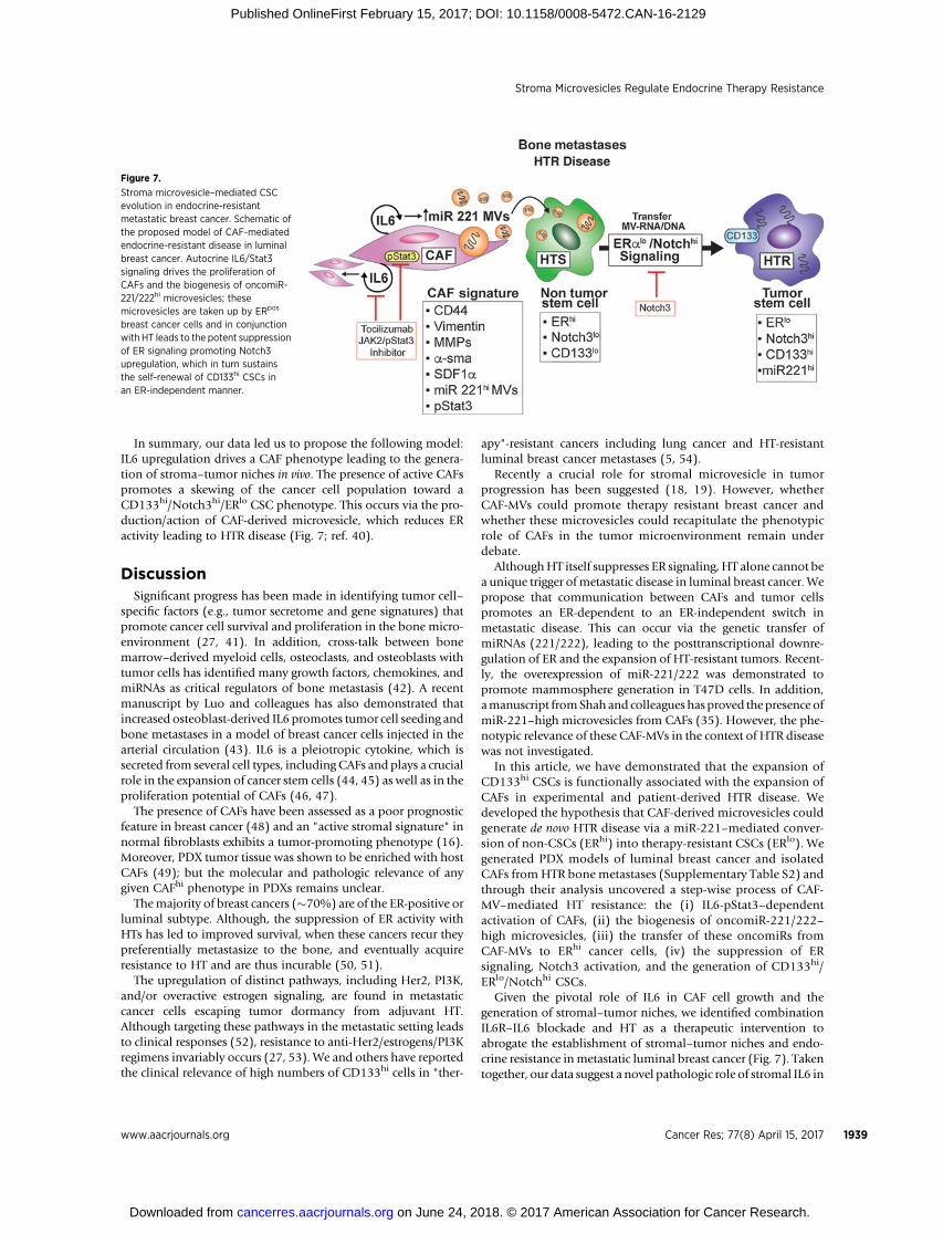
In summary, our data led us to propose the following model:IL6 upregulation drives a CAF phenotype leading to the genera-tion of stroma–tumor niches in vivo. The presence of active CAFspromotes a skewing of the cancer cell population toward aCD133hi/Notch3hi/ERlo CSC phenotype. This occurs via the pro-duction/action of CAF-derived microvesicle, which reduces ERactivity leading to HTR disease (Fig. 7; ref. 40).
DiscussionSignificant progress has been made in identifying tumor cell–
specific factors (e.g., tumor secretome and gene signatures) thatpromote cancer cell survival and proliferation in the bone micro-environment (27, 41). In addition, cross-talk between bonemarrow–derived myeloid cells, osteoclasts, and osteoblasts withtumor cells has identified many growth factors, chemokines, andmiRNAs as critical regulators of bone metastasis (42). A recentmanuscript by Luo and colleagues has also demonstrated thatincreased osteoblast-derived IL6 promotes tumor cell seeding andbone metastases in a model of breast cancer cells injected in thearterial circulation (43). IL6 is a pleiotropic cytokine, which issecreted from several cell types, including CAFs and plays a crucialrole in the expansion of cancer stem cells (44, 45) as well as in theproliferation potential of CAFs (46, 47).
The presence of CAFs have been assessed as a poor prognosticfeature in breast cancer (48) and an "active stromal signature" innormal fibroblasts exhibits a tumor-promoting phenotype (16).Moreover, PDX tumor tissue was shown to be enriched with hostCAFs (49); but the molecular and pathologic relevance of anygiven CAFhi phenotype in PDXs remains unclear.
Themajority of breast cancers (�70%) are of the ER-positive orluminal subtype. Although, the suppression of ER activity withHTs has led to improved survival, when these cancers recur theypreferentially metastasize to the bone, and eventually acquireresistance to HT and are thus incurable (50, 51).
The upregulation of distinct pathways, including Her2, PI3K,and/or overactive estrogen signaling, are found in metastaticcancer cells escaping tumor dormancy from adjuvant HT.Although targeting these pathways in the metastatic setting leadsto clinical responses (52), resistance to anti-Her2/estrogens/PI3Kregimens invariably occurs (27, 53). We and others have reportedthe clinical relevance of high numbers of CD133hi cells in "ther-
apy"-resistant cancers including lung cancer and HT-resistantluminal breast cancer metastases (5, 54).
Recently a crucial role for stromal microvesicle in tumorprogression has been suggested (18, 19). However, whetherCAF-MVs could promote therapy resistant breast cancer andwhether these microvesicles could recapitulate the phenotypicrole of CAFs in the tumor microenvironment remain underdebate.
AlthoughHT itself suppresses ER signaling, HT alone cannot bea unique trigger ofmetastatic disease in luminal breast cancer. Wepropose that communication between CAFs and tumor cellspromotes an ER-dependent to an ER-independent switch inmetastatic disease. This can occur via the genetic transfer ofmiRNAs (221/222), leading to the posttranscriptional downre-gulation of ER and the expansion of HT-resistant tumors. Recent-ly, the overexpression of miR-221/222 was demonstrated topromote mammosphere generation in T47D cells. In addition,amanuscript fromShahand colleagues has proved thepresence ofmiR-221–high microvesicles from CAFs (35). However, the phe-notypic relevance of these CAF-MVs in the context of HTR diseasewas not investigated.
In this article, we have demonstrated that the expansion ofCD133hi CSCs is functionally associated with the expansion ofCAFs in experimental and patient-derived HTR disease. Wedeveloped the hypothesis that CAF-derived microvesicles couldgenerate de novo HTR disease via a miR-221–mediated conver-sion of non-CSCs (ERhi) into therapy-resistant CSCs (ERlo). Wegenerated PDX models of luminal breast cancer and isolatedCAFs fromHTR bone metastases (Supplementary Table S2) andthrough their analysis uncovered a step-wise process of CAF-MV–mediated HT resistance: the (i) IL6-pStat3–dependentactivation of CAFs, (ii) the biogenesis of oncomiR-221/222–high microvesicles, (iii) the transfer of these oncomiRs fromCAF-MVs to ERhi cancer cells, (iv) the suppression of ERsignaling, Notch3 activation, and the generation of CD133hi/ERlo/Notchhi CSCs.
Given the pivotal role of IL6 in CAF cell growth and thegeneration of stromal–tumor niches, we identified combinationIL6R–IL6 blockade and HT as a therapeutic intervention toabrogate the establishment of stromal–tumor niches and endo-crine resistance inmetastatic luminal breast cancer (Fig. 7). Takentogether, our data suggest a novel pathologic role of stromal IL6 in
Figure 7.
Stroma microvesicle–mediated CSCevolution in endocrine-resistantmetastatic breast cancer. Schematic ofthe proposed model of CAF-mediatedendocrine-resistant disease in luminalbreast cancer. Autocrine IL6/Stat3signaling drives the proliferation ofCAFs and the biogenesis of oncomiR-221/222hi microvesicles; thesemicrovesicles are taken up by ERpos
breast cancer cells and in conjunctionwith HT leads to the potent suppressionof ER signaling promoting Notch3upregulation, which in turn sustainsthe self-renewal of CD133hi CSCs inan ER-independent manner.
Stroma Microvesicles Regulate Endocrine Therapy Resistance
www.aacrjournals.org Cancer Res; 77(8) April 15, 2017 1939
on June 24, 2018. © 2017 American Association for Cancer Research. cancerres.aacrjournals.org Downloaded from
Published OnlineFirst February 15, 2017; DOI: 10.1158/0008-5472.CAN-16-2129

luminal breast cancer: the secretion of oncomiR-221/222 highmicrovesicles leading to the evolution of therapy-resistant stro-mal–tumor niches. We characterized the cellular components ofthis stroma–tumor niche: "CD133hi CSCs and CAFs" and deter-mined the molecular machinery responsible for the niche gener-ation: autocrine IL6 inCAFs andCAF-derivedmicrovesicle-depen-dent downregulation of ER in cancer cells.
Disclosure of Potential Conflicts of InterestN. Fabbri is a consultant/advisory boardmember for IlluminossMedical Inc.
No potential conflicts of interest were disclosed for the other authors.
Authors' ContributionsConception and design: P. Sansone, D. Lyden, J. BrombergDevelopment of methodology: P. Sansone, M. Berishaj, C. SaviniAcquisition of data (provided animals, acquired and managed patients,provided facilities, etc.): P. Sansone, M. Berishaj, V.K. Rajasekhar, Q. Chang,A. Strillacci, L. Shapiro, A. Benito-Martin, N. Fabbri, J.H. Healey, J. BrombergAnalysis and interpretation of data (e.g., statistical analysis, biostatistics,computational analysis): P. Sansone, M. Berishaj, C. Ceccarelli, Q. Chang,A. Strillacci, R. Bowman, C. Mastroleo, F. Perna, E. Spisni, M. Cricca, D. Lyden,M. Bonaf�eWriting, review, and/or revision of the manuscript: P. Sansone, V.K. Rajase-khar, L. Shapiro, L. Daly, D. Lyden, M. Bonaf�e, J. BrombergAdministrative, technical, or material support (i.e., reporting or organizingdata, constructing databases): P. Sansone, M. Berishaj, S.D. CarolisStudy supervision: P. Sansone, D. Lyden, J. Bromberg
AcknowledgmentsWe are grateful to Mesruh Turkekul, Afsar Barlas, Sho Fujisawa, Romin
Yevgeniy (Molecular Cytology Core), and Donatella Santini (Department ofExperimental, Diagnostic and Specialty Medicine, University of Bologna, Italy)for advice and technical assistance.
Grant SupportThis study is supported by grants from Department of Defense
(W81XWH-10-1-1013 to P. Sansone) the NIH (R01: CA87637 to J. Brom-berg), Charles and Marjorie Holloway Foundation (J. Bromberg), SussmanFamily Fund (J. Bromberg), Lerner Foundation (J. Bromberg), The Beth C.Tortolani Foundation (J. Bromberg and D. Lyden), MSK Cancer CenterSupport Grant/Core Grant (P30 CA008748 to J. Bromberg), NIH (U01-CA169538 to D. Lyden), The Manning Foundation (D. Lyden), The HartwellFoundation (D. Lyden), Fundacao para aCiencia e a Tecnologia (D. Lyden),The Nancy C and Daniel P Paduano Foundation (D. Lyden), The Mary KayFoundation (D. Lyden), Pediatric Oncology Experimental Therapeutic Inves-tigator Consortium (POETIC; D. Lyden), James Paduano Foundation (D.Lyden), Malcolm Hewitt Weiner Foundation (D. Lyden), Theodore A RappFoundation (D. Lyden), American Hellenic Educational Progressive Associ-ation 5th District Cancer Research Foundation (D. Lyden). C. Savini won aMarco Polo fellowship from the University of Bologna. M. Bonaf�e issupported by the Cornelia and Roberto Pallotti Legacy.
The costs of publication of this articlewere defrayed inpart by the payment ofpage charges. This article must therefore be hereby marked advertisement inaccordance with 18 U.S.C. Section 1734 solely to indicate this fact.
ReceivedAugust 2, 2016; revised January 12, 2017; accepted January 12, 2017;published OnlineFirst February 15, 2017.
References1. Yu DD, Wu Y, Shen HY, Lv MM, Chen WX, Zhang XH, et al. Exosomes in
development, metastasis and drug resistance of breast cancer. Cancer Sci2015;106:959–64.
2. Challagundla KB, Wise PM, Neviani P, Chava H, Murtadha M, Xu T, et al.Exosome-mediated transfer of microRNAs within the tumor microenvi-ronment and neuroblastoma resistance to chemotherapy. J Natl CancerInst 2015;107:186–91.
3. Nouraee N, Mowla SJ, Calin GA. Tracking miRNAs' footprints intumor-microenvironment interactions: Insights and implicationsfor targeted cancer therapy. Genes Chromosomes Cancer 2015;54:335–52.
4. Haslam SZ, Woodward TL.Host microenvironment in breast cancer devel-opment: epithelial-cell-stromal-cell interactions and steroid hormoneaction in normal and cancerous mammary gland. Breast Cancer Res2003;5:208–15.
5. Sansone P, Ceccarelli C, Berishaj M, Chang Q, Rajasekhar VK, Perna F,et al. Self-renewal of CD133(hi) cells by IL6/Notch3 signalling regulatesendocrine resistance in metastatic breast cancer. Nat Commun2016;7:10442.
6. Dalerba P, Clarke MF. Cancer stem cells and tumor metastasis: first stepsinto uncharted territory. Cell Stem Cell 2007;1:241–2.
7. Hermann PC, Huber SL, Herrler T, Aicher A, Ellwart JW, Guba M, et al.Distinct populations of cancer stem cells determine tumor growth andmetastatic activity in human pancreatic cancer. Cell Stem Cell 2007;1:313–23.
8. Vermeulen L, de Sousa e Melo F, Richel DJ, Medema JP. The developingcancer stem-cell model: clinical challenges and opportunities. LancetOncol 2012;13:e83–9.
9. Borovski T, De Sousa E Melo F, Vermeulen L, Medema JP. Cancer stem cellniche: the place to be. Cancer Res 2011;71:634–9.
10. Sansone P, Storci G, Giovannini C, Pandolfi S, Pianetti S, Taffurelli M, et al.p66Shc/Notch-3 interplay controls self-renewal and hypoxia survival inhuman stem/progenitor cells of the mammary gland expanded in vitro asmammospheres. Stem Cells 2007;25:807–15.
11. ChangQ, Bournazou E, Sansone P, BerishajM,Gao SP,Daly L, et al. The IL-6/JAK/Stat3 feed-forward loop drives tumorigenesis and metastasis. Neo-plasia 2013;15:848–62.
12. Allinen M, Beroukhim R, Cai L, Brennan C, Lahti-Domenici J, Huang H,et al. Molecular characterization of the tumormicroenvironment in breastcancer. Cancer Cell 2004;6:17–32.
13. Strillacci A, Griffoni C, Sansone P, Paterini P, Piazzi G, Lazzarini G,et al. miR-101 downregulation is involved in cyclooxygenase-2overexpression in human colon cancer cells. Exp Cell Res 2009;315:1439–47.
14. Chen C, Ridzon DA, Broomer AJ, Zhou Z, Lee DH, Nguyen JT, et al. Real-time quantification ofmicroRNAs by stem-loop RT-PCR. Nucleic Acids Res2005;33:e179.
15. D'Anello L, Pasquale Sansone, Gianluca Storci, Valentina Mitrugno, Gab-rieleD'Uva, Pasquale Chieco, et al. Epigenetic control of the basal-like geneexpression profile via Interleukin-6 in breast cancer cells. Mol Cancer2010;9:300.
16. Al-Rakan MA, Colak D, Hendrayani SF, Al-Bakheet A, Al-Mohanna FH,Kaya N, et al. Breast stromal fibroblasts from histologically normal surgicalmargins are pro-carcinogenic. J Pathol 2013;231:457–65.
17. Au Yeung CL, Co NN, Tsuruga T, Yeung TL, Kwan SY, Leung CS, et al.Exosomal transfer of stroma-derived miR21 confers paclitaxel resistancein ovarian cancer cells through targeting APAF1. Nat Commun2016;7:11150.
18. Zhang L, Zhang S, Yao J, Lowery FJ, Zhang Q, Huang W-C, et al. Micro-environment-induced PTEN loss by exosomal microRNA primes brainmetastasis outgrowth. Nature 2015;527:100–4.
19. Boelens MC, Wu TJ, Nabet BY, Xu B, Qiu Y, Yoon T, et al. Exosome transferfrom stromal to breast cancer cells regulates therapy resistance pathways.Cell 2014;159:499–513.
20. ToyW, Shen Y,WonH,Green B, Sakr RA,WillM, et al. ESR1 ligand-bindingdomain mutations in hormone-resistant breast cancer. Nat Genet2013;45:1439–45.
21. OsborneCK, Schiff R.Mechanisms of endocrine resistance in breast cancer.Annu Rev Med 2011;62:233–47.
22. Leary AF, Drury S, Detre S, Pancholi S, Lykkesfeldt AE, Martin LA, et al.Lapatinib restores hormone sensitivity with differential effects on estrogenreceptor signaling in cell models of human epidermal growth factorreceptor 2-negative breast cancer with acquired endocrine resistance. ClinCancer Res 2010;16:1486–97.
Cancer Res; 77(8) April 15, 2017 Cancer Research1940
Sansone et al.
on June 24, 2018. © 2017 American Association for Cancer Research. cancerres.aacrjournals.org Downloaded from
Published OnlineFirst February 15, 2017; DOI: 10.1158/0008-5472.CAN-16-2129

23. Symmans WF, Hatzis C, Sotiriou C, Andre F, Peintinger F, Regitnig P, et al.Genomic index of sensitivity to endocrine therapy for breast cancer. J ClinOncol 2010;28:4111–9.
24. Rao X, Di Leva G, Li M, Fang F, Devlin C, Hartman-Frey C, et al. MicroRNA-221/222 confers breast cancer fulvestrant resistance by regulating multiplesignaling pathways. Oncogene 2011;30:1082–97.
25. Simoes BM, O'Brien CS, Eyre R, Silva A, Yu L, Sarmiento-Castro A, et al.Anti-estrogen resistance in human breast tumors is driven byJAG1-NOTCH4-dependent cancer stem cell activity. Cell Rep 2015;12:1968–77.
26. Pedersen LG, Castelruiz Y, Jacobsen S, Aasted B. Identification of mono-clonal antibodies that cross-react with cytokines from different animalspecies. Vet Immunol Immunopathol 2002;88:111–122.
27. Zhang XH, Jin X,Malladi S, Zou Y,Wen YH, Brogi E, et al. Selection of bonemetastasis seeds bymesenchymal signals in the primary tumor stroma. Cell2013;154:1060–73.
28. Zhao R, Wu J, Jia W, Gong C, Yu F, Ren Z, et al. Plasma miR-221 as apredictive biomarker for chemoresistance in breast cancer patients whopreviously received neoadjuvant chemotherapy. Onkologie 2011;34:675–80.
29. Haughian JM, Pinto MP, Harrell JC, Bliesner BS, Joensuu KM, Dye WW,et al. Maintenance of hormone responsiveness in luminal breastcancers by suppression of Notch. Proc Natl Acad Sci U S A 2012;109:2742–7.
30. Bergamaschi A, Madak-Erdogan Z, Kim YJ, Choi YL, Lu H, Katzenel-lenbogen BS. The forkhead transcription factor FOXM1 promotesendocrine resistance and invasiveness in estrogen receptor-positivebreast cancer by expansion of stem-like cancer cells. Breast Cancer Res2014;16:436.
31. Magnani L, Stoeck A, Zhang X, L�anczky A, Mirabella AC, Wang TL, et al.Genome-wide reprogrammingof the chromatin landscapeunderlies endo-crine therapy resistance in breast cancer. ProcNatl Acad SciU S A2013;110:E1490–9.
32. Lombardo Y, Faronato M, Filipovic A, Vircillo V, Magnani L, Coombes RC.Nicastrin and Notch4 drive endocrine therapy resistance and epithelial tomesenchymal transition in MCF7 breast cancer cells. Breast Cancer Res2014;16:R62.
33. Karnoub AE, Dash AB, Vo AP, Sullivan A, Brooks MW, Bell GW, et al.Mesenchymal stem cells within tumour stroma promote breast cancermetastasis. Nature 2007;449:557–63.
34. Nagasaki T, Hara M, Nakanishi H, Takahashi H, Sato M, Takeyama H.Interleukin-6 released by colon cancer-associated fibroblasts is critical fortumour angiogenesis: anti-interleukin-6 receptor antibody suppressedangiogenesis and inhibited tumour-stroma interaction. Br J Cancer2014;110:469–78.
35. Shah SH, Miller P, Garcia-Contreras M, Ao Z, Machlin L, Issa E, et al.Hierarchical paracrine interaction of breast cancer associated fibroblastswith cancer cells via hMAPK-microRNAs to drive ER-negative breast cancerphenotype. Cancer Biol Ther 2015;16:1671–81.
36. Gilani RA, Kazi AA, Shah P, Schech AJ, Chumsri S, Sabnis G, et al. Theimportance of HER2 signaling in the tumor-initiating cell population inaromatase inhibitor-resistant breast cancer. Breast Cancer Res Treat2012;135:681–92.
37. Liu S, Cong Y, Wang D, Sun Y, Deng L, Liu Y, et al. Breast cancer stem cellstransition between epithelial and mesenchymal states reflective of theirnormal counterparts. Stem Cell Reports 2014;2:78–91.
38. Calon A, Lonardo E, Berenguer-Llergo A, Espinet E, Hernando-MomblonaX, Iglesias M, et al. Stromal gene expression defines poor-prognosis sub-types in colorectal cancer. Nat Genet 2015;47:320–9.
39. Diaz LA Jr,Williams R,Wu J, Kinde I, Hecht JR, Berlin J, et al. Themolecularevolution of acquired resistance to targeted EGFR blockade in colorectalcancers. Nature 2012;486:537–40.
40. Sjostrom AE, Sandblad L, Uhlin BE, Wai SN. Membrane vesicle-mediatedrelease of bacterial RNA. Sci Rep 2015;5:15329.
41. RenG, EspositoM, Kang Y. Bonemetastasis and themetastatic niche. JMolMed 2015;93:1203–12.
42. Esposito M, Kang Y. Targeting tumor-stromal interactions in bone metas-tasis. Pharmacol Ther 2014;141:222–33.
43. Luo X, Fu Y, Loza AJ, Murali B, Leahy KM, Ruhland MK, et al. Stromal-initiated changes in the bone promote metastatic niche development. CellRep 2016;14:82–92.
44. Sansone P, Storci G, Tavolari S, Guarnieri T, Giovannini C, Taffurelli M,et al. IL-6 triggers malignant features in mammospheres from humanductal breast carcinoma and normal mammary gland. J Clin Invest2007;117:3988–4002.
45. Korkaya H, Kim G-i, Davis A, Malik F, Henry NL, Ithimakin S, et al.Activation of an IL6 inflammatory loop mediates trastuzumab resistanceinHER2þ breast cancer by expanding the cancer stem cell population. MolCell 2012;47:570–84.
46. Mace TA, Ameen Z, Collins A, Wojcik S, Mair M, Young GS, et al. Pancreaticcancer-associated stellate cells promote differentiation of myeloid-derivedsuppressorcellsinaSTAT3-dependentmanner.CancerRes2013;73:3007–18.
47. Shintani Y, Fujiwara A, Kimura T, Kawamura T, Funaki S, Minami M, et al.IL-6 Secreted from cancer-associated fibroblasts mediates chemoresistancein NSCLC by increasing epithelial-mesenchymal transition signaling. JThorac Oncol 2016;11:1482–92.
48. Paulsson J,Micke P. Prognostic relevance of cancer-associatedfibroblasts inhuman cancer. Semin Cancer Biol 2014;25:61–8.
49. Cassidy JW, Caldas C, Bruna A. Maintaining tumor heterogeneity inpatient-derived tumor xenografts. Cancer Res 2015;75:2963–8.
50. Coleman REClinical features ofmetastatic bone disease and risk of skeletalmorbidity. Clin Cancer Res 2006;12:6243s–6249s.
51. Davies C, Pan H, Godwin J, Gray R, Arriagada R, Raina V, et al. Long-termeffects of continuing adjuvant tamoxifen to 10 years versus stopping at 5years after diagnosis of oestrogen receptor-positive breast cancer: ATLAS, arandomised trial. Lancet 2013;381:805–16.
52. Baselga J, CamponeM, Piccart M, Burris HAIII, Rugo HS, Sahmoud T, et al.Everolimus in postmenopausal hormone-receptor-positive advancedbreast cancer. N Engl J Med 2012;366:520–9.
53. Arnedos M, Vicier C, Loi S, Lefebvre C, Michiels S, Bonnefoi H, et al.Precision medicine for metastatic breast cancer–limitations and solutions.Nat Rev Clin Oncol 2015;12:693–704.
54. Bertolini G, D'Amico L, Moro M, Landoni E, Perego P, Miceli R, et al.Microenvironment-modulated metastatic CD133þ/CXCR4þ/EpCAM-lung cancer-initiating cells sustain tumor dissemination and correlate withpoor prognosis. Cancer Res 2015;75:3636–49.
www.aacrjournals.org Cancer Res; 77(8) April 15, 2017 1941
Stroma Microvesicles Regulate Endocrine Therapy Resistance
on June 24, 2018. © 2017 American Association for Cancer Research. cancerres.aacrjournals.org Downloaded from
Published OnlineFirst February 15, 2017; DOI: 10.1158/0008-5472.CAN-16-2129

Correction
Evolution of Cancer Stem-like Cells inEndocrine-Resistant Metastatic BreastCancers Is Mediated by Stromal Microvesicles
In this article (Cancer Res 2017;77:1927–41), which appeared in the April 15,2017 issue of Cancer Research (1), the affiliations for Dr. Massimiliano Bonaf�ewere incorrect; Dr. Bonaf�e's correct affiliations are Department of Experimental,Diagnostic and Specialty Medicine, AlmaMater Studiorum, Universit�a di Bologna,Bologna, Italy and Center for Applied Biomedical Research Laboratory, PoliclinicoUniversitario S. Orsola-Malpighi AlmaMater Studiorum, Universit�a di Bologna,Bologna, Italy.
The online version of the article has been corrected and no longer matches the print.The authors regret this error.
Reference1. Sansone P, Berishaj M, Rajasekhar VK, Ceccarelli C, Chang Q, Strillacci A, et al. Evolution of cancer
stem-like cells in endocrine-resistantmetastatic breast cancers ismediated by stromalmicrovesicles.Cancer Res 2017;77:1927–41.
Published online October 2, 2017.doi: 10.1158/0008-5472.CAN-17-2427�2017 American Association for Cancer Research.
CancerResearch
Cancer Res; 77(19) October 1, 20175438

2017;77:1927-1941. Published OnlineFirst February 15, 2017.Cancer Res Pasquale Sansone, Marjan Berishaj, Vinagolu K. Rajasekhar, et al. Metastatic Breast Cancers Is Mediated by Stromal MicrovesiclesEvolution of Cancer Stem-like Cells in Endocrine-Resistant
Updated version
10.1158/0008-5472.CAN-16-2129doi:
Access the most recent version of this article at:
Material
Supplementary
http://cancerres.aacrjournals.org/content/suppl/2017/02/15/0008-5472.CAN-16-2129.DC1
Access the most recent supplemental material at:
Cited articles
http://cancerres.aacrjournals.org/content/77/8/1927.full#ref-list-1
This article cites 53 articles, 9 of which you can access for free at:
Citing articles
http://cancerres.aacrjournals.org/content/77/8/1927.full#related-urls
This article has been cited by 3 HighWire-hosted articles. Access the articles at:
E-mail alerts related to this article or journal.Sign up to receive free email-alerts
Subscriptions
Reprints and
To order reprints of this article or to subscribe to the journal, contact the AACR Publications Department at
Permissions
Rightslink site. Click on "Request Permissions" which will take you to the Copyright Clearance Center's (CCC)
.http://cancerres.aacrjournals.org/content/77/8/1927To request permission to re-use all or part of this article, use this link
on June 24, 2018. © 2017 American Association for Cancer Research. cancerres.aacrjournals.org Downloaded from
Published OnlineFirst February 15, 2017; DOI: 10.1158/0008-5472.CAN-16-2129