evolutionary convergence in conodonts revealed by ...palaeo-electronica.org/content/pdfs/681.pdf ·...
TRANSCRIPT

Palaeontologia Electronica palaeo-electronica.org
Evolutionary convergence in conodonts revealed by Synchrotron-based Tomographic Microscopy
Michele Mazza and Carlos Martínez-Pérez
ABSTRACT
The conodont fossil record is well known for its morphological diversity, but theiterative evolution that characterizes conodonts often avoids providing reliable phylo-genetic frameworks among species, making unclear if the diagnostic characters of thetaxa are indicative of common ancestry or evolutionary convergences. To distinguishhomologies from analogies in conodonts, the most reliable method is by studying theontogenetic development of the single taxa. Until now, the reconstruction of theontogenetic stages was based on the study of separate individuals at different agefrom single populations. Nevertheless, the only unequivocal way to describe theontogenesis of a conodont is to describe it from a single specimen. We achieve thisobjective by using Synchrotron Radiation X-ray Tomographic Microscopy applied to P1
elements of species belonging to the Upper Triassic genera Carnepigondolella andEpigondolella. Our analysis provided internal tomographic information for the recon-struction of the conodont ontogenesis. We focused our study on the posterior platform,where an accessorial node develops behind the cusp. This node resulted in an autapo-morphy of the genus Epigondolella from previous cladistic analyses and, thus, a diag-nostic character for the elaboration of phylogenetic models. The microtomographiesshowed that this character is instead an evolutionary convergence. These results sug-gest the revision of the Late Triassic conodont phylogenetic relationships, showing thatontogenesis can be used as a criterion for discriminating homology from homoplasy inconodonts, and demonstrating that Synchrotron Radiation X-ray Tomographic Micros-copy is a powerful and reliable tool to investigate conodont ontogenesis, evolutionaryprocesses and phylogenetic relationships.
Michele Mazza. Dipartimento di Scienze della Terra "A. Desio", Università degli Studi di Milano, Via Mangiagalli 34, 20133, Milano (Italy); [email protected]; [email protected] Martínez-Pérez. Departamento de Geología, Universidad de Valencia, Avda. Dr. Moliner, 50, 46100 Burjassot, Valencia (Spain); [email protected]; School of Earth Sciences, University of Bristol, Wills Memorial Building, Queen’s Road, Bristol BS8 1RJ, UK; [email protected]
Keywords: conodonts; X-ray tomographies; ultrastructure; ontogenesis; Triassic
Submission: 11 May 2016 Acceptance: 23 November 2016
Mazza, Michele and Martínez-Pérez, Carlos. 2016. Evolutionary convergence in conodonts revealed by Synchrotron-based Tomographic Microscopy. Palaeontologia Electronica 19.3.52A: 1-11palaeo-electronica.org/content/2016/1710-conodont-x-ray-tomographies
Copyright: Palaeontological Association December 2016

MAZZA & MARTÍNEZ-PÉREZ: CONODONT X-RAY TOMOGRAPHIES
INTRODUCTION
The conodont fossil record constitutes a richarchive of evolutionary history, in terms of its tem-poral extent, from Cambrian to Triassic, and itscompleteness (Foote and Sepkoski, 1999). Func-tional analyses have advanced to an extent whereconodont taxonomy, based on dental morphology,can be interpreted in terms of feeding ecology(Jones et al., 2012a, 2012b; Purnell and Jones,2012; Martínez-Pérez et al., 2014a, 2014b, 2016;Murdok et al., 2014). This opens the possibility ofinterpreting the conodont fossil record as a detailedand comparatively complete record of the evolutionof feeding ecology through much of the Phanero-zoic, including across some of the most dramaticecological and environmental crises that haveimpacted animal life, such as the end-Ordovician,Frasnian-Fammenian, Hangenberg, and Permian-Triassic mass extinction events. Realizing thisvision requires a coherent phylogeny for conodontson which to trace the evolution of feeding ecology.To this end, attempts have been made to establisha phylogeny for conodonts using cladistics (Dono-ghue, 2001; Zhang and Barnes, 2004; Wickströmand Donoghue, 2005; Donoghue et al., 2008;Mazza et al., 2012b), but this aim is challenged bywidespread morphological convergence in dispa-rate conodont lineages (Sweet, 1988; Dzik, 2005).Here, we explore the utility of ontogenetic evi-dence, in the form of growth arrest lines that recordthe morphogenesis of conodont elements, showingthat they can be used to discriminate betweenhomologous and convergent adult morphologies.Until now, the reconstruction of the differentontogenetic stages was based on the study of iso-lated specimens from single populations, but sev-eral problems normally arise in order to distinguishbetween juvenile specimens of related species thatdid not develop their characteristic morphologiesuntil more developed stages. Hence, the only une-quivocal way to describe the ontogenesis is bydescribing it from a single element. A first step for-ward in this direction has recently been made byMazza and Martínez-Pérez (2015), who used Syn-chrotron data to test and verify the reconstructionsof 10 ontogenetic series of Carnian-Norian (LateTriassic) platform conodont species. In this study ahigher level of detail is reached in the use of the X-Ray Tomography Microscopy, reconstructing theontogenetic mechanisms of a single morphologicalcharacter in order to unravel homology from homo-plasy in conodonts.
MATERIAL AND CASE STUDY
The present work is focused on the P1 ele-ments of Late Triassic gondolellid conodonts fromthe Neotethyan Province. Our study is based onseven phylogenetically related species belongingto the genera Carnepigondolella (C. pseudodiebeli)and Epigondolella (E. miettoi, E. quadrata, E. via-lovi, E. rigoi, E. triangularis, and E. uniformis),ranging from the Upper Carnian (Tuvalian) to theLower Norian (Lacian) (Table 1). The studied cono-donts have Colour Alteration Index (CAI) values of1.5 or 7 (Epstein et al., 1977). All the specimensare stored at the Dipartimento di Scienze dellaTerra “A. Desio” of the Università degli Studi diMilano (see Table 1 for the repository numbers).
The material comes entirely from the UpperTriassic (Upper Carnian-Rhaetian) section of PizzoMondello (western Sicily, Italy), GSSP candidatefor the Norian stage (Muttoni et al., 2001, 2004;Nicora et al., 2007; Balini et al., 2011, 2012, 2015;Mazza et al., 2012a, 2012b; Mazza and Krystyn,2015).
The Carnepigondolella-Epigondolella lineageconstitutes a worthy case of study, because it is theonly conodont phylogenetic lineage encompassingthe Carnian/Norian boundary and evolving into thelast Middle Norian and Rhaetian taxa (i.e., generaMockina, Parvigondolella, and Misikella), whichare the last conodont representatives (Martínez-Pérez et al., 2014c). In addition, previous innova-tive studies on this lineage (Mazza et al., 2012b)provide a solid evolutionary framework to betested. The phylogenetic relationships of the spe-cies composing this lineage were first investigatedusing numerical cladistic analyses (Mazza et al.,2012b; Figure 1). In these analyses, some morpho-logical characters of the posterior platform resultedin autapomorphies that defined the most derivedtaxa of the Epigondolella clade. These were thedevelopment of denticles on the posterior platformmargin together with the enlargement of the poste-rior platform, and the occurrence of a large carinalnode behind the cusp. In addition, the platformdevelopment (ontogenesis) of some carnepigon-dolellids and epigondolellids, considered also inthe present study, were recently described byMazza and Martínez-Pérez (2015), using bothgrowth series and X-ray tomographies. This studyalso provided the tools to give a specific taxonomicidentity to the most juvenile conodont forms. Nev-ertheless, those results produced some discrepan-cies between the new conodont ranges thatemerged with the classification of the juvenilespecimens and the previous cladistic phylogenetic
2
PALAEO-ELECTRONICA.ORG
model (Figure 1). In particular, the phylogeneticposition of E. vialovi, E. quadrata, E. rigoi, E. uni-formis, and E. triangularis became uncertain,because their stratigraphic ranges are now pro-longed downwards in the Upper Carnian and notlimited to the Lower Norian as before, questioningtheir previous phylogenetic relationships (Mazzaand Martínez-Pérez, 2015, figure 1). It is evidentthat convergences between species were misinter-preted as homologies with phylogenetic value. Inparticular, the accessorial node behind the cuspthat resulted in an autapomorphy in Mazza et al.(2012b) and defined the Epigondolella clade,seemed to be developed at different ontogeneticstages in the epigondolellids (see Mazza andMartínez-Pérez, 2015). Thus, in an attempt to dis-criminate between homology and homoplasy, weundertook a detailed analysis of the morphogene-sis of what seems to be a key character to shedlight on the evolutionary scenario of the Late Car-nian/Norian conodonts.
METHODS
We characterise the inner growth patterns ofthe different species using the synchrotron radia-tion X-Ray Tomography Microscope at the X02DATOMCAT beamline of the Swiss Light Source, PaulScherrer Institute (Villigen, Switzerland), a tech-nique that allows non-invasive, high resolution,quantitative and volumetric x-ray tomographies ondiverse samples. The specimens were scannedusing a 20x objective, with exposure time between83 to 270 ms at 10-14 keV, acquiring 1501 projec-tions equiangularly over 180 degrees. Isotropicvoxel dimensions are 0.325 µm. Projections werepost-processed and rearranged into flat- and dark-field-corrected sinograms, and reconstruction wasperformed on a 60-core Linux PC farm using aFourier transform routine and a regridding proce-dure (Marone and Stampanoni, 2012). The recon-structed files were analyzed and manipulated usingAVIZO v.8 (VSG), allowing us to extract accurate3D virtual models, and virtual thin sections thatwere created using the voltex module, which simu-lates the casting of light rays from preset sources
TABLE 1. List of the conodonts scanned at the X-ray Synchrotron Microscopy for analyses of the internal structure.Species for which more than one specimen was scanned are provided with a capital letter that, associated with the spe-cies name, identify the specimen in the text and in figures. All the specimens are from the Pizzo Mondello section
(western Sicily, Italy).
Species Epigondolella vialovi Epigondolella quadrata Epigondolella quadrata Epigondolella quadrata
Author and year (Burij, 1989) Orchard, 1991 Orchard, 1991 Orchard, 1991
Specimen A B C
Range of the species
Upper Carnian - Lower Norian
Upper Carnian - Middle Norian
Upper Carnian - Middle Norian
Upper Carnian - Middle Norian
Sample NA30 NA60 NA60 NA66
Age of the sample
Uppermost Tuvalian (Upper Carnian)
Upper Lacian (Lower Norian)
Upper Lacian (Lower Norian)
Upper Lacian (Lower Norian)
Repository number
Micro-Unimi no. 2010
Micro-Unimi no. 2011
Micro-Unimi no. 2012
Micro-Unimi no. 2017
CAI 1.5 7 7 1.5
Species Epigondolella rigoi Epigondolella rigoi Epigondolella uniformis Epigondolella
triangularis
Author and year Noyan and Kozur, 2007 Noyan and Kozur, 2007 (Orchard, 1991) (Budurov, 1972)
Specimen A B
Range of the species
Lower - Middle Norian Lower - Middle Norian Lower - Middle Norian Lower Norian
Sample NA61 NA60 NA42 NA44a
Age of the sample
Upper Lacian(Lower Norian)
Upper Lacian(Lower Norian)
Lower Lacian (Lower Norian)
Lower Lacian (Lower Norian)
Repository number
Micro-Unimi no. 2018
Micro-Unimi no. 2014
Micro-Unimi no. 2013
Micro-Unimi no. 2015
CAI 7 1.5 1.5 1.5
3

MAZZA & MARTÍNEZ-PÉREZ: CONODONT X-RAY TOMOGRAPHIES
through a volume of data. This technique permittedus to generate multiple sections of the analyzedspecimens in all the desired directions, providing acomplete control on the area of the conodont thatwe want to investigate (Figure 2). Horizontal sec-tions of the entire element of the epigondolellidsconsidered in this study were already employed inMazza and Martínez-Pérez (2015) to verify the reli-ability of the reconstructed conodont ontogeneticseries. The excellent correspondence between thejuvenile stages outlined by the growth lines in theX-ray sections and juvenile specimens photo-graphed at the SEM used for the growth series,proved the validity of the technique. In this work,we analyze for the first time cross and longitudinalsections perpendicular to the longitudinal axis ofthe platform and parallel to the platform surface,respectively (Figure 2).
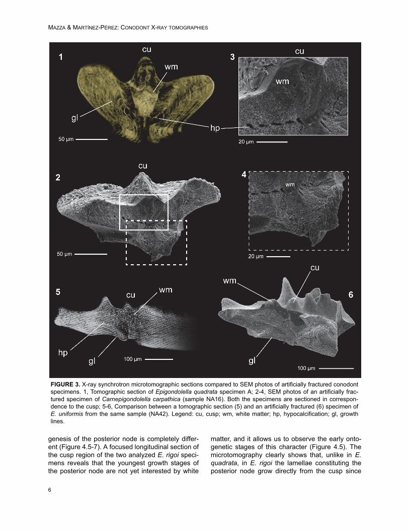
Microtomographic sections were comparedwith scanning electron microscopy (SEM) photos
of artificially fractured specimens from the samesamples in order to demonstrate the degree ofdetail of the synchrotron analyses (Figure 3). Theartificial fractures have been produced across thelongitudinal axes of the platforms with a commonneedle, etched with 0.5% orthophosphoric acid for2–4 minutes and coated with gold previous to theSEM analysis.
RESULTS
Tomographic images of the conodonts ultra-structure were obtained from all the scanned ele-ments, but with variable levels of detail fromspecimen to specimen. The best results wereobtained on conodonts with CAI value 7 (see Table1), probably induced by hydrothermal alteration(Rejebian, 1987). These conodonts show the bestcontrast between the growth lines and the highestnumber of visible details in the ultrastructure (Fig-
FIGURE 1. Cladogram representing the Epigondolella clade of Mazza et al. (2012b), illustrating the phylogeneticrelationships between the Upper Carnian-Lower Norian (Upper Triassic) carnepigondolellids and epigondolellids andtheir main evolutionary trends. Only the species analysed in this work are figured. The specimens of E. vialovi, E.quadrata, E. uniformis, and E. triangularis are from Mazza and Martínez-Pérez (2015). Vertical bars beside the spec-imens indicate the platform (P)/blade (B) length ratio; white circles mark the cusp. All the specimens are at the samescale.
4

PALAEO-ELECTRONICA.ORG
ure 3.1). No differences of preservation or othersign of alteration were observed on specimens withCAI 1.5 that could affect the X-ray analyses.
Conodont crown tissue is basically constitutedby two main components, the hyaline lamellar tis-sue and the white matter (Donoghue, 1998 and ref-erences herein), both clearly recognizable in thetomographic reconstructions. Lamellar tissue iscomposed by prismatic crystallites organized ingrowth lines (i.e., lamellae). In the microtomo-graphic sections, these growth lines appear asbright and parallel lines, separated by darker gaps(interlamellar space) of variable thickness (Figure3). On the other hand, the white matter, a hard tis-sue that shows a more compact structure than theprevious one, is characterized by a dense porousstructure that gives to this tissue a typical cancel-lated appearance (Donoghue, 1998). In the micro-tomographic sections the white matter is easilydistinguishable, being brighter than the lamellar tis-sue in all the analysed specimens and CAI values,and showing a coarse granulated appearancegiven by the porosity of the tissue (Figure 3). Asshown in the microtomographies, its distribution islimited only to the blade denticles and carinalnodes.
Ontogenesis of the Posterior Node
We focused our X-ray microtomographic anal-yses on the posterior platform, particularly diagnos-tic for the terminal taxa of the Epigondolella clade(Mazza et al., 2012b; Mazza and Martínez-Pérez,
2015). In Carnepigondolella pseudodiebeli and E.miettoi the cusp is the last node of the carina but, inthe descendant epigondolellids, the cusp shifts for-ward and a larger carinal node starts developingbehind the cusp (Figure 1). This character wasinterpreted by previous cladistic analyses as anautapomorphy shared by all the most derived epi-gondolellids (Mazza et al., 2012b). In all thescanned specimens the cusp is always easily dis-tinguishable in longitudinal section due to the mas-sive occurrence of white matter that permeates theentire node from its tip to the basal cavity (Figures4, 5). Local tomographic sections that longitudinallycuts the cusp and the posterior node of E. quadratashow, in all the three analyzed specimens of thisspecies, growth lines developing from the cusp andbuilding a short posterior platform margin (Figure4.1, 3-4). In the younger growth stages, no carinalnodes occur behind the cusp of E. quadrata, asseen in a juvenile specimen of the same species,sampled from an E. quadrata monospecific popula-tion (Figure 4.2) and illustrated also in the growthseries of Mazza and Martínez-Pérez (2015, plate3, p. 171). The posterior node starts developingonly later in age: a series of hypocalcifications,increasing in size with age, may be observed grow-ing from the juvenile posterior platform margin andoriginating what was previously regarded as a cari-nal node (Figure 4.4).
In Epigondolella rigoi, instead, the supposeddirect descendant species of E. quadrata (Noyanand Kozur, 2007; Mazza et al., 2012b), the onto-
FIGURE 2. X-ray Synchrotron microtomography of Epigondolella quadrata specimen A. Three possible sections thatcan be obtained with the X-ray synchrotron microscopy are shown. 1, 3D model of the specimen: 2, Longitudinal sec-tion; 3, Horizontal section; 4, Cross section. Scale bar equals 200 µm.
5
MAZZA & MARTÍNEZ-PÉREZ: CONODONT X-RAY TOMOGRAPHIES
genesis of the posterior node is completely differ-ent (Figure 4.5-7). A focused longitudinal section ofthe cusp region of the two analyzed E. rigoi speci-mens reveals that the youngest growth stages ofthe posterior node are not yet interested by white
matter, and it allows us to observe the early onto-genetic stages of this character (Figure 4.5). Themicrotomography clearly shows that, unlike in E.quadrata, in E. rigoi the lamellae constituting theposterior node grow directly from the cusp since
FIGURE 3. X-ray synchrotron microtomographic sections compared to SEM photos of artificially fractured conodontspecimens. 1, Tomographic section of Epigondolella quadrata specimen A; 2-4, SEM photos of an artificially frac-tured specimen of Carnepigondolella carpathica (sample NA16). Both the specimens are sectioned in correspon-dence to the cusp; 5-6, Comparison between a tomographic section (5) and an artificially fractured (6) specimen ofE. uniformis from the same sample (NA42). Legend: cu, cusp; wm, white matter; hp, hypocalcification; gl, growthlines.
6

PALAEO-ELECTRONICA.ORG
7
FIGURE 4. Microtomographic longitudinal sections focused on the posterior platform of Epigondolella quadrata andE. rigoi, aimed to show the ontogenesis of the posterior node growing behind the cusp. For each section a 3D modelof the correspondent conodont is provided. Beside the sections, the outlines of selected growth lines are reported, inorder to evidence some visible growth stages and show the different ontogenetic processes of E. quadrata and E.rigoi. Arrows and letters (a, b, c) mark the growth lines considered to draw the stages. 1, E. quadrata A; 2, juvenilespecimen of E. quadrata from sample NA60 (from Mazza and Martínez-Pérez, 2015; repository number Micro-Unimino. 2001); 3, E. quadrata B; 4, E. quadrata C; 5, E. rigoi A; 6, juvenile specimen of E. rigoi from sample NA68 (fromMazza and Martínez-Pérez, 2015; repository number Micro-Unimi no. 2003), showing that the posterior node isalready occurring; 7, E. rigoi B. Scale bars equal 200 µm. Legend: cu, cusp; pn, posterior node.
MAZZA & MARTÍNEZ-PÉREZ: CONODONT X-RAY TOMOGRAPHIES
the beginning of the ontogenetic process of thespecies. Very juvenile specimens of E. rigoi, in fact,already bear a large carinal node behind the cusp(Figure 4.6).
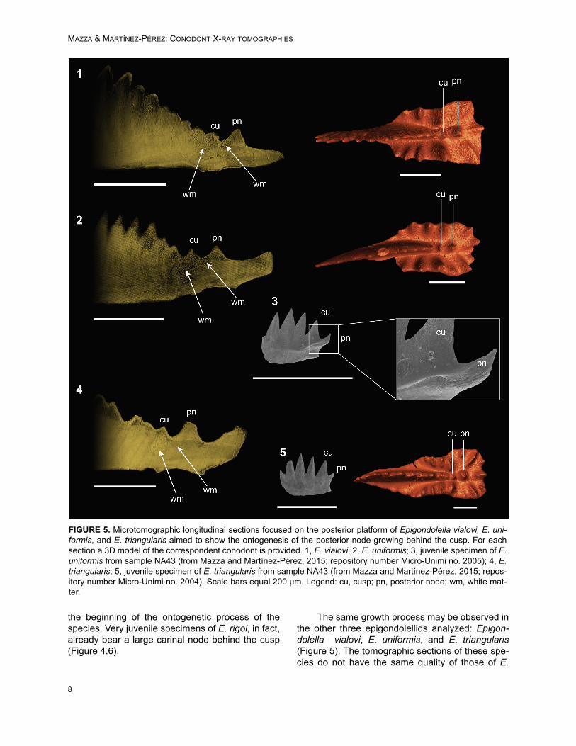
The same growth process may be observed inthe other three epigondolellids analyzed: Epigon-dolella vialovi, E. uniformis, and E. triangularis(Figure 5). The tomographic sections of these spe-cies do not have the same quality of those of E.
FIGURE 5. Microtomographic longitudinal sections focused on the posterior platform of Epigondolella vialovi, E. uni-formis, and E. triangularis aimed to show the ontogenesis of the posterior node growing behind the cusp. For eachsection a 3D model of the correspondent conodont is provided. 1, E. vialovi; 2, E. uniformis; 3, juvenile specimen of E.uniformis from sample NA43 (from Mazza and Martínez-Pérez, 2015; repository number Micro-Unimi no. 2005); 4, E.triangularis; 5, juvenile specimen of E. triangularis from sample NA43 (from Mazza and Martínez-Pérez, 2015; repos-itory number Micro-Unimi no. 2004). Scale bars equal 200 µm. Legend: cu, cusp; pn, posterior node; wm, white mat-ter.
8

PALAEO-ELECTRONICA.ORG
quadrata and E. rigoi, thus the lamellae of the cuspregion are not well distinguishable. Nevertheless,in all the three epigondolellids the posterior node ispermeated by a massive occurrence of white mat-ter as in E. rigoi and, in the same way, in the mostanterior part of the node the albid tissue appears tobe coalescent with that of the cusp (Figure 5).Thus, even if in E. vialovi, E. uniformis, and E. tri-angularis the growth lines are not well distinguish-able, the internal structure of their posterior node ishomologous to that of E. rigoi and its ontogenesisis seemingly the same. Comparisons with SEMphotos of specimens of E. uniformis and E. triangu-laris from monospecific populations, together withthe analyses of their growth series (Mazza andMartínez-Pérez, 2015), confirm the occurrence ofthe posterior denticle already from the early juve-nile stages of these species (Figure 5.3, 5.5).
DISCUSSION
The evolutionary trends of the Late Triassic P1
conodont elements are quite well defined (see Fig-ure 1; Mazza et al., 2012b). One of the most evi-dent trend is the forward shifting of cusp and pit,which is associated with the occurrence of nodesbehind the cusp (Orchard, 2014). This trend alsocharacterizes the evolution of the Late Carnian car-nepigondolellids into genera Epigondolella and,later in the Middle Norian, the development ofnodes behind the cusp is a character inherited alsoby the genus Mockina (Mazza et al., 2012a; Mazzaand Martínez-Pérez, 2015). The development ofthe large node behind the cusp in the epigondolel-lids considerably influenced the phylogeneticmodel obtained with the cladistic analyses (Mazzaet al., 2012b).
The Synchrotron Radiation X-ray Tomo-graphic Microscopy allowed us to investigate indetail the ontogenetic process of this character,revealing that the posterior node of Epigondolellavialovi, E. rigoi, E. uniformis, and E. triangularis is acarinal node because it grows directly from thecusp, while in E. quadrata this node, even if it hasthe same morphology and is located in the sameposition of that of the other mentioned epigondolel-lids, has the same ontogenetic process that leadsto the growth of the nodes/denticles developing onthe platform margins. For this reason, the posteriornode of E. quadrata is a different structure than acarinal node, analogous to other platform orna-mentation structures (Figures 4, 5). Thus, the largeposterior node growing behind the cusp cannot beconsidered as indicative of common ancestry, as
supposed before, but as a clear evolutionary con-vergence.
Consequently, since different ontogenetic pro-cesses for the same morphological character indi-cate evolutionary convergence, the main questionthat raises is if the position of Epigondolellaquadrata, E. rigoi, E. uniformis, and E. triangularisin the phylogenetic model previously proposed hasto be reconsidered. One hypothesis is that the evo-lution of E. quadrata into E. rigoi could proceed bythe conversion of the posterior node of E. quadratainto a carinal node in E. rigoi. Nevertheless, wehave to consider that the studies on the Late Trias-sic conodont growth series (Mazza and Martínez-Pérez, 2015) revealed that E. quadrata, E. rigoi,and E. uniformis first occur quite contemporane-ously in the uppermost Carnian and can be foundtogether in a long stratigraphic range until the Mid-dle Norian (Mazza et al., 2012a; Karádi et al.,2013; Mazza and Martinez-Perez, 2015). Juvenileepigondolellids now classifiable as E. triangularisare found in the Tuvalian even below the firstoccurrences of the other three species (Mazza andMartínez-Pérez, 2015). This suggests that morethan being phylogenetically related, these speciesevolve in parallel lineages, sharing analogouscharacters. This definitely confirms a new andmore articulated scenario for the evolutionary his-tory of the Upper Triassic epigondolellids. GenusEpigondolella cannot be considered anymore as amonophyletic group, but a paraphyletic assem-blage of species deriving from Carnian taxa thatdevelop similar morphologies but follow differentevolutionary and ontogenetic processes. The pre-vious phylogenetic model has necessarily to berevised.
This aspect raises a considerable transcen-dence in conodont, as far as their morphology isthe base for its systematics and phylogenetic rela-tionships, highlighting the importance of discrimi-nating homology from homoplasy. In this sense,Synchrotron Radiation X-ray Tomographic Micros-copy represents a unique and versatile instrumentfor these kind of investigations, providing data thatcan inform on conodont ontogenesis, evolutionaryprocesses and phylogenetic relationships.
CONCLUSIONS
The application of Synchrotron Radiation X-ray Tomographic Microscopy to reconstruct theontogenesis of single Late Triassic conodont P1
elements, suggests that using ontogenesis as a cri-terion for discriminating homology from homoplasyis actually effective, allowing to identify cryptic
9

MAZZA & MARTÍNEZ-PÉREZ: CONODONT X-RAY TOMOGRAPHIES
homology between apparently convergent charac-ters in the conodont platform morphology. In partic-ular, our results about the ontogenesis of theposterior node growing behind the cusp of Late Tri-assic conodont P1 elements, an autapomorphy
characterizing the Epigondolella clade, reveal thatthe studied character is not a homologous charac-ter of the epigondolellids but an evolutionary con-vergence. Therefore, it cannot be considered asevidence of common ancestry anymore, confirmingthat the genus Epigondolella is not a monophyleticclade as previously hypothesized, but a polyphy-letic assemblage of taxa with different possibleancestors among the carnepigondolellids as laterreconsidered. This result highlights the importanceto discriminate homology from homoplasy in cono-donts to depict more precisely their ontogeneticand evolutionary dynamics.
ACKNOWLEDGEMENTS
The authors are grateful to P. Donoghue (Uni-versity of Bristol) for his continuous support andadvice, and for giving us access to the TOMCATbeamline (SLS). We thank J. Cunningham, M.Rücklin, J. Keating, and F. Marone for beamlineassistance. The authors also thank the two anony-mous referees for reviewing the manuscript andthe editor C. Haug for her precious suggestions.Research was partially funded by MIUR PRIN(2008BEF5Z7_001; PI M. Balini, Università degliStudi di Milano), by a Marie Curie FP7-People IEF2011-299681 (CMP), and by the Research ProjectsCGL2014-52662-P (Spanish Ministry of Economyand Competitiveness) and GV/2016/102 (Generali-tat Valenciana).
REFERENCES
Balini, M., Bertinelli, A., Carter, E.S., Di Stefano, P., Krys-tyn, L., Levera, M., Mazza, M., McRoberts, C., Mut-toni, G., Nicora, A., Orchard, M.J., Preto, N., Rigo,M., Tripodo, A., and Zonneveld J.-P. 2012. Towardsthe definition of the GSSP of the Norian Stage (UpperTriassic): integrated stratigraphy and correlation ofthe two candidate sections Black Bear Ridge (BC,Canada) and Pizzo Mondello (Italy). 34th Interna-tional Geological Congress, Brisbane, Australia,abstract book: 753
Balini, M., Bertinelli, A., Di Stefano, P., Guaiumi, C.,Levera, M., Mazza, M., Muttoni, G., Nicora, A., PretoN., and Rigo, M. 2011. The Late Carnian-Rhaetiansuccession at Pizzo Mondello (Sicani Mountains).Albertiana, 39:36-58.
Balini, M., Di Stefano, P., Tripodo, A., Mazza, M., Levera,M., Muttoni, G., Nicora, A., Rigo, M., and Krystyn, L.
2015. High resolution integrated biomagnetostratig-raphy of the Carnian/Norian boundary at Pizzo Mon-dello and Pizzo Lupo (western Sicily, Italy). Berichtedes Institutes für Erdwissenschaften Karl-Franzens-Universität Graz, Band 21:28.
Budurov, K. 1972. Ancyrogondolella triangularis gen. etsp. n. (Conodonta). Mitteilungen der Gesellschaft derGeologie- und Bergbaustudenten in Wien, 21:853-860.
Burij, G.I. 1989. Konodonty i stratigrafija Sikhote-Alin. ANSSSR, dalnevostochnoe otdelenie, Vladivostok, Rus-sia. (In Russian)
Donoghue, P.C.J. 1998. Growth and patterning in theconodont skeleton. Philosophical Transactions of theRoyal Society, Series B, 353:633-666.
Donoghue, P.C.J. 2001. Conodonts meet cladistics:recovering relationships and assessing the complete-ness of the conodont fossil record. Palaeontology,44:65-93.
Donoghue, P.C.J. Purnell, M. A., Aldridge, R. J. andZhang, S. 2008. The interrelationships of ‘complex’conodonts (Vertebrata). Journal of Systematic Palae-ontology, 6:119-153.
Dzik, J. 2005. The chronophyletic approach: stratophe-netics facing an incomplete fossil record. SpecialPapers in Palaeontology, 73:159-183.
Epstein, A.G., Epstein, J.B., and Harris, L.D. 1977.Conodont color alteration an index to organic meta-morphism. U.S. Geological Survey ProfessionalPapers, 995:27.
Foote, M. and Sepkoski, J.J., Jr. 1999. Absolute mea-sures of the completeness of the fossilrecord. Nature, 398:415-417.
Jones, D., Evans, A.R., Rayfield, E.J., Siu, K.K.W., andDonoghue, P.C.J. 2012b. Testing micro structuraladaptation in the earliest dental tools. Biology Let-ters, 8:952-955.
Jones, D., Evans, A.R., Siu, K.K.W., Rayfield, E.J., andDonoghue, P.C.J. 2012a. The sharpest tools in thebox? Quantitative analysis of conodont element func-tional morphology. Proceedings of the Royal SocietyB: Biological Sciences, 279:2849-2854.
Karádi, V., Kozur, H.W,. and Görög, A. 2013. Stratigraph-ically important Lower Norian Conodonts from theCsovár borehole (CSV-1), Hungary - comparisonswith the conodont succession of the Norian GSSPcandidate Pizzo Mondello (Sicily, Italy), p. 284-295.In Tanner, L.H., Spielmann, J.A., and Lucas, S.G.(eds.), The Triassic System. New Mexico Museum ofNatural History and Science, Bulletin 61. New MexicoMuseum of Natural History and Science, Albuquer-que.
Marone, F. and Stampanoni, M. 2012. Regridding recon-struction algorithm for real-time tomographic imag-ing. Journal of Synchrotron Radiation, 19:1029-1037.
Martínez-Pérez, C., Plasencia, P., Jones, D., Kolar-Jurkovšek, T., Sha, J., Botella, H., and Donoghue,P.C.J. 2014b. There is no general model for occlusalkinematics in conodonts. Lethaia, 47:547-555.
10

PALAEO-ELECTRONICA.ORG
Martínez-Pérez, C., Plasencia, P., Cascales-Miñana, B.,Mazza, M., and Botella, H. 2014c. New insights intothe diversity dynamics of Triassic conodonts. Histori-cal Biology, 26(5):591-602. doi:10.1080/08912963.2013.808632
Martínez-Pérez, C., Rayfield, E.J., Botella, H., andDonoghue, P.C.J. 2016. Translating taxonomy intothe evolution of conodont feeding ecology. Geology.44(4):247-250.
Martínez-Pérez, C., Rayfield, E.J., Purnell, M.A., andDonoghue, P.C.J. 2014a. Finite element, occlusal,microwear and microstructural analyses indicate thatconodont microstructure is adapted to dental func-tion. Palaeontology, 57:1059-1066.
Mazza, M., Cau, A., and Rigo, M. 2012b. Application ofnumerical cladistic analyses to the Carnian-Norianconodonts: a new approach for phylogenetic inter-pretations. Journal of Systematic Palaeontology,10(3):401-422.
Mazza, M. and Krystyn, L. 2015. The revised Upper Tri-assic conodont record of the Tethys: a new steptowards a better definition of the conodont bioeventsaround the base of the Norian stage. Berichte desInstitutes für Erdwissenschaften Karl-Franzens-Uni-versität Graz, Band 21:243.
Mazza, M. and Martínez-Pérez, C. 2015. Unravellingconodont (Conodonta) ontogenetic processes in theLate Triassic through growth series reconstructionsand X-ray microtomography. Bollettino della SocietàPaleontologica Italiana, 54 (3):161-186. doi: 10.4435/BSPI.2015.10
Mazza, M., Rigo, M., and Gullo, M. 2012a. Taxonomyand stratigraphic record of the Upper Triassic cono-donts of the Pizzo Mondello section (Western Sicily,Italy), GSSP candidate for the base of the Norian.Rivista Italiana di Paleontologia e Stratigrafia,118(1):85-130.
Murdock, D.J.E., Rayfield, E.J., and Donoghue, P.C.J.2014. Functional adaptation underpinned the evolu-tionary assembly of the earliest vertebrate skeleton.Evolution and Development, 16:354-361.
Muttoni, G., Kent, D.V., Di Stefano, P., Gullo, M., Nicora,A., Tait, J., and Lowrie, W. 2001. Magnetostratigra-phy and biostratigraphy of the Carnian/Norian bound-ary interval from the Pizzo Mondello section (SicaniMountains, Sicily). Palaeogeography, Palaeoclima-tology, Palaeoecology, 166:383-399.
Muttoni, G., Kent, D.V., Olsen, P.E., Di Stefano, P., Low-rie, W., Bernasconi, S.M., and Hernández, F.M. 2004.Tethyan magnetostratigraphy from Pizzo Mondello(Sicily) and correlation to the Late Triassic Newark
astrochronological polarity time scale. GeologicalSociety of America Bulletin, 116:1043-1058.
Nicora, A., Balini, M., Bellanca, A., Bertinelli, A., Bow-ring, S.A., Di Stefano, P., Dumitrica, P., Guaiumi, C.,Gullo, M., Hungerbuehler, A., Levera, M., Mazza, M.,McRoberts, C.A., Muttoni, G., Preto, N., and Rigo, M.2007. The Carnian/Norian boundary interval at PizzoMondello (Sicani Mountains, Sicily) and its bearingfor the definition of the GSSP of the Norian Stage.Albertiana, 36:102-129.
Noyan, O. and Kozur, H. 2007. Revision of the late Car-nian-early Norian conodonts from the Stefanion sec-tion (Argolis, Greece) and their paleobiogeographicimplications. Neues Jahrbuch für Geologie und Pale-ontologie Abhandlungen, 245(2):159-178.
Orchard, M.J. 1991. Upper Triassic conodont biochronol-ogy and new index species from the Canadian Cor-dillera, p. 299-335. In Orchard, M.J. and McCracken,A.D. (eds.), Ordovician to Triassic conodont paleon-tology of the Canadian Cordillera. Geological Surveyof Canada, Vancouver.
Orchard, M.J. 2014. Conodonts from the Carnian-NorianBoundary (Upper Triassic) of Black Bear Ridge,Northeastern British Columbia, Canada. New MexicoMuseum of Natural History and Science Bulletin, 64.New Mexico Museum of Natural History and Science,Albuquerque.
Purnell, M.A. and Jones, D.O. 2012. Quantitative analy-sis of conodont tooth wear and damage as a test ofecological and functional hypotheses. Paleobiology,38:605-626.
Rejebian, V.A. 1987. Conodont color and textural alter-ation: an index to regional metamorphism, contactmetamorphism, and hydrothermal alteration. Geolog-ical Society of America Bulletin, 99:471-479.
Sweet, W.C. 1988. Oxford Monographs on Geology andGeophysics. The Conodonta: Morphology, Taxon-omy, Paleoecology, and Evolutionary History of aLong-Extinct Animal Phylum. Clarendon Press,Oxford.
Wickstrom, L.M. and Donoghue, P.C.J. 2005. Clado-grams, phylogenies and the veracity of the conodontfossil record. Special Papers in Palaeontology,73:185-218.
Zhang, S. and Barnes, C.R. 2004. Conodont bioevents,cladistics and response to glacio-eustasy, Ordovi-cian–Silurian boundary through Llandovery, AnticostiBasin, Que´bec, p. 73-104. In Beaudoin, A.B. andHead, M.J. (eds.), The Palynology and Micropalae-ontology of Boundaries, Geological Society of Lon-don, Special Publication vol. 230. Geological Society
of London, London.
11