evolutionary dynamics of cancer - harvard...
TRANSCRIPT
Evolutionary dynamics of cancer
A thesis presented
by
Franziska Michor
to
The Department of Organismic and Evolutionary Biology
in partial fulfillment of the requirements
for the degree of
Doctor of Philosophy
in the subject of
Organismic and Evolutionary Biology
Harvard University
Cambridge, Massachusetts
May 2005

c© 2005 Franziska Michor
All rights reserved.

Thesis Advisor: Professor Martin A. Nowak Franziska Michor
Evolutionary dynamics of cancer
Evolutionary dynamics of mutation and selection can be formulated by mathemat-
ical equations. Cancer arises as a consequence of somatic evolution. Therefore, a
mathematical approach can be used to understand cancer initiation and progression.
Tumorigenesis is driven by genetic alterations of oncogenes, tumor suppressor genes
and genetic instability genes. First, I discuss the fundamental principles that gov-
ern the dynamics of activating oncogenes and inactivating tumor suppressor genes
in populations of reproducing cells. A quantitative theory of mutation and selection
provides insights into the role of genetic instability in cancer initiation. Second,
I analyze the dynamics of cancer progression via inactivation of one or two tu-
mor suppressor genes. Third, I derive a mathematical framework for the complete
mutational sequence of colorectal tumorigenesis (as known today) and the effect
of chromosomal instability therein. Fourth, I present a new mathematical model
of colon cancer initiation that assumes a linear flow from stem cells to differenti-
ated cells to apoptosis. Finally, a mathematical model of chronic myeloid leukemia
(CML) and the molecular response to imatinib therapy suggests that leukemic stem
cells are spared by therapy. The model provides quantitative insights into the in
vivo kinetics of this cancer and determines the probability of disease relapse due to
resistance mutations.
iii

Contents
1 Introduction 1
2 Dynamics of cancer progression 5
2.1 Introduction . . . . . . . . . . . . . . . . . . . . . . . . . . . . . . . . 6
2.2 Oncogenes . . . . . . . . . . . . . . . . . . . . . . . . . . . . . . . . . 8
2.3 Tumor suppressor genes . . . . . . . . . . . . . . . . . . . . . . . . . 17
2.4 Genetic instability . . . . . . . . . . . . . . . . . . . . . . . . . . . . 23
2.5 Conclusions . . . . . . . . . . . . . . . . . . . . . . . . . . . . . . . . 35
3 Can chromosomal instability initiate cancer? 38
3.1 Introduction . . . . . . . . . . . . . . . . . . . . . . . . . . . . . . . . 39
3.2 Chromosomal instability before one tumor suppressor gene . . . . . . 42
3.3 Chromosomal instability before two tumor suppressor genes . . . . . 48
3.4 Conclusions . . . . . . . . . . . . . . . . . . . . . . . . . . . . . . . . 55
3.5 Methods . . . . . . . . . . . . . . . . . . . . . . . . . . . . . . . . . . 56
4 Dynamics of Colorectal Cancer 59
4.1 Introduction . . . . . . . . . . . . . . . . . . . . . . . . . . . . . . . . 60
4.2 Mutations in colorectal cancer cell lines . . . . . . . . . . . . . . . . . 64
4.3 Mathematical modeling . . . . . . . . . . . . . . . . . . . . . . . . . . 66
4.4 The model . . . . . . . . . . . . . . . . . . . . . . . . . . . . . . . . . 69
4.5 Numerical examples . . . . . . . . . . . . . . . . . . . . . . . . . . . . 79
iv

4.6 Discussion . . . . . . . . . . . . . . . . . . . . . . . . . . . . . . . . . 82
4.7 Appendix . . . . . . . . . . . . . . . . . . . . . . . . . . . . . . . . . 84
5 Linear model of colon cancer initiation 85
5.1 Introduction . . . . . . . . . . . . . . . . . . . . . . . . . . . . . . . . 86
5.2 Mechanism . . . . . . . . . . . . . . . . . . . . . . . . . . . . . . . . 88
5.3 Mathematical Analysis . . . . . . . . . . . . . . . . . . . . . . . . . . 97
5.4 Numerical Examples . . . . . . . . . . . . . . . . . . . . . . . . . . . 99
5.5 Discussion . . . . . . . . . . . . . . . . . . . . . . . . . . . . . . . . . 103
6 Dynamics of chronic myeloid leukemia 105
6.1 Introduction . . . . . . . . . . . . . . . . . . . . . . . . . . . . . . . . 106
6.2 Results . . . . . . . . . . . . . . . . . . . . . . . . . . . . . . . . . . . 114
6.3 Methods . . . . . . . . . . . . . . . . . . . . . . . . . . . . . . . . . . 131
7 References 142
v

Acknowledgments
Thanks to my committee members David Haig, Andrew Murray, Martin Nowak,
and John Wakeley for their advice and support. Thanks to the Department of Or-
ganismic and Evolutionary Biology and the Faculty of Arts and Sciences of Harvard
University. I thank my collaborators Yoh Iwasa, Christoph Lengauer, Bert Vogel-
stein, Robert May, Charles Sawyers, Tim Hughes, Neil Shah, Sue Branford, Steven
Frank, Natalia Komarova, Harith Rajagopalan, and Christoph Hauert for amazing
times. Thanks to the members of the Program for Evolutionary Dynamics at Har-
vard and the Program for Theoretical Biology at the Institute for Advanced Study;
they made my years in graduate school an unforgettable experience. Finally, I thank
my parents Elli and Peter, my sister Johanna, and also Mirra & Gosi for making
me what I am.
1
1 Introduction
This thesis is about the somatic evolutionary dynamics of cancer. Cancer results
from an accumulation of genetic alterations in somatic cells. This accumulation of
mutations leads to a breakdown of cooperation between cells in a tissue, such that
single cells revert to selfish behavior reminiscent of unicellular organisms. Mutation
and selection can best be described when represented as mathematical equations.
Hence, mathematical frameworks can be used to analyze the evolutionary dynamics
of cancer.
Cancer susceptibility genes can be subdivided into three categories1: (i) gate-
keepers, such as oncogenes and tumor suppressor genes, control growth and differen-
tiation pathways of the cell - alterations of such genes lead to increased growth rates
or decreased death rates as compared with neighboring cells; (ii) caretakers main-
tain the genomic integrity of the cell - alterations of such genes increase the rate of
accumulating further mutations and cause genetic instabilities; and (iii) landscapers
regulate the cell’s interaction with the surrounding microenvironment - alterations
of such genes contribute to the neoplastic transformation of the cell. Cancer results
from an accumulation of mutations in gatekeeper genes; at the time of diagnosis,
most cancers are genetically unstable. A major question and controversy in cancer
biology is whether genetic instability arises early and hence is a driving force of
tumorigenesis.
In Chapter 2, I present a mathematical framework designed to study the kinetics
1

of mutations in cancer-susceptibility genes and apply this framework to cancer initi-
ation and progression. I discuss the evolutionary dynamics of mutations activating
oncogenes and inactivating tumor suppressor genes in populations of reproducing
cells. The probability of activating an oncogene in a cellular population depends
on the tissue organization and population size: a population fed by a small number
of stem cells can dramatically reduce the risk of accumulating mutations in onco-
genes as compared to well-mixed cellular compartments. The analysis yields three
different kinetic laws for the probability of inactivating a tumor suppressor gene
in a population of cells: in dependence of the population size as compared to the
mutation rates inactivating the two alleles, the tumor suppressor gene is homozy-
gously inactivated in two, one, or zero rate-limiting hits. Later, this framework is
extended to include mutations causing genetic instability and to analyze the role of
genetic instability in human cancers. I discuss the conditions under which genetic
instability is likely to initiate tumorigenesis.
In Chapter 3, the mathematical framework of tumor suppressor gene inactivation
and genetic instability is extended to situations in which two tumor suppressor genes
need to be inactivated to drive tumorigenesis. Once the first tumor suppressor gene
has been inactivated in a cell, this cell might give rise to clonal expansion. In a
moderately large population of cells, it might take only one rate-limiting hit to
inactivate the second tumor suppressor gene. In this situation, genetic instability is
very likely to emerge before inactivation of the first tumor suppressor gene. If clonal
2
expansion after inactivation of the first gene gives rise to a very large population of
cells, however, then a further genetic alteration is not rate-limiting any longer and
genetic instability becomes less important.
In Chapter 4, I analyze the evolutionary dynamics of the complete mutational
sequence of colorectal tumorigenesis as known today. A literature review of math-
ematical models of colorectal tumorigenesis is provided, and the specific mutations
found in colorectal cancer cell lines are outlined. Then I discuss the dynamics of
colorectal tumorigenesis in well-mixed populations of (stem) cells and determine the
role and importance of genetic instability in this setting.
In Chapter 5, I design a new mathematical model of colorectal tumorigenesis
based on the cellular dynamics and tissue architecture of a colonic crypt. A stem
cell at the base of the crypt produces a cellular lineage that populates the crypt. The
differentiated cells are pushed to the top of the crypt by the continuous production
and migration of such cells and undergo apoptosis on top of the crypt. All of these
cells could in principle accumulate mutations that initiate tumorigenesis. I analyze
the mutational dynamics of tumor suppressor genes and genetic instability genes in
such a linear model and determine the mutations likely to initiate tumorigenesis.
In Chapter 6, I provide an analysis of the molecular response to imatinib (Gleevec),
a chemotherapeutic agent used to treat chronic myeloid leukemia (CML), in a 169
patient dataset. This dataset establishes that imatinib leads to a bi-phasic decline of
the leukemic cell burden and an overall 5000-fold decrease in the leukemic cell mass.
3
Upon discontinuation of therapy, however, the leukemic burden rapidly rebounds to
values beyond pre-treatment baseline, suggesting that leukemic stem cells are not
depleted during imatinib therapy. A mathematical model recovers all features of
the in vivo kinetics and can be used to analyze the disease kinetics and parameter
values. Acquired resistance mutations are similarly modeled. The probability of
resistance and time to treatment failure can be estimated. This model provides the
first quantitative analysis of a human cancer in vivo.
4

2 Dynamics of cancer progression
Michor F, Iwasa Y, Nowak MA
Dynamics of cancer progression2
Nature Reviews Cancer 4, 197-206 (2004)
Cancer arises from an accumulation of mutations in oncogenes, tumor
suppressor genes and genes that maintain the genomic integrity of the
cell. Oncogenes lead to increased net growth rates of the cell when ac-
tivated by a point mutation, amplified or overexpressed. We study the
evolutionary dynamics of oncogene activation in populations of reproduc-
ing cells. Tumor suppressor genes lead to increased net growth rates of
the cell when inactivated in both alleles. We analyze how the kinetics
of tumor suppressor gene inactivation depends on the population size of
cells and the mutation rates for the first and second hit. We find three
different kinetic laws: in small, intermediate and large populations, it
takes, respectively, two, one and zero rate limiting steps to inactivate a
tumor suppressor gene. Genetic instability refers to an increased mu-
tation rate across the genome and arises due to mutations in genetic
instability genes. A major question in cancer biology is whether genetic
instability is an early event in tumorigenesis. We analyze the role of ge-
netic instability in tumor initiation and progression and determine the
5
conditions under which genetic instability represents the first phenotypic
alteration on the road to cancer.
2.1 Introduction
Cancer is a genetic disease1. Although environmental and other nongenetic factors
have roles in many stages of tumorigenesis, it is widely accepted that cancer arises
due to mutations in cancer-susceptibility genes. These genes belong to one of three
classes1,3: gatekeepers, caretakers and landscapers. Gatekeepers directly regulate
growth and differentiation pathways of the cell and comprise oncogenes and tumor
suppressor genes. Caretakers, in contrast, promote tumorigenesis indirectly4,5. They
function in maintaining the genomic integrity of the cell. Mutation of caretakers can
lead to genetic instability, and the cell rapidly accumulates changes in other genes
that directly control cell birth and death. Landscaper defects do not directly affect
cellular growth, but generate an abnormal stromal environment that contributes to
the neoplastic transformation of cells6.
The alteration of one gene, however, does not suffice to give rise to full blown
cancer. For progression toward malignancy and invasion, further mutational hits
are necessary7–9. Hence the risk of cancer development does not only depend on
mutations initiating tumorigenesis, but also on subsequent mutations driving tumor
progression.
A quantitative understanding of cancer biology requires a mathematical frame-
6
work to describe the fundamental principles of population genetics and evolution
that govern tumor initiation and progression10,11. Mutation, selection and tissue
organization determine the dynamics of tumorigenesis12–15 and should be studied
quantitatively, both in terms of experiment and theory16.
The mathematical investigation of cancer began in the 1960s, when Nordling17,
Armitage and Doll18,19, and Fisher20 set out to explain the age-dependent incidence
curves of human cancers. These seminal studies led to the idea that multiple prob-
abilistic events are required for the somatic evolution of cancer21,22. In the early
1980s, Knudson used a statistical analysis of the incidence of retinoblastoma in chil-
dren to explain the role of tumor suppressor genes in sporadic and inherited cancers
10. This work was later extended to a two-stage stochastic model for the process
of cancer initiation and progression9,11, which inspired much subsequent work23–25.
Later on, considerable effort was devoted to the development of specific theories for
drug resistance26,27, angiogenesis28, immune responses against tumors29, and genetic
instabilities30–35.
Here, we address the following questions: What are the fundamental principles
that determine the dynamics of activating oncogenes and inactivating tumor sup-
pressor genes? How do mutation, selection and tissue architecture influence the rate
of tumor initiation and progression? And how do quantitative approaches help to
investigate the role of genetic instability in tumorigenesis?
7

2.2 Oncogenes
Oncogenes can contribute to tumorigenesis if one allele is mutated, amplified or in-
appropriately expressed1. In the last decades, many oncogenes have been discovered
that are involved in various stages of human cancers - tumor initiation, progression,
angiogenesis, and metastasis. Here, we discuss the basic aspects of evolutionary dy-
namics of oncogene activation and outline concepts such as selection, fixation, and
random drift.
Tissues of multicellular organisms are subdivided into compartments36, which
contain populations of cells that proliferate to fulfill organ-specific tasks. Compart-
ments are subject to homeostatic mechanisms which ensure that the cell number
remains approximately constant over time. Whenever a cell divides, another cell
has to die to keep the total population size the same. Cancer results if the equilib-
rium between cell birth and cell death is shifted toward uncontrolled proliferation.
Not all cells of a compartment, however, might be at risk of becoming cancer cells.
Differentiated cells, for example, might not have the capacity to divide often enough
to accumulate the necessary number of mutations in cancer susceptibility genes37.
The effective population size of a compartment describes those cells that are at risk
of becoming cancer cells. In the following, compartment size will be used synony-
mously with effective population size within a compartment.
Consider a compartment of replicating cells. During each cell division, there is a
small probability that a mistake will be made during DNA replication; in this case,
8

a mutated daughter cell is produced. The mutation might confer a fitness advantage
to the cell by ameliorating an existing function or inducing a new function. Then the
mutation is advantageous in terms of somatic selection. Alternatively, the mutation
might impair an important cellular function and confer a fitness disadvantage to the
cell. Then the cell proliferates more slowly or dies more quickly than its neighbors.
The net reproductive rate is decreased, and the mutation is deleterious in terms
of somatic selection. Finally, the mutation might not change the reproductive rate
of the cell. Then the cell proliferates at the same rate as its neighbors, and the
mutation is neutral in terms of somatic selection.
Let us discuss the dynamics of a particular mutation within a compartment.
Initially, all cells are unmutated. What is the probability that a single mutated
cell has arisen by time t? We measure time, t, in cell cycles. If the relevant cells
divide once per day, then the unit of time is one day. Denote by N the number
of cells in a compartment, and denote by u the mutation rate per gene per cell
division. The probability that at least one mutated cell has arisen by time t is given
by P (t) = 1 − exp(−Nut) (Figure 1a).
What is the fate of a single mutated cell? In the simplest scenario, there is
a constant probability, q, that this cell will not die, but will initiate a neoplasia.
The probability that a compartment has initiated a neoplasia by time t is given by
P (t) = 1 − exp(−Nuqt).
Alternatively, consider a scenario in which the mutated cell has a relative fitness
9

r compared to a wild-type cell with fitness 1. If r > 1, the mutation is advanta-
geous; if r < 1, the mutation is disadvantageous; if r = 1, the mutation is neutral.
Normally, we expect mutations in oncogenes to cause increased net growth rates,
r > 1; however, a mutation in an oncogene could be kept in check by apoptotic
defense mechanisms, so r could be less than one.
What is the probability that such a mutation will not die out, but will take
over the compartment? In order to calculate this probability, we consider a specific
stochastic process known as the Moran process38. At each time step, a cell is chosen
for reproduction at random, but proportional to fitness. If there are i mutated cells,
then the probability that a mutated cell is chosen for reproduction is ri/(ri+N− i).
The chosen cell produces a daughter cell that replaces another randomly chosen cell
that dies. The total number of cells remains strictly constant. The probability39 that
a single mutated cell with r > 1 or r < 1 takes over the whole compartment is given
by ρ = (1 − 1/r)/(1 − 1/rN ). For a neutral mutant40, r = 1, the evolutionary
dynamics are described by random drift in the frequency of the mutant in the
population; the fixation probability is ρ = 1/N . In this stochastic process, a cell can
either produce a lineage that dies out or that takes over the whole compartment.
Stable coexistence of different cell types is impossible. The quantity ρ is called
fixation probability. An advantageous mutation has a higher fixation probability
than a neutral mutation, which has a higher fixation probability than a deleterious
mutation. The events in a small compartment, however, are dominated by random
10

drift: if N is small, then even a deleterious mutation has a fairly high probability of
reaching fixation due to chance events.
The probability that a mutation destined to be fixed has arisen by time t is
given by P (t) = 1 − exp(−Nuρt) (Figure 1b). Here we neglect the time it takes
the mutant to reach fixation in the population and therefore, P (t) is also the prob-
ability that a mutant has been fixed by time t. Note that any mutation has a
higher fixation probability in a small compartment than in a large compartment,
but P (t) is an increasing function of N for r > 1 and a decreasing function of N
for r < 1. Thus, large compartments accelerate the accumulation of advantageous
mutations, but slow down the accumulation of deleterious mutations. Conversely,
small compartments slow down the accumulation of advantageous mutations, but
accelerate the accumulation of deleterious mutations. Therefore, the compartment
size is important in determining the types of mutations that are likely to occur41,42.
The linear process
So far, we have considered the evolutionary dynamics of a mutation that arises in a
well-mixed compartment. This approach describes a tissue compartment in which
all relevant cells are in equivalent positions and in direct reproductive competition
with each other - there are no spatial effects. However, we can also envisage theories
where cellular differentiation and spatial structure are explicitly modeled. One sim-
ple approach considers N cells in a linear array43 (Figure 1c). Again, at each time
step, a cell is chosen at random, but proportional to fitness. The cell is replaced
11

by two daughter cells, and all cells to its right are shifted by one place to the right.
The cell at the far right undergoes apoptosis; the cell at the far left acts as a stem
cell. In this approach, the fixation probability of a mutant cell is given by ρ = 1/N .
The probability of fixation does not depend on its relative fitness r because only
a mutation in the far left cell can reach fixation in the compartment. A mutation
arising in an offspring cell will eventually be ’washed out’ of the compartment by the
continuous production of cells and their migration from the stem cell to differentia-
tion and apoptosis. The probability that all cells of the compartment are mutated
at time t is given by P (t) = 1−exp(−ut); here we again neglect the time to fixation.
Time is measured in units of stem cell divisions. If the stem cell divides more slowly
than the other cells, then the accumulation of mutated cells is decelerated.
This ’linear process’ of cancer initiation has the important feature of balancing
out fitness differences between mutations43. Advantageous, deleterious and neutral
mutations all have the same fixation probability, ρ = 1/N . This is in contrast to a
well-mixed compartment, in which the fittest mutation has the highest probability
of fixation. In comparison with a well-mixed compartment, a linear compartment
delays the development of cancers that are initiated by advantageous mutations, such
as mutations in oncogenes and tumor suppressor genes. However, it can increase
the probability of cancer initiation via deleterious mutations, such as mutations in
genetic instability genes44. Note also that the linear tissue design does not change
the rate of accumulation of neutral mutations.
12

13

Figure 1: Oncogene dynamics. (a) The probability that at least one mutated cell has
arisen in a compartment of N cells before time t is given by P (t) = 1− exp(−Nut).
Here u denotes the mutation rate per gene per cell division, and time is measured
in units of cellular generations. (b) The probability that a compartment of N cells
has been taken over by mutated cells is given by P (t) = 1 − exp(−Nuρt). The
probability that a single mutant cell with relative fitness r reaches fixation is given
by ρ = (1− 1/r)/(1− 1/rN ). (c) N cells are arranged in a linear array. Whenever a
cell divides, one daughter cell is put to its right, and all cells to the right are shifted
by one place. The rightmost cell undergoes apoptosis. Here the fixation probability
is ρ = 1/N irrespective of r, because mutations have to arise in the leftmost cell
(the stem cell) for not being ’washed out’. The probability that the compartment
consists only of mutated cells at time t is given by P (t) = 1 − exp(−ut).
14
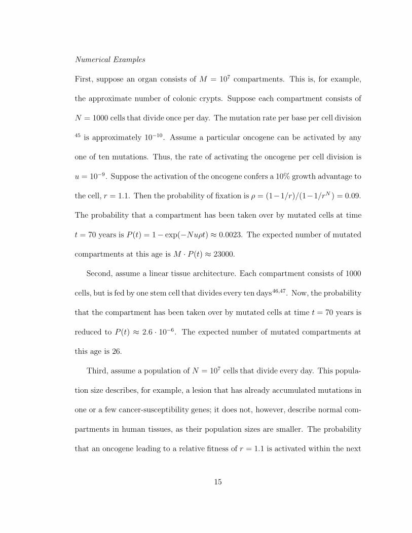
Numerical Examples
First, suppose an organ consists of M = 107 compartments. This is, for example,
the approximate number of colonic crypts. Suppose each compartment consists of
N = 1000 cells that divide once per day. The mutation rate per base per cell division
45 is approximately 10−10. Assume a particular oncogene can be activated by any
one of ten mutations. Thus, the rate of activating the oncogene per cell division is
u = 10−9. Suppose the activation of the oncogene confers a 10% growth advantage to
the cell, r = 1.1. Then the probability of fixation is ρ = (1−1/r)/(1−1/rN ) = 0.09.
The probability that a compartment has been taken over by mutated cells at time
t = 70 years is P (t) = 1− exp(−Nuρt) ≈ 0.0023. The expected number of mutated
compartments at this age is M · P (t) ≈ 23000.
Second, assume a linear tissue architecture. Each compartment consists of 1000
cells, but is fed by one stem cell that divides every ten days46,47. Now, the probability
that the compartment has been taken over by mutated cells at time t = 70 years is
reduced to P (t) ≈ 2.6 · 10−6. The expected number of mutated compartments at
this age is 26.
Third, assume a population of N = 107 cells that divide every day. This popula-
tion size describes, for example, a lesion that has already accumulated mutations in
one or a few cancer-susceptibility genes; it does not, however, describe normal com-
partments in human tissues, as their population sizes are smaller. The probability
that an oncogene leading to a relative fitness of r = 1.1 is activated within the next
15
year is given by P ≈ 0.28. The time until the probability of activating the oncogene
is 1/2 is obtained as T1/2 = 2.1 years.
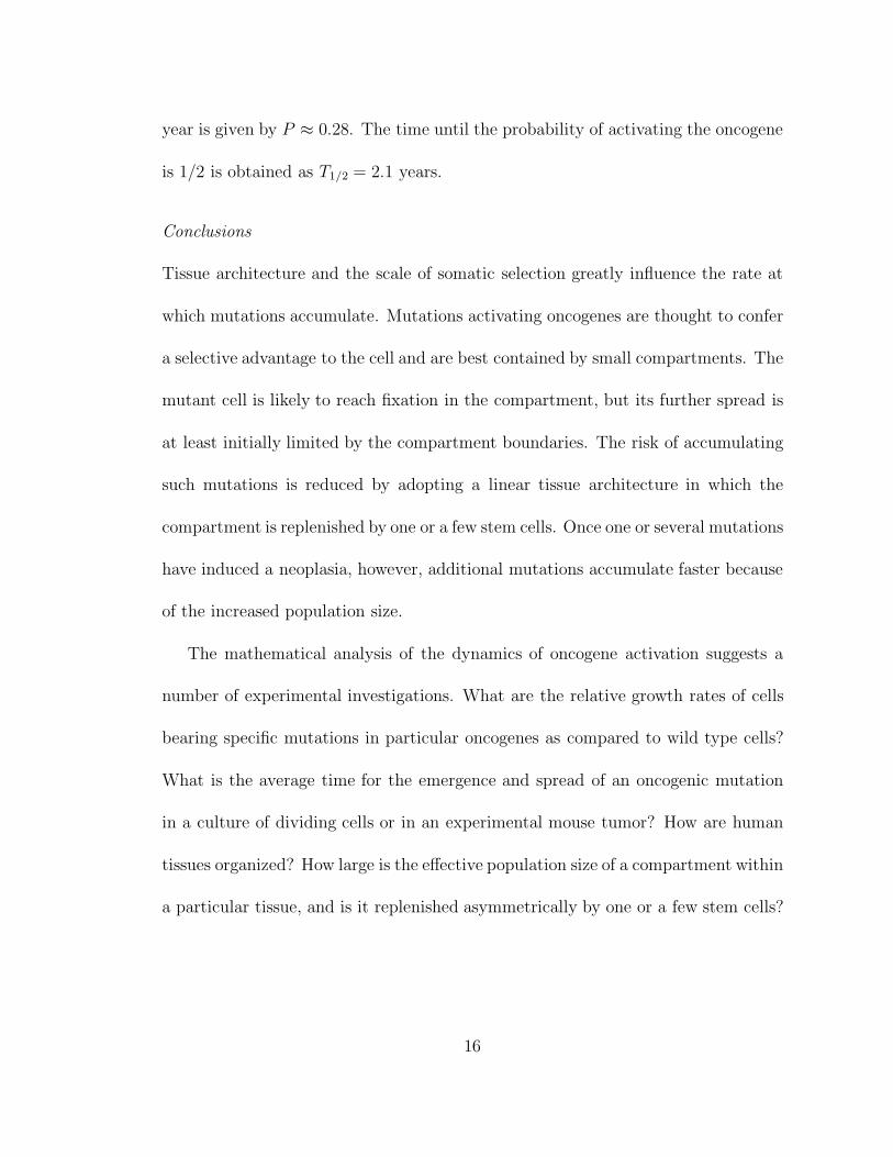
Conclusions
Tissue architecture and the scale of somatic selection greatly influence the rate at
which mutations accumulate. Mutations activating oncogenes are thought to confer
a selective advantage to the cell and are best contained by small compartments. The
mutant cell is likely to reach fixation in the compartment, but its further spread is
at least initially limited by the compartment boundaries. The risk of accumulating
such mutations is reduced by adopting a linear tissue architecture in which the
compartment is replenished by one or a few stem cells. Once one or several mutations
have induced a neoplasia, however, additional mutations accumulate faster because
of the increased population size.
The mathematical analysis of the dynamics of oncogene activation suggests a
number of experimental investigations. What are the relative growth rates of cells
bearing specific mutations in particular oncogenes as compared to wild type cells?
What is the average time for the emergence and spread of an oncogenic mutation
in a culture of dividing cells or in an experimental mouse tumor? How are human
tissues organized? How large is the effective population size of a compartment within
a particular tissue, and is it replenished asymmetrically by one or a few stem cells?
16

2.3 Tumor suppressor genes
So far, we have discussed genes that confer an altered phenotype to the cell when
mutated in one allele, and are dominant at the cellular level. Some genes, however,
are recessive at the cellular level, and have to be mutated in both alleles to cause
a phenotypic change of the cell. Examples of recessive mutations are those that
inactivate some tumor suppressor genes1,48,49 (TSGs).
The concept of a TSG emerged from a statistical analysis of retinoblastoma inci-
dence in children10. This study and subsequent work1,11,50 led to the 2-hit hypoth-
esis, which proposes that two hits in the retinoblastoma gene are the rate limiting
steps of this particular cancer. In the inherited form, the first mutation is present in
the germ line, whereas the second mutation emerges during somatic cell divisions.
In the sporadic form, both mutations arise during somatic cell divisions. In the
meanwhile, a large number of TSGs has been discovered that function in apoptosis,
cell senescence, and other signaling pathways1.
A normal cell has two alleles of a TSG. The mutation that inactivates the first
allele can be neutral, disadvantageous, or advantageous, and a cell with one inacti-
vated allele correspondingly has a normal, decreased, or increased net reproductive
rate. The first hit is neutral if the TSG is strictly recessive: the remaining wild type
allele has sufficient tumor suppressing function. The first hit is disadvantageous
if the TSG is checked by apoptotic defense mechanisms: as soon as surveillance
mechanisms discover an imbalance in the TSG product, apoptosis is triggered. The
17

first hit is advantageous if the TSG is haploinsufficient: the remaining wild type
allele has insufficient tumor suppressing function. Here, we consider TSGs whose
first hit is neutral. The mutation that inactivates the second allele is advantageous,
and a cell with two inactivated alleles has an increased net reproductive rate. The
mutation rates for the first and the second hit are denoted by u1 and u2, respec-
tively. We assume u1 < u2 because some mutational mechanisms, such as mitotic
recombination, can only constitute the second hit.
What is the probability that a single cell with two inactivated TSG alleles has
arisen by time t in a population of N cells39,51–53? Interestingly, the answer depends
on the population size, N , as compared with the mutation rates that constitute the
first and second hit, u1 and u2. There are three different cases (Figure 2).
First, in small populations, N < 1/√
u2, a cell with one inactivated allele reaches
fixation in the population before a cell with two inactivated alleles arises. The
probability that at least one cell with two hits emerges before time t is
P (t) = 1 − Nu2 exp(−u1t)− u1 exp(−Nu2t)
Nu2 − u1(1)
For very short times, t < 1/Nu2, we can approximate P (t) ≈ Nu1u2t2/2. Therefore,
this probability accumulates as second order of time: it takes two rate limiting hits
to inactivate a TSG in a small population of cells.
Second, in populations of intermediate size, 1/√
u2 < N < 1/u1, a cell with two
inactivated alleles emerges before a cell clone with one inactivated allele has taken
over the population. The population ’tunnels’ from wild type directly to the second
18
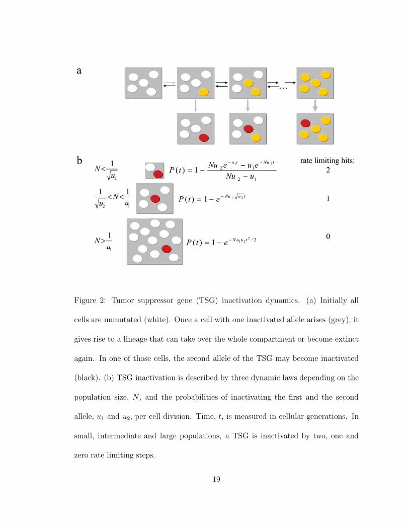
Figure 2: Tumor suppressor gene (TSG) inactivation dynamics. (a) Initially all
cells are unmutated (white). Once a cell with one inactivated allele arises (grey), it
gives rise to a lineage that can take over the whole compartment or become extinct
again. In one of those cells, the second allele of the TSG may become inactivated
(black). (b) TSG inactivation is described by three dynamic laws depending on the
population size, N , and the probabilities of inactivating the first and the second
allele, u1 and u2, per cell division. Time, t, is measured in cellular generations. In
small, intermediate and large populations, a TSG is inactivated by two, one and
zero rate limiting steps.
19

hit without ever having fixed the first hit51,53. The probability that at least one cell
with two hits has arisen before time t is
P (t) = 1 − exp(−Nu1
√u2t) (2)
This probability accumulates as a first order of time: it takes only one rate limiting
hit to inactivate a TSG in a population of intermediate size.
Third, in very large populations, N > 1/u1, cells with one inactivated allele arise
immediately and the waiting time for a cell with two inactivated alleles dominates
the dynamics. The probability that at least one cell with two hits has arisen before
time t is
P (t) = 1 − exp(−Nu1u2t2/2) (3)
This probability again accumulates as a second order of time. Eliminating a TSG
in a large population of cells is, however, not rate limiting for the overall process of
tumorigenesis. Due to the large population size, mutated cells are constantly being
produced, and the inactivation of a TSG is not rate limiting (Figure 2b).
These three dynamic laws provide a complete description of TSG inactivation.
In a normal tissue consisting of small compartments of cells, a TSG is eliminated by
two rate limiting hits. The overall rate of inactivation is proportional to the second
order of time. In small neoplasias, only one rate limiting hit is needed to inactivate
a TSG. The rate of inactivation is proportional to the first order of time. In large
tumors, it again takes two hits to inactivate a TSG, but neither of them is rate
20
limiting for the overall process of tumorigenesis. Therefore, as the population size
increases, a TSG is inactivated in two, one or zero rate limiting steps.
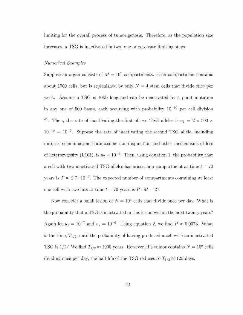
Numerical Examples
Suppose an organ consists of M = 107 compartments. Each compartment contains
about 1000 cells, but is replenished by only N = 4 stem cells that divide once per
week. Assume a TSG is 10kb long and can be inactivated by a point mutation
in any one of 500 bases, each occurring with probability 10−10 per cell division
45. Then, the rate of inactivating the first of two TSG alleles is u1 = 2 × 500 ×
10−10 = 10−7. Suppose the rate of inactivating the second TSG allele, including
mitotic recombination, chromosome non-disjunction and other mechanisms of loss
of heterozygosity (LOH), is u2 = 10−6. Then, using equation 1, the probability that
a cell with two inactivated TSG alleles has arisen in a compartment at time t = 70
years is P ≈ 2.7 · 10−6. The expected number of compartments containing at least
one cell with two hits at time t = 70 years is P ·M = 27.
Now consider a small lesion of N = 104 cells that divide once per day. What is
the probability that a TSG is inactivated in this lesion within the next twenty years?
Again let u1 = 10−7 and u2 = 10−6. Using equation 2, we find P ≈ 0.0073. What
is the time, T1/2, until the probability of having produced a cell with an inactivated
TSG is 1/2? We find T1/2 ≈ 1900 years. However, if a tumor contains N = 109 cells
dividing once per day, the half life of the TSG reduces to T1/2 ≈ 120 days.
21

Conclusions
The dynamics of TSG inactivation are described by three laws concerning the de-
pendence of the population size and the mutation rates constituting the first and
the second hit. In a small compartment, a TSG is inactivated by two rate limiting
hits. In a small lesion, it is inactivated by one, and in a large tumor, by zero rate
limiting hits.
Mutations inactivating TSGs, like mutations activating oncogenes, are best con-
tained by small compartments that are replenished by a small number of stem
cells43. Once a compartment has accumulated one or a few mutations in cancer-
susceptibility genes, a neoplasia develops in which other TSGs might have to be
inactivated for further tumor progression. Small neoplasias, however, are unlikely
to succeed in inactivating a further TSG within a human lifespan under the assump-
tion of normal mutation rates. Increased mutation rates due to genetic instability
might be necessary for the further progression of some small lesions.
The mathematical analysis of TSG inactivation suggests several new experimen-
tal studies. How does the inactivation of one or two alleles of a TSG change the net
growth rate of the cell? Is the first step indeed neutral or does it slightly modify the
fitness of the cell? Such fitness differences can be measured in cell cultures or animal
models. Furthermore, how long does it take for a population of cells to inactivate a
TSG? How does the time until inactivation of a TSG depend on the population size
and the mutation rates? Can the three dynamical regimes that we predict from the
22

theoretical analysis be verified? What are the relative rates of the various mecha-
nisms that contribute to LOH? Precise kinetic measurements are needed in order to
obtain quantitative insights into cancer progression.
2.4 Genetic instability
Genetic instability is a defining characteristic of most human cancers and represents
one of the most active research areas in cancer biology4. Two major types of genetic
instabilities have been identified54. In a small fraction of colorectal and some other
cancers, a defect in mismatch repair results in an increased rate of point mutations
and consequent widespread microsatellite instability. The majority of colorectal and
most other cancers, however, have chromosomal instability (CIN), which refers to an
increased rate of losing or gaining whole chromosomes or large parts of chromosomes
during cell division. The consequence of CIN is an imbalance in chromosome number
(aneuploidy) and an increased rate of LOH, which is an important property of CIN
because it accelerates the rate of TSG inactivation.
The molecular basis for CIN is just beginning to be understood. A large number
of genes that trigger CIN when mutated have been discovered in Saccharomyces
cerevisiae55–57. These so-called ’CIN genes’ are involved in chromosome conden-
sation, sister-chromatid cohesion, kinetochore structure and function, microtubule
formation and cell cycle checkpoints. By comparison with yeast, we expect several
hundred human CIN genes, but only a few have been identified so far. These genes
23

include hBUB1, MAD2, BRCA2, and hCDC458. The classification of CIN genes
is based on the mutational events that are required to trigger CIN4. Class I CIN
genes, such as MAD2, trigger CIN if one allele of the gene is mutated or lost. Class
II CIN genes, such as hBUB1, trigger CIN if one allele is mutated in a dominant-
negative fashion. Class III CIN genes, such as BRCA2, trigger CIN if both alleles
are mutated.
A number of hereditary syndromes are known that stem from germ line mutations
in what might be CIN genes. Inherited mutations in the RECQ-like helicases BLM
and WRT give rise to the Bloom and Werner Syndromes, respectively. An inherited
deficiency in nucleotide excision repair causes Xeroderma pigmentosum. Germ line
mutations in the gene ATM give rise to Ataxia telangiectasia. These syndromes are
all characterized by a high incidence of multiple types of cancer, but the mechanistic
connection between these genes and CIN is still somewhat unclear.
An important question in cancer genetics is to what extent CIN, or any genetic
instability, is an early event and driving force of tumorigenesis5,22,59. The investiga-
tion of the role of genetic instability requires a quantitative theory of how mutation
and selection of gatekeeper and caretaker genes contribute to cancer initiation and
progression51,60–62. Here, we discuss the role of CIN in cancers that are initiated by
inactivation of one or two TSGs51,63,64.
24

CIN before one TSG
Consider a case where tumorigenesis is initiated by inactivating a TSG, A, in a
small compartment of cells51. An appropriate example is the inactivation of APC in
a colonic crypt1. If the mutation rate is less than the inverse of the compartment size,
then the compartment almost always consists of a single type of cells - a mutated cell
either reaches fixation or goes extinct before another mutated cell arises. Initially, all
cells are wild type, A+/+. The compartment evolves from A+/+, via A+/−, to A−/−.
A mutation that triggers CIN can arise at any stage of this process. The crucial
effect of CIN is to increase the rate of LOH, thereby accelerating the transition from
A+/− to A−/−. Stochastic tunneling can lead from A+/− without CIN directly to
A−/− with CIN53.
Figure 3a shows the rates of evolution of TSG inactivation with or without CIN
in a small compartment of cells. There are two rate limiting hits for inactivating the
TSG without CIN. Interestingly, there are also two rate limiting hits for inactivating
the TSG with CIN51: one rate limiting step is needed for inactivating the first
allele of the TSG, another rate limiting step is needed for the CIN mutation. The
inactivation of the second TSG allele is greatly accelerated in the presence of CIN
and is therefore not rate limiting51.
There are three evolutionary trajectories, all of which contain two rate limiting
steps (Figure 3b): the TSG can be inactivated without CIN; the CIN mutation can
occur first, followed by the inactivation of the two TSG alleles; one TSG allele can be
25
mutated first, followed by a CIN mutation, followed by the inactivation of the second
TSG allele. The third trajectory contains a tunnel if there is a significant cost of CIN,
i.e. if a CIN cell has a high probability of undergoing apoptosis due to deleterious
and lethal mutations. Such a cell might not reach fixation in the compartment,
but might nevertheless produce a cell with two inactivated TSG alleles that reaches
fixation. The relative rates of these trajectories depend on the compartment size,
N , the mutation rates, the number of CIN genes, and the cost of CIN (Figure 3b).
We can use these rates to calculate the fraction of mutated compartments with
or without CIN. We can also estimate the minimum number of CIN genes in the
genome that are required to ensure that CIN precedes the inactivation of the first
or second TSG allele51,63.
Numerical Examples of CIN before one TSG
Again, consider an organ that consists of M = 107 compartments. Suppose each
compartment contains 1000 cells, but is replenished by a small pool of N = 4 stem
cells. Assume that the probabilities of inactivating the first and the second allele of
a TSG per cell division are u1 = 10−7 and u2 = 10−6, respectively. We assume that
the inactivation of both TSG alleles confers a big selective advantage to the cell,
such that the probability of fixation is 1.
Assume there is one class I CIN gene in the genome that does not change the
fitness of the cell when mutated, r = 1. What is the ratio of the probabilities
of inactivating the TSG with CIN versus without CIN? We find that 50% of all
26

27

Figure 3: Emergence of chromosomal instability (CIN) during inactivation of one
tumor suppressor gene (TSG). (a) The alleles of TSG A are inactivated at rates u1
and u2, respectively, per cell division. The probability of mutating a CIN gene is
given by uc. A CIN cell has relative fitness r and reaches fixation in a compartment
of N cells with probability ρ = (1 − 1/r)/(1 − 1/rN ). CIN increases the rate
of inactivating the second TSG allele to u3 >> u2. The probabilities that the
compartment is in state A−/− without and with CIN at time t are given by X(t)
and Y (t), respectively. (b) The compartment can evolve along three evolutionary
trajectories, all of which contain two rate limiting hits: the TSG can be inactivated
without CIN; CIN can arise before the inactivation of the first TSG allele; or CIN can
arise between the inactivation of the first and the second TSG allele. The relative
weights of each trajectory for neutral CIN, r ≈ 1, and costly CIN, r < 1, allow us
to calculate their respective contributions to tumorigenesis.
28
mutated compartments have inactivated the TSG in a cell with CIN. In 25%, the
CIN mutation occurred before the first TSG mutation, whereas in 25%, the CIN
mutation occurred between the first and the second TSG mutation. In 50% of
mutated compartments, there was no CIN mutation prior to the inactivation of the
TSG. These results are largely independent of time.
Instead, assume that there are two class I CIN genes in the genome, each of
which is sufficient to trigger CIN when mutated. Mutation of either gene reduces
the fitness of the cell to r = 0.7. In this case, 54% of all mutated compartments
have inactivated the TSG in a cell with CIN. In 25%, the CIN mutation occurred
before the first TSG mutation. In 25%, the CIN mutation occurred between the
first and the second TSG mutation without a tunnel, in 4% with a tunnel. In 46%
of mutated compartments, there was no CIN mutation before the TSG inactivation.
Alternatively, we can calculate the critical number of CIN genes in the genome
that are needed to ensure that CIN arises before the inactivation of the TSG. If CIN
is neutral, r = 1, then one class I CIN gene or 11 class II CIN genes in the genome
are needed. If CIN has a selective disadvantage of r = 0.7, then two class I CIN
genes or 19 class II CIN genes in the genome are needed to make sure that CIN
arises first. Note again that in yeast, more than hundred CIN genes are known, and
it is possible that an even larger number exists in the human genome.
29

CIN in inherited cancers
In some genetic diseases, one allele of a TSG is mutated in the germ line. For
example, patients with familial adenomatous polyposis (FAP) inherit a mutation in
one allele of the tumor suppressor gene APC. By their teens, they harbor hundreds
to thousands of colorectal polyps. In terms of the model, the mutational path starts
with a compartment that consists only of A+/− cells without CIN. There are two
evolutionary trajectories: the somatic mutation that inactivates the second TSG
allele occurs in a cell without CIN, or a CIN mutation precedes the inactivation of
the second TSG allele. Both trajectories require one rate limiting step63. Again,
we can calculate the relative rates of these two possibilities. We can also calculate
the critical number of CIN genes in the genome that are needed to ensure that
CIN arises before the inactivation of the second allele. If CIN has a negligible cost,
then twice as many CIN genes as before are needed to make sure that CIN comes
first. The factor two comes from the fact that during the inactivation of two TSG
alleles, CIN can arise either before the first or before the second hit. If the first
hit is already present in the germ line, CIN can arise only before the second hit.
This reduces the probability that CIN arises before the inactivation of the TSG by
a factor 1/2. If CIN has a substantial cost, then the same number of CIN genes
is needed as in the sporadic case with neutral CIN. Hence, there is only a small
difference in the relative importance of CIN in those cases where both TSG alleles
have to be inactivated somatically and where one TSG allele is already inactivated
30

in the germ line63.
CIN before two TSGs
Consider a path to cancer in which two TSGs, A and B, have to be eliminated
in rate limiting steps. Initially, the compartment consists of N0 wild type cells,
A+/+B+/+, that divide every τ0 days. Suppose gene A has to be inactivated first,
so the evolutionary pathway proceeds from A+/+B+/+ via A+/−B+/+ to A−/−B+/+,
and subsequently to A−/−B+/− and A−/−B−/− (Figure 4). CIN can emerge at
any stage of this pathway; once arisen, CIN accelerates the transitions from A+/−
to A−/− and from B+/− to B−/−. Inactivation of the first TSG induces neoplastic
growth. We assume that the A−/− compartment gives rise to a small lesion of N1 cells
that divide every τ1 days. In this lesion, the second TSG has to be inactivated for
further tumor progression. Due to the increased compartment size, the evolutionary
trajectory tunnels53 from A−/−B+/+ directly to A−/−B−/− (Figure 4).
Without CIN, inactivation of two TSGs requires three rate limiting hits. It takes
two hits to inactivate the first TSG - if this leads to a moderate clonal expansion,
then the second TSG can be inactivated in one rate limiting step (Equation 2). If the
inactivation of the first TSG leads to a vast clonal expansion, then the inactivation
of a further TSG is not rate limiting, and CIN becomes less important. With CIN,
inactivation of two TSGs also requires three rate limiting hits. It takes two hits
to inactivate the first TSG (one in a TSG allele and one in a CIN gene), and one
further hit to inactivate the second TSG.
31

Figure 4: Emergence of chromosomal instability (CIN) during inactivation of two
tumor suppressor genes (TSGs). Inactivation of TSG A in a compartment of N0
cells causes a clonal expansion to N1 cells in which TSG B is inactivated. Due to
the increased compartment size, the evolutionary trajectory tunnels from A−/−B+/+
directly to A−/−B−/−. CIN can arise at any stage of tumorigenesis and increases the
rate of TSG inactivation. It takes three rate-limiting hits to inactivate two TSGs
both with and without CIN.
32
Again, we can calculate the minimum number of CIN genes in the genome that
are needed to ensure that CIN arises before the inactivation of the first TSG in
paths to cancer in which two TSGs have to be eliminated in rate limiting steps. We
find that very few CIN genes in the genome are necessary to make sure that CIN
arises early (Figure 5). The cost of CIN is compensated by an acceleration of every
successive TSG inactivation. It is possible that the first TSG, A, is predominantly
inactivated in cells without CIN. Thus, most lesions which are caused by inactivation
of TSG A do not have CIN, but only the small fraction of lesions with CIN will
eliminate TSG B within the time scale of a human life. In such a situation all
(or almost all) cancers will derive from lesions in which a CIN mutation preceded
inactivation of the first TSG.
Conclusions
The role of CIN in tumorigenesis is one of the most active and controversial fields
of all of biology. In order to evaluate hypotheses and interpret experimental obser-
vations, a quantitative model is required for understanding how tissue organization,
mutation rates, and selection determine the role of CIN in tumorigenesis. Here we
have outlined a mathematical approach for evaluating the importance of early CIN
in tumorigenesis initiated by inactivation of ‘half’, one or two TSGs in rate limiting
situations. We find that one or a few neutral CIN genes in the genome are sufficient
to ensure the emergence of CIN before the inactivation of one TSG. One or a few
costly CIN genes in the genome are sufficient to ensure the emergence of CIN before
33

Figure 5: Minimum number of CIN genes needed to ensure that CIN arises before
the inactivation of one or two TSGs. The relative selective fitness of CIN cells is
denoted by r. Class I CIN genes, n1, trigger CIN if one allele is mutated or lost.
Class II CIN genes, n2, trigger CIN if one allele is mutated in a dominant negative
fashion. Class III CIN genes, n3, trigger CIN if both alleles are mutated. Parameter
values are u1 = 10−7, u2 = 10−6, u3 = 10−2, N0 = 4, and t = 80 years.
34

the inactivation of the first TSG if two TSGs have to be eliminated in rate limiting
steps.
The theoretical investigation of CIN suggests a number of new experiments. Can
one demonstrate in mouse that the two rate limiting steps consist of inactivating
one allele of the TSG and one CIN mutation? What is the selective cost of a CIN
mutation? Can CIN be neutral or even advantageous65? Is it possible to find CIN
mutations in dysplastic crypts, which represent the first stage of colon cancer58?
What is the number of CIN genes in the human genome? How many of them fall
into classes I, II or III? Is it possible to show that CIN is essential for inactivating
two TSGs in small populations of cells?
2.5 Conclusions
We have outlined some fundamental principles of somatic evolutionary dynamics.
We have discussed the rates at which mutations in oncogenes and tumor suppressor
genes (TSGs) accumulate in small compartments of cells, early lesions, and large
cancers. We have explained how the relevant time scales depend on population
size, mutation rates, and fitness differences. We have studied the role of tissue
architecture and genetic instability.
Tissue architecture and compartment size determine the rates at which different
types of mutations accumulate. Cells with mutations in oncogenes or TSGs can have
an increased somatic fitness. Such mutations are best contained when the tissue is
35
organized into small compartments and each compartment is fed by one (or a few)
stem cells, as in the linear process43. Cells with mutations in genetic instability
genes are likely to have a reduced somatic fitness. Such mutations accumulate
faster in small compartments than in large compartments. Therefore, the optimum
compartment size is a tradeoff between preventing mutations in oncogenes, tumor
suppressor genes and genetic instability genes41,42.
Chromosomal instability (CIN) confers an increased probability of gaining or
losing whole chromosomes or arms of chromosomes. CIN accelerates the rate of
inactivating TSGs. A major debate in cancer genetics is to what extent CIN is an
early event and hence a driving force of cancer progression. We have shown that in
a small compartment of cells, it takes two hits to inactivate a TSG both with and
without CIN. Whether or not CIN occurs before the first TSG depends on the cost
of CIN, the mutation rate, the rate of LOH, the number of CIN genes in the genome,
and the population size. For a wide range of parameters, one (or a few) neutral CIN
genes are enough to ensure that CIN initiates tumor formation in a pathway where
one TSG needs to be eliminated in a rate limiting situation. One (or a few) costly
CIN genes are enough to ensure that CIN initiates tumor formation in a pathway
where two TSGs need to be eliminated in rate limiting situations.
We have reviewed models of somatic evolution with constant selection, which
are based on the assumption that the fitness of mutants neither depends on their
own relative abundance nor on the relative abundance of other mutants. Cancer
36
progression, however, might often include a complex interplay of various types of
cells. Mutations in landscaper genes, alterations promoting angiogenesis and mu-
tations enabling the cell to fight off the immune system might be examples. These
complex dependencies require a game theoretical analysis51, and we are still only at
the beginning of a long endeavor.
Mathematical models of cancer evolution are essential for quantifying the effects
of mutation, selection, and tissue architecture. The dynamical laws of tumor sup-
pressor gene inactivation, oncogene activation, and emergence of genetic instability
provide fundamental new insights into tumorigenesis. Theory can help to clarify
concepts, interpret experimental data, and suggest new experiments. Mathematical
models describing tumor initiation, progression, invasion and metastasis contribute
toward a quantitative understanding of cancer biology.
37

3 Can chromosomal instability initiate cancer?
Michor F, Iwasa Y, Lengauer C, Vogelstein B, Nowak MA
Can chromosomal instability initiate tumorigenesis?63
Seminars in Cancer Biology 15, 43-49 (2005)
Cancers result from the accumulation of inherited and somatic mutations
in oncogenes and tumor suppressor genes. These genes encode proteins
that function in growth regulatory and differentiation pathways. Muta-
tions in those genes increase the net reproductive rate of cells. Chromo-
somal instability (CIN) is a feature of most human cancers. Mutations in
CIN genes increase the rate at which whole chromosomes or large parts
of chromosomes are lost or gained during cell division. CIN causes an
imbalance in chromosome number (aneuploidy) and an enhanced rate of
loss of heterozygosity, which is an important mechanism of inactivating
tumor suppressor genes. A crucial question of cancer biology is whether
CIN is an early event and thus a driving force of tumorigenesis. Here we
discuss mathematical models of situations where inactivation of one or
two tumor suppressor genes is required for tumorigenesis. If two tumor
suppressor genes have to be inactivated in rate-limiting steps, then CIN
is likely to emerge before the inactivation of the first tumor suppressor
gene.
38
3.1 Introduction
Tumor suppressor genes (TSGs) are negative regulators of cellular growth1,48,50,66,67.
Loss of their function contributes to tumorigenesis10,23,68. The concept of a TSG
emerged from a statistical analysis of retinoblastoma incidence in children10. This
study and subsequent work led to the 2-hit hypothesis, which proposes that two
mutations in the retinoblastoma gene are the rate limiting steps of this particular
cancer1. In the inherited form, the first mutation is present in the germ line, whereas
the second mutation emerges during somatic cell divisions. In the sporadic form,
both mutations arise during somatic cell divisions. Inactivation of the first allele
of a TSG does not normally change the phenotype of the cell and is considered
an (almost) neutral mutation. Inactivation of the second allele confers a fitness
advantage to the cell and can promote neoplastic growth. Human cancers result
from the accumulation of mutations in several TSGs, oncogenes, and genes that are
involved in maintaining genomic stability. Here, we discuss mathematical models of
cancers that require the inactivation of one or two TSGs in rate limiting steps.
Genetic instability is a defining characteristic of most human cancers4,69. Two
types of genetic instability have been identified54. A defect in mismatch repair
(MMR) results in an elevated mutation rate at the nucleotide level leading to mi-
crosatellite instability70,71 (MIN). MIN is only found in a small fraction of colorectal
and some other cancers. The majority of colorectal and most other solid cancers,
however, have chromosomal instability (CIN). CIN refers to an increased rate of los-
39
ing or gaining whole chromosomes or large parts of chromosomes during cell division.
CIN causes an imbalance in chromosome number (aneuploidy) and an increased rate
of loss of heterozygosity (LOH). An elevated rate of LOH is an important property
of CIN, because it accelerates the inactivation of TSGs. The potential cost of CIN
is discussed in section 3.2
Note that CIN refers to an enhanced rate of accumulating gross chromosomal
changes during cell division; it is not synonymous with the state of aneuploidy that
is observed in a static image of the cell’s chromosomal content. While CIN is a
process that drives most cells to aneuploidy, aneuploidy per se does not prove the
existence of CIN. There are several ways in which a cell can become aneuploid in the
absence of CIN. The cell could have divided many more times than other cells in the
tissue; as gross chromosomal changes (rarely) occur in normal cells, this could lead
to aneuploidy without CIN. Alternatively, aneuploidy could be due to exposure to
endogenous or exogenous agents that disrupt spindle formation. It is also possible
that cancer cells accumulate gross chromosomal changes at the same rate as normal
cells, but that these changes are only lethal in normal cells. This hypothesis is
strengthened by the knowledge that mutations in cancer cells disrupt the capacity
of cells to undergo apoptosis.
Although CIN is defined as an increased rate of losing or gaining parts of chro-
mosomes or whole chromosomes during cell division, CIN has only been formally
demonstrated for whole chromosome losses54. There is no assay at present that can
40

reliably measure the rate of formation of changes at the subchromosomal level, such
as deletions, insertions, inversions, rearrangements, amplifications, unequal sister
chromatid exchange, and gene conversion. These changes are at least as common as
losses or gains of whole chromosomes.
A large number of genes that trigger CIN when mutated have been identified in
the yeast Saccharomyces cerevisiae55–57. These so-called ‘CIN genes’ are involved
in chromosome condensation, sister-chromatid cohesion, kinetochore structure and
function, and microtubule formation as well as in cell cycle checkpoints. Similarly,
several genes can cause CIN in Drosophila melanogaster when genetically altered
72,73. By analogy, we expect several hundred human CIN genes, but only few have
been identified so far74. These genes include hBUB1, MAD2, BRCA1, BRCA2 and
hCDC458,75–78. The genes hBUB1 and MAD2 encode proteins that are required
for function of the spindle assembly checkpoint75,76. This checkpoint modulates the
timing of anaphase initiation in mitotic cells containing improperly aligned chromo-
somes and increases the probability of successful delivery of a correct chromosome set
to each daughter cell. The genes BRCA1 and BRCA2 encode proteins that are im-
plicated in DNA repair and recombination, checkpoint control of the cell cycle, and
transcription76,78. hCDC4 is an E3 ubiquitin ligase that is thought to be involved
in regulating the G1-S cell-cycle checkpoint by targeting proteins for destruction
58. Inactivation of hCDC4 leads to increased levels of cyclin E, the formation of
micronuclei, defects in the execution of anaphase, and CIN.
41
The classification of CIN genes is based on the mutational events required to
engage CIN4. Class I CIN genes, such as MAD2, trigger CIN if one allele of the
gene is mutated or lost. Class II CIN genes, such as hBUB1, trigger CIN if one
allele is mutated in a dominant negative fashion. Both class I and II are ‘single hit’
CIN genes. Class III CIN genes, such as BRCA1 and BRCA2, trigger CIN if both
alleles are mutated.
A major question in cancer genetics is to what extent CIN, or any genetic in-
stability, is an early event and thus a driving force in tumorigenesis2,39,41,51,59,60,79.
Experimental evidence shows that early colorectal adenomas have allelic imbalance
80 (Figure 6), but this provides no proof of underlying CIN. hCDC4 mutations have
been shown to occur early in colorectal tumorigenesis58. Here, we discuss a mathe-
matical framework for the evolutionary dynamics of tumorigenesis offering insights
into the causal relations between CIN and cancers initiated by inactivation of one
or two TSGs.
3.2 Chromosomal instability before one tumor suppressor
gene
Consider a path to cancer where both alleles of a TSG, A, have to be inactivated in
a single cell (Figure 7a). Initially, the cell is wild type, A+/+. The cell evolves from
A+/+ via A+/− to A−/−. The first allele of a TSG is typically inactivated by a point
mutation. The second allele can be inactivated by one of several possible mechanisms
42

Figure 6: Experimental evidence of aneuploidy in early tumors. The table shows the
percentage of early colorectal adenomas (size 1-3 mm, total number 32) with allelic
imbalance in five different chromosome arms. Ninety percent of the adenomas have
allelic imbalance in any one of the five chromosome arms. Data from Shih et al.80.
43

(Figure 8). It can be inactivated by a second point mutation, leading to two distinct
point mutations in the two heterozygous alleles. Alternatively, the chromosome on
which the second allele resides can be lost; this chromosome loss might be deleterious
or lethal. The two alleles at the TSG locus can recombine mitotically, leading to two
identical point mutations on partly heterozygous alleles. Finally, the chromosome
harboring the wild type allele can be lost and the chromosome harboring the first
point mutation duplicated. This mechanism may require two steps, occurring in two
distinct cell divisions, but could also occur in a single division through a process
broadly related to mitotic recombination.
CIN can emerge at any stage of tumorigenesis. First, assume that CIN is domi-
nant, i.e. triggered by mutation of a class I or II CIN gene4,51. The crucial effect of
CIN is to increase the rate of LOH, thereby accelerating the transition from A+/− to
A−/−. CIN can have a cost for the cell by increasing the rate of accumulating lethal
mutations and triggering apoptosis. CIN, however, can also be advantageous by
reducing the duration of the cell cycle (if certain checkpoints have been eliminated)
or by providing increased evolvability in detrimental environments65,81. Therefore,
it makes sense to consider possibilities of costly, neutral, and advantageous CIN
phenotypes.
Tissues are organized into small compartments of cells12,36,46,47. Not all cells of
a compartment might be at risk of becoming cancer cells, however, because certain
mutations might only have an effect if they occur in stem cells37. Suppose N0 cells
44

45
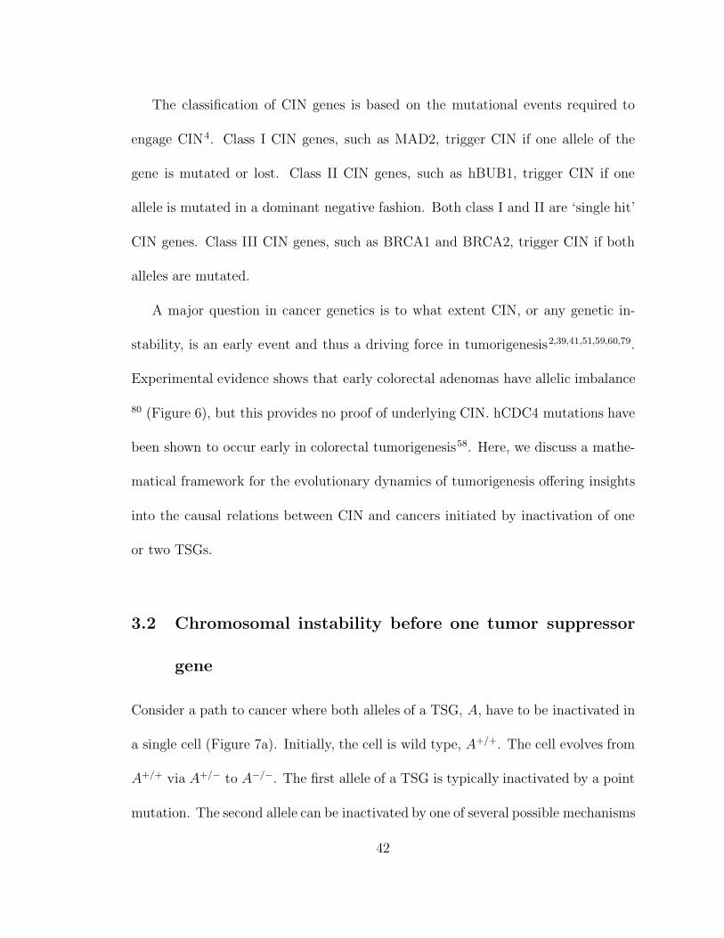
Figure 7: Inactivation dynamics of a tumor suppressor gene (TSG), A, with and
without chromosomal instability (CIN). (a) The wild type cell (‘start’) has two
unmutated alleles of TSG A, A+/+. The cell evolves from A+/+ via A+/− to A−/−.
CIN can arise at any stage of TSG inactivation and causes very fast LOH. (b) A
compartment of cells evolves along the evolutionary trajectory. If an A+/− cell clone
with CIN produces an A−/− cell before taking over the compartment, a stochastic
tunnel arises (diagonal arrow); the compartment evolves from A+/− without CIN to
A−/− with CIN without ever visiting A+/− with CIN.
46

Figure 8: Mechanisms of tumor suppressor gene (TSG) inactivation. The first TSG
allele is typically inactivated by a point mutation. The second allele can be inacti-
vated by a second point mutation, by chromosome loss, mitotic recombination, or
chromosome loss followed by duplication of the mutated allele.
47

are at risk in any one compartment. If the mutation rate is smaller than the inverse
of N0, then the approximation of homogeneous compartments holds: a mutated cell
will either take over the compartment or go extinct before the next mutation arises
(Figure 7b). If an A+/− cell clone with CIN produces an A−/− cell before taking
over the compartment, then the phenomenon of ‘stochastic tunneling’ arises51,53:
the compartment moves from A+/− without CIN to A−/− with CIN without ever
visiting A+/− with CIN. If CIN is neutral or advantageous, then the tunnel does not
arise. Mathematical procedures are outlined in Section 3.5.
3.3 Chromosomal instability before two tumor suppressor
genes
Consider a path to cancer where two TSGs, A and B, have to be inactivated in a
single cell (Figure 9). Initially, the cell is wild type, A+/+B+/+. Suppose gene A
has to be inactivated first. Hence the cell evolves from A+/+B+/+ via A+/−B+/+
to A−/−B+/+, and subsequently to A−/−B+/− and A−/−B−/−. CIN can emerge at
any stage of tumorigenesis due to mutations of class I, II or III CIN genes. Once
CIN has emerged, it accelerates the transitions from A+/− to A−/− and from B+/−
to B−/−.
Evolutionary dynamics within a compartment of cells are illustrated in Figure
10. CIN might emerge before gene A has been inactivated. A stochastic tunnel
arises if an A+/−B+/+ cell clone with CIN produces an A−/−B+/+ cell before taking
48

Figure 9: Mutational network of inactivating two tumor suppressor genes (TSGs), A
and B, with and without chromosomal instability (CIN). The wild type cell (‘start’)
has two unmutated alleles of both TSGs, A+/+B+/+. Mutations lead via A+/−B+/+,
A−/−B+/+ and A−/−B+/− to A−/−B−/−. Without CIN, the cell maintains a stable
genotype (second row). CIN can arise at any stage of tumorigenesis due to mutations
in a class I, II, or III CIN gene. Class I and II CIN genes require one hit to trigger
CIN (first row). Class III CIN genes require two hits to trigger CIN, one in each allele
(third and forth row). Once arisen, CIN accelerates the inactivation of the second
allele of each following TSG. CIN, however, has a cost for the cell by increasing the
chance of lethal mutations and apoptosis.
49
over the compartment51,53. Then the compartment moves from A+/−B+/+ without
CIN to A−/−B+/+ with CIN without ever visiting A+/−B+/+ with CIN.
Inactivation of the first TSG can induce neoplastic growth. We assume that the
A−/− compartment gives rise to a small lesion of N1 cells. In this lesion the second
TSG has to be inactivated for further tumor progression. Due to the increased
compartment size, the evolutionary pathway might tunnel from A−/−B+/+ directly
to A−/−B−/−. This means that the A−/−B+/− cell does not reach fixation before
the A−/−B−/− cell arises. Mathematical procedures are outlined in Section 3.5.
The importance of early CIN in tumorigenesis depends on the number of possible
CIN mutations, which in turn depends on the number of CIN genes in the human
genome. We can calculate the minimum number of CIN genes in the genome that are
needed to ensure that a CIN mutation precedes the inactivation of one or two TSGs.
Let us discuss some plausible parameter choices (Figure 11). The mutation rate45
per base per cell division is 10−10 to 10−11. If an average TSG can be inactivated
by any one of 1000 point mutations, the mutation rate per gene per cell division is
u = 10−7. Estimates of the rate of LOH in non-CIN cells range from p0 = 10−7 to
p0 = 10−5. In our opinion, the most likely scenario is that p0 has the same order
of magnitude as u. In this case and in the absence of CIN, the hit inactivating the
second TSG allele is sometimes LOH and sometimes a point mutation. If p0 >> u,
then two distinct point mutations should never be observed in the two TSG alleles.
The rate of LOH in CIN cells82 is p = 10−2. Consider a compartment size of N0 = 4.
50

/A
/B
/A
/B
CIN(I, II) /B /B
/A /A
/A
/B
/A
/B
/A/B
/A/B
stable
/A
/B
/A
/B
stable( )/III
/A/B
/A/B
/A
/B
/A
/B
CIN(III) /B /B
/A /A
Figure 10: Inactivation dynamics starting with a population of N0 wild type cells.
CIN can emerge at any stage of tumorigenesis. If an A+/−B+/+ cell clone without
CIN produces an A−/−B+/+ cell with CIN before reaching fixation, the compartment
tunnels to A−/−B+/+ with CIN. Inactivation of TSG A leads to a small lesion of
N1 cells in which TSG B is eliminated. Due to the increased compartment size, the
evolutionary pathway tunnels directly to A−/−B−/−.
51
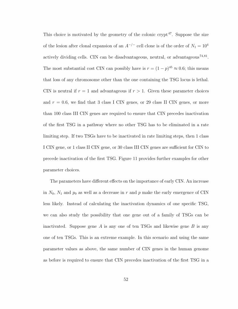
This choice is motivated by the geometry of the colonic crypt47. Suppose the size
of the lesion after clonal expansion of an A−/− cell clone is of the order of N1 = 104
actively dividing cells. CIN can be disadvantageous, neutral, or advantageous74,81.
The most substantial cost CIN can possibly have is r = (1 − p)45 ≈ 0.6; this means
that loss of any chromosome other than the one containing the TSG locus is lethal.
CIN is neutral if r = 1 and advantageous if r > 1. Given these parameter choices
and r = 0.6, we find that 3 class I CIN genes, or 29 class II CIN genes, or more
than 100 class III CIN genes are required to ensure that CIN precedes inactivation
of the first TSG in a pathway where no other TSG has to be eliminated in a rate
limiting step. If two TSGs have to be inactivated in rate limiting steps, then 1 class
I CIN gene, or 1 class II CIN gene, or 30 class III CIN genes are sufficient for CIN to
precede inactivation of the first TSG. Figure 11 provides further examples for other
parameter choices.
The parameters have different effects on the importance of early CIN. An increase
in N0, N1 and p0 as well as a decrease in r and p make the early emergence of CIN
less likely. Instead of calculating the inactivation dynamics of one specific TSG,
we can also study the possibility that one gene out of a family of TSGs can be
inactivated. Suppose gene A is any one of ten TSGs and likewise gene B is any
one of ten TSGs. This is an extreme example. In this scenario and using the same
parameter values as above, the same number of CIN genes in the human genome
as before is required to ensure that CIN precedes inactivation of the first TSG in a
52
23111011211.4
42112011630.6
26111111321.0
382116111221.0
31211311811.4
>10042591154270.6
41 10N 5
1 10N1oftindependen N
CIN before 1 TSG CIN before 2 TSGs
2111811211.4
40111711631.0
a
b
c
d 76401318121.0
65271245111.4
615130112930.6
>10010015425130.6
66
>100
>100
95
>100
>100
>100
>100
>100
>100
>100
>100
>100
>100
>100
1n 2n 3n 1n 2n 3n 1n 2n 3nr
53
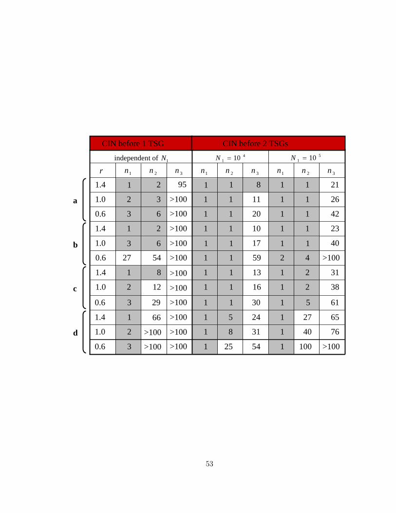
Figure 11: Minimum number of CIN genes needed to ensure that CIN emerges
before one or two TSGs. Class I CIN genes, n1, trigger CIN if one allele is mutated
or lost. Class II CIN genes, n2, trigger CIN if one allele is mutated in a dominant
negative fashion. Class III CIN genes, n3, trigger CIN if both alleles are mutated.
The relative somatic fitness of CIN cells is denoted by r, and the compartment
size after clonal expansion by N1. Results are obtained by numerical simulation of
Equation 6. Parameter values are u = 10−7, p = 10−2, t = 80 years, and p0 = 10−7
and N0 = 4 in (a), p0 = 10−7 and N0 = 10 in (b), p0 = 10−6 and N0 = 4 in (c), and
p0 = 10−5 and N0 = 4 in (d).
54
pathway where no other TSG has to be eliminated in a rate limiting step. If two
TSGs have to be inactivated in rate limiting steps, then 1 class I CIN gene, or 5 class
II CIN gene, or 62 class III CIN genes are sufficient for CIN to precede inactivation
of the first TSG. Hence, a large number of alternative TSGs increases the number
of class II and III CIN genes needed, but does not significantly alter the number of
class I CIN genes required for CIN to arise early.
3.4 Conclusions
Even if many alternative CIN genes are needed to ensure that costly CIN emerges
before one TSG, only very few CIN genes suffice for CIN to precede two TSGs.
This effect is especially strong if inactivation of the first TSG leads to a moderate
clonal expansion. In this case, the second TSG must be inactivated in a rate limiting
fashion. If, on the other hand, the inactivation of the first TSG causes a rapid clonal
expansion (N1 >> 105), then the inactivation of the second TSG does not occur in a
rate limiting step, and CIN can only accelerate inactivation of the first TSG. A wide
range of plausible parameter values, which are conservatively biased against CIN,
all give the same message: of the order of 1 (to 10) neutral CIN genes are needed to
ensure that CIN emerges before the inactivation of one TSG; of the order of 1 (to
10) costly CIN genes are needed to ensure that CIN emerges before the inactivation
of the first of two TSGs. By analogy with yeast, we expect several hundreds of CIN
genes in the human genome. Therefore it is likely that in any one human tissue a
55
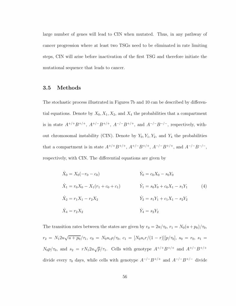
large number of genes will lead to CIN when mutated. Thus, in any pathway of
cancer progression where at least two TSGs need to be eliminated in rate limiting
steps, CIN will arise before inactivation of the first TSG and therefore initiate the
mutational sequence that leads to cancer.
3.5 Methods
The stochastic process illustrated in Figures 7b and 10 can be described by differen-
tial equations. Denote by X0, X1, X2, and X4 the probabilities that a compartment
is in state A+/+B+/+, A+/−B+/+, A−/−B+/+, and A−/−B−/−, respectively, with-
out chromosomal instability (CIN). Denote by Y0, Y1, Y2, and Y4 the probabilities
that a compartment is in state A+/+B+/+, A+/−B+/+, A−/−B+/+, and A−/−B−/−,
respectively, with CIN. The differential equations are given by
X0 = X0(−r0 − c0) Y0 = c0X0 − s0Y0
X1 = r0X0 − X1(r1 + c0 + c1) Y1 = s0Y0 + c0X1 − s1Y1 (4)
X2 = r1X1 − r2X2 Y2 = s1Y1 + c1X1 − s2Y2
X4 = r2X2 Y4 = s2Y2
The transition rates between the states are given by r0 = 2u/τ0, r1 = N0(u+p0)/τ0,
r2 = N12u√
u + p0/τ1, c0 = N0ucρ/τ0, c1 = [N0ucr/(1 − r)][p/τ0], s0 = r0, s1 =
N0p/τ0, and s2 = rN12u√
p/τ1. Cells with genotype A+/+B+/+ and A+/−B+/+
divide every τ0 days, while cells with genotype A−/−B+/+ and A−/−B+/− divide
56

every τ1 days. Let us first discuss the mutation-selection network leading from X0
via X1 and X2 to X4. The rate of change, r0, is equal to the rate that a mutation
occurs in the first allele of TSG A times the probability that the mutant cell will
take over the compartment. The mutation occurs at rate 2N0u/τ0, because (i) the
mutation can arise in any one of N0 cells, (ii) the cells divide once every τ0 days,
(iii) the mutation rate per gene per cell division is given by u and (iv) there are
2 alleles. The probability that the mutated cell takes over the compartment (i.e.,
reaches fixation) is given by 1/N0, if we assume that the mutation is neutral40. Thus,
the population size, N0, cancels in the product, and we simply obtain 2u/τ0.
Once the first allele of TSG A has been inactivated, the second allele can be
inactivated either by another point mutation or an LOH event. This process occurs
at rate r1 = N0(u + p0)/τ0, where p0 is the rate of LOH in normal cells. We
assume that A−/− cells have a large fitness advantage, which means the probability
of fixation of such a cell is close to one. Inactivation of both alleles of TSG A leads to
a small lesion of N1 cells dividing every τ1 days. In this lesion, TSG B is inactivated
at rate r2 = N12u√
u + p0/τ1.
CIN can arise at any stage of tumorigenesis and causes very fast LOH82. Denote
the rate of LOH in CIN cells by p; we have p >> p0. The rate of change, c0, is equal
to the rate at which a mutation triggering CIN occurs, N0ucτ0, times the probability
that the mutant cell will take over the compartment. The mutation rate triggering
CIN, uc, depends on the number of available class I, II and III CIN genes in the
57

human genome, n1, n2 and n3, respectively. The mutation rates of class I and II
CIN genes are uc = 2n1(u + p0) and uc = 2n2u, respectively. The mutation rate of
the first allele of class III CIN genes is 2n3u, and the mutation rate of the second
allele of class III CIN genes is u + p0. The probability of fixation of a CIN cell
depends on its somatic fitness, r, and is given by ρ = (1 − 1/r)/(1 − 1/rN0 ). This
is the standard fixation probability of a Moran process38,43. Therefore, the rate of
change is given by c0 = N0ucρ/τ0. A stochastic tunnel arises if an A−/−B+/+ cell
with CIN reaches fixation before an A+/−B+/+ cell does. The rate of tunneling is
given by c1 = [N0ucr/(1 − r)][p/τ0].
Let us now discuss the mutation-selection network leading from Y0 via Y1 and Y2
to Y4. The rate of change, s0, is equal to the rate r0, because CIN does not change
the probability of point mutations. The rate at which the second allele of TSG A
is inactivated is given by s1 = N0p/τ0. The rate at which TSG B is inactivated is
given by s2 = rN12u√
p/τ1.
Equation 4 is a system of linear differential equations which can be solved ana-
lytically using standard techniques.
58

4 Dynamics of Colorectal Cancer
Michor F, Iwasa Y, Lengauer C, Nowak MA
Dynamics of Colorectal Cancer83
Seminars in Cancer Biology, in press (2005)
Colorectal cancer results from an accumulation of mutations in tumor
suppressor genes and oncogenes. An additional defining characteristic
of colorectal cancer is its genetic instability. Two main types of genetic
instability have been identified. Microsatellite instability leads to an in-
creased point mutation rate, whereas chromosomal instability refers to
an enhanced rate of accumulating gross chromosomal aberrations. All
colon cancer cell lines are genetically unstable. An interesting question is
whether genetic instability arises early in tumorigenesis. An early emer-
gence of genetic instability could drive most of the somatic evolution of
cancer. Here, we review mathematical models of colorectal tumorigenesis
and discuss the role of genetic instability.
59
4.1 Introduction
’I wish I had the voice of Homer, to sing of rectal carcinoma’ are the opening lines
of a poem by British mathematical biologist J. B. S. Haldane, who was diagnosed
with this disease in 1964 and died a few years later. Haldane, together with Ronald
Fisher and Sewell Wright, was one of the founding fathers of population genetics. He
unified Darwinian evolution with Mendelian genetics and brought about the current
understanding of evolutionary biology. Here, we apply population genetics to the
evolutionary dynamics of colorectal cancer.
Colorectal cancer is the second leading cause of cancer death in the United States.
In 2004, there were an estimated 100,000 new cases and 57,000 deaths from this dis-
ease84. The incidence of colorectal cancer, however, could be reduced dramatically
by preventative methods such as colonoscopy and detection of mutations in fecal
DNA85,86.
Colorectal cancer progresses through a series of clinical and histopathological
stages ranging from single crypt lesions through small benign tumors (adenomatous
polyps) to malignant cancers (carcinomas)1. The model of colorectal tumorigenesis
includes several genetic changes that are required for cancer initiation and progres-
sion87 (Figure 12). The earliest and most prevalent genetic event yet identified in col-
orectal tumorigenesis is genetic disruption of the APC (adenomatous polyposis coli)
pathway66. This pathway is documented to be altered in approx. 95% of colorectal
tumors88. A critical function of APC is the inhibition of β-catenin/Tcf-mediated
60

transcription89. Inactivation of APC leads to increased β-catenin/Tcf-mediated
transcription of growth-promoting genes90. In tumors with wild-type APC, mu-
tations of β-catenin have been observed that render it resistant to the inhibitory
effects of APC91. Loss of APC function or gain of β-catenin function leads to clonal
expansion of the mutated epithelial cell, giving rise to a small adenoma92. Muta-
tions in the RAS/RAF pathway, the p53 pathway, and several other genes/pathways
drive tumor progression towards malignancy and metastasis1. Figure 13 shows the
age-specific incidence of colorectal cancer.
Two types of genetic instability have been identified in colorectal cancer54. In
about 15% of all sporadic colorectal cancers, mismatch-repair deficiency leads to
microsatellite instability (MIN) at the nucleotide level93. The remaining 85% of
sporadic colorectal cancers have chromosomal instability (CIN), which refers to an
increased rate of losing or gaining whole chromosomes or parts of chromosomes dur-
ing cell division82. CIN can contribute to an increased rate of loss of heterozygosity
(LOH), which is considered an important mechanism of tumor suppressor gene in-
activation. More than a hundred genes can cause CIN when mutated in yeast cells,
many of which have several homologues in humans94. These include genes that
are involved in chromosome metabolism, spindle assembly and dynamics, cell-cycle
regulation and mitotic checkpoint control74.
The classification of CIN genes is based on the mutational events that are re-
quired to trigger CIN4. Class I CIN genes, such as MAD2, trigger CIN if one allele
61

Figure 12: The genetic model of colorectal tumorigenesis. Colorectal cancers develop
over the course of 20-40 years due to genetic disruption of the APC, RAS and p53
pathways. Genetic instability arises somewhere during the process of colorectal
tumorigenesis, but whether it is the first event and therefore initiates and drives the
neoplastic transformation is still a matter of much debate.
62
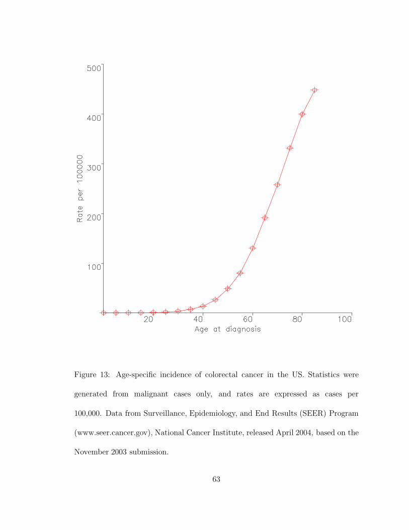
Figure 13: Age-specific incidence of colorectal cancer in the US. Statistics were
generated from malignant cases only, and rates are expressed as cases per
100,000. Data from Surveillance, Epidemiology, and End Results (SEER) Program
(www.seer.cancer.gov), National Cancer Institute, released April 2004, based on the
November 2003 submission.
63
of the gene is inactivated or lost. Class II CIN genes, such as hBUB1, trigger CIN if
one allele is mutated, and this mutation exerts an effect. Class III CIN genes, such
as BRCA2, trigger CIN only if both alleles are mutated.
The role of genetic instability in colorectal tumorigenesis is an active research
area of cancer biology4,5. Loeb was the first to propose that cancer cells acquire
a ’mutator phenotype’ early in tumor progression69,95. Several lines of evidence,
experimental and theoretical, indicate that genetic instability can indeed arise early
in colorectal tumorigenesis and subsequently drive tumor progression4. This view
is widely, however not universally accepted5,96.
Here, we address the following questions: How can the dynamics of colorectal
tumorigenesis be described with mathematical models? And what is the role of
genetic instability in tumor initiation?
4.2 Mutations in colorectal cancer cell lines
The genetic model of colorectal tumorigenesis (Figure 12) is based on decades of
investigation into the mutational sequence of colorectal tumorigenesis87. The frac-
tion of tumors with mutations in the APC pathway is approximately 95%; the same
number holds for the fraction of tumors with mutations in the p53 pathway. Somatic
mutations that lead to alteration of the Ras/Raf pathway have so far been found
in about 70% of tumors. There are additional genes with somatic mutations in
colorectal cancers, but their respective roles and pathways are less well understood
64

and examined.
Mutational studies require both tumor and normal tissue material for compar-
ison. The attainment of tumor material poses a significant technical difficulty, as
contamination with normal tissue renders signal and noise indistinguishable. Only a
few colorectal cancers and their corresponding normal tissue counterparts have been
analyzed systematically to determine the spectrum of specific mutations in a series
of genes, i.e. all APC pathway genes (APC, β-catenin, and axin), p53 pathway genes
(p53, MDM2, and BAX), and RAS pathway genes (K-Ras and B-Raf) in the same
tumors. Though such data are sparse, profound differences between the mutations
found in CIN and MIN tumors have been reported. At least a third of MIN can-
cers contain mutations in β-catenin instead of APC, whereas β-catenin mutations
are extremely rare in non-MIN cancers97. This is indirect evidence suggesting that
MIN arises before inactivation of the APC pathway. Additionally, the spectrum
of APC mutations that occur in most MIN cancers without β-catenin mutations is
different from that in non-MIN cancers98. In particular, there is a higher frequency
of alterations in simple repeat sequences in the MIN than in the non-MIN cancers.
65

4.3 Mathematical modeling
Mathematical modeling of colorectal tumorigenesis has provided insights into the
dynamics and statistics of the disease. Luebeck and Moolgavkar23 subdivided the
multi-step model into two stages; during the first stage, accumulation of neutral
mutations does not lead to any phenotypic changes; during the second phase, ad-
vantageous mutations drive clonal expansion of the colorectal tissue. By fitting the
age-specific incidence data of colorectal cancer, they found that two rare events (in-
activation of both APC alleles) are needed for tumor initiation and only one more
event is necessary for malignant transformation. Luebeck and Moolgavkar conclude
that genetic instability is not necessary for fitting the incidence curve of colorectal
cancer.
The difference between their approach and ours is that our model includes specific
genetic mutations and the effective population sizes of cells where these mutations
have to occur. We discuss possible parameter values for the rates of cell division in
various stages of tumor progression and the selective advantages or disadvantages
of mutated cells. We argue that age incidence curves of cancers can be fitted by
many different models with or without genetic instability. For example, we have
shown that two rate limiting hits for inactivation of a tumor suppressor gene can be
interpreted as two stochastic events inactivating each of the two alleles, or as one
stochastic event inactivating the first allele followed by a CIN mutation followed by
very fast inactivation (LOH) of the second allele51. Both evolutionary trajectories
66
have two rate limiting steps. Therefore, age incidence curves alone do not provide
enough information to decide whether genetic instability is early or not.
Herrero-Jimenez et al.99 provided a statistical analysis of colorectal cancer inci-
dence data accounting for demographic stochasticity, i.e. human heterogeneity for
inherited traits and environmental experiences. They fitted the two-stage initiation-
promotion model to the incidence data and calculated adenoma growth rates, the
number of initiating mutations and the rate of LOH, which was found to be sig-
nificantly higher than in normal cells. Pinsky100 adapted the multi-stage model to
the problem of development and growth of adenomas, both in sporadic and inher-
ited colorectal cancers. He fitted the model to adenoma data including adenoma
prevalence by age, the size distribution of adenomas, clustering of adenomas within
individuals and the correlation between proximal and distal adenomas.
Little and Wright60 extended the multi-stage model to incorporate genomic in-
stability and an arbitrary number of mutational stages. They found that a model
with five stages and two levels of genomic destabilization fits colon cancer incidence
data. Yatabe et al.47 investigated stem cells in the human colon by using methyla-
tion patterns and statistics. They found that colonic stem cells follow a stochastic
rather than deterministic model, i.e. each stem cell division produces two, one, or
zero daughter stem cells and zero, one, or two differentiated cells, respectively. This
leads to drift in the number of descendents of each stem cell lineage over time. Zahl
101 developed a proportional regression model for colon cancer in Norway. Mehl102
67
presented a mathematical computer simulation model to predict the natural history
of colon polyp and cancer development. He incorporated different cell types and
two kinetic processes, mutation and promotion, into his model.
Most mathematical analyses consist of fitting a stochastic model to colorectal
cancer incidence data. The number of steps is then extracted from the fitting instead
of from cell-biological and genetic assumptions of the necessary number of mutations,
rate constants and fitness values. Quantitative estimates, however, are essential to
correctly infer the mechanistic sequence of events. Population genetic studies have
shown that the effective number of steps observed from the probability distribution
of the time to cancer initiation can be smaller than the actual number2. This is
demonstrated by stochastic tunneling in which two mutations occur in one rate-
limiting process51,53. The effective step number also depends on the population size
and the possibility of somatic selection2,41.
Colorectal cancer incidence data can be explained by many alternative mod-
els. Most models are not easily falsifiable, as the assumptions of intermediate steps
and rate constants are not explicitly stated. The identification of the number of
rate-limiting steps in a particular model cannot justify the rejection of other inter-
pretations of the incidence data. The assumptions about intermediate steps and rate
constants must be identified to render mathematical models testable by experiments.
A careful discussion of parameter values and their likely ranges is needed for test-
ing hypotheses. Hence, the specification of intermediate steps and their parameter
68
values is essential for a full understanding of colorectal tumorigenesis.
Our group has developed a mathematical model of colorectal cancer that investi-
gates the role of genetic instability in tumorigenesis2,39,51,63,64. We have studied the
question whether a CIN mutation induces the first phenotypic change on the way to
colon cancer. We have shown in various models that this is indeed highly probably if
there are enough ’CIN genes’ in the human genome. We have also shown that tissue
organization tremendously influences the probability of emergence of genetic insta-
bility41. Furthermore, we have developed a specific model including cell-biological
and mechanical assumptions of a colonic crypt to describe colorectal tumorigenesis
44.
In the following, we offer a perspective of colorectal tumorigenesis including all
sequential mutations needed for tumor initiation and progression and determine the
importance of genetic instabilities in colorectal cancer.
4.4 The model
Here, we discuss the evolutionary dynamics of a model of colorectal tumorigenesis
including alterations of four genes (Figure 12). Inactivation of the tumor suppres-
sor gene APC initiates tumorigenesis. Subsequent tumor progression is driven by
activation of the oncogene RAS. Finally, inactivation of the tumor suppressor gene
p53 gives rise to a carcinoma. Somewhere CIN or MIN emerges.
We will analyze the radical hypothesis that CIN precedes inactivation of the
69

APC tumor suppressor gene. Here, we do not discuss MIN because it occurs only
in a minority of sporadic colorectal cancers and is rarely found in other cancers.
A theoretical investigation of MIN in sporadic and inherited colorectal cancer is
provided elsewhere64.
The mammalian colon is lined with a rapidly proliferating epithelium. This
epithelium is subdivided into 107 compartments of cells called crypts. Each crypt
contains about 1000 to 4000 cells. Approximately 1 - 10 of those cells are stem cells,
residing at the base of each crypt46,47. The progeny of stem cells migrate up the
crypt, continuing to divide until they reach its mid-portion. Then they stop dividing
and differentiate into mature cells. When the cells reach the top of the crypt, they
undergo apoptosis and are engulfed by stromal cells or shed into the gut lumen. The
cell migration from the base to the top of the crypt takes about 3 - 6 days103.
In this setting, two scenarios of cellular dynamics within a colonic crypt are
possible2,44. In the first scenario, only colorectal stem cells are considered to be at
risk of becoming cancer cells. Differentiated cells are assumed not to live long enough
to accumulate the necessary number of mutations that lead to cancer. Mutated
differentiated cells are washed out of the crypt by the continuous production and
migration of cells to the top of the crypt where they undergo apoptosis37 . In this
case, the effective population size of each colonic crypt is equal to the number of stem
cells. The stem cell compartment is assumed to be well-mixed, i.e. all stem cells
reside in equivalent positions and are in direct reproductive competition with each
70

other. There are no spatial effects. In the second scenario, all cells of the colonic
crypt are assumed to be at risk of accumulating mutations that lead to cancer44.
Mutations in specific genes might confer an increased probability to the cell to stick
on top of the crypt instead of undergoing apoptosis. Here, we use the first scenario
to discuss the evolutionary dynamics of mutations in APC, RAS, and p53. The
second scenario is outlined in the next chapter.
APC inactivation
We will now describe the dynamics of APC inactivation in a well-mixed compartment
of cells. Denote the number of stem cells by N0; stem cells divide every τ0 days.
Denote the probabilities of inactivating the first and second APC allele per cell
division by u1 and u2, respectively. We have u1 < u2 because there are more
possibilities, such as LOH and mitotic recombination, for the second hit. If the
mutation rate is less than the inverse of the population size, then the approximation
of homogeneous compartments holds: at any time, all cells of the compartment will
have 0, 1 or 2 inactivated alleles of APC. Denote the probabilities that all cells of the
compartment have 0, 1 or 2 inactivated alleles of APC at time t by X0, X1 and X2,
respectively (Figure 14a). The transition rate between states APC+/+ and APC+/−
is equal to the rate at which a mutation occurs in the first APC allele times the
probability that the mutant cell takes over the compartment. The mutation occurs
at rate N0u1/τ0, because (i) the mutation can arise in any one of N0 cells, (ii) the
rate at which the first APC allele is inactivated per cell division is u1, and (iii) the
71

cells divide once every τ0 days. The probability that the mutated cell takes over
the compartment (i.e., reaches fixation) is given by 1/N0, if we assume that the
mutation is neutral40. Thus, the compartment size, N0, cancels in the product, and
we obtain u1/τ0 as the transition rate between states APC+/+ and APC+/−. The
transition rate between states APC+/− and APC−/− is equal to the rate that a
mutation occurs in the second APC allele times the probability that the mutant cell
will take over the compartment. We assume that a cell with two inactivated APC
alleles has a large fitness advantage, which means the fixation probability of such
a cell is close to one. Therefore, we obtain N0u2/τ0 as the transition rate between
states APC+/− and APC−/− (Figure 14a). The differential equation describing the
stochastic process is outlined in the Appendix. The overall probability that APC
has been inactivated by time t is approximately given by X2(t) = N0u1u2t2/(2τ 2
0 ).
This probability accumulates as a second order of time, which means that it takes
two rate-limiting hits to inactivate APC in the compartment of cells2,10.
Chromosomal instability (CIN)
CIN could arise at any stage of tumorigenesis. Here we discuss the possibility
that CIN emerges before inactivation of APC (Figure 14b). The crucial effect of
CIN is to accelerate the inactivation rate of the second APC allele. Denote the
probability of inactivating the second APC allele per CIN cell division by u3; we
have u3 >> u2. This increased rate, however, might confer a selective cost to the
CIN cell, because such a cell has an increased probability of lethal mutations and
72

73
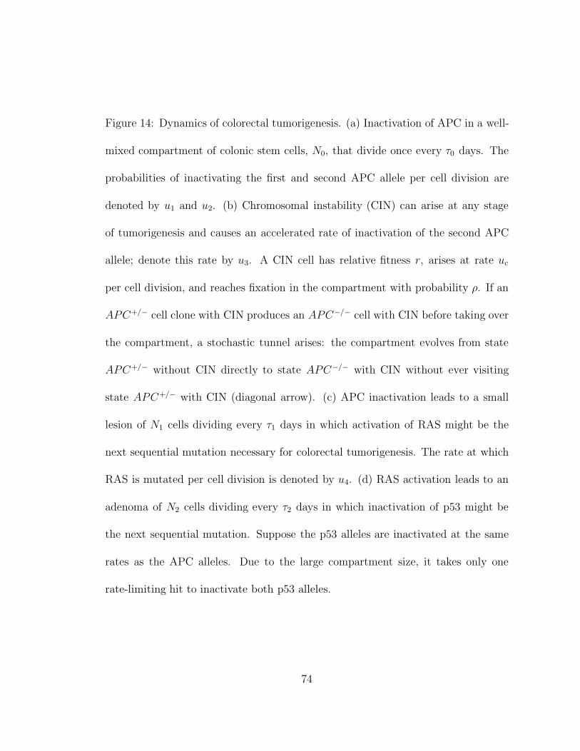
Figure 14: Dynamics of colorectal tumorigenesis. (a) Inactivation of APC in a well-
mixed compartment of colonic stem cells, N0, that divide once every τ0 days. The
probabilities of inactivating the first and second APC allele per cell division are
denoted by u1 and u2. (b) Chromosomal instability (CIN) can arise at any stage
of tumorigenesis and causes an accelerated rate of inactivation of the second APC
allele; denote this rate by u3. A CIN cell has relative fitness r, arises at rate uc
per cell division, and reaches fixation in the compartment with probability ρ. If an
APC+/− cell clone with CIN produces an APC−/− cell with CIN before taking over
the compartment, a stochastic tunnel arises: the compartment evolves from state
APC+/− without CIN directly to state APC−/− with CIN without ever visiting
state APC+/− with CIN (diagonal arrow). (c) APC inactivation leads to a small
lesion of N1 cells dividing every τ1 days in which activation of RAS might be the
next sequential mutation necessary for colorectal tumorigenesis. The rate at which
RAS is mutated per cell division is denoted by u4. (d) RAS activation leads to an
adenoma of N2 cells dividing every τ2 days in which inactivation of p53 might be
the next sequential mutation. Suppose the p53 alleles are inactivated at the same
rates as the APC alleles. Due to the large compartment size, it takes only one
rate-limiting hit to inactivate both p53 alleles.
74

apoptosis. Conversely, it is possible that a CIN cell has a faster cell cycling time
because of avoiding certain checkpoints. Denote the relative fitness of a CIN cell
by r. If r = 1, then CIN is neutral; if r < 1, it is disadvantageous, and a CIN
cell has a lower growth rate than a cell with stable karyotype; if r > 1, then it
is advantageous, and a CIN cell has a larger growth rate than a cell with stable
karyotype. Denote the probabilities that all cells of the compartment have CIN and
0, 1 or 2 inactivated APC alleles at time t by Y0(t), Y1(t) and Y2(t), respectively. The
transition rate between states APC+/+ without CIN and APC+/+ with CIN is the
same as the transition rate between states APC+/− without CIN and APC+/− with
CIN and equal to the rate at which a mutation triggering CIN occurs, N0uc/τ0, times
the probability that the mutant cell will take over the compartment. The fixation
probability of a CIN cell depends on its somatic fitness, r, and the compartment
size, N0, and is given by ρ = (1 − 1/r)/(1 − 1/rN0 ). This is the standard fixation
probability of a Moran process39. Therefore, the transition rate is given by N0ucρ/τ0.
If an APC+/− cell clone with CIN produces an APC−/− cell with CIN before tak-
ing over the compartment, a stochastic tunnel arises2,53: the compartment evolves
from state APC+/− without CIN directly to state APC−/− with CIN without ever
visiting state APC+/− with CIN. This tunnel occurs at rate N0ucru3/[(1−r)τ0] (Fig-
ure 14b). If CIN is neutral, r = 1, then the tunnel does not occur. The transition
rate between states APC+/+ with CIN and APC+/− with CIN is u1/τ0 as before,
because CIN is not assumed to cause increased point mutation rates. The transition
75

rate between states APC+/− with CIN and APC−/− with CIN is given by N0u3/τ0;
this rate is fast in the sense that N0u3/τ0 >> 1, and hence is not rate-limiting.
The overall probability that APC has been inactivated with CIN by time t is
given by Y2(t) = N0u1uc[2ρ + ru3/(1 − r)]t2/(2τ 20 ). This probability again accumu-
lates as a second order of time, so it also takes two rate-limiting hits to inactivate
APC in a compartment with CIN. Hence, Knudson’s two-hit hypothesis10 is com-
patible with tumor suppressor gene inactivation both without and with CIN2.
RAS activation
Inactivation of APC leads to clonal expansion of the colonic stem cell compartment
to a small lesion of N1 cells dividing every τ1 days. It is in this lesion that other
genes must be mutated for further tumor progression. The next genetic event in the
sequence might be activation of the oncogene RAS87.
Let us discuss the activation dynamics of RAS. Denote the probability that
a RAS mutation occurs per cell division by u4. Denote the probabilities that
a lesion consists only of APC−/−RAS+/− cells without and with CIN at time t
by X3(t) and Y3(t), respectively (Figure 14c). The transition rate between states
APC−/−RAS+/+ and APC−/−RAS+/− without CIN is given by N1u4/τ1, because
(i) the mutation can arise in any one of N1 cells, (ii) the rate at which the RAS
allele is mutated per cell division is u4, (iii) the cells divide once every τ1 days,
and (iv) a cell with a RAS mutation is assumed to have a large fitness advantage,
such that its fixation probability is close to 1. The transition rate between states
76
APC−/−RAS+/+ and APC−/−RAS+/− with CIN is the same as the transition rate
between states APC−/−RAS+/+ and APC−/−RAS+/− without CIN, because the
CIN phenotype does not alter the point mutation rate.
p53 inactivation
Activation of RAS leads to clonal expansion of the compartment to a late adenoma
of N2 cells dividing every τ2 days. In this adenoma, other genes must be mutated
for progression to a carcinoma. The next genetic event in the sequence might be
inactivation of the tumor suppressor gene p531.
We will now discuss the inactivation dynamics of p53 in the context of our model.
Denote the probability of inactivating the first p53 allele per cell division by u1 and
the probabilities of inactivating the second p53 allele per normal and CIN cell divi-
sion by u1 and u3, respectively. Again we have u3 >> u2 > u1. Here, we assume
that the p53 alleles are inactivated at the same rates as the APC alleles. Denote the
probabilities that a lesion consists only of APC−/−RAS+/−p53−/− cells without and
with CIN at time t by X5(t) and Y5(t), respectively (Figure 14d). The transition
rate between states APC−/−RAS+/−p53+/+ and APC−/−RAS+/−p53−/− without
CIN is N2u1√
u2/τ2, because (i) the mutation can arise in any of N2 cells, (ii) the
rate at which the two p53 alleles are inactivated per cell division is u1√
u2 due to
the increased compartment size53, and (iii) the cells divide once every τ2 days. The
transition rate between states APC−/−RAS+/−p53+/+ and APC−/−RAS+/−p53−/−
with CIN is N2u1√
u3/τ2, because the rate at which the two p53 alleles are inac-
77
tivated per CIN cell division is u1√
u3. The differential equation is given in the
Appendix and can be used to calculate the probability as a function of time to
develop a carcinoma. We can also determine the role of CIN in tumorigenesis. In
section 4.5, we will provide numerical examples of our model.
The cost of CIN
Once a CIN phenotype has reached fixation in a pre-cancer lesion, the question
remains how to quantify the further cost of CIN. Comparing two lesions, one without
CIN and one with CIN, we might be tempted to argue that the rate of evolution of
the CIN lesion has to be multiplied by a factor (given by a number between 0 and
1) which takes into account the increased death rate caused by CIN. Therefore, the
rate of evolution of the CIN lesion is reduced by a factor describing the cost of CIN.
On the other hand, it is conceivable that the increased rate of cell death in the
CIN lesion allows for an increased number of cell divisions (to make up for the cells
that have died) and therefore even accelerates the rate of evolution. A variety of
simple mathematical models can be constructed that demonstrate that the ‘cost of
CIN’ can be a net selective advantage when comparing the competition between
CIN and non-CIN lesions.
Since both scenarios are possible, here we assume that CIN lesions and non-CIN
lesions are neutral with respect to the cost of CIN during further tumor progression.
The cost of CIN is only considered when calculating the probability that a CIN cell
reaches fixation in the compartment where it first arises.
78
4.5 Numerical examples
The human colon consists of approximately Z = 107 crypts, each of which is replen-
ished by about 1-10 stem cells47,63. Let us choose N0 = 4. Later, we will discuss
numerical examples with N0 = 1, 4 and 10 stem cells. The mutation rate45 per
base per cell division is approximately 10−10. Suppose the APC tumor suppressor
gene can be inactivated by point mutations in any of about 500 bases104; hence the
rate of inactivating the first of two alleles is u1 = 2 × 500 × 10−10 = 10−7 per cell
division both in normal and CIN cells. The rate of inactivating the second allele per
cell division, u2, is the sum of the rate of LOH and the rate of a point mutation.
Assume the rate of inactivating the second allele is about u2 = 10−6 per cell division
in normal cells. The rate of inactivating the second allele in CIN cells82 has been
measured to be u3 = 10−2.
Assume clonal expansion of the stem cell compartment to N1 = 103 cells after
APC inactivation and to N2 = 105 cells after RAS activation. Suppose normal stem
cells divide once every τ0 = 10 days47. Assume that APC−/− cells divide every
τ1 = 5 days and APC−/−RAS+/− cells every τ2 = 1 day. The most substantial cost
CIN can possibly have is r = (1−u3)45 ≈ 0.6; this means that loss of any chromosome
other than the one containing the tumor suppressor gene locus is lethal.
(i) Assume there is only one class I CIN gene that triggers CIN when mutated;
then uc = 2u2 = 2 · 10−6, because either allele can be inactivated. The expected
number of carcinomas that were initiated by inactivation of APC in a t = 70 year-
79

old is 0.0014. The expected number of carcinomas that were initiated by CIN in a
t = 70 year-old is 0.0082 (see Appendix). Hence, 85% of carcinomas were initiated
by CIN. Among 1000 people, there are 9 colorectal cancer cases, 8 of which were
initiated by CIN. However, all of them might have CIN in the end.
(ii) Assume there is one class II CIN gene that triggers CIN when mutated; then
uc = 10−7. The expected number of carcinomas that were initiated by inactivation
of APC in a t = 70 year-old is 0.0014. The expected number of carcinomas that
were initiated by CIN is 0.0004 (see Appendix). Hence, 22% of carcinomas were
initiated by CIN.
(iii) Alternatively, we can calculate the minimum number of CIN genes in the
human genome needed to make sure CIN arises before the inactivation of APC.
Given the parameter values above, one class I CIN gene or four class II CIN genes
are necessary to ensure that CIN arises early in colorectal tumorigenesis. Note that
there are hundreds of CIN genes in yeast and even larger numbers are expected in
humans.
Figure 15 provides further examples of parameter values.
80

81%90%0.681%90%0.881%89%1.0
r 102n11n
62
410 10,10,1 NNN
33%50%0.647%64%0.854%70%1.0
r 102n11n
62
410 10,10,4 NNNa b
75%86%0.685%92%0.888%93%1.0
r 102n11n
52
310 10,10,4 NNN
25%40%0.659%74%0.874%85%1.0
r 102n11n
52
310 10,10,10 NNNc d
Figure 15: Percentage of carcinomas that were initiated by chromosomal instability
(CIN), if there is n1 = 1 class I CIN gene or n2 = 10 class II CIN genes in the human
genome. Class I CIN genes, n1, trigger CIN if one allele is mutated or lost. Class II
CIN genes, n2, trigger CIN if one allele is mutated in a dominant negative fashion.
The relative fitness of CIN cells is denoted by r. Parameter values are u1 = 10−7,
u2 = 10−6, u3 = 10−2, u4 = 10−9, τ0 = 10, τ1 = 5, τ2 = 1, t = 70 years, and (a)
N0 = 1, N1 = 104 and N2 = 106, (b) N0 = 4, N1 = 104 and N2 = 106, (c) N0 = 4,
N1 = 103 and N2 = 105, and (d) N0 = 10, N1 = 103 and N2 = 105.
81

4.6 Discussion
Here, we have discussed the mutational sequence that leads to colorectal cancer.
The traditional model of colorectal tumorigenesis87 describes several genetic changes
that are required for cancer initiation and progression (Figure 12). However, no large
scale sequencing of genes like APC or p53 has been done. An important goal of the
field should be detailed experimental analyses of the mutations found in colorectal
tumors.
We have provided a review of the literature of mathematical and statistical anal-
yses of colon cancer. Several excellent statistical studies have been performed to
analyze colorectal cancer incidence data. Stochastic multi-stage and population
genetic models have been developed and been fitted to incidence data. Although
this effort determined the number of stages needed for initiation and promotion of
colon cancer, the biological implications are unclear; biologically and clinically useful
investigation of colorectal tumorigenesis must include the exact specification of indi-
vidual steps. Experimental input of parameter values such as mutation rates, fitness
values and rate of population growth are required for a quantitative understanding
of cancer progression.
We have reviewed our mathematical approach to colon cancer by means of a
specific model of colorectal tumorigenesis. We have assumed that colorectal tumori-
genesis is driven by subsequent genetic alterations of APC, RAS, and p53. Naturally,
the model describes only a subset of cancers and does not attempt to provide a uni-
82

versally found sequence of mutations leading to colorectal cancer.
On the basis of this case study, we developed a mathematical representation of
the evolutionary dynamics of colorectal tumorigenesis. We specified the dependence
of the transition rates between stages on mutation rates, population sizes, fitness
values and cell division times. We introduced mutation of chromosomal instability
(CIN) genes that lead to an increased rate of loss of heterozygosity (LOH), thereby
accelerating the inactivation of tumor suppressor genes. We investigated the role of
CIN in initiating and driving tumor progression in dependence of the number of genes
that can initiate CIN when mutated in colorectal tissue. Given the experimental
input of rate constants and parameter values, we found that one or two CIN genes
in the genome are enough to make sure CIN emerges early. Therefore, it is very
likely that a CIN mutation initiates colorectal tumorigenesis.
There is the possibility of a second tumor suppressor gene that needs to be
genetically altered after inactivation of APC and before activation of RAS. In that
case and if both tumor suppressor genes are inactivated in rate-limiting situations,
i.e. if inactivation of APC alone does not lead to a large tumor, then chromosomal
instability it very likely to initiate the mutational sequence that leads to colorectal
cancer.
83
4.7 Appendix
The stochastic process illustrated in Figure 14 can be described by differential equa-
tions. Denote the probabilities that a compartment is in state APC+/+, APC+/−,
APC−/−, APC−/−RAS+/−, and APC−/−RAS+/−p53−/− without CIN by X0, X1,
X2, X3, and X5, respectively. Denote the probabilities that a compartment is in
the corresponding states with CIN by Y0, Y1, Y2, Y3, and Y5, respectively. The
differential equations are given by
X0 = −(a + b)X0 Y0 = bX0 − aY0
X1 = aX0 − (b + c + d)X1 Y1 = aY0 + bX1 − gY1 (5)
X2 = cX1 − eX2 Y2 = gY1 + dX1 − eY2
X3 = eX2 − fX3 Y3 = eY2 − hY3
X5 = fX3 Y5 = hY3
Here a = u1/τ0, b = N0ucρ(r), c = N0u2/τ0, d = N0ucru3/[(1 − r)τ0], e =
N1u4/τ1, f = N2u1√
u2/τ2, g = N0u3/τ0, and h = N2u1√
u3/τ2. See the main
text for an explanation of the parameter values. Equation 5 is a system of linear
differential equations which can be solved analytically using standard techniques.
The numerical examples contain numerical simulation of system 5.
84

5 Linear model of colon cancer initiation
Michor F, Iwasa Y, Rajagopalan H, Lengauer C, Nowak MA
Linear model of colon cancer initiation44
Cell Cycle 3, 358-362 (2004)
Cancer results if regulatory mechanisms of cell birth and death are dis-
rupted. Colorectal tumorigenesis is initiated by somatic or inherited mu-
tations in the APC tumor suppressor gene pathway. Several additional
genetic hits in other tumor suppressor genes and oncogenes drive the
progression from polyps to malignant, invasive cancer. The majority of
colorectal cancers present chromosomal instability, CIN, which is caused
by mutations in genes that are required to maintain chromosomal stabil-
ity. A major question in cancer genetics is whether CIN is an early event
and thus a driving force of tumor progression. We present a new mathe-
matical model of colon cancer initiation assuming a linear flow from stem
cells to differentiated cells to apoptosis. We study the consequences of
mutations in different cell types and calculate the conditions for CIN to
precede APC inactivation. We find that early emergence of CIN is very
likely in colorectal tumorigenesis.
85

5.1 Introduction
The mammalian colon is lined with a rapidly proliferating epithelium. This ep-
ithelium is organized into compartments of cells called crypts. Intestinal stem cells
reside at the base of each crypt46. The progeny of stem cells migrate up the crypt,
continuing to divide until they reach its mid-portion. Then they stop dividing and
differentiate to mature cells. When the cells reach the top of the crypt, they undergo
apoptosis and are engulfed by stromal cells or shed into the gut lumen. The cell
migration from the base to the top of the crypt takes about 3 - 6 days105. In normal
crypts, the rate of cellular death balances the rate of cellular proliferation. If this
homeostatic mechanism is shifted toward cellular growth, neoplasia results.
Colorectal tumors progress through four distinct clinical stages that describe
dysplastic crypts, small benign tumors, malignant tumors invading surrounding tis-
sues, and finally metastatic cancer. This progression involves several genetic changes
such as inactivation of tumor suppressor genes and activation of oncogenes87. Mu-
tations of the adenomatous polyposis coli (APC) gene are considered the earliest88
and most prevalent genetic changes in colorectal tumorigenesis. More than 95% of
colon cancers are estimated to have a somatic mutation of APC1. APC is a tumor
suppressor gene on chromosome 5q21. The APC gene product plays an important
role in modulating the Wnt/β-catenin signal transduction pathway that regulates,
among several other genes, the transcription of the oncogene c-Myc90,91. APC has
been shown to reduce net cellular proliferation through an increased rate of apopto-
86

sis106. Most APC mutations found in cancer cells lead to a truncation of the protein,
thereby preventing its regulating function. An inherited APC germ line mutation
leads to the development of hundreds to thousands of benign colorectal tumors,
some of which progress to malignancy (familial adenomatous polyposis, FAP)107.
Genetic instability is a defining characteristic of most human cancers. Two
forms of genetic instability have been identified: chromosomal instability, CIN, and
microsatellite instability, MIN54. About 85% of sporadic colorectal cancers and
most other solid tumors have CIN. CIN refers to an increased rate of losing or
gaining whole chromosomes or large parts of chromosomes during cell division. It
also increases the rate of loss of heterozygosity (LOH). LOH is thought to be an
important property of CIN, because tumor suppressor genes have to be inactivated
in both alleles. CIN accelerates the rate of inactivation of tumor suppressor genes.
The less prevalent form of genetic instability, MIN, is caused by alterations in
mismatch repair genes54. These alterations lead to an approximately 1000-fold in-
creased point mutation rate in MIN cancer cells. No increased rate of gross chromo-
somal changes is observed. MIN occurs in about 15% of sporadic colorectal cancers,
but seldom happens in other cancer types. CIN and MIN are generally mutually
exclusive.
A major question in cancer genetics is to what extent CIN, or any genetic insta-
bility, is an early event and thus a driving force of tumorigenesis2,23,39,41,51,59,60,64,79.
The precise timing of the emergence of CIN is difficult to measure experimentally,
87
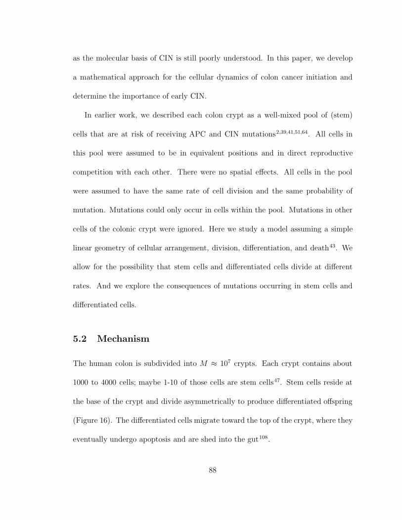
as the molecular basis of CIN is still poorly understood. In this paper, we develop
a mathematical approach for the cellular dynamics of colon cancer initiation and
determine the importance of early CIN.
In earlier work, we described each colon crypt as a well-mixed pool of (stem)
cells that are at risk of receiving APC and CIN mutations2,39,41,51,64. All cells in
this pool were assumed to be in equivalent positions and in direct reproductive
competition with each other. There were no spatial effects. All cells in the pool
were assumed to have the same rate of cell division and the same probability of
mutation. Mutations could only occur in cells within the pool. Mutations in other
cells of the colonic crypt were ignored. Here we study a model assuming a simple
linear geometry of cellular arrangement, division, differentiation, and death43. We
allow for the possibility that stem cells and differentiated cells divide at different
rates. And we explore the consequences of mutations occurring in stem cells and
differentiated cells.
5.2 Mechanism
The human colon is subdivided into M ≈ 107 crypts. Each crypt contains about
1000 to 4000 cells; maybe 1-10 of those cells are stem cells47. Stem cells reside at
the base of the crypt and divide asymmetrically to produce differentiated offspring
(Figure 16). The differentiated cells migrate toward the top of the crypt, where they
eventually undergo apoptosis and are shed into the gut108.
88

Figure 16: A colonic crypt. Colonic stem cells reside at the base of the crypt
and divide asymmetrically to produce differentiated progeny. The progeny migrate
toward the top of the crypt where they undergo apoptosis and are shed into the gut
lumen or engulfed by stromal cells.
89

In this setting, different scenarios of tumorigenesis are plausible (Figures 17
to 19). The ’traditional’ view of colorectal tumor initiation does not involve CIN
(Figure 17). Initially, the crypt is unmutated and contains only wild type cells,
APC+/+ without CIN. The inactivation of the first APC allele has to happen in
the stem cell in order to take over the whole crypt. If the mutation happens in a
differentiated cell, the mutated cell will be pushed to the top of the crypt by the
continuous production and migration of differentiated cells and will be ’washed out’
of the crypt12,14,37.
Once the mutation has happened in the stem cell, the stem cell produces mutated
progeny that eventually populates the whole crypt. The crypt moves from state
APC+/+ to state APC+/−. The inactivation of the remaining APC allele might take
place in any cell. The inactivation of both APC alleles confers a certain probability
to the cell to stick on top of the crypt instead of undergoing apoptosis. There are
two possibilities: (i) the cell fails to stick on top of the crypt, undergoes apoptosis,
and the crypt stays in state APC+/−, or (ii) the cell succeeds to stick on top of the
crypt and initiates dysplastic growth. We call this state APC−/−.
CIN can emerge before the inactivation of either APC allele. Figures 18 and 19
show two different mutational pathways in which the CIN mutation precedes the
inactivation of the second APC allele. The cell lineage is in state APC+/− without
CIN. Assume the mutation triggering CIN has to happen in the stem cell (Figure 18).
Then the mutated stem cell produces progeny that eventually populate the whole
90
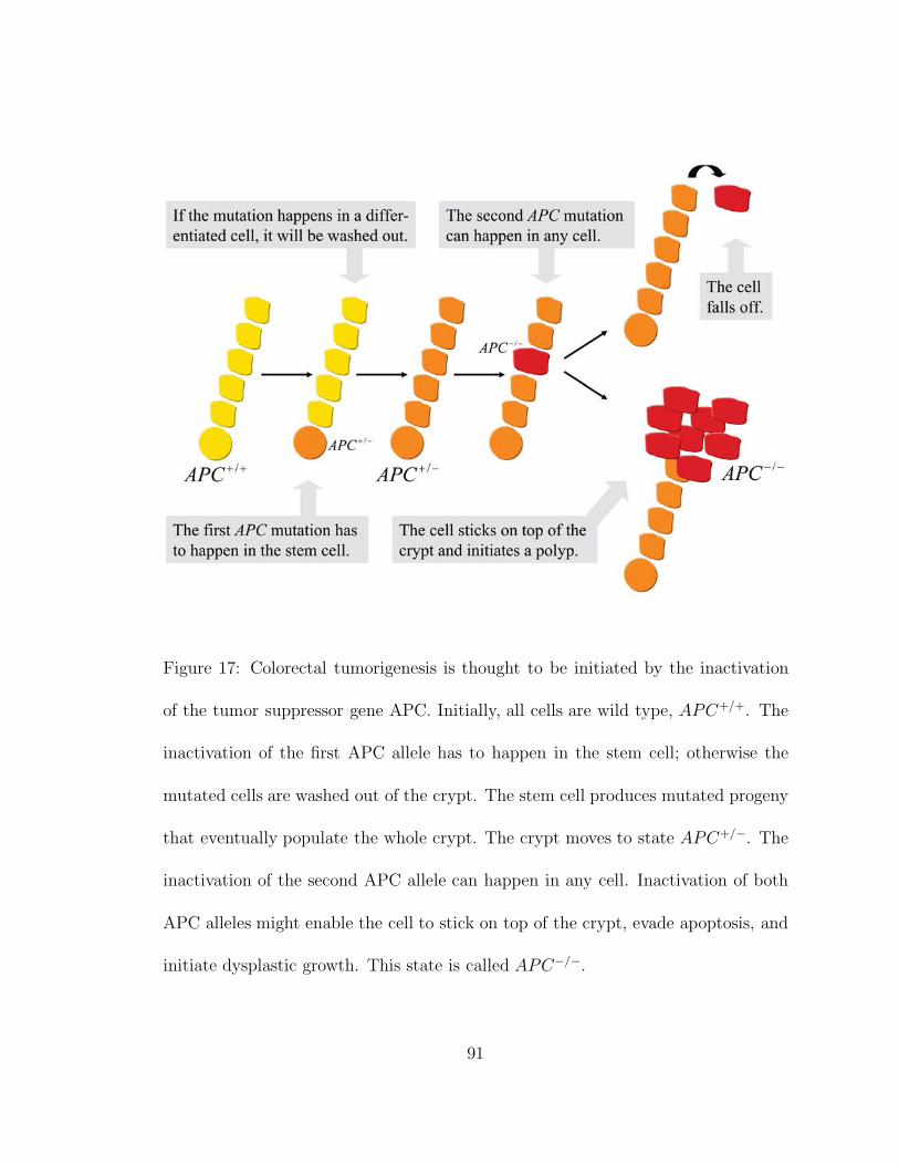
Figure 17: Colorectal tumorigenesis is thought to be initiated by the inactivation
of the tumor suppressor gene APC. Initially, all cells are wild type, APC+/+. The
inactivation of the first APC allele has to happen in the stem cell; otherwise the
mutated cells are washed out of the crypt. The stem cell produces mutated progeny
that eventually populate the whole crypt. The crypt moves to state APC+/−. The
inactivation of the second APC allele can happen in any cell. Inactivation of both
APC alleles might enable the cell to stick on top of the crypt, evade apoptosis, and
initiate dysplastic growth. This state is called APC−/−.
91

crypt, and the cell lineage moves to state APC+/− with CIN. The crucial effect of
CIN is to increase the rate of LOH, thereby greatly accelerating the inactivation of
the second APC allele. Thus, the inactivation of the second allele can happen in
any cell. If a cell with two inactivated alleles of APC succeeds to stick on top of the
crypt, dysplastic growth is initiated.
Assume the CIN mutation happens in a differentiated cell (Figure 19). The CIN
cell produces mutated progeny that populates only part of the crypt. The CIN
cells will eventually be washed out of the crypt, unless the second APC allele is
inactivated in a CIN cell on its way up the crypt. If this cell then succeeds to stick
on top of the crypt, it initiates dysplastic growth.
Figure 20 shows all pathways of how a crypt consisting of wild type cells can
mutate into a dysplastic crypt. A mathematical analysis of the model reveals the
transition rates between different mutational states and the probabilities of APC
inactivation without and with CIN. These probabilities specify the importance of
early CIN and determine the minimum number of CIN genes in the human genome
needed to ensure that CIN arises before the inactivation of APC.
92
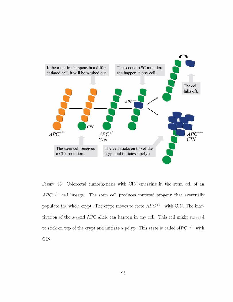
Figure 18: Colorectal tumorigenesis with CIN emerging in the stem cell of an
APC+/− cell lineage. The stem cell produces mutated progeny that eventually
populate the whole crypt. The crypt moves to state APC+/− with CIN. The inac-
tivation of the second APC allele can happen in any cell. This cell might succeed
to stick on top of the crypt and initiate a polyp. This state is called APC−/− with
CIN.
93

Figure 19: Colorectal tumorigenesis with CIN emerging in a differentiated cell of
an APC+/− cell lineage. The mutated cell produces progeny that populate part
of the crypt. The CIN cells will eventually be washed out of the crypt, unless the
inactivation of the second APC allele happens in a CIN cell while it moves up the
crypt. This cell might succeed to stick on top of the crypt and initiate a polyp. The
rest of the crypt is repopulated by APC+/− cells without CIN. This state is called
APC−/− partly with CIN.
94
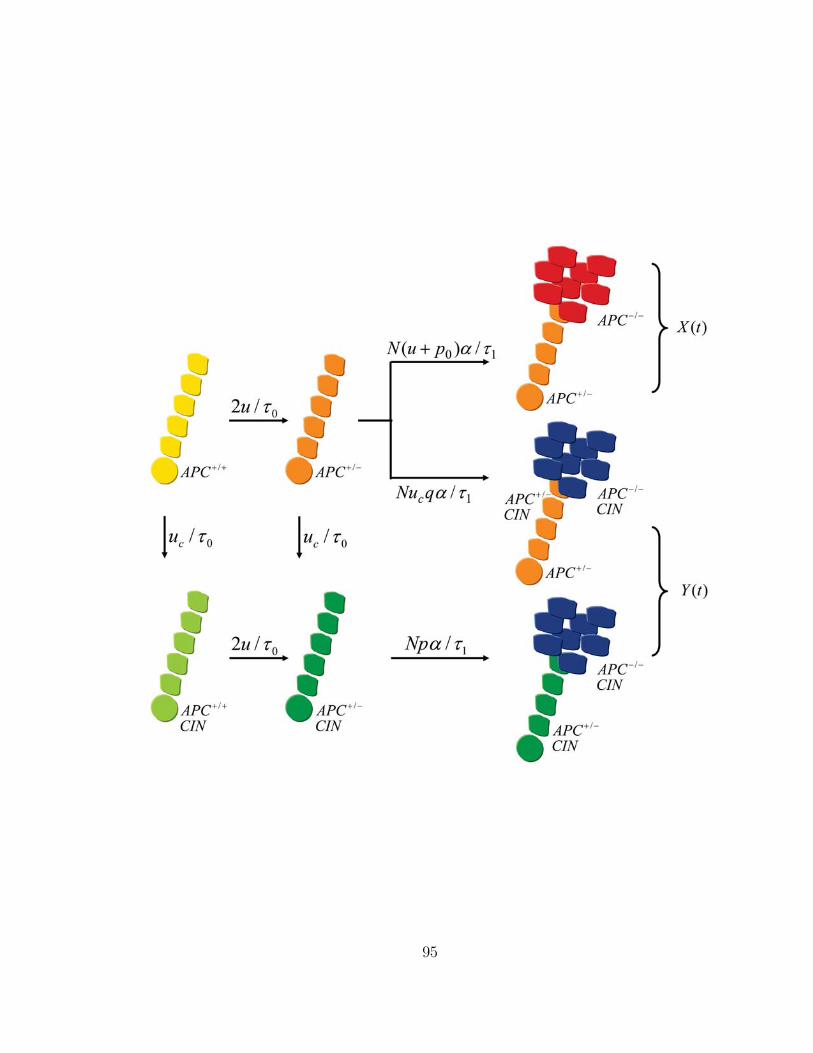
95
Figure 20: Pathways to dysplasia. Colorectal tumorigenesis might be initiated with-
out CIN. Then the crypt evolves from APC+/+ via APC+/− to APC−/− without
CIN. CIN can emerge before the inactivation of either APC allele. If CIN arises
in the stem cell, the crypt evolves from APC+/+ either via APC+/+ with CIN or
via APC+/− without CIN to APC+/− with CIN, and subsequently to APC−/−
with CIN. If CIN arises in a differentiated cell, the crypt evolves from APC+/+ via
APC+/− without CIN to APC−/− partly with CIN. The transition rates are out-
lined in the text and specify the probabilities of tumor initiation without and with
CIN.
96

5.3 Mathematical Analysis
The mathematical and statistical analysis of cancer progression using stochastic
processes has a long tradition11,18,23. Here we develop a new approach for studying
cancer initiation by describing mutational events and cellular reproduction in well-
defined populations of stem cells and differentiated cells.
The mutational network of colorectal tumorigenesis is illustrated in Figure 20.
First, consider tumor initiation without CIN. The inactivation of the first APC allele
has to happen in the stem cell. The crypt moves at rate 2u/τ0 from state APC+/+ to
state APC+/−: the stem cell divides every τ0 days; the mutation rate per APC allele
per cell division is denoted by u; and there are two APC alleles. The inactivation
of the second APC allele can happen either in the stem cell or in a differentiated
cell. The crypt moves at rate (u + p0)[1/τ0 + Nα/τ1] from state APC+/− to state
APC−/−: the rate of LOH is denoted by p0; there are N differentiated cells in the
crypt; they divide every τ1 days; and the probability that a cell with two inactivated
APC alleles sticks on top of the crypt and initiates dysplastic growth is denoted by
α. For cell-biological reasons, the second APC mutation might have to occur in the
stem cell; in that case, we can set α = 0. The probability that a crypt is dysplastic
without CIN at time t is given by
X(t) =2u
τ0
(u + p0
τ0+
N(u + p0)α
τ1
)t2
2(6)
CIN can emerge before the inactivation of either APC allele. Suppose the mu-
tation triggering CIN has to occur in the stem cell. Then the crypt moves at rate
97

uc/τ0 from state APC+/+ or APC+/− without CIN to state APC+/+ or APC+/−
with CIN: the probability that a CIN mutation arises per cell division is denoted by
uc. Both normal and CIN crypts move at rate 2u/τ0 from state APC+/+ to state
APC+/−. The inactivation of the second APC allele can happen in any cell, either
in the stem cell or in a differentiated cell. The crypt moves at rate p(1/τ0 + Nα/τ1)
from state APC+/− to state APC−/−: the rate of LOH in CIN cells is denoted by
p; we have p >> p0. This rate is fast in the sense that p(1/τ0 + Nα/τ1) >> 1. If
the second APC mutation has to occur in the stem cell, we again set α = 0. The
probability that a crypt is dysplastic and contains only CIN cells at time t is given
by Y1(t) = 2uuct2/τ 2
0 . This probability contains both the case in which the CIN
mutation precedes the first APC mutation and the case in which the CIN mutation
precedes the second APC mutation.
Suppose the mutation triggering CIN occurs in a differentiated cell. Then the
crypt moves at rate Nucqα/τ1 from state APC+/− without CIN to state APC−/−
partly with CIN: the expected probability that a CIN cell encounters an LOH event
on its way up the crypt is q = 1−[1−2n(1−p)n]/[(2n−1)(1−2(1−p))]; the maximum
number of cell divisions a differentiated cell undergoes before being shed off the top
of the crypt is denoted by n. If the cell with two inactivated APC alleles succeeds
to stick on top of the crypt, the APC+/− stem cell repopulates the remainder of the
crypt. The probability that a crypt is dysplastic and partly consists of CIN cells
at time t is given by Y2 = Nuucqαt2/(τ0τ1). The overall probability that a crypt is
98
dysplastic with CIN at time t is given by
Y (t) = Y1(t) + Y2(t) =2u
τ0
(2uc
τ0
+Nucqα
τ1
)t2
2(7)
CIN precedes the inactivation of APC if Y (t) > X(t). The inequality specifies
the minimum number of CIN genes needed to make sure CIN emerges before the
inactivation of APC. Suppose there are n1 class I genes that trigger CIN if one
allele is mutated or lost, and n2 class II genes that trigger CIN if one allele is
mutated; then the probability that a CIN mutation arises per cell division is given
by uc = 2n1(u + p0) + 2n2u. Either n1 > (Nατ0 + τ1)[2Nqατ0 + 4τ1]−1 class I CIN
genes or n2 > (u+p0)(Nατ0 +τ1)[2u(Nqατ0+2τ1]−1 class II CIN genes are sufficient
to make sure that CIN arises before the inactivation of APC.
5.4 Numerical Examples
The human colon contains about M = 107 crypts. Each crypt consists of approxi-
mately N = 1000 differentiated cells47. The mutation rate per APC allele per cell
division is u = 10−7: the mutation rate per base per cell division45 is about 10−10,
and we assume an APC allele can be inactivated by point mutations in approxi-
mately 1000 bases. The rate of LOH in normal cells, p0, might be of the order of u.
The rate of LOH in CIN cells82 is p = 0.01. Assume stem cells divide every τ0 = 10
days and differentiated cells divide every τ1 = 1 day. The maximum number of cell
divisions a colonic cell undergoes is n ≈ 10, because ten cell divisions are sufficient
to populate a crypt of about N = 1000 cells (Figure 21). Hence, the probability
99

that a CIN cell with one inactivated APC allele encounters an LOH event in the
second APC allele on its way up the crypt is q ≈ 0.01. Note that the probability
α that a cell with two inactivated APC alleles sticks on top of the crypt cannot be
too high, otherwise too many dysplastic crypts develop.
(i) Assume α = 0.001. Then a 70-year old has about 14 dysplastic crypts without
CIN and 5 dysplastic crypts with CIN if there is n1 = 1 class I CIN gene, or 3
dysplastic crypts with CIN if there is n2 = 1 class II CIN gene.
(ii) Assume α = 0.01. Then a 70-year old has about 132 dysplastic crypts
without CIN and 8 dysplastic crypts with CIN if there is n1 = 1 class I CIN gene,
or 4 dysplastic crypts with CIN if there is n2 = 1 class II CIN gene.
(iii) We can calculate the minimum number of CIN genes needed to ensure that
CIN emerges before the inactivation of APC. If α = 0.01, then n1 = 17 class I CIN
genes or n2 = 34 class II CIN genes are needed. If α = 0.01, then n1 = 3 class I
CIN genes or n1 = 6 class II CIN genes are needed. If α = 0, i.e. if the inactivation
of the second APC allele has to happen in the stem cell, then n1 = 1 class I CIN
gene or n2 = 1 class II CIN gene is needed to ensure that CIN emerges before the
inactivation of APC. Figure 22 shows the number of class I CIN genes, n1, needed
for a 10%, 50%, and 90% probability that CIN emerges before the inactivation of
APC in dependence of α.
100

Figure 21: Cell division dynamics in the colonic crypt. Ten cell divisions suffice to
populate the whole crypt of about 1000 differentiated cells. After having completed
ten cell divisions, differentiated cells cease to proliferate, are pushed to the top of
the crypt, and undergo apoptosis. The cell division dynamics specify the probability
that a differentiated CIN cell receives the second APC mutation while moving up
the crypt.
101

Figure 22: Number of class I CIN genes, n1, needed for a 10%, 50%, and 90%
probability that CIN emerges before the inactivation of APC, in dependence of the
probability α that a cell with two inactivated APC alleles, APC−/−, sticks on top of
the crypt and initiates dysplastic growth. Parameter values are N = 1000, τ0 = 10,
τ1 = 1, q = 0.01, and t = 70 years.
102
5.5 Discussion
In this paper, we develop a new mathematical approach for cellular dynamics, tissue
architecture, and differentiation patterns of colonic crypts. The model is based on
simple genetic and mechanical assumptions. A colonic stem cell gives rise to a lineage
of differentiated cells that move up the crypt and undergo apoptosis on top of the
crypt. Stem cells are assumed not to be subject to somatic selection; each stem
cell and its offspring can be described as an independent cell lineage. All of these
cells can receive mutations inactivating one or both alleles of the tumor suppressor
gene APC, which is thought to initiate tumorigenesis. The cells can also receive
mutations in genes that give rise to chromosomal instability, CIN. CIN might have
a cost as it increases the chance of lethal mutations and apoptosis. If CIN causes the
stem cell to divide more slowly, the stem cell still populates the crypt with mutated
progeny. If CIN causes the stem cell to undergo apoptosis, the crypt dies out and
might be replaced by a neighbor crypt.
We study the effect of various mutations that arise in stem cells or differentiated
cells. Some mutations, such as the inactivation of the first APC allele, have to
happen in the stem cell; otherwise the mutated cells are pushed to the top of the
crypt and ’washed out’ by the continuous production and migration of wild type
differentiated cells. Other mutations, such as the inactivation of the second APC
allele, can also happen in a differentiated cell. The inactivation of both APC alleles
enables the cell to stick on top of the crypt, evade apoptosis, and initiate dysplastic
103

growth.
A major question in cancer genetics is whether genetic instability is an early
event and thus a driving force of tumorigenesis. The present paper shows how to
calculate the rate of colon cancer initiation and the conditions for chromosomal
instability to precede the inactivation of APC. For plausible parameter values, we
obtain that a small number of CIN genes is sufficient to ensure that CIN emerges
before the inactivation of APC. CIN, however, accelerates the rate of inactivation
of every successive tumor suppressor gene needed for further tumor progression. If
colorectal tumorigenesis requires the inactivation of two or more tumor suppressor
genes in rate limiting situations, then even fewer CIN genes are needed to ensure
that CIN arises before the inactivation of the first tumor suppressor gene63. It is
therefore very likely that a CIN mutation initiates colorectal tumorigenesis.
104

6 Dynamics of chronic myeloid leukemia
Michor F, Hughes TP, Iwasa Y, Branford S,
Shah NP, Sawyers CL, Nowak MA
Dynamics of chronic myeloid leukemia109
Nature, in press (2005)
Chronic myeloid leukemia (CML) is associated with the oncogenic fusion
gene BCR-ABL. The clinical success of the ABL tyrosine kinase inhibitor
imatinib in CML serves as a model for molecularly targeted therapy of
cancer, but at least two critical questions remain. Can imatinib erad-
icate leukemic stem cells? What are the dynamics of relapse due to
imatinib resistance, which is caused by mutations in the ABL kinase do-
main? The precise understanding of how imatinib exerts its therapeutic
effect in CML and the ability to measure disease burden by quantita-
tive PCR provide an opportunity to develop a mathematical approach.
We find that a 4-compartment model, based on the known biology of
hematopoietic differentiation, can explain the kinetics of the molecular
response to imatinib in a 169 patient dataset. Successful therapy leads
to a bi-phasic exponential decline of leukemic cells. The first slope of
0.05 per day represents the turnover rate of differentiated leukemic cells,
105

while the second slope of 0.008 per day represents the turnover rate of
leukemic progenitors. The model suggests that imatinib is a potent in-
hibitor of the production of differentiated leukemic cells, but does not
deplete leukemic stem cells. We calculate the probability of developing
imatinib resistance mutations and estimate the time until detection of
resistance. Our model provides the first quantitative insights into the
in-vivo kinetics of a human cancer.
6.1 Introduction
Peripheral blood consists of terminally differentiated hematopoietic cells such as
erythrocytes, myelocytes, lymphocytes, and thrombocytes110. These cells have half
lives of hours (granulocytes) to months (erythrocytes) and thus need to be contin-
uously replenished. Their production is organized into the multiple differentiation
and expansion steps that make up the hierarchy of the hematopoietic system (Figure
23).
The most primitive cells, long-term hematopoietic stem cells (LT-HSCs), are re-
sponsible for life-long blood cell production111–113. LT-HSCs are (i) multipotential -
they give rise to all cells found in the blood114,115 and can also transdifferentiate to
produce non-hematopoietic tissues116 such as muscle cells117, and (ii) self-renewing
- during normal stem cell proliferation, lineage commitment and differentiation are
balanced by the production of additional stem cells, such that their total num-
106
ber remains constant118,119. Throughout adult life, the LT-HSC population resides
predominantly in the bone marrow and is associated with a highly organized mi-
croenvironment120–123. Low numbers of stem cells, however, are found migrating in
peripheral blood124.
LT-HSCs give rise to short-term HSCs (ST-HSCs), which give rise to multipotent
progenitors (MPPs)125. Dedifferentiation cannot be detected126. As HSCs mature
from the long-term self-renewing pool to multipotent progenitors, they progressively
lose their potential to self-renew, but become more mitotically active. In mice, it
has been showsn that LT-HSCs produce mature hematopoietic cells for the lifetime
of the mouse. ST-HSCs and MPPs, however, reconstitute lethally irradiated mice
for less than eight weeks126.
Common lymphoid progenitors (CLPs) and common myeloid progenitors (CMPs)
are oligolineage-restricted progeny of MPPs and precursors of all lymphoid and
myeloid cells, respectively. CMPs give rise to megakaryocyte/erythrocyte progeni-
tors (MEPs) or granulocyte/monocyte progenitors (GMPs). These cells eventually
produce all mature myelocytes found in the blood. CLPs give rise to further pre-
cursors which in turn produce all mature lymphoid cells.
Hematopoietic cell numbers are proportional to their differentiation stage. The
most primitive stem cells are the least abundant, and it has been estimated that only
one in 104 to 105 bone marrow cells is a stem cell127,128. Terminally differentiated
blood cells are the most abundant and account for 1012 cells in total.
107

108
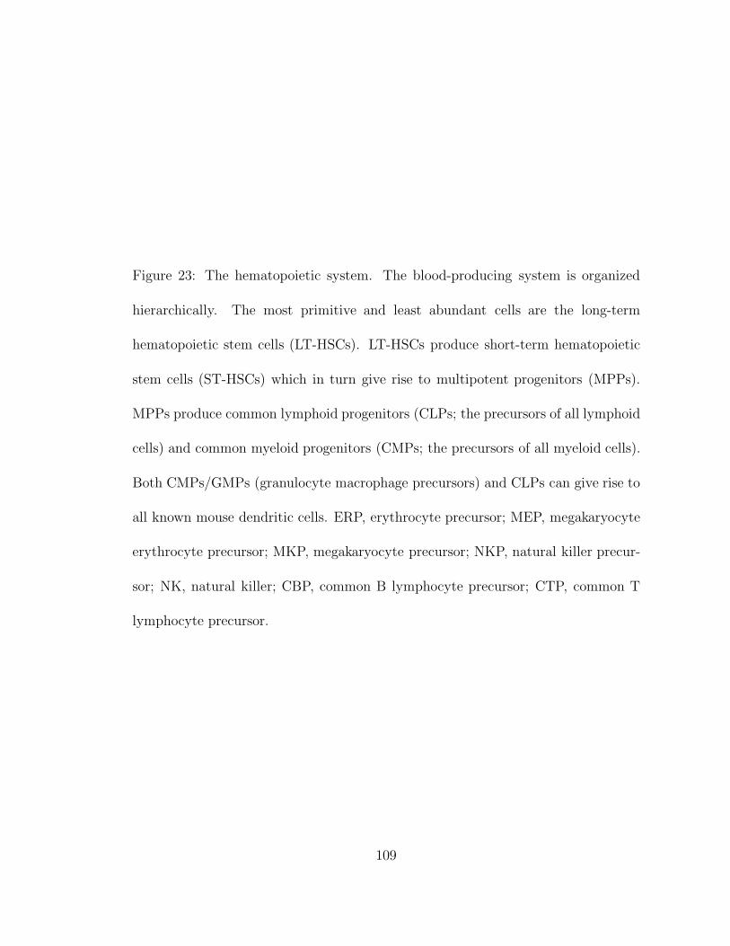
Figure 23: The hematopoietic system. The blood-producing system is organized
hierarchically. The most primitive and least abundant cells are the long-term
hematopoietic stem cells (LT-HSCs). LT-HSCs produce short-term hematopoietic
stem cells (ST-HSCs) which in turn give rise to multipotent progenitors (MPPs).
MPPs produce common lymphoid progenitors (CLPs; the precursors of all lymphoid
cells) and common myeloid progenitors (CMPs; the precursors of all myeloid cells).
Both CMPs/GMPs (granulocyte macrophage precursors) and CLPs can give rise to
all known mouse dendritic cells. ERP, erythrocyte precursor; MEP, megakaryocyte
erythrocyte precursor; MKP, megakaryocyte precursor; NKP, natural killer precur-
sor; NK, natural killer; CBP, common B lymphocyte precursor; CTP, common T
lymphocyte precursor.
109

Chronic myeloid leukemia (CML) is a malignant clonal disorder of hematopoietic
stem cells that leads to increased numbers of myelocytes, erythrocytes and thrombo-
cytes in peripheral blood129,130. The molecular hallmark of CML is the Philadelphia
(Ph) chromosome, first described as shortened chromosome 22 in 1960131 and then
as a t(9;22) translocation in 1973132. This abnormal chromosome is found in cells
from the myeloid, erythroid, megakaryocytic, and B lymphoid lineages, indicating
that CML is a stem cell disease130. The Ph chromosome is present in 95% of pa-
tients. The remaining 5% have complex or variant translocations that have the
same result: fusion of the BCR (breakpoint cluster region) gene on chromosome
22 to the ABL (Ableson leukemia virus) gene on chromosome 9 (Figure 24a). The
chimeric oncogene BCR-ABL encodes a constitutively active cytoplasmic tyrosine
kinase. This protein activates growth and differentiation pathways in hematopoietic
cells; its effectors include RAS, RAF, MYC, JUN, STAT and PI3K129. BCR-ABL
initiates a process to transform hematopoietic cells such that their growth and sur-
vival become independent of cytokines133. Transgenic expression of BCR-ABL in
mice causes acute leukemia at birth134.
CML is recognized when the BCR-ABL clone has expanded to an extent that
allows about 1012 leukemic cells to be produced daily. This occurs only after a
prolonged latent period - on average 6 to 7 years, as calculated from the atomic
bomb survivor data135. The disease then progresses from an initial chronic phase
via an accelerated phase to the final blast crisis within three to five years. During
110
chronic phase, leukemic myeloid progenitors, so-called blasts, increase in number to
constitute up to five percent of the total blood cell population. Progression of the
disease is marked by accelerated growth of immature myelocytes that are incapable
of differentiation; this progression might be due to further genetic alterations such as
duplication of the Ph chromosome or inactivation of tumor suppressor genes136–138.
During accelerated phase, blasts accumulate to abundances of five to thirty percent.
CML then progresses to the rapidly fatal blast crisis which often transforms into an
acute leukemia139.
Traditional treatment of CML includes killing of stem cells by chemotherapy and
subsequent bone marrow transplantation129. Due to age limitations of patients and
the lack of appropriate bone marrow donors, only 15 to 20% of CML patients are
candidates for transplantation. The survival rates of these patients are 50 to 60%
140. Patients not eligible for transplantations are treated with interferon alfa, alone
or in combination with other drugs, but this treatment fails to induce long-term
remissions in most patients.
The revolution in CML treatment came about with the discovery of the tyrosine
kinase inhibitor imatinib (STI-571, Gleevec)141. This compound was demonstrated
to be effective in all stages of the disease142. Therefore, CML represents the first
human cancer in which molecularly targeted therapy leads to a dramatic clinical
response142–148. Imatinib binds competitively with ATP to BCR-ABL and blocks
its abnormal signaling (Figure 24b). It selectively inhibits the proliferation of BCR-
111
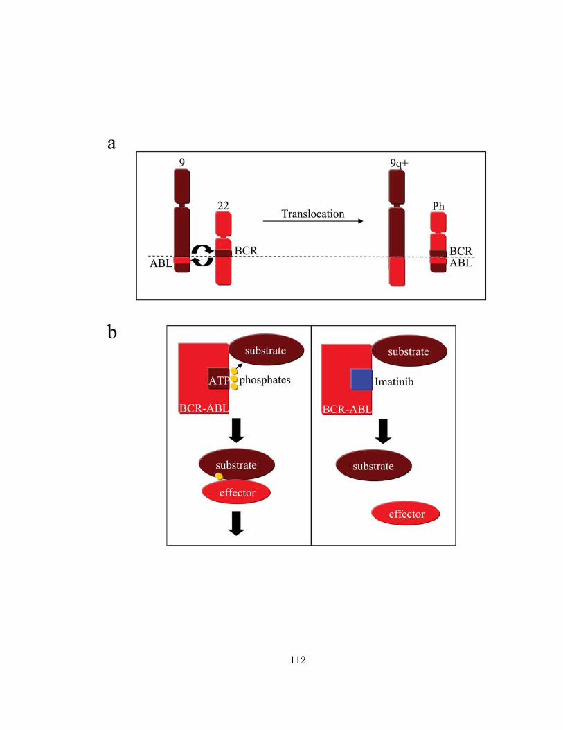
112

Figure 24: The Philadelphia chromosome and imatinib (Gleevec). (a) The Philadel-
phia chromosome results from a t(9;22) translocation that creates the BCR-ABL fu-
sion gene. The BCR-ABL gene encodes a constitutively active cytoplasmic tyrosine
kinase. This protein activates growth and differentiation pathways in hematopoi-
etic cells and causes chronic myeloid leukemia (CML). (b) Imatinib (Gleevec) binds
competitively with ATP to BCR-ABL and blocks its abnormal signaling.
113

ABL positive cell lines. In most patients, however, the rapid decline of the leukemic
cell burden induced by the ABL tyrosine kinase inhibitor imatinib fails to eliminate
residual disease. Bone marrow studies have shown that the residual cells are part
of the stem cell compartment149,150. This observation raises the question whether
imatinib is capable of impairing the proliferation of leukemic stem cells. Moreover, a
substantial fraction of patients develops acquired resistance to imatinib. Mutations
in the ABL kinase domain are the main mechanism of resistance and account for
70 - 80% of cases with treatment failure151–159 (Figure 25). Sometimes, resistance
mutations are present in leukemic cells prior to imatinib therapy159–161.
6.2 Results
We design a mathematical model which describes four layers of the differentiation
hierarchy of the hematopoietic system (see Section 6.3). Stem cells give rise to pro-
genitors, which produce differentiated cells, which produce terminally differentiated
cells. This hierarchy applies both to normal and leukemic cells. The BCR-ABL
oncogene is present in all leukemic cells. It leads to a slow clonal expansion of
leukemic stem cells and accelerates the rate at which these cells produce progenitors
and differentiated cells.
We analyze 169 CML patients. The levels of BCR-ABL transcripts in the blood
of the patients is measured by a quantitative real-time polymerase-chain-reaction
(RQ-PCR) assay154,162. BCR is used as the control gene and BCR-ABL values are
114

Figure 25: Resistance mutations. Acquired resistance to imatinib usually develops
due to single point mutations in the BCR-ABL kinase domain. Each point mutation
is sufficient to confer resistance to imatinib; the degree of resistance, however, differs
between the point mutations. About 40 different resistance mutations have been
discovered so far. In some patients, multiple resistance mutations were detected.
The labels denote the individual mutations that are detected; for example, E255K
means that at position 255 the amino acid glutamic acid was replaced by lysine. The
number of circles represent the number of patients in which the respective mutation
was found. Data from T. P. Hughes.
115

expressed as a percentage of the BCR transcript levels to compensate for variations
in the RNA quality and efficiency of reverse transcription. As the blood predomi-
nantly contains terminally differentiated cells, the obtained values give an estimate
of the fraction of terminally differentiated leukemic cells.
Successful therapy leads to a bi-phasic exponential decline of leukemic cells (Fig-
ure 26). The first slope is determined by calculating the exponential decline between
0 and 3 months; a mean value of 0.05± 0.02 per day is obtained, which corresponds
to a decline of 5% per day. The second slope is determined by calculating the ex-
ponential decline between 6 and 12 months; a mean value of 0.008 ± 0.004 per day
is obtained, which corresponds to a decline of 0.8% per day. This analysis considers
only patients who do not have any rise in the leukemic cell burden during the first
12 months of therapy to exclude the effect of acquired resistance.
Fitting our model to the data, we conclude that the first slope represents the
turnover rate of differentiated leukemic cells. Therefore, these cells have an average
life-span of 1/0.05 = 20 days. Upon reaching a steady-state with the leukemic
progenitors, the number of differentiated leukemic cells decreases at the same rate
as the number of leukemic progenitors. The second slope represents the turnover rate
of leukemic progenitors. Hence, these cells have an average life-span of 1/0.008 =
125 days. Both estimates denote average life-spans during imatinib therapy. Any
process that removes cells from the corresponding subpopulation (including further
differentiation) contributes to the average life-span.
116
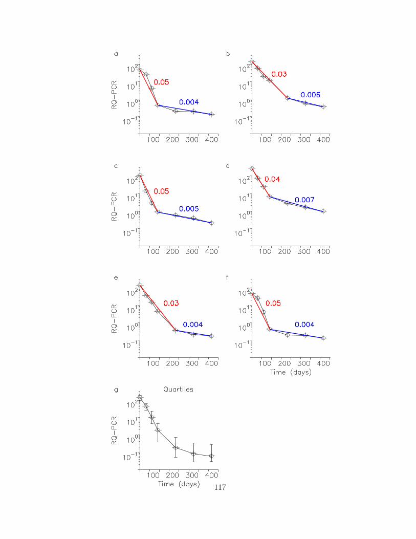
117

Figure 26: Imatinib leads to a bi-phasic decline of leukemic cells. (a-f) The levels
of BCR-ABL transcripts in the blood of 6 patients are shown during 12 months
of therapy starting at day 0. The BCR-ABL transcript levels are determined by
quantitative PCR (RQ-PCR) and are expressed as percentage of the values of BCR.
Every leukemic cell has one copy of the normal BCR gene and at least one copy
of BCR-ABL. The values might exceed 100% because BCR-ABL expression can be
upregulated in leukemic cells. In the 6 patients shown here the first slope ranges
from 0.03 to 0.05 per day. The second slope ranges from 0.004 to 0.007 per day. On
average, we find that the first slope is 0.05 ± 0.02 per day while the second slope
is 0.008 ± 0.004 per day. In the context of our model, the first slope represents the
death rate of leukemic differentiated cells and the second slope the death rate of
leukemic progenitors during imatinib therapy. On average, the first slope leads to
a 1000-fold decline in the leukemic cell burden. Panel (g) shows the median with
quartiles taken over all patients who do not have a rise in the leukemic cell burden
during the first 12 months of therapy. This approach attempts to exclude the effect
of acquired resistance.
118

The first slope leads to an approximately 1000-fold decline in the leukemic cell
burden. Therefore, imatinib reduces the rate at which leukemic differentiated cells
arise from leukemic progenitors about 1000-fold. This effect is as if imatinib pre-
vented about 10 rounds of cell division of leukemic cells (210 = 1024), either by
increasing their death rate or by reducing their division rate.
Some patients ceased imatinib therapy due to complications or side effects (Fig-
ure 27). Even if imatinib had been administered for many months (up to three
years), the numbers of BCR-ABL transcripts in those patients rose within 3 months
after discontinuation of therapy to levels at pre-treatment baseline or above. Other
studies of patients who ceased imatinib led to similar findings163,164. We conclude
that long-term imatinib treatment does not deplete the cell population that drives
this disease. This conclusion is consistent with the hypothesis that leukemic stem
cells are insensitive to chemotherapy165. The average rate of the exponential in-
crease after stop of treatment is 0.09 ± 0.05 per day, corresponding to a doubling
time of 8 days. This time scale indicates the rate at which terminally differentiated
leukemic cells arise from leukemic stem cells.
A comparison between model and data suggests the following two concepts:
(i) Imatinib treatment leads to a competitive disadvantage of leukemic progenitors
and differentiated cells; their production rates are dramatically reduced. The con-
sequence is a bi-phasic decline of the abundance of the BCR-ABL transcript in
response to therapy. (ii) Leukemic stem cells, however, are not depleted during
119

120
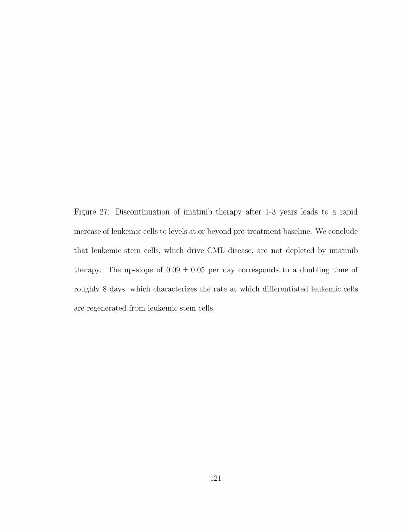
Figure 27: Discontinuation of imatinib therapy after 1-3 years leads to a rapid
increase of leukemic cells to levels at or beyond pre-treatment baseline. We conclude
that leukemic stem cells, which drive CML disease, are not depleted by imatinib
therapy. The up-slope of 0.09 ± 0.05 per day corresponds to a doubling time of
roughly 8 days, which characterizes the rate at which differentiated leukemic cells
are regenerated from leukemic stem cells.
121

imatinib treatment. The total leukemic cell burden rapidly returns to the baseline
value (or beyond) when imatinib is discontinued.
Acquired imatinib resistance usually develops due to mutations in the ABL ki-
nase domain151–159. Resistant leukemic cells emerge after an initially successful
response to imatinib therapy and lead to a relapse of disease (Figure 28). The av-
erage slope was determined by calculating the exponential increase after the first
appearance of resistance mutations in 30 patients; a value of 0.02±0.01 per day was
obtained. Of those patients who start imatinib in the early chronic, late chronic,
and accelerated phase of CML, respectively, 12%, 32%, and 62% develop detectable
resistance mutations within two years of treatment166 (Figure 30a).
Our basic model can be extended to include the stochastic evolution of resistance.
Consider an exponentially growing leukemia continually producing resistant cells at
rate u per cell division. The probability of having resistance mutations once leukemic
stem cells have reached a certain abundance, y0, is given by P = 1 − exp(−uy0σ)
where σ = (1 + s) log(1 + 1/s). The parameter s denotes the excess reproductive
ratio of the exponentially growing leukemia, which is the relative difference between
birth and death rates, and is given by s = (ry − d0)/d0. This calculation assumes
that resistance mutations are neutral prior to therapy.
If the point mutation rate is about 10−8 per base per cell division45, then resis-
tance due to any one of about forty known mutations150–160 arises at rate u = 4·10−7
per cell division. The abundance of leukemic stem cells at diagnosis in early chronic
122

123

Figure 28: About 40 different point mutations in BCR-ABL have been identified
that confer various degrees of resistance to imatinib therapy. We show the evolution
of resistance in six patients. The labels denote the individual mutations that are
detected at various time points. For example, E255K means that at position 255
the amino acid glutamic acid was replaced by lysine. Resistance mutations lead to
a relapse of leukemic cells. The up-slope ranges from 0.003 to 0.06 per day in 30
patients. The average up-slope is 0.02 ± 0.01 per day. Our mathematical analysis
suggests that the characteristic time scale for the rise of resistance is given by the
rate at which resistant leukemic stem cells expand during therapy. Therefore, more
aggressive leukemias should lead to a faster emergence of resistance, while slowly
growing leukemias should allow many years of successful therapy even if resistance
mutations are present in a patient at low frequencies.
124
phase167–169 is estimated to be about y0 = 2.5 · 105. For these values and s = 1, we
obtain that about 13% of patients harbor resistance mutations at the time of diag-
nosis. If imatinib therapy commences at a later stage in disease, when the leukemic
stem cell population has expanded to 107 cells, for example, then 100% of those
patients have some resistant leukemic stem cells. Therefore, the higher incidence of
resistance in patients who start imatinib therapy in a later phase of disease can be
explained by an increased leukemic cell burden (Figure 30b).
We expect that the frequency of resistance mutations at the start of imatinib
therapy is below detection limit in most patients. We can use the deterministic
model to calculate the time until detection of resistance mutations and treatment
failure (Figure 30c). In our model, the characteristic time scale is given by the rate
at which the resistant cancer stem cells are expanding during therapy. Therefore, a
faster growing leukemia leads to an earlier emergence of resistance, while a slower
growing leukemia might allow many years of successful therapy, even if resistant
cancer cells are present at low frequencies. The reason for this unusual behavior is
that leukemic stem cells, with or without resistance mutations, continue to expand
during treatment. Imatinib primarily acts by reducing the rate of differentiation of
those leukemic cells that do not have resistance mutations.
Figure 29 summarizes the dynamical features of our mathematical model. Nor-
mal hematopoietic cells are in a steady state. Leukemic stem cells expand exponen-
tially at a slow rate. Imatinib reduces the rate at which leukemic stem cells produce
125

126
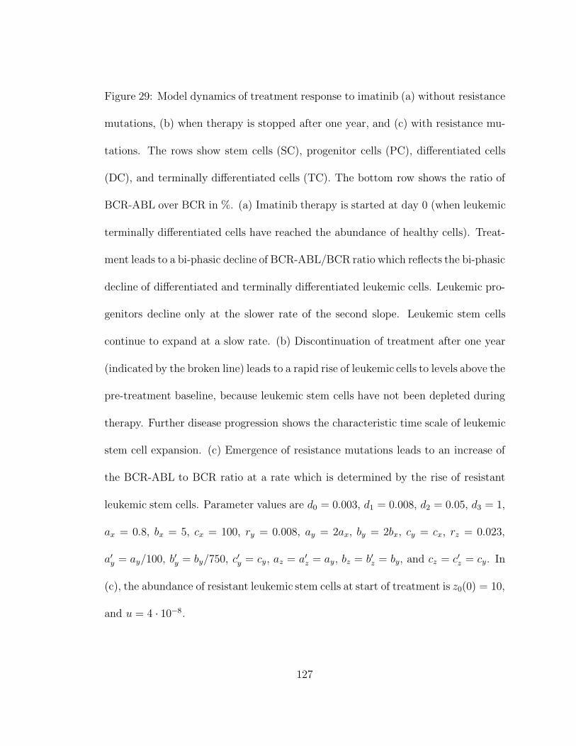
Figure 29: Model dynamics of treatment response to imatinib (a) without resistance
mutations, (b) when therapy is stopped after one year, and (c) with resistance mu-
tations. The rows show stem cells (SC), progenitor cells (PC), differentiated cells
(DC), and terminally differentiated cells (TC). The bottom row shows the ratio of
BCR-ABL over BCR in %. (a) Imatinib therapy is started at day 0 (when leukemic
terminally differentiated cells have reached the abundance of healthy cells). Treat-
ment leads to a bi-phasic decline of BCR-ABL/BCR ratio which reflects the bi-phasic
decline of differentiated and terminally differentiated leukemic cells. Leukemic pro-
genitors decline only at the slower rate of the second slope. Leukemic stem cells
continue to expand at a slow rate. (b) Discontinuation of treatment after one year
(indicated by the broken line) leads to a rapid rise of leukemic cells to levels above the
pre-treatment baseline, because leukemic stem cells have not been depleted during
therapy. Further disease progression shows the characteristic time scale of leukemic
stem cell expansion. (c) Emergence of resistance mutations leads to an increase of
the BCR-ABL to BCR ratio at a rate which is determined by the rise of resistant
leukemic stem cells. Parameter values are d0 = 0.003, d1 = 0.008, d2 = 0.05, d3 = 1,
ax = 0.8, bx = 5, cx = 100, ry = 0.008, ay = 2ax, by = 2bx, cy = cx, rz = 0.023,
a′y = ay/100, b′y = by/750, c′y = cy, az = a′
z = ay, bz = b′z = by, and cz = c′z = cy. In
(c), the abundance of resistant leukemic stem cells at start of treatment is z0(0) = 10,
and u = 4 · 10−8.
127

a
b
c
62%32%12%2 years38%14%5.9%1 year
acceleratedphase
late chronic phase
early chronic phase
43%5.4%0.6%
100%43%5.4%u 0y 510 610 710
71048104
810
100%
100%
119 d9.2 d100 000241 d61 d10 000357 d101 d1000470 d141 d100566 d186 d10
failuredetection)0(0z
128
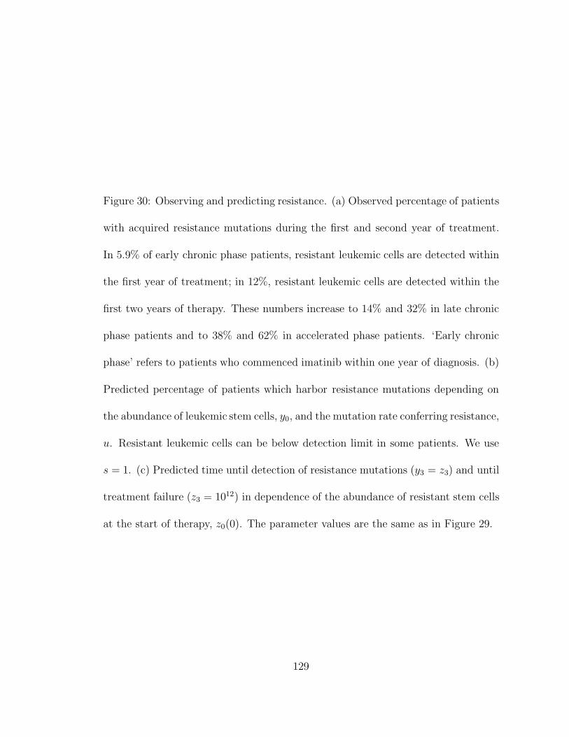
Figure 30: Observing and predicting resistance. (a) Observed percentage of patients
with acquired resistance mutations during the first and second year of treatment.
In 5.9% of early chronic phase patients, resistant leukemic cells are detected within
the first year of treatment; in 12%, resistant leukemic cells are detected within the
first two years of therapy. These numbers increase to 14% and 32% in late chronic
phase patients and to 38% and 62% in accelerated phase patients. ‘Early chronic
phase’ refers to patients who commenced imatinib within one year of diagnosis. (b)
Predicted percentage of patients which harbor resistance mutations depending on
the abundance of leukemic stem cells, y0, and the mutation rate conferring resistance,
u. Resistant leukemic cells can be below detection limit in some patients. We use
s = 1. (c) Predicted time until detection of resistance mutations (y3 = z3) and until
treatment failure (z3 = 1012) in dependence of the abundance of resistant stem cells
at the start of therapy, z0(0). The parameter values are the same as in Figure 29.
129

progenitors. Hence, the abundance of leukemic progenitors declines once treatment
is started. Similarly, imatinib reduces the rate at which leukemic progenitors pro-
duce differentiated cells. The abundance of differentiated leukemic cells shows a
bi-phasic decline. The first slope is determined by the average life-span of leukemic
differentiated cells (20 days), while the second slope reflects the longer life-span of
leukemic progenitors (125 days). We assume that imatinib does not affect the rate
at which leukemic differentiated cells produce terminally differentiated cells. This
cell population has a fast turnover rate - on a time scale of one day, and simply
tracks the biphasic decline of leukemic differentiated cells. If imatinib is discontin-
ued, there is a rapid resurgence of the leukemic load because the cell population
which drives the disease, the leukemic stem cells, was not depleted during therapy.
Resistant leukemic stem cells might expand faster than leukemic stem cells during
therapy for two reasons: (i) either they have an inherent selective advantage; or (ii)
imatinib somewhat reduces the growth rate of leukemic stem cells without depleting
them. In any case, resistant leukemic stem cells continue to produce large amounts
of progenitors and differentiated cells during therapy. The total leukemic cell burden
declines initially, but rises again once resistant differentiated cells become abundant.
In this case, the time to treatment failure is determined by the rate of expansion of
resistant leukemic stem cells.
In summary, the comparison between model and data provides quantitative in-
sights into the in-vivo kinetics of CML. We obtain numerical estimates for the
130

turnover rates of leukemic progenitors and differentiated cells. Imatinib reduces
the rate at which those cells are being produced from stem cells, but does not lead
to an observable decline of leukemic stem cells. The probability of harboring resis-
tance mutations increases with disease progression as a consequence of an increased
leukemic stem cell abundance. The characteristic time to treatment failure caused
by acquired resistance is given by the growth rate of the leukemia. Thus, mul-
tiple drug therapy170 is especially important for patients who are diagnosed with
advanced and rapidly growing disease.
6.3 Methods
The basic model. Our mathematical model is based on the architecture of the
hematopoietic system as proposed by Weissman112,114. Denote by x0, x1, x2, and x3
the abundances of normal hematopoietic stem cells, progenitors, differentiated cells,
and terminally differentiated cells. Their respective leukemic abundances are given
by y0, y1, y2, and y3 for cells without resistance mutations and by z0, z1, z2, and z3
for cells with imatinib resistance mutations. We assume that normal hematopoietic
stem cells are held at a constant level by a homeostatic mechanism. The growing
leukemic cell population might eventually upset this homeostasis, but we do not
consider this effect here. Leukemic stem cells escape from the homeostatic control
and grow at a slow pace; it takes about 6-7 years from the first occurence of the
BCR-ABL oncogene to detection of disease135. If imatinib resistance mutations are
131
neutral in the absence of treatment, then leukemic stem cells with these mutations
grow at the same rate as the other leukemic stem cells. It is conceivable that
some resistance mutations confer a selective advantage or disadvantage leading to
different expansion rates of resistant clones. We assume, for simplicity, that the
leukemia follows an exponential growth law; other dynamics are possible and need
separate investigation.
Stem cells produce progenitors which produce differentiated cells which pro-
duce terminally differentiated cells. The rate constants are given by a, b, and c
with subscripts x, y and z distinguishing between healthy, leukemic, and resistant
cell lineages. The current understanding is that the BCR-ABL oncogene increases
the rate at which leukemic stem cells proliferate and differentiate into progenitors.
Furthermore, the BCR-ABL oncogene is thought to increase the rate at which pro-
genitors proliferate and turn into differentiated leukemic cells. Therefore, we expect
that ay > ax and by > bx. Imatinib counteracts this effect and reduces the rates to
a′y < ay and b′y < by. The death rates of stem cells, progenitors, differentiated, and
terminally differentiated cells are denoted by d0, d1, d2, and d3. The ‘death’ rates
can include further differentiation of some cell types. Progenitors, differentiated,
and terminally differentiated cells have a limited potential for cell division. They
divide a certain number of times before they differentiate further or undergo apop-
tosis. The actual dynamics of this process is more complicated than described in
our simple model, but the essential features are captured: we assume that a stem
132

cell leads to a progenitor lineage that divides i times thereby generating 2i cells.
The number 2i is embedded in the rate constant a. Equally, a progenitor leads to a
differentiated cell that generates 2j cells, and this number is embedded in the rate
constant b. Imatinib reduces the rate constants a and b, thereby preventing a certain
number of cell divisions in each stage.
Homeostasis of normal stem cells is achieved by an appropriate declining func-
tion, λ. Leukemic stem cells divide at rate ry and give rise to resistant stem cells at
rate u per cell division. Resistant stem cells divide at rate rz. With these assump-
tions, we have the following system of differential equations:
x0 = [λ(x0) − d0]x0 y0 = [ry(1 − u) − d0]y0 z0 = (rz − d0)z0 + ryy0u
x1 = axx0 − d1x1 y1 = ayy0 − d1y1 z1 = azz0 − d1z1 (8)
x2 = bxx1 − d2x2 y2 = byy1 − d2y2 z2 = bzz1 − d2z2
x3 = cxx2 − d3x3 y3 = cyy2 − d3y3 z3 = czz2 − d3z3
We assume that normal cells remain at their equilibrium values, x0, x1 = axx0/d1,
x2 = bxx1/d2, and x3 = cxx2/d3. At the start of therapy, leukemic cells are in steady
state ratios with their precursors: y3 = cyy2/d3, y2 = byy1/d2, and y1 = ayy0/d1. The
leukemic stem cell population expands as y0(t) = exp[(ry−d0)t] (ignoring resistance
mutations). Imatinib dramatically reduces the rate constants, ay to a′y and by to b′y.
This leads to a bi-phasic decline. The first slope describes the exponential decline of
differentiated leukemic cells to their new steady state, y2 = b′yy1/d2. The magnitude
of this decline is around 1000-fold, suggesting that b′y is 1000 times smaller than by.
133
Thus, imatinib prevents about 10 rounds of cell division of differentiated leukemic
cells. Note that 210 = 1024. The exponential decline is given by exp(−d2t). The
observed slope establishes that the turnover rate of differentiated leukemic cells
in the presence of imatinib is d2 = 0.05 per day. Hence the average life-time of
these cells is about 1/d2 = 20 days. After about 200 days of therapy, differentiated
leukemic cells have reached their new steady state with leukemic progenitors and
follow their decline that is given by exp(−d1t). The data suggest that the turnover
rate of leukemic progenitors is d1 = 0.008 per day corresponding to an average
life-time of about 1/d1 = 125 days. A minimum estimate of the magnitude of this
decline is about 7-fold, suggesting that a′y is at the very least 7 times smaller than
ay. The overall 5000-fold decline is the combined effect of a′y and b′y. Figure 31
shows the molecular response to imatinib in all patients whose leukemic cell load
was used to calculate the bi-phasic decline.
We have assumed that imatinib primarily acts by reducing the proliferation rate
of leukemic cells, but it is in principle possible that imatinib also increases the death
rates of those cells. In this case, we have to assign different death rates to the x,
y and z populations, but all our conclusions remain the same; the numerical esti-
mates for the decay slopes refer to the death rates of the corresponding cells during
imatinib therapy. ‘Death’ includes any process that removes cells from the rele-
vant subpopulation and does include cellular differentiation. It is also conceivable
that the BCR-ABL oncogene changes the death rates of leukemic cells. In this case
134

135

136

137

Figure 31: Molecular response to imatinib. The figure shows the leukemic cell load
in all patients used to calculate the bi-phasic decline. This analysis excludes patients
who had a rise in their leukemic burden during the first twelve months of therapy.
This approach attempts to exclude the effect of acquired resistance mutations.
138
again, the observed slopes indicate the turnover rates of leukemic cells during ima-
tinib therapy. Should BCR-ABL and imatinib, however, not affect the death rates
of these cells, then our results apply to the turnover rates of healthy hematopoietic
cells, too.
If therapy is interrupted, the model predicts an explosive recurrence of cancer
cells. Since imatinib leads to an at least 5000-fold reduction of the production of
differentiated leukemic cells, stopping therapy leads to a sudden 5000-fold increase
of differentiated leukemic cells at the time scale of their cell division. If imatinib
led to a decline in leukemic stem cells, then the rebound after therapy should be
to a level below baseline (by whatever amount leukemic stem cells have declined
during therapy). If imatinib does not lead to a decline of leukemic stem cells, the
rebound should lead to baseline levels or beyond. The latter is observed in all three
patients who stopped therapy (Figure 27). Once terminally differentiated leukemic
cells reach their new steady state, the further increase of the disease burden follows
the characteristic time scale of leukemic stem cell expansion.
For comparison with the experimental PCR data, we calculate the BCR-ABL to
BCR ratio as (y3 + z3)/(2x3 + y3 + z3) times 100%. A healthy cell has two copies of
BCR. A leukemic cell normally has one copy of BCR and one copy of BCR-ABL.
Most cells that are sampled by the PCR assay are terminally differentiated cells.
Imatinib and stem cells. Finding a cellular mechanism for drug resistance of
stem cells is a very important goal for future experimental research. Imatinib is a
139

substrate for the multidrug resistance protein MDR p-glycoprotein and will therefore
be excluded from cells that express significant MDR levels171. Stem cells naturally
express higher levels of MDR172. It is unknown if this is the actual mechanism for
sparing stem cells, because it has not been possible to accurately measure imatinib
concentration in stem cells nor to measure BCR-ABL inhibition selectively in this
compartment. Another possibility is that leukemic stem cells are less dependent on
BCR-ABL for growth and survival than committed progenitors are, and therefore
BCR-ABL inhibition does not eliminate leukemic stem cells.
Oncogene addiction. There is a fundamental unanswered question at the heart
of molecularly targeted cancer therapy: if drugs impair the action of an oncogene
and thereby reduce the growth of tumor cells, then why do these tumor cells have
a competitive disadvantage relative to normal cells? The term ‘oncogene addiction’
has been coined to describe the phenomenon of tumor cells becoming dependent on
a single molecular driver, but its cellular basis is unknown173. Most hypotheses are
focused on the notion that tumor cells lose the redundant signaling machinery that
normal cells retain, but this remains to be proven.
The probability of resistance mutations. We use a continuous-time branch-
ing process to calculate the probability that a patient has resistance mutations at
the beginning of imatinib therapy. Assume that resistance mutations are neutral
prior to therapy: this means mutated and unmutated stem cells expand at the same
rate in the absence of imatinib. Leukemic stem cells, y0, follow a branching process
140
starting with a single cell at time t = 0. They go extinct with probability d0/ay and
grow exponentially to give rise to leukemia with probability 1 − d0/ay. Resistant
mutants are produced from normal leukemic cells with probability u per cell divi-
sion. They reproduce and die at the same rate as normal leukemic cells. For the
probability of having resistance mutations once the stem cell population has reached
a certain size, y0, we obtain P = 1 − exp(−uy0σ). Here σ = (1 + s) log(1 + 1/s),
where s = (ay − d0)/d0 denotes the excess reproductive ratio of leukemic stem cells.
For a wide range of plausible values of s, there is only little variation in σ: if s
changes from 0.1 to infinity, then σ changes from 2.64 to 1.
We expect that patients who are diagnosed at later stages of CML disease tend
to have a larger population size, y0, of leukemic stem cells and consequently a
higher chance of harboring resistance mutations at the time when therapy is started.
In addition, these patients might already have a more aggressive, faster growing
leukemia and therefore the time to detection of resistance and treatment failure can
be shortened (see Figure 30c). Finally, especially in blast crisis it is conceivable that
the mutation rate of leukemic cells is increased, which could also contribute to a
higher incidence of resistance mutations.
141
7 References
[1] Vogelstein, B. & Kinzler, K. W. 2002. The genetic basis of human cancer. New
York: McGraw-Hill.
[2] Michor, F., Iwasa, Y., & Nowak, M. A. 2004 Dynamics of cancer progression.
Nat Rev Can 4, 197–205.
[3] Kinzler, K. W. & Vogelstein, B. 1997 Gatekeepers and caretakers. Nature 386,
761–763.
[4] Rajagopalan, H., Nowak, M. A., Vogelstein, B., & Lengauer, C. 2003 The sig-
nificance of unstable chromosomes in colorectal cancer. Nat Rev Can 3, 675–701.
[5] Sieber, O. M., Heinimann, K., & Tomlinson, I. P. 2003 Genomic instability - the
engine of tumorigenesis?. Nat Rev Can 3, 701–708.
[6] Bissell, M. J. & Radisky, D. 2001 Putting tumors in context. Nat Rev Can 1,
46–54.
[7] Boveri, T. 1914. Zur Frage der Entstehung maligner Tumoren. Jena: Gustav
Fischer.
[8] Muller, H. J. 1927 Artificial transmutation of the gene. Science 46, 84–87.
[9] Knudson, A. G. 2001 Two genetic hits to cancer. Nat Rev Can 1, 157–162.
142

[10] Knudson, A. G. 1971 Mutation and cancer: statistical study of retinoblastoma.
Proc Natl Acad Sci U S A 68, 820–823.
[11] Moolgavkar, S. H. & Knudson, A. G. 1981 Mutation and cancer: a model for
human carcinogenesis. J Natl Can Inst 66, 1037–1052.
[12] Cairns, J. 1975 Mutation, selection and the natural history of cancer. Nature
255, 197–200.
[13] Cairns, J. 2002 Somatic stem cells and the kinetics of mutagenesis and carcino-
genesis. Proc Natl Acad Sci U S A 99, 10567–10570.
[14] Frank, S. A., Iwasa, Y., & Nowak, M. A. 2003 Patterns of cell division and the
risk of cancer. Genetics 163, 1527–1532.
[15] Frank, S. A. & Nowak, M. A. 2003 Cell biology: developmental predisposition
to cancer. Nature 422, 494.
[16] Gatenby, R. A. & Maini, P. K. 2003 Mathematical oncology: cancer summed
up. Nature 421, 321.
[17] Nordling, C. O. 1953 A new theory on cancer-inducing mechanism. Br J Cancer
7, 68–72.
[18] Armitage, P. & Doll, R. 1954 The age distribution of cancer and a multi-stage
theory of carcinogenesis. Br J Cancer 8, 1–12.
143

[19] Armitage, P. & Doll, R. 1957 A two-stage theory of carcinogenesis in relation
to the age distribution of human cancer. Br J Cancer 11, 161–169.
[20] Fisher, J. C. 1959 Multiple-mutation theory of carcinogenesis. Nature 181,
651–652.
[21] Nunney, L. 1999 Lineage selection and the evolution of multistage carcinogen-
esis. Proc Roy Soc Lond B 266, 493–498.
[22] Tomlinson, I., Sasieni, P., & Bodmer, W. 2002 How many mutations in a can-
cer?. Am J Pathol 160, 755–758.
[23] Luebeck, E. G. & Moolgavkar, S. H. 2002 Multistage carcinogenesis and the
incidence of colorectal cancer. Proc Natl Acad Sci U S A 99, 15095–15100.
[24] Grist, S. A. & al. 1992 In vivo human somatic mutation: frequency and spec-
trum with age. Mutat Res 266, 189–196.
[25] Gatenby, R. A. & Vincent, T. L. 2003 An evolutionary model of carcinogenesis.
Cancer Res 63, 6212–6220.
[26] Goldie, J. H. & Coldman, A. J. 1979 A mathematical model for relating the
drug sensitivity of tumors to their spontaneous mutation rate. Cancer Treat Rep
63, 1727–1733.
[27] Goldie, J. H. & Coldman, A. J. 1983 Quantitative model for multiple levels of
drug resistance in clinical tumors. Cancer Treat Rep 67, 923–931.
144

[28] Anderson, A. R. & Chaplain, M. A. 1998 Continuous and discrete mathematical
models of tumor-induced angiogenesis. Bull Math Biol 60, 857–899.
[29] Owen, M. R. & Sherrat, J. A. 1999 Mathematical modeling of macrophage
dynamics in tumors. Math Models Methods Appl Biol Chem 377, 675–684.
[30] Chang, S., Khoo, C., & DePinho, R. A. 2001 Modeling chromosomal instability
and epithelial carcinogenesis in the telomerase-deficient mouse. Semin Cancer Biol
11, 227–239.
[31] Chang, S., Khoo, C., Naylor, M. L., Maser, R. S., & DePinho, R. A. 2003
Telomere-based crisis: functional differences between telomerase activation and
ALT in tumor progression. Genes Dev 17, 88–100.
[32] Maser, R. S. & DePinho, R. A. 2002 Connecting chromosomes, crisis, and
cancer. Science 297, 565–569.
[33] Taddei, F. & al. 1997 Role of mutator alleles in adaptive evolution. Nature 387,
700–702.
[34] Otsuka, K. & al. 2003 Analysis of the human APC mutation spectrum in a
saccharomyces cerevisiae strain with a mismatch repair defect. Int J Cancer 103,
624–630.
[35] Strauss, B. S. 1998 Hypermutability in carcinogenesis. Genetics 148, 1619–
1626.
145
[36] Mintz, B. 1971 Clonal basis of mammalian differentiation. Symp Soc Exp Biol
25, 345–370.
[37] Michor, F., Nowak, M. A., Frank, S. A., & Iwasa, Y. 2003 Stochastic elimination
of cancer cells. Proc Roy Soc Lond B 270, 2017–2024.
[38] Moran, P. 1962. The statistical processes of evolutionary theory. : Clarendon
Press.
[39] Komarova, N. L., Sengupta, A., & Nowak, M. A. 2003 Mutation-selection net-
works of cancer initiation: tumor suppressor genes and chromosomal instability.
Jour Theor Biol 223, 433–450.
[40] Kimura, M. 1985 The role of compensatory neutral mutations in molecular
evolution. Jour Genetics 64, 7–19.
[41] Michor, F., Iwasa, Y., Komarova, N. L., & Nowak, M. A. 2003a Local regulation
of homeostasis favors chromosomal instability. Curr Biol 13, 581–584.
[42] Michor, F., Frank, S. A., May, R. M., Iwasa, Y., & Nowak, M. A. 2003b Somatic
selection for and against cancer. Jour Theor Biol 225, 337–382.
[43] Nowak, M. A., Michor, F., & Iwasa, Y. 2003 The linear process of somatic
evolution. Proc Natl Acad Sci U S A 100, 14966–14969.
[44] Michor, F., Iwasa, Y., Rajagopalan, H., Lengauer, C., & Nowak, M. A. 2004
Linear model of colon cancer initiation. Cell Cycle 3, 358–362.
146
[45] Kunkel, T. A. & Bebenek, K. 2000 DNA replication fidelity. Annu Rev Biochem
69, 497–529.
[46] Bach, S. P., Renehan, A. G., & Potten, C. S. 2000 Stem cells: the intestinal
stem cell as a paradigm. Carcinogenesis 21, 469–476.
[47] Yatabe, Y., Tavare, S., & Shibata, D. 2001 Investigating stem cells in human
colon by using methylation patterns. Proc Natl Acad Sci U S A 98, 10839–10844.
[48] Weinberg, R. A. 1991 Tumor suppressor genes. Science 254, 1138–1146.
[49] Levine, A. J. 1993 The tumor suppressor genes. Annu Rev Biochem 62, 623–
651.
[50] Friend, S. H. & al. 1986 A human DNA segment with properties of the gene
that predisposes to retinoblastoma and osteosarcoma. Nature 323, 643–646.
[51] Nowak, M. A. & al. 2002 The role of chromosomal instability in tumor initiation.
Proc Natl Acad Sci U S A 99, 16226–16231.
[52] Nowak, M. A., Michor, F., Komarova, N. L., & Iwasa, Y. 2004 Evolutionary
dynamics of tumor suppressor gene inactivation. Proc Natl Acad Sci U S A 101,
10635–10638.
[53] Iwasa, Y., Michor, F., & Nowak, M. A. 2004 Stochastic tunnels in evolutionary
dynamics. Genetics 166, 1571–1579.
147
[54] Lengauer, C., Kinzler, K. W., & Vogelstein, B. 1998 Genetic instabilities in
human cancers. Nature 396, 643–649.
[55] Nasmyth, K. 2002 Segregating sister genomes: the molecular biology of chro-
mosome separation. Science 297, 559–565.
[56] Kolodner, R. D., Putnam, C. D., & Myung, K. 2002 Maintenance of genome
stability in saccharomyces cerevisiae. Science 297, 552–557.
[57] Shonn, M. A., McCarroll, R., & Murray, A. W. 2000 Requirement of the spindle
checkpoint for proper chromosome segregation in budding yeast meiosis. Science
289, 300–303.
[58] Rajagopalan, H., Jallepalli, P. V., Rago, C., Velculescu, V. E., Kinzler, K. W.,
Vogelstein, B., & Lengauer, C. 2004 Inactivation of hCDC4 can cause chromoso-
mal instability. Nature 428, 77–81.
[59] Loeb, L. A. 1991 Mutator phenotype may be required for multistage carcino-
genesis. Cancer Res 51, 3075–3079.
[60] Little, M. P. & Wright, E. G. 2003 A stochastic carcinogenesis model incor-
porating genomic instability fitted to colon cancer data. Math Biosciences 183,
111–134.
[61] Wodarz, D. & Krakauer, D. C. 2001 Genetic instability and the evolution of
angiogenic tumor cell lines. Oncol Rep 8, 1195–1201.
148
[62] Plotkin, J. B. & Nowak, M. A. 2002 The different effects of apoptosis and DNA
repair on tumorigenesis. J Theor Biol 214, 452–467.
[63] Michor, F., Iwasa, Y., Lengauer, C., Vogelstein, B., & Nowak, M. A. 2005 Can
chromosomal instability initiate tumorigenesis?. Seminars in Cancer Biology 15,
43–49.
[64] Komarova, N. L., Lengauer, C., Vogelstein, B., & Nowak, M. A. 2002 Dynamics
of genetic instability in sporadic and familial colorectal cancer. Cancer Biol Ther
1, 685–692.
[65] Bardelli, A. & al. 2001 Carcinogen-specific induction of genetic instability. Proc
Natl Acad Sci U S A 98, 5770–5775.
[66] Kinzler, K. W. & al. 1991 Identification of a gene located at chromosome 5q21
that is mutated in colorectal cancers. Science 251, 1366–1370.
[67] Knudson, A. G. 1993 Antioncogenes and human cancer. Proc Natl Acad Sci U
S A 90, 10914–10921.
[68] Park, B. H. & Vogelstein, B. 2000. Clinical Oncology. Editor: M. D. Abeloff. :
Churchill Livingston.
[69] Loeb, L. A. 2001 A mutator phenotype in cancer. Cancer Res 61, 3230–3239.
[70] Kinzler, K. W. & Vogelstein, B. 1996 Lessons from hereditary colon cancer.
Cell 87, 159–170.
149
[71] Perucho, M. 1996 Cancer of the microsatellite phenotype. Biol Chem 377,
675–684.
[72] Mihaylov, I. S. & al. 2002 Control of DNA replication and chromosome ploidy
by geminin and cyclin A. Mol Cell Biol 22, 1868–1880.
[73] Fung, S. M., Ramsay, G., & Katzen, A. L. 2002 Mutations in drosophila myb
lead to centrosome amplification and genomic instability. Development 129, 347–
359.
[74] Cahill, D. P. & al. 1998 Mutations of mitotic checkpoint genes in human cancers.
Nature 392, 300–303.
[75] Li, Y. & Benezra, R. 1996 Identification of a human mitotic checkpoint gene:
hsMAD2. Science 274, 246–248.
[76] Milner, J. & al. 1997 Transcriptional activation functions in BRCA2. Nature
386, 772–773.
[77] Pangilinan, F. & al. 1997 Mamalian BUB1 protein kinases: map positions and
in vivo expression. Genomics 46, 379–388.
[78] Yarden, R. I. & al. 2002 BRCA1 regulates the G2/M checkpoint by activating
Chk1 kinase upon DNA damage. Nat Genet 30, 265–269.
[79] Sieber, O. M. & al. 2002 Analysis of chromosomal instability in human colorec-
150
tal adenomas with two mutational hits at APC. Proc Natl Acad Sci U S A 99,
14800–14803.
[80] Shih, I. M. & al. 2001 Evidence that genetic instability occurs at an early stage
of colorectal tumorigenesis. Cancer Res 61, 818–822.
[81] Breivik, J. 2001 Don’t stop for repairs in a war zone: Darwinian evolution
unites genes and environment in cancer development. Proc Natl Acad Sci U S A
98, 5379–5381.
[82] Lengauer, C., Kinzler, K. W., & Vogelstein, B. 1997 Genetic instability in
colorectal cancer. Nature 386, 623–627.
[83] Michor, F., Iwasa, Y., Lengauer, C., & Nowak, M. A. 2005 Dynamics of col-
orectal cancer. Seminars in Cancer Biology, in press.
[84] Society, A. C. 2005. Cancer Facts and Figures 2005. Atlanta: American Cancer
Society.
[85] Sidransky, D. & al. 1992 Identification of ras oncogene mutations in the stool
of patients with curable colorectal tumors. Science 256, 102–105.
[86] Traverso, G. & al. 2002 Detection of APC mutations in fecal DNA from patients
with colorectal tumors. N E J M 346, 311–320.
[87] Fearon, E. R. & Vogelstein, B. 1990 A genetic model for colorectal tumorigen-
esis. Cell 61, 759–767.
151

[88] Powell, S. M. & al. 1992 APC mutations occur early during colorectal tumori-
genesis. Nature 359, 235–237.
[89] Su, L. K., Vogelstein, B., & Kinzler, K. W. 1993 Association of the APC tumor
suppressor protein with catenins. Science 262, 1734–1737.
[90] He, T. C. & al. 1998 Identification of c-MYC as a target of the APC pathway.
Science 281, 1509–1512.
[91] Morin, P. J. & al. 1997 Activation of beta-catenin-Tcf signaling in colon cancer
by mutations in beta catenin or APC. Science 275, 1787–1790.
[92] Su, L. K. & al. 1992 Multiple intestinal neoplasia caused by a mutation in the
murine homolog of the APC gene. Science 256, 669–670.
[93] Aaltonen, L. A. & al. 1993 Clues to the pathogenesis of familial colorectal
cancer. Science 260, 812–816.
[94] Jallepalli, P. V. & Lengauer, C. 2001 Chromosome segregation and cancer:
cutting through the mystery. Nat Rev Can 1, 109–117.
[95] Loeb, L. A., Springgate, C. F., & Battula, N. 1974 Errors in DNA replication
as a basis of malignant changes. Cancer Res 34, 2311–2321.
[96] Duesberg, P. H. 2003 Are cancers dependent on oncogenes or on aneuploidy?.
Cancer Genet Cytogenet 143, 89–91.
152

[97] Sparks, A. B. & al. 1998 Mutational analysis of the APC/beta-catenin/Tcf
pathway in colorectal cancer. Cancer Res 58, 1130–1134.
[98] Huang, J. & al. 1996 Apc mutations in colorectal tumors with mismatch repair
deficiency. P N A S 93, 9049–9054.
[99] Herrero-Jimenez, P. & al. 2000 Population risk and physiological rate parame-
ters for colon cancer. the union of an explicit model for carcinogenesis with the
public health records of the united states. Mut Res 447, 73–116.
[100] Pinsky, P. F. 2000 A multi-stage model of adenoma development. JTB 207,
129–143.
[101] Zahl, P. H. 1995 A proportional regression model for 20 year survival of colon
cancer in norway. Stat Med 14, 1249–1261.
[102] Mehl, L. E. 1991 A mathematical computer simulation model for the develop-
ment of colonic polyps and colon cancer. J Surg Oncol 47, 243–252.
[103] Lipkin, M., Shelrock, P., & Bell, B. 1962 Generation time of epithelial cells in
the human colon. Nature 195, 175–177.
[104] Nagase, H. & Nakamura, Y. 1993 Mutations of the APC gene. Hum Mut 2,
425–434.
[105] Lipkin, M., Bell, B., & Shelrock, P. 1963 Cell proliferation kinetics in the
153
gastrointestinal tract of man. I. cell renewal in colon and rectum. J Clin Invest
42, 767–776.
[106] Morin, P. J., Vogelstein, B., & Kinzler, K. W. 1996 Apoptosis and APC in
colorectal tumorigenesis. Proc Natl Acad Sci U S A 93, 7950–7954.
[107] Kinzler, K. W. & al. 1991 Identification of FAP locus genes from chromosome
5q21. Science 253, 661–665.
[108] Meineke, F. A., Potten, C. S., & Loeffler, M. 2001 Cell migration and organi-
zation in the intestinal crypt using a lattice-free model. Cell Prolif 34, 253–266.
[109] Michor, F., Iwasa, Y., Hughes, T. P., Branford, S., Shah, N. P., Sawyers, C. L.,
& Nowak, M. A. 2005 Dynamics of chronic myeloid leukemia. Nature, in press.
[110] McKenzie, S. 2004. Clinical Laboratory Hematology. : Prentice Hall.
[111] Domen, J. & Weissman, I. L. 1999 Self-renewal, differentiation or death: reg-
ulation and manipulation of hematopoietic stem cell fate. Mol Med Today 5,
201–208.
[112] Spangrude, G. J., Heimfeld, S., & Weissman, I. L. 1988 Purification and char-
acterization of mouse hematopoietic stem cells. Science 241, 58–62.
[113] Reya, T., Morrison, S. J., Clarke, M. C., & Weissman, I. L. 2001 Stem cells,
cancer, and cancer stem cells. Nature 414, 105–111.
154
[114] Morrison, S. J., Uchida, N., & Weissman, I. L. 1995 The biology of hematopoi-
etic stem cells. Annu Rev Cell Dev Biol 11, 35–71.
[115] Akashi, K., Traver, D., Miyamoto, T., & Weissman, I. L. 2000 A clonogenic
common myeloid progenitor that gives rise to all myeloid lineages. Nature 404,
193–197.
[116] Zubair, A. C., Silberstein, L., & Ritz, J. 2002 Adult hematopoietic stem cell
plasticity. Transfusion 42, 1096–1101.
[117] Gussoni, E., Soneoka, Y., Strickland, C. D., Buzney, E. A., Khan, M. K.,
Flint, A. F., Kunkel, L. M., & Mulligan, R. C. 1999 Dystrophin expression in the
mdx mouse restored by stem cell transplantation. Nature 401, 390–394.
[118] Jordan, C. T. & Lemischka, I. R. 1990 Clonal and systemic analysis of long-
term hematopoiesis in the mouse. Genes Dev 4, 220–232.
[119] Potten, C. S. & Loeffler, M. 1990 Stem cells: attributes, cycles, spirals, pitfalls
and uncertainties. lessons for and from the crypt. Development 110, 1001–1020.
[120] Nilsson, S. K., Johnston, H. M., & COverdale, J. A. 2001 Spatial localization
of transplanted hemopoietic stem cells: inferences for the localization of stem cell
niches. Blood 97, 2293–2299.
[121] Schofield, R. 1978 The relationship between the spleen colony-forming cell and
the haemopoietic stem cell. Blood Cells 4, 7–25.
155

[122] Gidali, J. & Lajtha, L. G. 1972 Regulation of haemopoietic stem cell turnover
in partially irradiated mice. Cell Tissue Kinet 5, 147–157.
[123] Moore, K. A., Ema, H., & Lemischka, I. R. 1997 In vitro maintenance of highly
purified, transplantable hematopoietic stem cells. Blood 89, 4337–4347.
[124] Wright, D. E., Wagers, A. J., Gulati, A. P., Johnson, F. L., & Weissman, I. L.
2001 Physiological migration of hematopoietic stem and progenitor cells. Science
294, 1933–1936.
[125] Morrison, S. J. & Weissman, I. L. 1994 The long-term repopulating subset of
hematopoietic stem cells is deterministic and isolatable by phenotype. Immunity
1, 661.
[126] Morrison, S. J., Wandycz, A. M., Hemmati, H. D., Wright, D. E., & Weissman,
I. L. 1997 Identification of a lineage of multipotent hematopoietic progenitors.
Development 124, 1929–1939.
[127] Harrison, D. E. & Astle, C. M. 1997 Short- and long-term multilineage re-
populating hematopoietic stem cells in late fetal and newborn mice: models for
human umbilical cord blood. Blood 90, 174–181.
[128] Cheshier, S. H., Morrison, S. J., Liao, X., & Weissman, I. L. 1999 In vivo
proliferation and cell cycle kinetics of long-term self-renewing hematopoietic stem
cells. Proc Natl Acad Sci USA 96, 3120–3125.
156

[129] Sawyers, C. L. 1999 Chronic myeloid leukemia. N E J M 340, 1330–1340.
[130] Reya, T., Morrison, S. J., Clarke, M. F., & Weissman, I. L. 2001 Stem cells,
cancer, and cancer stem cells. Nature 414, 105–111.
[131] Nowell, P. C. 1997 Genetic alterations in leukemias and lymphomas: impres-
sive progress and continuing complexity. Cancer Genet Cytogenet 94, 13–19.
[132] Rowley, J. D. 1973 Letter: A new consistent chromosomal abnormality in
chronic myelogenous leukaemia identified by quinacrine fluorescence and giemsa
staining. Nature 243, 290–293.
[133] Gishizky, M. L. & Witte, O. N. 1992 Initiation of deregulated growth of mul-
tipotent progenitor cells by BCR-ABL in vitro. Science 256, 836–839.
[134] Heisterkamp, N., Jenster, G., ten Hoeve, J., Zovich, D., Pattengale, P. K.,
& Groffen, J. 1990 Acute leukaemia in BCR/ABL transgenic mice. Nature 344,
251–253.
[135] Ichimaru, M., Ishimaru, T., Mikami, M., Yamada, Y., & Ohkita, T. 1981.
Incidence of leukemia in a fixed cohort of atomic bomn survivors and controls,
Hiroshima and Nagasaki October 1950-December 1978: Technical Report RERF
TR 13-81. Hiroshima: Radiation Effects Research Foundation.
[136] Bernstein, R. 1988 Cytogenetics of chronic myelogenous leukemia. Semin
Hematol 25, 20–34.
157
[137] Sill, H., Goldman, J. M., & Cross, N. C. 1995 Homozygous deletions of the p16
tumor-suppressor gene are associated with lymphoid transformation of chronic
myeloid leukemia. Blood 85, 2013–2016.
[138] Ahuja, H., Bar-Eli, M., Arlin, Z., Advani, S., Allen, S. L., Goldman, J., Snyder,
D., Foti, A., & Cline, M. 1991 The spectrum of molecular alterations in the
evolution of chronic myelocytic leukemia. J Clin Invest 87, 2042–2047.
[139] Dutcher, J. P. & Wiernik, P. H. 2000 Accelerated and blastic phase of chronic
myeloid leukemia. Curr Treat Options Oncol 1, 51–62.
[140] Horowitz, M. M., Rowlings, P. A., & Passweg, J. R. 1996 Allogeneic bone
marrow transplantation for CML: a report from the international bone marrow
transplant registry. Bone Marrow Transplant 17 Suppl 3, S5–6.
[141] Druker, B. J. & al. 2001 Activity of a specific inhibitor of the BCR-ABL tyro-
sine kinase in the blast crisis of chronic myeloid leukemia and acute lymphoblastic
leukemia with the philadelphia chromosome. N E J M 344, 1038–1042.
[142] Sawyers, C. L. & al. 2002 Imatinib induces hematologic and cytogenetic re-
sponses in patients with chronic myelogenous leukemia in myeloid blast crisis:
results of a phase ii study. Blood 99, 3530–3539.
[143] Goldman, J. M. & Melo, J. V. 2003 Chronic myeloid leukemia - advances in
bology and new approaches to treatment. N E J M 349, 1451–1464.
158
[144] Druker, B. J., Tamura, S., Buchdunger, E., Ohno, S., Segal, G. M., Fanning,
S., Zimmermann, J., & Lydon, S. B. 1996 Effects of a selective inhibitor of the
ABL tyrosine kinase on the growth of BCR-ABL positive cells. Nat Med 2, 561–
566.
[145] Deininger, M. W., Goldman, J. M., Lydon, N., & Melo, J. V. 1997 The tyrosine
kinase inhibitor CGP57148B selectively inhibits the growth of BCR-ABL positive
cells. Blood 90, 3691–3698.
[146] S, B, M. & al. 2000 The tyrosine kinase inhibitor STI571, like interferon-alpha,
preferentially reduces the capacity for amplification of granulocyte-macrophage
progenitors from patients with chronic myeloid leukemia. Exp Hematol 28, 551–
557.
[147] Holtz, M. S. & al. 2002 Imatinib mesylate (STI571) inhibits growth of prim-
itive malignant progenitors in chronic myelogenous leukemia through reversal of
abnormally increased proliferation. Blood 99, 3792–3800.
[148] Hughes, T. P. & al. 2003 Frequency of major molecular responses to imatinib
or interferon alfa plus cytarabine in newly diagnosed chronic myeloid leukemia.
N E J M 349, 1423–1432.
[149] Bhatia, R. & al. 2003 Persistence of malignant hematopoietic progenitors in
chronic myelogenous leukemia patients in complete cytogenetic remission follow-
ing imatinib mesylate treatment. Blood 101, 4701–4707.
159
[150] Chu, S., Xu, H., Shah, N. P., Snyder, D. S., Forman, S. J., Sawyers, C. L., &
Bhatia, R. 2004 Detection of BCR-ABL kinase mutations in CD34+ cells from
chronic myelogenous leukemia patients in complete cytogenetic remission on ima-
tinib mesylate treatment.. Blood.
[151] Gorre, M. E., Mohammed, M., Ellwood, K., Hsu, N., Paquette, R., Rao, P. N.,
& Sawyers, C. L. 2001 Clinical resistance to STI-571 cancer therapy caused by
bcr-abl gene mutation or amplification. Science 293, 876–880.
[152] Branford, S. & al. 2002 High frequency of point mutations clustered within
the adenosine triphosphate-binding region of BCR/ABL in patients with chronic
myeloid leukemia or ph-positive acute lymphoblastic leukemia who develop ima-
tinib (STI571) resistance. Blood 99, 3472–3475.
[153] Branford, S. & al. 2003 Detection of BCR-ABL mutations in patients with
CML treated with imatinib is virtually always accompanied by clinical resistance,
and mutations in the ATP phosphate-binding loop (P-loop) are associated with
a poor prognosis. Blood 102.
[154] Branford, S. & al. 2004 Real-time quantitative PCR analysis can be used as
a primary screen to identify imatinib-treated patients with CML who have BCR-
ABL kinase domain mutations. Blood.
[155] Hochhaus, A. & al. 2002 Molecular and chromosomal mechanisms of resistance
to imatinib (STI571) therapy. Leukemia 16, 2190–2196.
160
[156] von Bubnoff, N. & al. 2002 BCR-ABL gene mutations in relation to clinical re-
sistance of philadelphia-chromosome-positive leukaemia to STI571: a prospective
study. Lancet 359, 487–491.
[157] Kreill, S. & al. 2003 Management and clinical outcome of CML patients after
imatinib resistance associated with ABL kinase domain mutations. Blood 102,
71a.
[158] Al-Ali, H. K. & al. 2004 High incidence of BCR-ABL kinase domain mutations
and absence of mutations of the pdgfr and kit activation loops in cml patients
with secondary resistance to imatinib. Hematol J 5, 55–60.
[159] Shah, N. P., Nicoll, J. M., Nagar, B., Gorre, M. E., Paquette, R. L., Kuriyan,
J., & Sawyers, C. L. 2002 Multiple BCR-ABL kinase domain mutations confer
polyclonal resistance to the tyrosine kinase inhibitor imatinib (STI571) in chronic
phase and blast crisis chronic myeloid leukemia. Cancer Cell 2, 117–125.
[160] Roche-Lestienne, C., Soenen-Cornu, V., Grardel-Duflos, N., Lai, J. L.,
Philippe, N., Facon, T., Fenaux, P., & Preudhomme, C. 2002 Several types of
mutations of the ABL gene can be found in chronic myeloid leukemia patients
resistant to STI571, and they can pre-exist to the onset of treatment. Blood 100,
1014–1018.
[161] Roche-Lestienne, C. & al. 2003 A mutation conferring resistance to imatinib
at the time of diagnosis of chronic myeloid leukemia. N E J M 348, 2265–2266.
161
[162] Branford, S. & al. 1999 Monitoring chronic myeloid leukaemia therapy by
real-time quantitative PCR in blood is a reliable alternative to bone marrow
cytogenetics. Br J Haematol 107, 587–599.
[163] Cortes, J., O’Brien, S., & Kantarjian, H. 2004 Discontinuation of imatinib
therapy after achieving molecular response. Blood 104, 2204–2205.
[164] Higashi, T. & al. 2004 Imatinib mesylate-sensitive blast crisis immediately af-
ter discontinuation of imatinib mesylate therapy in chronic myelogenous leukemia:
report of two cases. Am J Hematol 76, 275–278.
[165] Graham, S. M. & al. 2002 Primitive, quiescent, philadelphia-positive stem cells
from patients with chronic myeloid leukemia are insensitive to STI571 in vitro.
Blood 99, 319–325.
[166] Branford, S. & al. 2003 The incidence of BCR-ABL kinase mutations in chronic
myeloid leukemia patients is as high in the second year of imatinib therapy as the
first but survival after mutation detection is significantly longer for patients with
mutations detected in the second year of therapy. Blood 102, 414.
[167] Holyoake, T. L., Jiang, X., Drummond, M. W., Eaves, A. C., & Eaves, C. J.
2002 Elucidating critical mechanisms of deregulated stem cell turnover in the
chronic phase of chronic myeloid leukemia. Leukemia 16, 549–558.
[168] Holyoake, T. L., Jiang, X., Eaves, C., & Eaves, A. 1999 Isolation of a highly
162

quiescent subpopulation of primitive leukemic cells in chronic myeloid leukemia.
Blood 94, 2056–2064.
[169] Holyoake, T. L. & al. 2001 Primitive quiescent leukemic cells from patients
with chronic myeloid leukemia spontaneously initiate factor-independent growth
in vitro in association with up-regulation of expression of interleukin-3. Blood 97,
720–728.
[170] Shah, N. P. & al. 2004 Overriding imatinib resistance with a novel ABL kinase
inhibitor. Science 305, 399–401.
[171] Mahon, F. X. & al. 2003 MDR1 gene overexpression confers resistance to
imatinib mesylate in leukemia cell line models. Blood 101, 2368–2373.
[172] Chaudhary, P. M. & Roninson, I. B. 1991 Expression and activity of p-
glycoprotein, a multidrug efflux pump, in human hematopoietic stem cells. Cell
66, 85.
[173] Weinstein, I. B. 2002 Addiction to oncogenes–the achilles heal of cancer. Sci-
ence 297, 63–64.
163