evolutionary negative design? ard louis jonathan doye, michele vendruscolo, janet thornton
TRANSCRIPT

Evolutionary Negative Design?
Ard LouisJonathan Doye, Michele Vendruscolo,
Janet Thornton

Why are proteins hard to crystallize?
Hypothesis: Proteins have evolved to avoid native state aggregation in vivo This “evolutionary negative design” also affects crystallization
experiments in vitro
Negative Design: The design of an object not to do something
Similar negative design principles in nature: Protein folding Non-native state aggregation (amyloid formation); different
evolutionary pressures Biological self-assembly (positive and negative design)
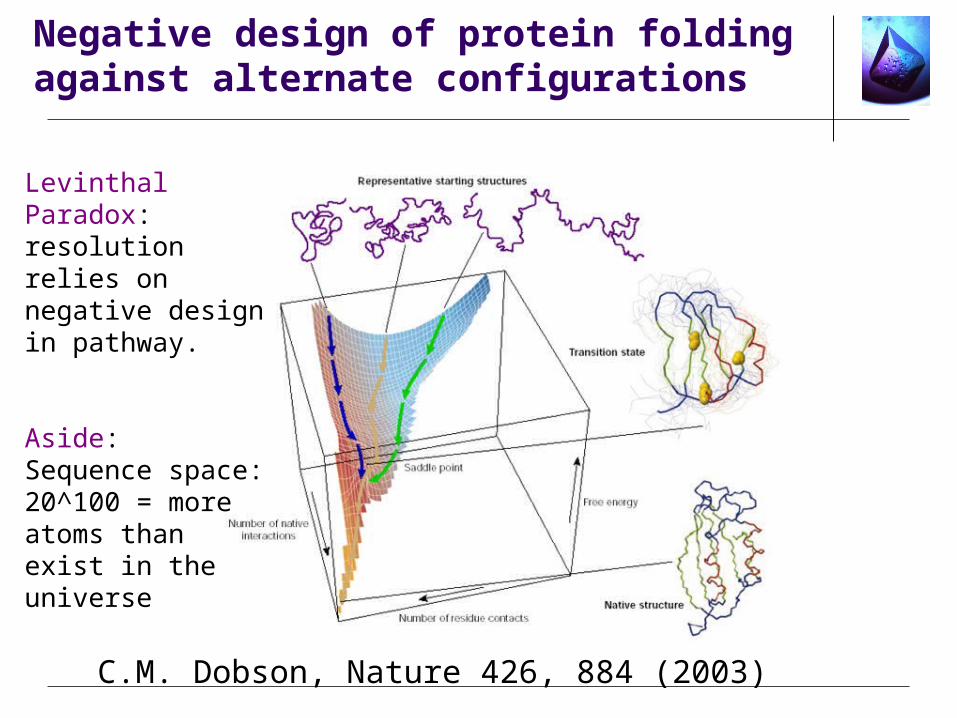
Negative design of protein folding against alternate configurations
C.M. Dobson, Nature 426, 884 (2003)
Levinthal Paradox: resolution relies on negative design in pathway.
Aside: Sequence space:20^100 = more atoms than exist in the universe

Negative design in mis-folded proteins; amyloid formation
C.M. Dobson, Nature 426, 884 (2003)

Copyright ©2002 by the National Academy of Sciences
Richardson, Jane S. and Richardson, David C. (2002) Proc. Natl. Acad. Sci. USA 99, 2754-2759
Fig. 3. (a-d) A set of equivalent, paired sheetedges from four different beta-sandwich protein families having thelectin/glucanase fold
Negative design in mis-folded proteins; amyloid formation

Copyright ©2002 by the National Academy of Sciences
Wang, Weixun and Hecht, Michael H. (2002) Proc. Natl. Acad. Sci. USA 99, 2760-2765
Fig. 1. (A) Schematic representation of a fibril formed byopen ended oligomerization of a six-stranded beta-sheet protein.beta-strands are shown in green, and turns in silver
Negative design in mis-folded proteins; amyloid formation

Normally self-assembly of surfactants etc… is very polydisperse
•Viruses•Nanotechnology?•Colloidal “molecules”?
Pauline Wong: to make these monodisperse icosohedra, the dimers must be designed out.
Positive and Negative Design for Self-Assembly

Protein crystallization is fundamentally different!(notwithstanding “crystallization slot”)
Colloids are artificially stablized (negative design against aggregation)
2. steric stabilization1. charge stabilization 3. nanohalo stabilization
Dynamic Nanoparticle Halos, S. Karanikas and AAL, Phys. Rev. Lett 93, 248303 (2004)
P. Pusey and W. van Megen, Nature 320,340 (1986)
Analogies with colloidal crystallization?

Cells are crowded environments
David Goodsell http://www.scripps.edu/mb/goodsell/gallery/patterson.html
proteins in blue, DNA and RNA in red and orange, lipids in yellow, and carbohydrates green Ribosomes are colored magenta.

Cells are crowded environments
Network of protein-protein interactions must have bothSpecific functional interactions (> 100) (positive design)Generic repulsion to avoid aggregation (negative design)
Remarkable control of this interactome -- many constraints!
With colloids such high concentrations would result in gunk,
….unless you stabilize them

Negative design and protein crystallization
Most likely through surface properties•Surface residues are not random (J. Thornton)
Key question: are the design mechanisms against aggregation robust enough to affect protein crystallization in vitro?(are you fighting evolution?)
Yes: Evidence? •Protein as a variable (G.E. Dale, C. Oefner & A. D’Arcy, J. Struc. Biol 142, 88 (2003))•Random mutagenesis of surface residues•Directed mutagenesis of Lys etc… (Derewenda)•In-vivo crystallization; when nature wants to it can
How do proteins achieve negative design?

• Other study: Human thymidylate synthase:11 surface mutations: most led to enhanced crystallizability (McElroy et al J. Crys. Growth 122, 265 (1992))
DNA gyrase B subunit from E-coli. Crystals grown in the presence of novobiocin; (a)-(c) crystals from original microbatch,(d) optimized for X-ray analysis.(a) Wild type,(b) K14R/F104Y mutant, (c) K57R/I82N mutant,(d) F104Y mutant.
Mutagenesis studies (protein as a variable)
A. D'Arcy, M. Stihle, D.Kostrewa and G. Dale,Acta Cryst. D 55, 1623 (1999)

Rational mutagenesis studies
Derewenda group: Mutations of Lys -> Ala etc….
Is Lysine like a steric stabilizer (entropy)?
Role of water??
Mechanism?

Protein crystallization in vivo
Protein crystallization, and more generally native state aggregation, is likely to be harmful to the cell
Examples•Cataracts: crystallization and aggregation of the gamma crystallins•Sickle-cell anaemia: ordered aggregation of hemoglobin•Hemoglobin C disease: crystallization of hemoglobin•(many more in A. McPherson’s book)
Non-native state aggregation is more common in part because it is harder for evolution to control disordered proteins. Also, many of these diseases may be post-evolutionary (C. Dobson) and related to the breakdown of regulatory machinery.
J. Doye

•Protein crystallization in vivo
Storage•Seed proteins•Insulin granules•Baccillus thuringiensis•Ribosome crystals in hibernation animals•Crystals in egg yolks
Encapsulation•Baculoviruses
Obstruction•Hex-1 protein crystals in Woronin bodies in fungi•Serum albumim crystals at wound?
Accidental?•Iridoviruses•Inclusion bodies in expression systems

•Baculoviruses etc.. Virus-insect relationships
Encapsulated virus rod of P. interpunctella (army worm) granulovirus
Virus-insect relationshipsKenneth M. SmithLongman (New York 1976)
“this book has been written by one of the few remaining old-fashioned virologists…”

•Baculoviruses etc.. Virus-insect relationships
Nuclear Polyhedron (NPV)(micron size crystals)
Cytoplastic Polyhedron (CPV)
Granulovirus (Smaller crystals)
Release of a viron in alkali
“Wipfelkrankheit” in catepillars“Tree-top disease”“catepillar wilt”

•Baculoviruses etc.. Virus-insect relationships
Insect cells full of baculovirus containing polyhedra
“Wipfelkrankheit” in catepillars“Tree-top disease”“catepillar wilt”
Nuclear Polyhedron (NPV)(micron size crystals)
Cytoplastic Polyhedron (CPV)
Granulovirus (Smaller crystals)

•Baculoviruses etc.. Virus-insect relationships
Relatively monodisperse polyhedral crystals associated with Anopholes quadrimaculatus CPV

•Bacillus Thuringiensis
Insectial bacterium (commonly known as Bt)Production of protein toxins are associated with sporulation.Crystals involve a significant number of interprotein disulphide bridgesCrystals dissolve in alkaline environment of the insect gut, and aid entry
of germinating spores into the host

•Serum Albumin
“some wounds in the blood plasma harden in air to form crystals over the wound. Other blood proteins then help form a clot over the wound, preventing excessive blood loss.

•Serum Albumin
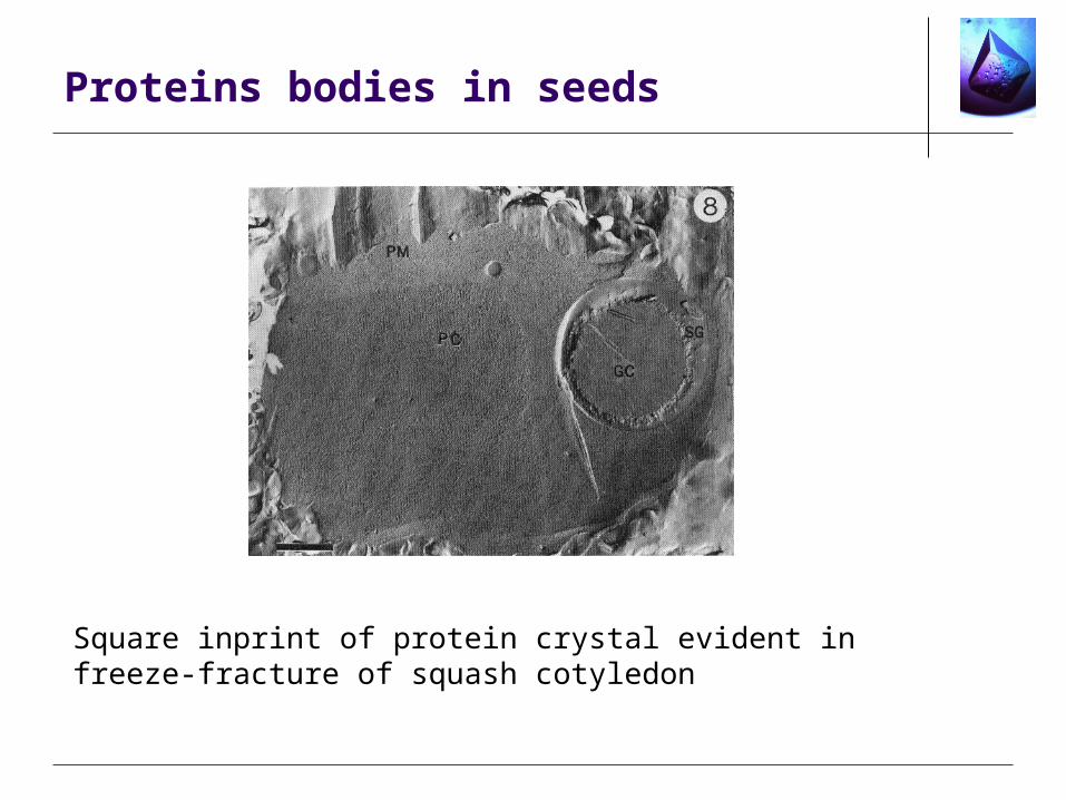
Proteins bodies in seeds
Square inprint of protein crystal evident in freeze-fracture of squash cotyledon

•Peroxisomes
Crystals of oxidative ensymes (catalase and urate oxidase) from a Peroxisome. Suggestion that these are storage crystals to guarantee a stable minimum level of catalase activity in the antioxidative system of plant cells.

•Conclusions
•Negative Design is ubiquitous in nature
•Understanding the mechanisms of this negative design may lead to rational methods for protein crystallization that seek to overcome this
•You’re probably fighting evolution
•Protein crystallization in vivo shows that low crystallizability is not an intrinsic property of proteins

•Future directions
with Janet Thornton, Jonathan Doye and Michele VendruscoloEBI and Dept. of Chemistry, Cambridge U.
J.P.K. Doye, A.A. Louis and M. Vendruscolo,Inhibition of protein crystallization by evolutionary negative design, Phys. Bio 1, P9-p113 (2004)
Combination of bioinformatics plus modelling approaches at various length scales
•Crystal contacts <--> crystal structures • role of mutations•Frustration•Minimal models
•Lysine etc …. (why so disfavoured at contacts?)•H20 and polar/hydrophobic surface patterns
We greatly value your input!
Also, we are collecting examples of in-vivo crystallisation

2-D simple models of randomness