examination of the pharmacodynamics and … · evaluated using an easylyte analyzer (medica...
TRANSCRIPT
5021
Key Words: NSAIDs, Nanoparticles, Diclofenac, Kidney, Pharma-
cokinetics, PLGA.
Introduction
Nonsteroidal anti-inflammatory drugs (NSAIDs) are categorized into two classes, cyclooxygenase (COX)-2-selective inhibitors and NSAIDs which non-selectively inhibit both COX-1 and COX-21. Preventing biosynthesis of prostaglandins (PGs) and prostanoids through COX enzyme inhibition, NSAIDs can reduce the inflammation, fever, and pain which may present in many disease states. Side effects may arise when NSAIDs are used, including but not limited to gastrointestinal and renal dysfunction. Such adverse events are even asso-ciated with diclofenac (DICLO), a non-selective NSAID which ranks as the most frequently used NSAID worldwide2.
Numerous conditions are implicated in the development of NSAID-induced intestinal dam-age, including a shortage of PGs due to the inhi-bition of COX3. PGs, such as PGE2, can defend the digestive tract through stabilizing the muco-sal integrity of intestinal cells4. Increased levels of myeloperoxidase (MPO) in the intestine have been utilized as a biomarker for inflammation and oxidative stress5,6. Thus for duodenal and gastric tissue, PGE2 and MPO were examined to assess inflammation in this study.
Abstract. – OBJECTIVE: Nonsteroidal anti-in-flammatory drugs (NSAIDs) are assembled into two categories; cyclooxygenase (COX-1) sparing inhibitors of COX-2 and non-selective NSAIDs. Diclofenac (DICLO) is a non-selective NSAID that has been linked to serious side effects includ-ing gastric ulcers and renal injury. In this study, we examine the effect of poly(lactic-co-glycol-ic) acid nanoformulation on DICLO-associated adverse events and pharmacokinetics using a nanoparticle (NP) formulation previously devel-oped in our laboratory.
MATERIALS AND METHODS: Rats were ad-ministered a single dose of methylcellulose (VEH), blank NP, DICLO (10 mg/kg), or a DIC-LO-NP suspension equivalent to the DICLO dose. Urinary and blood parameters were mea-sured at baseline and following treatment. Du-odenal and gastric prostaglandin E2 (PGE2) and duodenal myeloperoxidase (MPO) were collect-ed to assess inflammation at 24 hrs post-treat-ment.
RESULTS: The mean percent change from baseline in sodium excretion rate (µmol/min/100 g body weight) differed significantly from VEH in the NP (p < 0.0001), DICLO (p < 0.0001), and DICLO-NP (p = 0.0001) groups. The differenc-es among groups did not reach significance for plasma sodium or potassium concentrations, potassium excretion rate, gastric PGE2, or in-testinal biomarker concentrations. Regarding renal histopathology, DICLO produced consid-erably more necrosis compared to VEH; while DICLO-NP did not elicit notable differences from VEH.
CONCLUSIONS: Our results suggest that over the duration and dosage examined, DICLO-NP may reduce renal necrosis without influencing other side effects or drug characteristics.
European Review for Medical and Pharmacological Sciences 2016; 20: 5021-5031
S. HARIRFOROOSH1, K.O. WEST2, D.E. MURRELL1, J.W. DENHAM3, P.C. PANUS1, G.A. HANLEY4
1Department of Pharmaceutical Sciences, Gatton College of Pharmacy, East Tennessee State University, Johnson City, TN, USA2College of Public Health, East Tennessee State University, Johnson City, TN, USA3Department of Pathology, Quillen College of Medicine, East Tennessee State University, Johnson City, TN, USA4Division of Laboratory Animal Resources, East Tennessee State University, Johnson City, TN, USA
Corresponding Author: Sam Harirforoosh, Ph.D; e-mail: [email protected]
Examination of the pharmacodynamics and pharmacokinetics of a diclofenacpoly(lactic-co-glycolic) acid nanoparticle formulation in the rat

S. Harirforoosh, K.O. West, D.E. Murrell, J.W. Denham, P.C. Panus, G.A. Hanley
5022
Promising results, in terms of renal and gas-trointestinal side effect reduction, have been seen with the use of drug nanoformulation7. Nanoparti-cles (NPs) can be formulated to exhibit favorable qualities, such as large surface area or tissue-spe-cific targeting, which contribute to the utility of this drug delivery system8,9. Several types of NPs have been devised using various materials including the polymer, poly(lactic-co-glycolic) acid (PLGA). In an effort to alter the side effect profile of DICLO, a NP formulation (DICLO-NP) was recently developed in our laboratory7. This study sought to evaluate the gastrointestinal and renal side effects as well as the pharmacokinetics of the new DICLO dosage form.
Materials and Methods
ChemicalsThe drug of interest, DICLO, and methylcel-
lulose were acquired from MP Biomedical (So-lon, OH, USA) and Science Stuff Inc. (Austin, TX, USA), respectively. Flufenamic acid, PLGA (MW 30,000 Da, 50:50 copolymer composition), and didodecyldimethylammonium bromide (DMAB) were purchased from Sigma-Aldrich (St. Louis, MO, USA). Fischer Scientific Lab-oratory (Fair Lawn, NJ, USA) was the vendor for high-performance liquid chromatography (HPLC)-grade water, acetonitrile, acetic acid, and ethyl acetate.
Preparation and Characterizationof PLGA-NP Formulation
This NP formulation was prepared using a method previously optimized by our laboratory7. Briefly, fifty milligrams of PLGA was dissolved, stirred for 30 min at 750 rpm, in 3 mL of ethyl acetate along with 45 milligrams of DICLO. DMAB (0.25% w/v) was stirred and heated in 6 mL of water until dissolved. The organic phase was added under moderate stirring to the aque-ous phase in a drop wise manner. The emulsions were sonicated for 5 min (20 kHz) after which 25 mL of water was added. The organic phase was allowed to evaporate over 4 hrs under constant stirring. Following centrifugation at 18,665 g, the finished product (supernatant) was collected. NP parameters, including size (diameter) and zeta potential, were determined on a NICOMP particle sizer (Particle Sizing Systems, Port Richy, FL, USA). Drug entrapment efficiency was calculated based on the drug entrapped in
NP, as evaluated by UV spectroscopy, relative to total drug utilized.
Animals and Drug AdministrationMale Sprague-Dawley rats (265-300 g) from
Charles River Laboratories (Raleigh, NC, USA) were used for all experiments. Each animal was housed in a 12 hrs light-dark cycle with ambient humidity and temperature along with unrestricted access to food (2020X, Harlan Teklad) and water; however, upon dosing, food access was removed. Each animal was fitted with jugular vein cannula by the vendor. The study protocol was reviewed and approved by the Animal Care Committee of East Tennessee State University.
Study DesignAll four treatment groups (n = 6), 0.5% meth-
ylcellulose solution (VEH), blank NP, DICLO (10 mg/kg) in methylcellulose, and a PLGA-NP equivalent dose (DICLO-NP), were administered respective treatments using a stainless steel oral gavage tube. The VEH group rats were selected from a group of animals which would serve as controls for two studies. Administration of NP allowed for an examination of PLGA pharmaco-dynamics. The DICLO dose chosen for this study was therapeutically equivalent to a rofecoxib dose which had previously elicited sodium excretion rate changes in rats1. As such this dose would be sufficient to serve as a positive control for an adverse effect which may be alleviated through nanoformulation. Urine samples (12 hrs) were collected through housing the animals in meta-bolic cages subsequent to dosing.
Isoflurane was used to anesthetize the rats twenty-four hours post-treatment during which animals were exsanguinated using cardiac punc-ture. Tissues (stomach, proximal 8 cm of duode-num, and kidneys) were collected for analysis.
Renal Function Parameters
Change in Urine Flow Rate The total urine volume in milliliters was divided
by collection duration (12 hrs) to assay urine flow rate. Each value was normalized using 100 g body weight (B.W.) before the mean percent change from baseline was calculated for each group.
Change in Urinary and Plasma Electrolytes In addition to baseline and post-treatment urine
collection, one blood sample was taken prior to dosing and another prior to euthanasia to examine
Effect of PLGA diclofenac nanoparticles in rats
5023
electrolyte concentrations. Millimolar electrolyte (sodium and potassium) concentrations were evaluated using an EasyLyte analyzer (Medica Corporation, Bedford, MA, USA). Electrolyte excretion rates were determined using concen-tration (mM) detected in urine, urine volume (mL), and collection duration (hr). Rates were normalized by 100 g B.W. and presented as mean percent change from baseline.
Kidney Histopathological Assessment A board certified pathologist (blinded to treat-
ment groups) evaluated paraffin embedded, he-matoxylin and eosin stained kidney sections. Each section was given a score ranging from 0 to 3 in terms of tubular dilatation (normal, mild, moderate, or severe) and necrosis (0, 10, 25, > 25%).
Gastrointestinal Inflammatory Factors
Gastric and Intestinal PGE2An enzyme-linked immunosorbent assay
(ELISA) kit for detecting picograms per millili-ter concentrations of PGE2 was purchased from Antibodies-Online Inc. (Atlanta, GA, USA). The assay was carried out in accord with manufactur-er’s instructions using homogenized gastric and duodenal tissue then assayed using MyAssays software (MyAssays Ltd, Sussex, UK).
Intestinal MPOLevels of myeloperoxidase (MPO) were mea-
sured with an ELISA kit from Kamiya Biomedical Company (Seattle, WA, USA). Homogenized sam-ples were assayed following manufacturer’s instruc-tions with ng per mL reported. The standard curve used was determined using MyAssays software.
Chromatographic Conditions
Analysis Equipment and Solution Preparation
HPLC was orchestrated on a Shimadzu system (Shimadzu Scientific Instruments Inc., Columbia, MD, USA) fitted with a LC020AB solvent deliv-ery system coupled with a DGU-20A Prominence degasser. A SIL-20A HT auto sampler was used to load samples which then flowed through a CTO-20A column oven (C18 column). Drug sig-nal was observed using a SPD-M20A diode array detector (280 nm) and the system was controlled using a CBM-20A communication bus module.
DICLO concentrations were determined by a method described previously10. Using a mobile phase consisting of acetonitrile, water, and acetic acid (50:50:0.25) and a flow rate of 0.75 mL/min, chromatographic separation of DICLO was ac-complished. One hundred microliters of DICLO in methanol (ranging from 50 to 50,000 ng/mL) and 50 microliters of flufenamic acid (10,000 ng/mL) in acetonitrile, the internal standard, were added to the blank plasma. Following the addition of 2 mL of acetonitrile, samples were vortexed (30 sec) then centrifuged for 15 minutes (2,500 g). A Labconco CentriVap Concentrator (Kansas City, MO, USA) was used to evaporate the col-lected organic phase. Samples were reconstituted with 200 µL mobile phase; then 115 µL was transferred to injection vials in order to inject 100 µL. Signal height ratios were used to quantify drug concentration. This assay had a lower limit of detection of 50 ng/mL and a lower limit of quantitation of 100 ng/mL with a coefficient of variation of 2.67%.
Pharmacokinetic AnalysisNine-time points (0, 0.5, 1, 2, 4, 6, 8, 12, and
24 hrs) were examined in the pharmacokinetic blood sampling scheme; however, DICLO was undetectable at time points 12 and 24 in both for-mulations. Various pharmacokinetic parameters, half-life (t1/2), maximum plasma concentration (Cmax), area under the plasma concentration-time curve from time zero to infinity (AUC0-∞), appar-ent oral clearance (CLoral), and apparent volume of distribution (Vd/F), were calculated using the non-compartment component of Phoenix Win-Nonlin 6.3 (Certara USA, Inc., Princeton, NJ, USA). In the absence of a calculable elimination phase rate constant, rats were removed from the analysis.
Data Treatment and Statistical AnalysisThe mean percent change from baseline,
((post-treatment – baseline)/baseline) x 100, and standard error of the mean (SEM) were calculated for the rate of urine flow and electrolyte excretion along with plasma electrolyte concentrations. All data, unless otherwise stated, are presented as mean ± SEM. One-way ANOVA through PROC GLM in SAS (SAS Institute Inc., Cary, NC, USA) was used for gastrointestinal and urinary param-eter analysis. Pharmacokinetic parameters were analyzed using Student’s t-test. IBM SPSS Sta-tistics software version 21 (Armonk, NY, USA) was used to identify outliers in each data set apart

S. Harirforoosh, K.O. West, D.E. Murrell, J.W. Denham, P.C. Panus, G.A. Hanley
5024
from histological examination. Significance in this study was set at p < 0.05.
Kruskal-Wallis one-way analysis utilizing pairwise comparisons, post hoc testing which detected minimal significant difference between groups, was conducted on histological values11. The tabulated familywise error rate (significance at 0.05 and adjusted for sample size) was used for comparisons of the mean-of-ranks differences. Using a “z” of 2.576, five comparisons were made. Three outliers, two in dilatation and one in necrosis, were removed prior to statistical analysis.
Results
Characteristics of Diclofenac-loadedPLGA-NPs
The DICLO-NP formulation (n = 3) for this study presented with a mean diameter of 221.03 ± 1.71 nm and a drug entrapment efficiency of 76.38 ± 0.33%. The zeta-sizer also indicated a mean zeta potential of 20.86 ± 0.47 mV.
Renal Function Assessments Mean percent changes (compared to baseline)
in urine flow rate did not attain to significance (Figure 1; p = 0.2790). Urinary sodium excretion rate mean percent changes (Figure 2) presented with significant differences among groups (p =
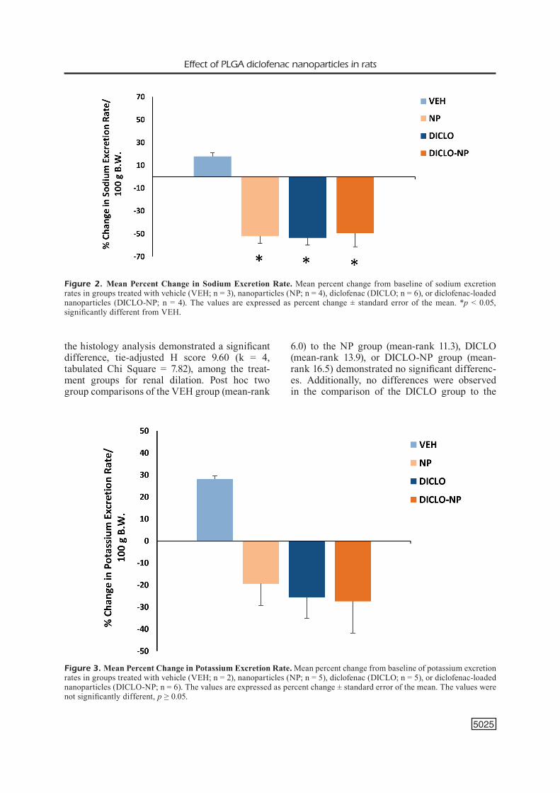
0.0002). The NP (-52.18 ± 6.14%; p < 0.0001), DICLO (-53.55 ± 6.39%; p < 0.0001), and DI-CLO-NP (-49.40 ± 11.98%; p = 0.0001) groups were each significantly decreased compared to VEH (17.82 ± 3.14%). While, as seen in Figure 3, the mean percent changes in urinary potassium excretion rate showed no significant difference (p = 0.1185) among groups. No significant dif-ference was found in either sodium (p = 0.2638) or potassium (p = 0.0929) plasma concentration mean percent change values among treatment groups, Figures 4 and 5, respectively.
Histopathological Assessments Sections from the VEH group (Figure 6-A)
showed mild tubular dilation in every kidney sampled; however, no necrosis was observed in any kidney from the VEH group. The NP group (Figure 6-B) showed tubular dilatation ranging from mild to moderate with necrosis ranging from none to moderate. Represented in Figure 6-C, sections from the DICLO group showed tu-bular dilatation and necrosis ranging from mild to severe. Finally, the DICLO-NP group, as shown in Figure 6-D, presented moderate tubular dilata-tion and mild necrosis in every kidney. Overall, the DICLO group had significantly more necrosis compared to VEH; while DICLO-NP group did not differ significantly from the VEH.
Specific histological mean-rank scores are enumerated in Table I. Statistical analysis of
Figure 1. Mean Percent Change in Urine Flow Rate. Mean percent change from baseline of urine flow rate in groups treated with vehicle (VEH; n = 6), nanoparticles (NP; n = 6), diclofenac (DICLO; n = 6), or diclofenac-loaded nanoparticles (DICLO-NP; n = 6). The values are expressed as percent change ± standard error of the mean. The values were not signifi-cantly different, p ≥ 0.05.
Effect of PLGA diclofenac nanoparticles in rats
5025
the histology analysis demonstrated a significant difference, tie-adjusted H score 9.60 (k = 4, tabulated Chi Square = 7.82), among the treat-ment groups for renal dilation. Post hoc two group comparisons of the VEH group (mean-rank
6.0) to the NP group (mean-rank 11.3), DICLO (mean-rank 13.9), or DICLO-NP group (mean-rank 16.5) demonstrated no significant differenc-es. Additionally, no differences were observed in the comparison of the DICLO group to the
Figure 2. Mean Percent Change in Sodium Excretion Rate. Mean percent change from baseline of sodium excretion rates in groups treated with vehicle (VEH; n = 3), nanoparticles (NP; n = 4), diclofenac (DICLO; n = 6), or diclofenac-loaded nanoparticles (DICLO-NP; n = 4). The values are expressed as percent change ± standard error of the mean. *p < 0.05, significantly different from VEH.
Figure 3. Mean Percent Change in Potassium Excretion Rate. Mean percent change from baseline of potassium excretion rates in groups treated with vehicle (VEH; n = 2), nanoparticles (NP; n = 5), diclofenac (DICLO; n = 5), or diclofenac-loaded nanoparticles (DICLO-NP; n = 6). The values are expressed as percent change ± standard error of the mean. The values were not significantly different, p ≥ 0.05.

S. Harirforoosh, K.O. West, D.E. Murrell, J.W. Denham, P.C. Panus, G.A. Hanley
5026
DICLO-NP group (mean ranks 13.9 vs. 16.5), or NP group to the DICLO-NP group (mean ranks 11.3 vs. 16.5).
For the renal necrosis histology analysis, again there was a tie-adjusted H score of 12.05 that was significant (k = 4, tabulated = 7.82). Post hoc group comparisons of the VEH group (mean-rank 5.0) to the NP (mean-rank 11.9) and DICLO-NP (mean-rank 14.5) groups demonstrated no signifi-
cant differences. However, there was a significant difference between the VEH group (mean-rank 5.0) and the DICLO (mean-rank 17.0) in the two group comparison.
Gastrointestinal Inflammatory FactorsThe differences in gastric PGE2 (Figure 7)
among groups did not reach significance (p = 0.5345). As shown in Figure 8, no significant
Figure 4. Mean Percent Change in Plasma Sodium Concentration. Mean percent change from baseline of plasma sodium concentration in groups treated with vehicle (VEH; n = 6), nanoparticles (NP; n = 6), diclofenac (DICLO; n = 5), or diclofenac-loaded nanoparticles (DICLO-NP; n = 6). The values are expressed as percent change ± standard error of the mean. The values were not significantly different, p ≥ 0.05.
Figure 5. Mean Percent Change in Plasma Potassium Concentration. Mean percent change from baseline of plasma potassium concentration in groups treated with vehicle (VEH; n = 6), nanoparticles (NP; n = 5), diclofenac (DICLO; n = 6), or diclofenac-loaded nanoparticles (DICLO-NP; n = 5). The values are expressed as percent change ± standard error of the mean. The values were not significantly different, p ≥ 0.05.

Effect of PLGA diclofenac nanoparticles in rats
5027
changes were noted in the groups when compar-ing intestinal PGE2 concentrations (p = 0.9963). There was also no notable difference (Figure 9) when comparisons were made within the treat-ment groups in regard to intestinal MPO (p = 0.2623).
Pharmacokinetics of DiclofenacThe plasma concentration time curves for both
formulations are given in Figure 10, while phar-macokinetic parameters are presented in Table II. The t1/2 (p = 0.2395), Cmax (p = 0.9134), AUC0-∞ (p = 0.7258), CLoral (p = 0.6650), and Vd/F (p = 0.5331) were not significantly changed between formulations.
Discussion
The use of NSAIDs is limited by two major side effects: gastrointestinal erosion and diminished re-nal function12. Nanoparticles are a group of colloidal drug delivery systems which may be used to alter
drug side effects13,14. By performing analysis of renal and gastrointestinal parameters, and pharma-cokinetics, we compared DICLO and DICLO-NP formulations.
Urine flow rate did not differ significantly among groups (Figure 1). This result is supported by a pre-vious study in which urine flow rate was unchanged in rats 24 hrs following a 30 mg/kg dose1. In that study, no significant change from baseline occurred until the fourth day of dosing. Sodium excretion rate exhibited notable differences among NP, DICLO, and DICLO-NP when compared to VEH (Figure 2). The 30 mg/kg DICLO dose from a previous study also produced a decrease in sodium excretion at 24 hrs post-dose1. As the effect of DICLO and DICLO-NP did not vary from each other, this parameter may not be altered by nanoformulation. As displayed in Figure 3, potassium excretion rate demonstrated no significant changes. Similarly, no change was seen in the 30 mg/kg dose study at 24 hrs either1. For this study, electrolytes were analyzed after only one day of exposure while the study
Figure 6. Kidney Histopathology. Kidney cross sections (hematoxylin & eosin stained) from rat groups treated with A (vehicle) showing mild tubular dilatation (arrow) and no areas of ne-crosis. B (blank nanoparticles) yielded mild dilatation (arrow) and necrosis (arrowheads). C (diclofenac) showed moderate tubular dilatation (arrow) and areas of necrosis (arrowheads). D (diclofenac-loaded nanoparticles) presented with moderate tubular dil-atation (arrow) and mild necrosis (ar-rowheads). 20× magnification.
Table I. Histopathological assessment of tubular dilatation and necrosis.
Tubular dilatation scores Tubular necrosis scores
Group 0 1 2 3 n Mean-Rank 0 1 2 3 n Mean-Rank
VEH 0 6 0 0 6 6.0 6 0 0 0 6 5.0NP 0 3 3 0 6 11.3 3 1 2 0 6 11.9DICLO 0 2 3 1 6 13.9 0 4 1 1 6 17.0*DICLO-NP 0 0 4 0 4 16.5 0 5 0 0 5 14.5
Tubular dilatation and necrosis scores in the groups treated with methylcellulose (VEH), empty nanoparticles (NP), diclofenac (DICLO), diclofenac-loaded nanoparticles (DICLO-NP). *p < 0.05, significantly different from VEH.

S. Harirforoosh, K.O. West, D.E. Murrell, J.W. Denham, P.C. Panus, G.A. Hanley
5028
mentioned collected electrolyte rates following up to four days of exposure at which point significant change was detected. Therefore, these results coin-cide with research that, like other NSAIDs, DIC-LO-associated side effects are dose-dependent and affected by exposure time15. Short exposure and single dosing may have limited the observable effect of DICLO or DICLO-NP on electrolyte excretion rates in this study.
Neither sodium nor potassium plasma con-centrations displayed any significant difference among groups as shown in Figure 4 and Figure
5. These results are supported by Stokes et al16 who found unaltered plasma electrolyte concen-trations when utilizing DICLO (50 mg; given 3 times-a-day for 14 days) in patients with hy-pertension controlled by a diuretic and/or a beta blocker. The lack of change in plasma electrolytes concentrations could also be indicative of the body’s capacity of maintaining homeostasis of electrolytes and the work of hormone mediators.
Histological assessment demonstrated a sig-nificant difference between the treatment groups for renal necrosis as seen in Table I. Overall, the
Figure 7. Gastric PGE2 Concentration. Effect of treatment with vehicle (VEH; n = 5), nanoparticles (NP; n = 6), diclofenac (DICLO; n = 6), or diclofenac-loaded nanoparticles (DICLO-NP; n = 6) on gastric PGE2 concentration. The values are expressed as mean ± standard error of the mean. The values were not significantly different, p ≥ 0.05.
Figure 8. Intestinal PGE2 Concentration. Effect of treatment with vehicle (VEH; n = 5), nanoparticles (NP; n = 6), di-clofenac (DICLO; n = 5), or diclofenac-loaded nanoparticles (DICLO-NP; n = 6) on intestinal PGE2 concentration. The values are expressed as mean ± standard error of the mean. The values were not significantly different, p ≥ 0.05.

Effect of PLGA diclofenac nanoparticles in rats
5029
DICLO group had significantly more necrosis compared to VEH; while the DICLO-NP group did not differ significantly from the VEH as shown in Figure 6. Renal papillary necrosis has been associated with the use of nonselective NSAIDs, such as DICLO17. These results imply
that DICLO administration amplified necrosis levels compared to VEH; while DICLO-NP did not at this dosage and interval of exposure.
There were no significant changes among groups in regard to gastric PGE2, intestinal PGE2, or intestinal MPO as seen in Figures 7, 8, and 9,
Figure 9. Intestinal MPO Concentration. Effect of treatment with vehicle (VEH; n = 6), nanoparticles (NP; n = 6), di-clofenac (DICLO; n = 6), or diclofenac-loaded nanoparticles (DICLO-NP; n = 6) on intestinal MPO concentration. The values are expressed as mean ± standard error of the mean. The values were not significantly different, p ≥ 0.05.
Figure 10. Diclofenac Plasma Concentration-time Curve. Plasma concentration-time profile of diclofenac following a single 10 mg/kg oral dose of DICLO (DICLO; n = 5) or a diclofenac-loaded nanoparticle (DICLO-NP; n = 4) equivalent.
DICLO-diclofenac; DICLO-NP-diclofenac-loaded nanoparticles. Values expressed as mean ± standard error of the mean. Values were not significantly different, p ≥ 0.05.
Table II. The pharmacokinetic parameters of diclofenac following a single oral dose of diclofenac (10 mg/kg) or a PLGA nanoparticle equivalent.
Formulation n t1/2 Cmax AUC0-∞ Cloral Vd/F
(hr) (µg/mL) (µg.h/mL) (L/h/kg) (L/kg) DICLO 5 4.44 ± 0.58 1.25 ± 0.28 6.18 ± 0.78 1.75 ± 0.26 11.52 ± 2.54DICLO-NP 5 6.36 ± 1.40 1.22 ± 0.16 6.55 ± 0.65 1.60 ± 0.19 13.78 ± 2.37

S. Harirforoosh, K.O. West, D.E. Murrell, J.W. Denham, P.C. Panus, G.A. Hanley
5030
respectively. In another study, DICLO (50 mg/kg with six hours of exposure) treatment reduced PGE2 gastric production and exhibited the max-imum rating of gastric lesions in rats18. In the same study, statistically significant increases in gastric MPO were detected after DICLO was administered. As found by Fornai et al19, fourteen days of DICLO (8 mg/kg), given daily reduced in-testinal PGE2 levels in rats. Additionally, DICLO increased MPO levels in both the jejunum and ileum. Our results do not correspond with these outcomes possibly due to differences in treatment duration and dosing level. This may explain why the PGE2 and MPO level changes were nonsignif-icant among groups in our study.
The pharmacokinetics of DICLO-NP showed no significant changes in any of the measures recorded (Figure 10). In a study conducted by Manvelian et al20, a proprietary nanoformu-lation of DICLO (35 mg) yielded an AUC0-∞ similar to a 50 mg dose in humans under fasted and fed conditions. The change in bioavailabil-ity is difficult to determine because different doses were compared. Other studies21,22, ex-amining DICLO ophthalmic nanoformulations (NP or nano-composite) in rabbits, have found increased bioavailability. This suggests that increased bioavailability is possible; however, in our study, this particular PLGA-NP formu-lation of DICLO did not change the systemic exposure of the drug.
Conclusions
In this work, nanoformulation of DICLO did not significantly alter expected DICLO-associat-ed effects or systemic exposure; however, there was a reduction in renal necrosis. Altogether, the DICLO-NP dosage form appears to be an im-provement upon the typical formulation, DICLO.
Acknowledgements We would like to thank Dustin Cooper, Angela Han-ley, and Kenny Bullins for their technical assistance.
FundingThis study was funded by a grant from the East Ten-nessee State University Research Development Com-mittee Major Grants Program. This research was sup-ported in part by the National Institutes of Health grant C06RR0306551.
Conflicts of interestThe authors declare no conflicts of interest.
References
1) HarirforoosH s, Jamali f. Effect of nonsteroidal anti-inflammatory drugs with varying extent of COX-2-COX-1 selectivity on urinary sodium and potassium excretion in the rat. Can J Physiol Pharmacol 2005; 83: 85-90.
2) Gan TJ. Diclofenac: an update on its mechanism of action and safety profile. Curr Med Res Opin 2010; 26: 1715-1731.
3) TakeucHi k, saToH H. NSAID-induced small intesti-nal damage--roles of various pathogenic factors. Digestion 2015; 91: 218-232.
4) monTrose Dc, nakanisHi m, murpHy rc, Zarini s, mcaleer Jp, Vella aT, rosenberG DW. The role of PGE2 in intestinal inflammation and tumorigen-esis. Prostaglandins Other Lipid Mediat 2015; 116-117: 26-36.
5) loria V, DaTo i, GraZiani f, biasucci lm. Myelop-eroxidase: a new biomarker of inflammation in ischemic heart disease and acute coronary syndromes. Mediators Inflamm 2008; 2008: 135625.
6) HanifeH m, Heilmann rm, sankari s, raJamaki mm, makiTalo l, syrJa p, kilpinen s, sucHoDolski Js, sTeiner Jm, spillmann T. S100A12 concentrations and my-eloperoxidase activity in the intestinal mucosa of healthy dogs. BMC Vet Res 2015; 11: 234.
7) cooper Dl, HarirforoosH s. Design and optimiza-tion of PLGA-based diclofenac loaded nanoparti-cles. PLoS One 2014; 9:e87326.
8) muDsHinGe sr, Deore ab, paTil s, bHalGaT cm. Nanoparticles: Emerging carriers for drug deliv-ery. Saudi Pharm J 2011; 19: 129-141.
9) kamaly n, Xiao Z, Valencia pm, raDoVic-moreno af, farokHZaD oc. Targeted polymeric thera-peutic nanoparticles: design, development and clinical translation. Chem Soc Rev 2012; 41: 2971-3010.
10) el-sayeD ym, abDel-HameeD me, suleiman ms, na-Jib nm. A rapid and sensitive high-performance liquid chromatographic method for the determi-nation of diclofenac sodium in serum and its use in pharmacokinetic studies. J Pharm Pharmacol 1988; 40: 727-729.
11) porTney lG, WaTkins mp. 3 ed. Norwalk, CN: Apple-ton & Lange, 2009.
12) HarirforoosH s, asGHar W, Jamali f. Adverse ef-fects of nonsteroidal antiinflammatory drugs: an update of gastrointestinal, cardiovascular and renal complications. J Pharm Pharm Sci 2013; 16: 821-847.
13) sinGH r, lillarD JW, Jr. Nanoparticle-based tar-geted drug delivery. Exp Mol Pathol 2009; 86: 215-223.

Effect of PLGA diclofenac nanoparticles in rats
5031
14) cooper Dl, carmical Ja, panus pc, HarirforoosH s. Formulation and in vitro evaluation of niacin-loaded nanoparticles to reduce prosta-glandin mediated vasodilatory flushing. Eur Rev Med Pharmacol Sci 2015; 19: 3977-3988.
15) alTman r, boscH b, brune k, paTriGnani p, younG c. Advances in NSAID development: evolution of diclofenac products using pharmaceutical tech-nology. Drugs 2015; 75: 859-877.
16) sTokes Gs, brooks pm, JoHnsTon HJ, monaGHan Jc, okoro eo, kelly D. The effects of sulindac and diclofenac in essential hypertension con-trolled by treatment with a beta blocker and/or diuretic. Clin Exp Hypertens A 1991; 13: 1169-1178.
17) WHelTon a. Nephrotoxicity of nonsteroidal an-ti-inflammatory drugs: physiologic foundations and clinical implications. Am J Med 1999; 106: 13S-24S.
18) sancHeZ s, marTin mJ, orTiZ p, moTilVa V, Herrerias Jm, alarcon De la lasTra c. Role of prostaglandins and nitric oxide in gastric damage induced by
metamizol in rats. Inflamm Res 2002; 51: 385-392.
19) fornai m, anTonioli l, colucci r, pelleGrini c, Gius-Tarini G, TesTai l, marTelli a, maTaranGasi a, naTale G, calDerone V, Tuccori m, scarpiGnaTo c, blanDiZZi c. NSAID-induced enteropathy: are the currently available selective COX-2 inhibitors all the same? J Pharmacol Exp Ther 2014; 348: 86-95.
20) manVelian G, Daniels s, Gibofsky a. The pharma-cokinetic parameters of a single dose of a novel nano-formulated, lower-dose oral diclofenac. Postgrad Med 2012; 124: 117-123.
21) asasuTJariT r, THeeracHayanan T, keWsuWan p, Veer-anoDHa s, fuonGfucHaT a, riTTHiDeJ Gc. Devel-opment and evaluation of diclofenac sodium loaded-N-trimethyl chitosan nanoparticles for ophthalmic use. AAPS PharmSciTech 2015; 16: 1013-1024.
22) li X, ZHanG Z, cHen H. Development and evalua-tion of fast forming nano-composite hydrogel for ocular delivery of diclofenac. Int J Pharm 2013; 448: 96-100.