excitotoxicity and human disease: part i: the cns
TRANSCRIPT

Excitotoxicity and Human
Disease:
The CNS
Russell L. Blaylock, MD, CCN
Editorial Board, Neuroscience
Surgical Neurology International
www.russellblaylockmd.com

Concept of Excitotoxicity
• In the 1969, Dr. John Olney discovered that MSG could cause certain neurons to become excessively excited, to the point that they would quickly die within 1 to 2 hours of exposure.
• Since this early discovery, several compounds have been discovered to have this property- mainly acidic proteins such as glutamate and aspartate.
• Cysteine in the presence of bicarbonate is also excitotoxic. Homocysteine is also an excitotoxin.

Concept of Excitotoxicity
• Olney discovered that certain areas of the brain were more
sensitive than others -arcuate nucleus, supraoptic nucleus,
paraventricular nucleus of the hypothalamus, hippocampus,
amygdala, locus ceruleus, etc.
• The animals developed a particular set of findings
• short statue, gross obesity
• shrunken endocrine glands and significant reductions in
neurohormones, especially prolactin, FSH, LH, ACTH and
HGH.

Special Characteristics of
Excitotoxicity
• The immature brain is 4X more sensitive to excitotoxins than is the mature brain.
• Increased sensitivity again arises in the aged brain
• Humans are the most sensitive to oral MSG of any known species
• 5X more sensitive than mice
• 20X sensitive than Rhesus monkeys
• The dose of MSG causing these lesion is equalivent to amounts humans receive in their diets.

Excitotoxic Disorders • Acute disorders:
– Seizures
– Brain trauma
– Strokes (ischemia)
– Hypoglycemia
• Chronic Disorders: – Alzheimer’s disease
– Parkinson’s disease
– Huntington’s disease
– ALS
– Viral encephilits
– HIV dementia
– Autism
– Heavy metal poisoning
– Aluminum toxicity
– Fluoride toxicity
– Mitochondrial disorders
– Depression/anxiety
– Schizophrenia
– Bacterial meningitis
– Prion diseases
• Metabolic Disorders
– Hyperammonemia
– Hyperglycemia
– hypoglycemia

Importance of Excitotoxicity • Effects on Brain Development
• Normal Functioning of the brain
– Learning and memory
– Attention
– Limbic control
– Regulation of other neurotransmitters
• Immune-excitotoxicity interactions
(immunoexcitotoxicity)
• Pathophysiology of: Alzheimer’s and Parkinson’s disease
• Ways to reduce excitotoxicity

Factors in MSG Toxicity
• Humans absorb MSG much easier than animals
• Human blood levels after dosing have variable absorption levels-from 19-fold to 50-fold increases in blood levels.
• Stegink found that adding aspartame to MSG dosing doubled the blood glutamate level.
• Repetitive dosing can cause perturbations in neuroendocrine system without microscopic damage to the hypothalamus.

MSG Toxicity • Viral infections can elevate blood glutamate levels
• MSG can produce silent lesions in brain
• MSG alters brain serotonin, norepinephrine, dopamine, GABA and acetylcholine levels during brain development
• Glutamate levels in fetus are twice the level in mothers following maternal MSG feeding
– Lowers seizure threshold
– Impairs learning
– Behavioral aggressiveness and anxiety
– Even without damage under light microscopy

Glutamate and Brain Development
• Found that fluctuations in brain levels of glutamate were critical in brain development. High levels over short period needed to prune excess synaptic connections and remove redundant pathways.
• Lower levels stimulated neuron migration and pathway development and eventual consolidation of connections.
• Excessive glutamate (excitotoxins) during neuronal migrations can halt cell migration leading to grossly abnormal brain development-
hererotopias and intracortical arrest.
• High excitotoxin levels can cause arrest of neuron migration at all levels, producing a wide spectrum of architectonic patterns seen in human malformation- microgyria, pachygyrias, double cortex and lissencephalies.

Circumventricular Organs

Neuroendocrine Effects of
Excitotoxins
• Van den Pol, et al -demonstrated that glutamate was the dominant excitatory transmitter in neuroendocrine regulation. ( Science 250:1990)
• Olney found that even in subtoxic doses (1/4 dose) induced rapid elevation of LH and prolactin and depressed pulsatile growth hormone release.
• In fully toxic doses- depressed LH, TSH, ACTH, GH and prolactin
• Perinatal exposure to MSG caused shrinkage of ovaries and uteri and lower estradiol levels.


Excitotoxins and Gross Obesity
• In real life situation, pregnant women, infants and children are exposed to numerous sources of excitotoxins.
• MSG and related disguised products
• Aspartame-40% aspartic acid
• Naturally occurring excitotoxins
• Spontaneously generated excitotoxins- cysteine-S-sulfonic acid from sulfite interaction with cysteine. (10X more potent as an excitotoxin as glutamate.)

Excitotoxins and Gross Obesity: Summery of
the Evidence
• Consumption of food-borne excitotoxins has increased dramatically over last 20 years.
• Additive toxic effects of subtoxic doses of individual excitotoxins becomes fully toxic
• Transplacental concentration of glutamate assures maximum toxicity to unborn infant.
• Infant and small child 4X more sensitive than adult
• Dramatic increased use of soy based infant formulas
• Use of aspartame products by mother during pregnancy
• Proven hypothalamic damage in areas known to produce gross obesity.

Individual Variations in Glutamate Blood
Levels Following a Meal
• People with gout or migraine headaches have higher blood glutamate levels.
• Those with ALS have 2x higher glutamate blood levels after a meal containing MSG than normal.
• People with even subclinical viral infection have 10-fold higher glutamate than normal.
• People exposed to mercury have higher brain glutamate levels.
• Among adults, blood glutamate levels can vary from 19X to 50X elevations with same dose of glutamate.
• Some infants develop 50-fold elevations in blood glutamate with glutamate-containing foods.
• Humans eat larger meals, test animals nibble.

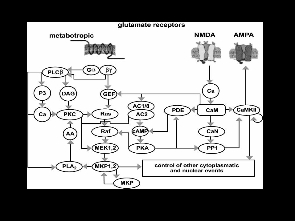
Glutamate Receptors

Glutamate Receptors
• Ionotropic
– NMDA
– AMPA
– Kainate
• Metabotropic
– Group I-III with eight subtypes
• Glutamate receptors are linked via lipid Rafts and protein
Scaffolding and are inducible
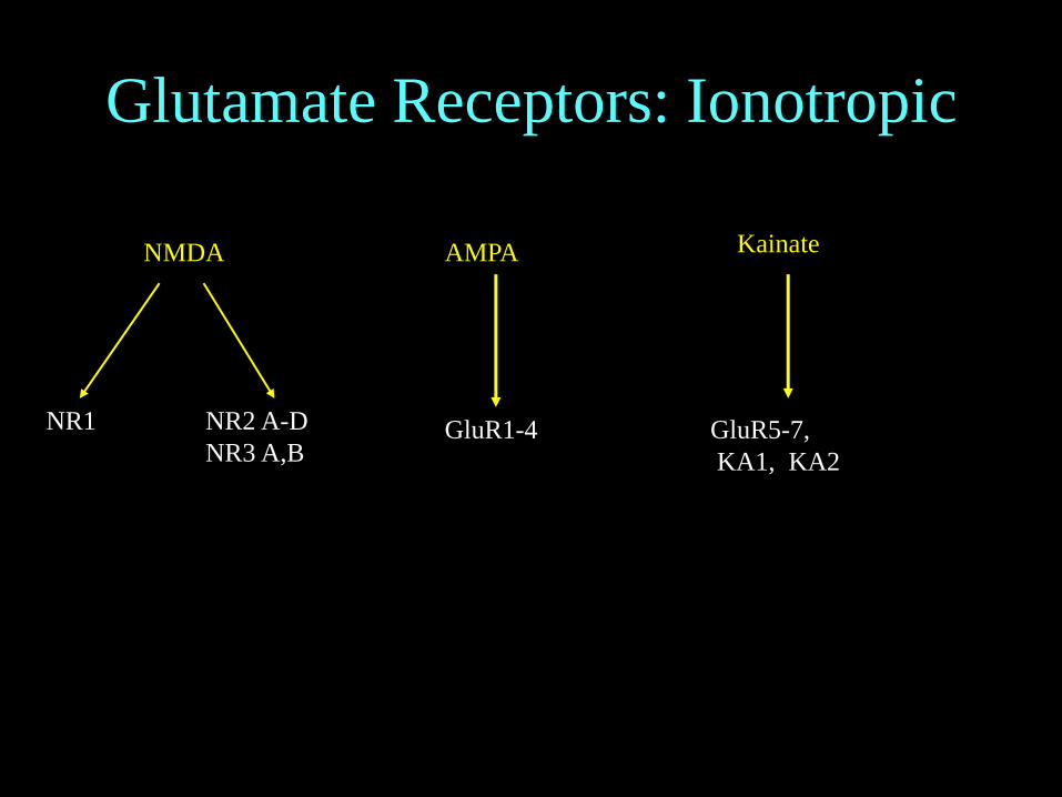
Glutamate Receptors: Ionotropic
NMDA AMPA Kainate
NR1 NR2 A-D
NR3 A,B GluR1-4 GluR5-7,
KA1, KA2

Arachidonic acid
Ca++
Ca++ PK-C
PL-A2
LOX COX II
PGE2
PGD2
Leukotriene
Inflammatory Cascade
iNOS Nitric Oxide
peroxynitrite
Mitochondrial
decreased energy
ROS, RNS
Glutamate transporter Glutamate
SO
(+)
(-)
D-serine
Mg++ Zn 2+
glycine
NFkB
A
B
C
D
E
F

NMDA Receptor Physiology
• Ligand-gated ion channel
• Requires simultaneous activation by glutamate and glycine
• Magnesium blocks ion channel opening-relieved by depolarization
• Modulated by polyamines-spermine and spermidine
• Zinc and NO inhibit NMDA activity

Glutamate NMDA Receptor
Subunits
• NR1 is seen in all NMDARs and accounts for most of receptor properties
• NR2 exist in 4 forms (NR2A-D)
– Interacts with NR1 for full activity
– Heterogeneously distributed in brain
– Accounts for variations in NMDAR function
• NR2C selectively made in cerebellum-less sensitive to glutamate antagonists
• Neonatal animals typically express NR2B and NR2D forms

Glutamate NMDA Receptor Subunits
• In adult:
– NR2A predominates in forebrain, hippocampus, cerebellum,
– NR2B highly represented in olfactory tubercle, hippocampus, olfactory bulb and cerebral cortex
– NR2C- highest in cerebellum
– NR2D-low levels in thalamus, brain stem, olfactory bulb and spinal cord.
• Functional receptors require NR1 and variable combinations of NR2 subunits

AMPA Receptor Function
• Exist both presynaptically and post-synaptically
• Responsible for fast transmission
• Trafficking between intracellular pool and membrane play major role in development and synaptic plasticity
• Intracellular sites major pool of GluR1 and GluR2 subunits utilized by AMPAR
• Glutamate receptors regulate actin-based motility of dendritic spines and growth cones
• AMPAR stimulation completely inhibits motility of growth cones in hippocampus

AMPA Receptor Subunits
• Has four subunits (GluR1-4)
• GluR2 is mainly repressed during brain development
• GluR1/GluR2 associated with adult brain function
• GluR2/GluR3 clustered on post-synaptic membrane

AMPA Receptor Function
• Exist both presynaptically and post-synaptically
• Responsible for fast transmission
• Trafficking between intracellular pool and membrane play major role in development and synaptic plasticity
• Intracellular sites major pool of GluR1 and GluR2 subunits utilized by AMPAR
• Glutamate receptors regulate actin-based motility of dendritic spines and growth cones
• AMPAR stimulation completely inhibits motility of growth cones in hippocampus

AMPA Receptor
• AMPA receptors normally are not calcium permeable
• AMPA receptors that do not contain GluR2 subunit makes them permeable to calcium
• See decreasing GluR2 subunit expression in a number of neurological diseases (epilepsy, ALS, etc)
• AMPA receptors interact with NMDA receptors and can lead to excitotoxicity


GlutamateReceptors:
Metabotropic
Metabotropic Receptors
Group I Group II Group III
mGlu 1,5 mGlu 2, 3 mGlu 4,6,7,8

Metabotropic Receptors
• Group I (mGluR 1/5) generally enhance iGluR activity
• Group II (mGluR2/3) induce microglial activation, mitochondrial dysfunction and apoptosis. Induces release of TNF-
• Group III (mGluR 4,6,7,8) protect against microglial toxicity
• Group II mGluR are activated by sustained elevated levels of glutamate

How the Brain Protects Itself From Glutamate Excitotoxicity
Extracellular Glutamate
Glutamate Transport Proteins: GLAST and GLT-1 (EAAT1-5)
Glutamate/cystine
XC- antiporter
Glutamine synthetase
Glutamate dehydrogenase
Glutamic acid decarboxylase
Enters astrocyte Exchange of
Cystine for glutamate
increasing astrocyte
glutathione
Metabolism of
glutamate


Excitotoxicity
Classic Excitotoxic
Cascade
XC- Antiporter
Oxidative Stress

Xc- Antiporter
Glutamate Cystine
Glutamate Cystine
2-Cysteines
Glutathione

Glutamate Transporters
• EAAT1-5
• EAAT1 and EAAT2 are predominately glial
(GLAST and GLT-1)
• EAAT3, EAAT4 and EAAT5 expressed in
neurons throughout brain
• EAAT4 and EAAT5 are specifically located on
Purkinje cells and retinal neurons

Glutamate Transporters • Protein Kinase C and Protein Kinase A control
transporter trafficking
• NMDAR interact with EAAT3 to control its surface expression
• With induction of LTP - glutamate uptake is markedly increased
• Transporter levels are altered by kindling, seizures and drug abuse
• Transporter activity necessary for synaptic independence and input specificity


Things Decreasing Glutamate Re-
Uptake
• ß-amyloid
• Oxidative stress
• Mitochondrial dysfunction
• Pro-inflammatory cytokines
• IL-1ß
• TNF-alpha
• IFN-alpha
• Mercury
• Lead
• aluminum

Immunoexcitotoxicity

The Aging Brain
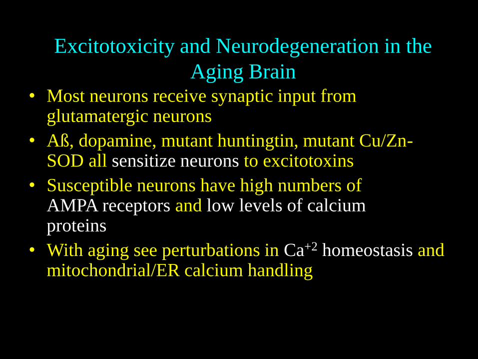
Excitotoxicity and Neurodegeneration in the
Aging Brain • Most neurons receive synaptic input from
glutamatergic neurons
• Aß, dopamine, mutant huntingtin, mutant Cu/Zn-SOD all sensitize neurons to excitotoxins
• Susceptible neurons have high numbers of NMDA & AMPA receptors and low levels of calcium binding proteins
• With aging see perturbations in Ca+2 homeostasis and mitochondrial/ER calcium handling

Excitotoxicity and Neurodegeneration in the
Aging Brain
• Genes associated with PD, AD and ALS are related to Ca+2 perturbations.
• Mutations in -synuclein associated with perturbations in Ca+2 regulation
• Dentate cells in cerebellum (protected in AD) have high levels of calbindin
• Aged-related loss of calbinding linked to loss of basal forebrain cholinergic neurons and entorhinal cortex layer II in AD
• Pyramidal neurons (vulnerable in AD) have little or no calbindin

Aged Brain and Inflammation
• Interleukin proteins
– IL-1ß was elevated the same in young and aged
– IL-6 much higher in aged
• Plasma cytokines
– IL-6 higher in aged
– IL-1ß higher in young
• Plasma cytokine levels did not reflect brain cytokine levels
• LPS caused only transient sickness behavior
– Young fully recovered in 24 hours
– Aged -57% still sick after 24 hours

Aged Brain and Inflammation • In older mice vs younger
– 38 genes controlling inflammation either up or
down regulated (control complement cascade)
• After exposure to LPS (Lipopolysacchride)
– 903 genes upregulated in aged brain only
– Transcripts for mRNA for IL-1ß, IL-6 showed
greatest increase in aged brains
– Transcripts for APP processing, INF-
increased in aged brain only

Aged Brain and Inflammation
• See acute cognitive impairment after systemic infection
• Inflammatory cytokines known to inhibit LTP
• IL-6 neutralizing antibody prolongs LTP and facilitates recovery from LPS-sickness behavior
• IL-1ß and TNF- can also induce cognitive defect
• Humans given very low doses of LPS have no sickness behavior but do have impaired declarative memory

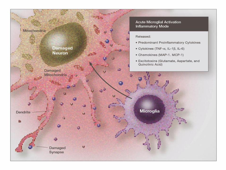
CNS Immunity
• Microglia are the resident immune cell in
the CNS
– Resting (ramified)-basal cytokines and growth
factors
–Amoeboid(activated) -secretes cytokines,
chemokines, complement, etc


Activated Microglia
Microglia
Reactive Oxygen
& Nitrogen Species Lipid
Peroxidation
Products
(4-HNE & Acrolein)
Excitotoxins:
• Quinolinic acid
• Glutamate
• Aspartate

Inflammation/Excitotoxicity
Interaction • COX-1 is constitutive in microglia
• COX-2 is inducible in glutamatergic neurons
• COX-2 induced by NFkB
• PARP-1 plays a central role in NFkB expression of COX-2
• iNOS, IL-1ß, TNF- and glutamate act via PARP-1 to induce inflammation
• Prostaglandins (PGE2)inhibit glutamate re-uptake
• With infarction see robust activation of COX-2
• Dramatic generation of ROS/RNS and LPO
• NMDAR activation stimulates COX-2 activity

Microglial Activation and Glutamate
Receptors
• MSG can activate resting (ramified) microglia
• MSG shown to significantly increase brain TNF-alpha, IL-1ß and IL-6 in neonatal rats
• See widespread loss of neocortical cells with accompanying increase in NR1 expression and GluR2 expression at PND-14 (NMDA and AMPA receptors)

Microglial Activation
• Changes in cell phenotype and gene expression
• Expression of MHC I and II
• Expression of cell adhesion molecules
• Elevation of secretion of pro-inflammatory cytokines and
chemokines
• Induction of iNOS and nNOS
• Release of ROS/RNS and LPO
• Release of excitotoxins

Microglial Neurotoxicity
• Release storms of SO., H202, 0H. , peroxynitrite and 4-HNE
• Release of excitotoxins: glutamate and quinolinic acid in excitotoxic concentrations
• Nitric oxide released from astrocytes and microglia
• Inhibition of mitochondrial respiration by NO
• Mitochondrial suppression stimulates glutamate release from synapse and dramatic elevation in glutamate excitotoxic sensitivity


Inflammation/Excitotoxicity
Interaction
• Reduced energy generation by mitochondria increase the sensitivity of NMDA and AMPA receptors -induced excitotoxicity
• Quinolinic acid increases as much as 300-fold with immune stimulation
• Subcytotoxic levels of glutamate can destroy synaptic connections and lead to dendritic retraction

Microglial Neurotoxicity • NO competes with oxygen at cytochrome binding site -
suppressing mitochondrial respiration
• This increases NMDA receptor sensitivity dramatically
• Shown that even nanomolar concentrations of NO produce
rapid and reversible inhibition of mitochondrial respiration
in brain synaptosomes and astrocytes
• Also see a dramatic suppression of neuronal respiration
after exposure to activated microglia

Microglial Neurotoxicity and
Mitochondrial Respiration
• Inhibition of mitochondrial respiration using specific inhibitors
– Early neuronal death secondary to excitotoxicity and blocked by MK-801
– Prolonged mitochondrial suppression- necrotic death not related to excitotoxicity
– Astrocytes are less dependent on mitochondrial respiration than neurons with glycolysis sufficient for astrocyte energy production
Microglial Neurotoxicity and
Mitochondrial Respiration
• NO release causes immediate glutamate release from a culture of mixed neurons and astrocytes
• Glutamate levels undetectable before NO added
• Glutamate re-uptake prevented by ROS/RNS/LPO, mercury, aluminum and pro-inflammatory cytokines


Sources of TNF-
• Both neurons and glia can produce TNF-
• The main sources are microglia and astrocytes
• Blocking TNF- also blocks AMPAR trafficking and obliterates effect on synaptic strength
• Cortical neurons less sensitive to TNF- effect than hippocampal neurons
TNF-Alpha effects on AMPA Receptor
Trafficking
• AMPA receptor trafficking controls synaptic strength for LTP and LTD
• Trafficking mediated by endocytosis and exocytosis at postsynaptic site
• Mediated by synaptic activity
• Dynamics of AMPAR trafficking is very complex
• Silent synapses have no membrane associated AMPAR. Movement to membrane “un-silences” these receptors.

AMPA Receptor Trafficking

TNF-Alpha and Excitotoxicity • Glutaminase converts glutamine to glutamate
• Using glutaminase -free medium abolished neurotoxic effect of TNF- via microglia almost completely
• Glutaminase inhibitor suppressed TNF- -induced glutamate release and neurotoxicity
• LPS and TNF- both up-regulated mRNA expression of glutaminase in microglia
• TNF- enhances excitotoxicity by synergistic stimulation of both the TNFR1 and NMDAR.
• Also inhibits astrocytic glutamate re-uptake

Microglial Activation and Cellular
Energy
• NMDAR signal induces a rapid drop in intracellular ATP Low
Low energy levels by inhibiting complex IV of mitochondria
• Conditioned media with LPS or TNF-- treated microglia also
induced rapid drop in intracellular ATP and induced
mitochondrial dysfunction in neurons
• Low intracellular energy production dramatically increases
sensitivity to excitotoxicity


Part II:
Alzheimer’s Disease
and
Immunoexcitotoxicity


Hippocampus

Pathophysiology of Alzheimer’s
Disease • Earliest change is synaptic disruption and neurite shrinkage
• Chronic microglial activation within affected areas
• Altered membrane phospholipids (DHA)
• Elevated homocysteine
• Endocrine dysfunction (elevated LH)
• Excitotoxicity
• Widespread oxidative stress and lipid peroxidation
• Elevated inflammatory prostaglandins
• Ref: Atwood CS et al. Neuroinflammation, 2nd Ed , 2003, pp249-266

Pathophysiology of Alzheimer’s
Disease: Soluble vs insoluble Aß
• Normally secrete low levels of soluble Abeta
• With aging begin to accumulate insoluble Aß 1-42 (from
neuron)
• Aß1-40 is more abundant in AD brain
• Concentration of amyloid is not directly correlated with
progression of AD
• Strong correlation between NFT and elevated soluble Aß.
• Ref: McLean C et al. Ann Neurol 1999; 46; 860-866

Things that stimulate APP processing
• Proinflammatory cytokines/ prostaglandins
• MSG (dietary excitotoxins)
• Activation of NMDAR and mGLuR
• Mercury and aluminum
• Low DHA levels
• Estrogen loss
• Elevated levels of Lutenizing hormone
• Oxidative stress


Alzheimer’s Disease and Microglial
Activation
• See high density of activated microglia in diffuse
plaque and throughout brain
• Aß peptide is neither necessary or sufficient for
microglial activation
• Most dying neurons not associated with amyloid
plaque
• Correlation between plaque burden and between
Aß1-42 and synaptic loss is rather weak
• Ref: Walker DG, Lue L-F Microglial response in Alzheimer’s disease. In, Neuroinflammation, 2003,
pp 267-282.


Alzheimer’s Disease and Microglial
Activation
• See elevated IL-1ß, TNF- and IL-6 in plaques
• IL-1 appears early in plaque formation
• Microglia secrete IL-1 as an early event around diffuse plaque.
• IL-1 activates microglia and microglia secrete IL-6 the main stimulus to astrocyte activation
• Systemic infection can produce prolonged microglial activation
• Ref Fillit H et al. Neurosci Lett 1991; 129: 318-320.

Alzheimer’s Disease and Microglial
Activation
• IL-6 is elevated in AD brain as early event
• Microglial activation is associated with accelerated APP processing
• Aß peptide activates p38MAPK and this activates the microglia.
• Microglial NADPH oxidase generates superoxide and NO (peroxynitrite), which inhibits mitochondrial function and enhances excitotoxic sensitivity
• Ref: Lue LF et al. Amer J Path 1999; 155: 853-862.
• McLean Ca et al. Ann Neurol 1999;46: 860-866.

Alzheimer’s Disease and IL-6
• Animals with overexpression of IL-6
– Decreased dendritic arborizations
– Loss of cholinergic hippocampal innervation
– Astrogliosis and microglial activation
– Deficit in LTP and memory
– Ref: Kettenmann H et al. Neuroinflammation-From Bench to Bedside. Springer, NY, 2002

Alzheimer’s Brain
• Elevation in NFkB activation
• Dramatic increase in ROS/RNS and LPO in
brain and systemically
• Increase in anti-inflammatory PPAR-
• Low levels of IL-10 (anti-inflammatory)
• Elevation in brain iron, aluminum and mercury

Alzheimer’s Brain
• Elevated protein oxidation and nitration levels
• Elevated AGEs
• Increased DNA oxidation
• Impaired glutamate transport
• Evidence of extensive excitotoxicity
• Ref. Floden AM et al. J Neuroscience 2005; 25: 2566-77.

Dysfunctional Glutamatergic system in
AD
• GLAST and GLT-1 are abnormally low in AD
• NMDA receptor and mGlu5R activation stimulates APP processing
• See decreased NMDAR, but remaining receptors are overstimulated by glutamate
• Glutamine synthetase activity is significantly reduced in AD (oxidatvely modified)
• Mixing Aß solutions with GS produces aggregates of Aß
• Non-toxic concentration of Aß has synergistic effect on excitotoxicity

Homocysteine in AD
• Alone, homocysteine has little toxicity but in presence of excitotoxicity and oxidative injury, greatly accelerates both processes.
• Homocysteine is converted into homocysteine sulfonic acid and homocysteine sulfinic acid, which are more toxic than glutamate
• Ref. Clark R et al. Arch Neurol 1998;55: 1449-1455.


Chronic Microglial
Activation
Vaccines and
infections
Mercury
Aluminum
Cadmium
Lead
Fluoride (?)
Excitotoxins Food allergens
Gliadin
Gluten
Casein
Glutamate
aspartate
Excitotoxicity
Androgens and LH
ROS/RNS/LPO
Inflammation:
TNFR1
PGE2

Neurodegenerative diseases
Chronic Microglial Activation
Inflammatory
cytokines Inflammatory
prostaglandins
Excitotoxins:
•Glutamate
•Aspartate
•Quniolinic acid
Mitochondrial Dysfunction
Lipid peroxidation products-
4-hydroxynonenal ROS/RNS:
peroxynitrite
Depression
and
Immunoexcitotoxicity

Depression and Immunoexcitotoxicity
• Depression and anxiety frequently co-exist
• Affects 20% of population and appears to be growing
in incidence
• Risk appears to be determined early in life
• Twin studies show 30 to 40% genetic influence
• Those carrying the 5-HT transporter (5-HTT) gene
variant have high anxiety as infants and children.

Depression and Immunoexcitotoxicity
• See shrinkage of hippocampus, also a deficit in working memory (even without atrophy)
• In primate, hippocampal circuits do not fully mature until adolescence
• Major depressive disorder (MDD) resembles sickness behavior – Aversion to food
– Fatigue
– Insomnia or daytime sleepiness
– Irritability
– Social disinterest
– Memory difficulties

Depression and Immunoexcitotoxicity
• The behavioral effects of sickness behavior are secondary to inflammatory cytokines and excitotoxcity
• Ketamine shown to dramatically improve depression and long after medication was stopped
• Patients treated with inflammatory cytokines often develop MDD


Depression and Immunoexcitotoxicity
• See major depression with inflammatory diseases:
– Rheumatoid arthritis
– Cardiovascular disease
– Type 2 diabetes
• With aging see increasing inflammation and increasing depression/anxiety
• Animals injected with IL-1ß or TNF- show depressive behavior

Depression and Immunoexcitotoxicity
• Peripheral immune links to CNS: – Vagal
– Trigeminal
– Humoral TLR on macrophages lining the CVO and choroid plexus
– Cytokine transporter on BBB
– Il-1 receptor on perivascular macrophages and endothelial cells of brain
venules
• Subseptic doses of LPS induces expression of IL-1ß, TNF- in
brain

Depression and Immunoexcitotoxicity
• In older mice see an exaggerated sickness behavior on exposure to LPS
• Low IL-10 worsens response
• Mice carrying diabetic genes (db/db) respond to LPS with exaggerated sickness behavior
• Aging itself can prime microglia
• In aged see more prolonged and profound depression
• Lemstra et al first to show that systemic infection in humans can result in microglial activation.
• Ref: Godbout JP et al. FASAB J

Depression and Immunoexcitotoxicity
• Inflammatory cytokines lower plasma tryptophan
• See increased tryptophan metabolism
• Increases quinolinic acid in brain (kynurenine pathway)
• Increases uptake of serotonin
• Chronic inflammatory cytokine elevation increase cortisol receptor resistance and this increases cortisol
• Cortisol enhances excitotoxicity in hippocampus

Depression and Immunoexcitotoxicity
• Antagonist of GluR 2,3,5 of mGluR has antidepressant effect
• Zinc acts as an antidepressant by suppressing NMDA receptor activity
• SSRI medications modulate AMPA receptors and this reduces depression
• Interaction with other neurotransmitters: dopamine, locus coeruleus (norepinephrine) and nAchR.
• Study of 38 depressed patients found higher glutamate and lower GABA in brain

Multiple Sclerosis
and
Immunoexcitotoxicity

Multiple Sclerosis and
Immunoexcitotoxicity
• In animal model of MS, EAE, blocking
AMPA/Kainate receptors significantly
ameliorated the disease
• It did so without reducing the intensity of the
immune reaction.
• There is an extensive presence of AMPA
receptors on oligodendroglia

Multiple Sclerosis and
Immunoexcitotoxicity
• See microglia activation at all active stages of
multiple sclerosis
• IL-1ß in a mixed culture will kill oligodendroglia
• Antagonist of the AMPA receptor will prevent this
• TNF- will also kill oligodendroglia (IL-1ß
stimulates release of TNF)
• Source of IL-1ß and TNF- is the microglia

Multiple Sclerosis and
Immunoexcitotoxicity
• PET scanning using peripheral benzodiazapine receptor (PBR) on 7 healthy controls and 22 patients with MS
• During disease relapse see increased PBR scanning with disease progression
• See low levels of GLT-1 around active MS lesions
• This raises the glutamate level around the affected neuron fiber

Multiple Sclerosis and
Immunoexcitotoxicity
• See absence of glutamine synthetase and glutamate
dehydrogenase around active and chronically silent
lesions
• This elevated local glutamate levels
• TNF- elevates glutaminase and this increases local
glutamate generation and secretion
• TNF- also increases trafficking of AMPA receptors
(increases excitotoxicity)

Protection from
Immunoexcitotoxicity

Things That Enhance Excitotoxicity
• Low mitochondrial energy production
• Low magnesium in CNS
• Systemic immune activation
• Mercury (all sources- ionic most damaging)
• Histamine excess
• Fluoroaluminum, lead, cadmium, triethyl tin
• Pesticides/herbicides and neurotoxic chemicals

Protection from Excitotoxicity
• Reduce Inflammation: Supplements
– Buffered vitamin C
– Natural form vitamin E (high gamma-E)
– Silymarin
– Curcumin
– Quercetin
– Resveratrol
– Ellagic acid
– Boswellia
– Magnesium citrate/malate

Protection from Excitotoxicity • Increase Cellular Energy
– Riboflavin-5-PO4
– Pyridoxal-5-PO4
– Niacinamide
– Vitamin K
– Thiamine (Benfotiamine)
– CoQ10 (ubiquinol)
– R-lipoic acid
– Acetyl-L-carnitine
– Acetyl-L-carnosine
– Pyruvate

Protection from Excitotoxicity
• Reduce Immune Overactivity – Silymarin
– Vitamin D3
– Magnesium
– Omega-3 oils
• Directly block excitotoxicity » Magnesium
– DHA
– Tetracycline antibiotics
– Dextromethorphan
– Prescription drugs