exercise in the treatment of rotator cuff impingement: a ... review and a synthesized evidence-based...
TRANSCRIPT
ARTICLE IN PRESS
Exercise in the treatment of rotator cuff impingement: Asystematic review and a synthesized evidence-basedrehabilitation protocol
John E. Kuhn, MD, Nashville, TN
A systematic review of the literature was performed toevaluate the role of exercise in treating rotator cuffimpingement and to synthesize a standard evidence-based rehabilitation protocol. Eleven randomized,controlled trials (level 1 and 2) evaluating the effect ofexercise in the treatment of impingement were identified.Data regarding demographics, methodology, andoutcomes of pain, range of motion, strength, and functionwere recorded. Individual components of eachrehabilitation program were catalogued. Effectivenesswas determined by statistical and clinical significance.Although many articles had methodologic concerns, thedata demonstrate that exercise has statistically andclinically significant effects on pain reduction andimproving function, but not on range of motion orstrength. Manual therapy augments the effects ofexercise, yet supervised exercise was not different thanhome exercise programs. Information regarding specificcomponents of the exercise programs was synthesizedinto a gold standard rehabilitation protocol for futurestudies on the nonoperative treatment of rotator cuffimpingement. (J Shoulder Elbow Surg 2008;-:---.)
Systematic reviews of interventions for rotator cuff pa-thology and shoulder pain suggest that exercise maybe an effective treatment,1,12,15,16 whereas ultra-sound therapy is of little benefit.16,28,37 Exercise isa broad term and includes the following interventions:range of motion, stretching and flexibility, andstrengthening exercises, with manual therapy and mo-dalities. Variations on individual exercises and these
From the Vanderbilt Shoulder Center.This work was funded by an unrestricted research grant from the
Arthrex Corporation.Reprint requests: John E. Kuhn, MD, Associate Professor and Chief
of Shoulder Surgery, Vanderbilt Sports Medicine, 4200 MCE,South Tower, 1215 21st Ave S, Nashville, TN 37232 (E-mail:[email protected]).
Copyright ª 2008 by Journal of Shoulder and Elbow SurgeryBoard of Trustees.
1058-2746/2008/$34.00doi:10.1016/j.jse.2008.06.004
components have been promoted by a number of au-thors who offer rehabilitation protocol sugges-tions.4,6,7,10,13,20,21,22,23,26,29,30,31 These protocolsare typically extrapolated from animal studies, ca-daver biomechanics studies, and studies of healthysubjects by using magnetic resonance imaging, videokinematics, electromyography, and strength measure-ments. As such, the protocols recommended by theseauthors are not based on high levels of evidence.
Not surprisingly, there is no consensus on an idealexercise program to treat patients with rotator cuff dis-ease, leading researchers who wish to conduct ran-domized trials to resort to using expert opinion (level5 evidence) when developing protocols.3 The purposeof this systematic review is evaluate the role of exercisein treating rotator cuff impingement and to develop anevidence-based gold standard, physical therapy, ex-ercise program for the treatment of rotator cuff im-pingement syndrome by synthesizing the features ofexercise protocols from clinical studies with the highestlevels of evidence.
MATERIALS AND METHODS
Before the search was initiated, inclusion and exclusioncriteria for articles were defined. Articles were included ifthey were level 1 or level 2 studies (randomized controlledtrials), compared physical therapy with other treatments orplacebo, used outcome measures of pain, function, or dis-ability with validated assessment tools, and were restrictedto patients with a diagnosis of impingement syndrome, asdetermined by positive a impingement sign by Neer32 orHawkins18 criteria, or both. Articles were excluded if theywere concerned with other shoulder conditions (calcific ten-dinosis, full thickness rotator cuff tears, adhesive capsulitis,osteoarthritis, etc), addressed postoperative management,were retrospective studies or case series, or used other out-come measures.
A computer search was conducted using the followingdatabases: PubMed, Ovid, the Cochrane Central Registerof Controlled Trials, Cochrane Database of Systematic Re-views, American College of Physicians (ACP) JournalClub, and Database of Abstracts of Reviews of Effects.Search words included shoulder, impingement, rotatorcuff, rehabilitation, physical therapy, physiotherapy, or exer-cise. The combined search produced 12,428 articles. Thetitles and abstracts were each reviewed to identify those of
1
2 Kuhn J Shoulder Elbow Surg-/- 2008
ARTICLE IN PRESS
interest for in-depth review. Eighty articles were retrieved,and their bibliographies were also reviewed to identify otherpotential articles for inclusion.
From 80 manuscripts, 11 met the inclusion crite-ria2,8,9,11,17,24,33,34,37,38,39 and were then reviewed usinga standard worksheet.36 The worksheet uses evidence-based guidelines to assist in the systematic review of ortho-pedic literature. In addition to recording practical informa-tion, such as title, author, journal, citation, primary andsecondary hypotheses, type of study, and results, the work-sheet also assists in identifying and recording sources ofbias, methods of randomization, follow-up, and other detailsimportant in assessing the methodologic design and identify-ing the level of evidence. Each of these 11 manuscripts wasthen reviewed in an evidence-based medicine journal clubby 9 faculty members and fellows familiar with evidence-based medicine concepts.
The Methods and Results sections of these 11 manuscriptswere then reviewed. Data regarding study demographicsand methodology were extracted and placed in tableform. Individual outcomes for pain, range of motion,strength, and function were catalogued. Outcomes were as-sessed for the effectiveness of each treatment over time (intra-group evaluation) and when different treatments werecompared (between-group comparisons). Statistical out-comes were recorded when available. Clinical significancewas found when statistical significance was P < .05 and theeffect size or difference between treatments was 20% ormore. Elements of the physical therapy programs used byeach study were collected and divided into five major cate-gories: range of motion, flexibility and stretching, strength-ening techniques, therapist-driven manual therapy,modalities, and schedule, which were placed in tableform. This information was used to develop a synthesizedphysical therapy program.
RESULTS
Demographics
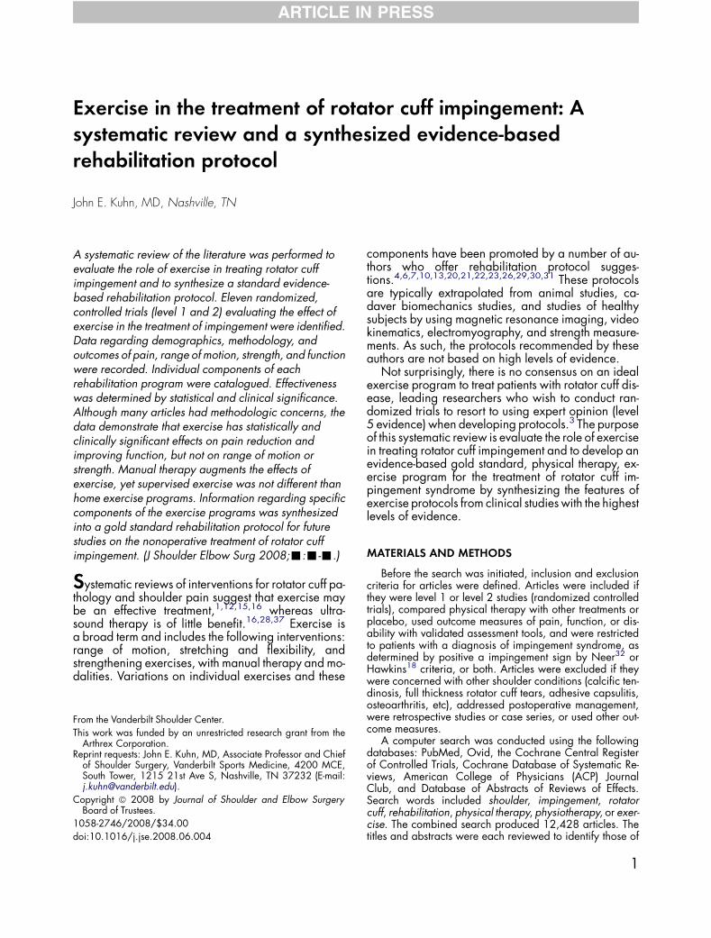
Patient demographics are summarized in Table I.Patient ages (range, 42-58 years) were typical for im-pingement syndrome.32 Workers’ compensation datawere frequently missing, yet because these studiescame from a number of different countries with differ-ent benefits and incentives for work-related injuries,these data may not translate across studies. The diag-nosis of impingement in all 11 studies was made byphysical examination using the impingement signs ofNeer32 or Hawkins,18 or both. Confirmation with animpingement test, consisting of an injection of lido-caine in the subacromial space with elimination ofthe pain with the impingement sign,32 was used in 5studies.
Methodology
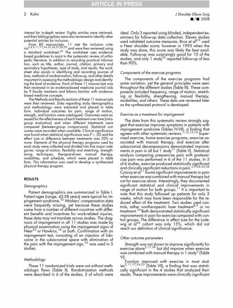
These 11 randomized trials were not without meth-odologic flaws (Table II). Randomization methodswere described in 6 of the studies, 5 of which were
ideal. Only 3 reported using blinded, independent ex-aminers for follow-up data collection. Eleven studiesused validated outcome measures. Brox et al8,9 useda Neer shoulder score; however in 1993 when thestudy was done, this score was likely the best avail-able. Follow-up was surprisingly good for 10 of thestudies, and only 1 study33 reported follow-up of lessthan 90%.
Components of the exercise programs
The components of the exercise programs hadsome variation, yet the general principles were seenthroughout the different studies (Table III). These com-ponents included frequency, range of motion, stretch-ing or flexibility, strengthening, manual therapy,modalities, and others. These data are reviewed lateras the synthesized protocol is developed.
Exercise as a treatment for impingement
The data from this systematic review strongly sug-gest that exercise improves symptoms in patients withimpingement syndrome (Tables IV-VII), a finding thatagrees with other systematic reviews.1,12,15,16 Super-vised exercise, home exercise programs, exercise as-sociated with manual therapy, and exercise aftersubacromial decompressions demonstrated improve-ments in pain in all but 1 study11 (Table V). Statisticalanalysis comparing preexercise pain with postexer-cise pain was performed in 6 of the 11 studies. In 5of 6 studies, exercise produced statistically significantand clinically significant reductions in pain.2,9,24,35,38
Conroy et al11 found significant improvements in painwhen exercise was combined with manual therapy butnot for exercise alone. Interestingly, they documentedsignificant statistical and clinical improvements inrange of motion for both groups.11 It is important tonote that this study followed up patients for only 3weeks, which may have been responsible for the re-duced effect of the treatment. Two studies used con-trols, either nontherapeutic laser treatment8,9 or notreatment.24 Both demonstrated statistically significantimprovements in pain for exercise compared with con-trol groups. The difference in effect size for the Lude-wig et al24 cohort was only 15%, which did notreach our definition of clinical significance.
Other outcome parameters
Strength was not shown to improve significantly forexercise alone2,17,38 but did improve when exercisewas combined with manual therapy in 1 study2 (TableVI).
Function improved with exercise in most stud-ies2,11,17,24,33 (Table VII), a finding that was statisti-cally significant in the 4 studies that analyzed theirresults. These improvements were clinically significant
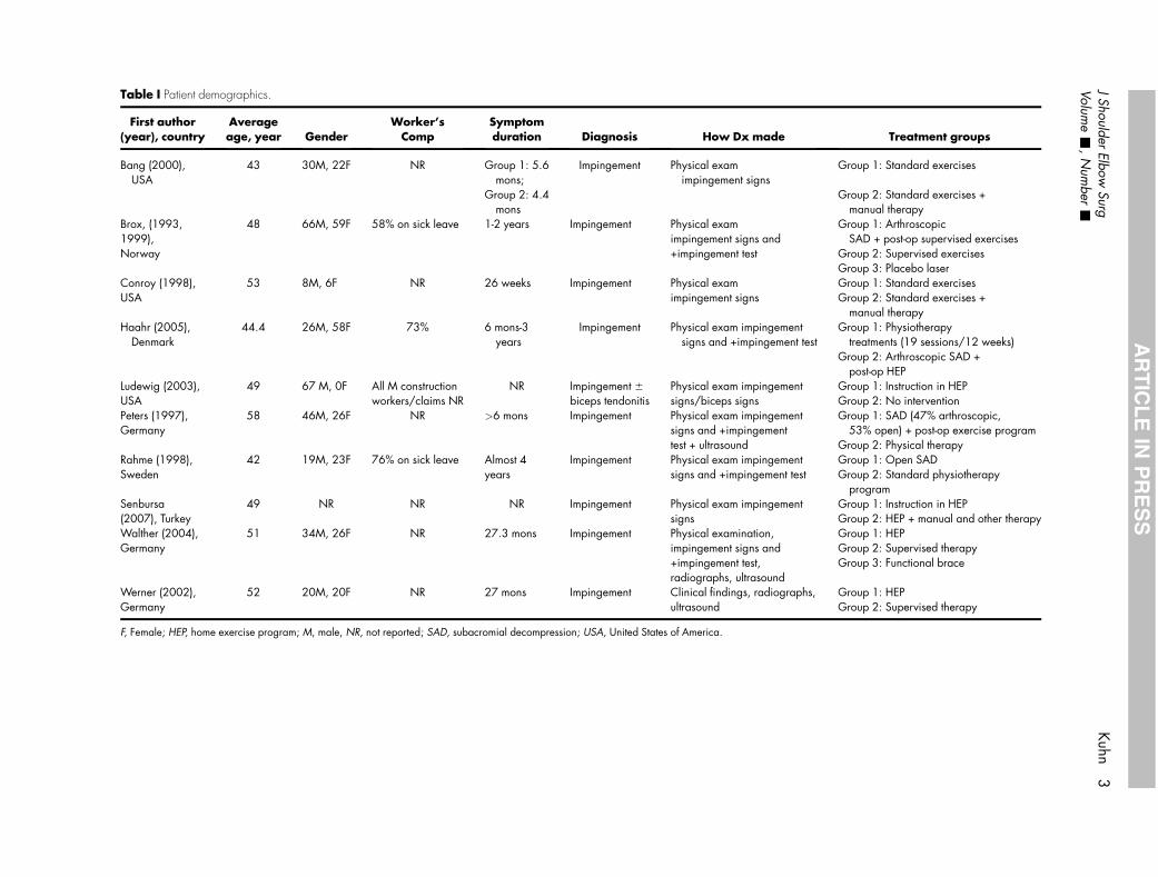
Table I Patient demographics.
First author(year), country
Averageage, year Gender
Worker’sComp
Symptomduration Diagnosis How Dx made Treatment groups
Bang (2000),USA
43 30M, 22F NR Group 1: 5.6mons;
Impingement Physical examimpingement signs
Group 1: Standard exercises
Group 2: 4.4mons
Group 2: Standard exercises +manual therapy
Brox, (1993,1999),Norway
48 66M, 59F 58% on sick leave 1-2 years Impingement Physical examimpingement signs and+impingement test
Group 1: ArthroscopicSAD + post-op supervised exercises
Group 2: Supervised exercisesGroup 3: Placebo laser
Conroy (1998),USA
53 8M, 6F NR 26 weeks Impingement Physical examimpingement signs
Group 1: Standard exercisesGroup 2: Standard exercises +
manual therapyHaahr (2005),
Denmark44.4 26M, 58F 73% 6 mons-3
yearsImpingement Physical exam impingement
signs and +impingement testGroup 1: Physiotherapy
treatments (19 sessions/12 weeks)Group 2: Arthroscopic SAD +
post-op HEPLudewig (2003),USA
49 67 M, 0F All M constructionworkers/claims NR
NR Impingement 6
biceps tendonitisPhysical exam impingementsigns/biceps signs
Group 1: Instruction in HEPGroup 2: No intervention
Peters (1997),Germany
58 46M, 26F NR >6 mons Impingement Physical exam impingementsigns and +impingementtest + ultrasound
Group 1: SAD (47% arthroscopic,53% open) + post-op exercise program
Group 2: Physical therapyRahme (1998),Sweden
42 19M, 23F 76% on sick leave Almost 4years
Impingement Physical exam impingementsigns and +impingement test
Group 1: Open SADGroup 2: Standard physiotherapy
programSenbursa(2007), Turkey
49 NR NR NR Impingement Physical exam impingementsigns
Group 1: Instruction in HEPGroup 2: HEP + manual and other therapy
Walther (2004),Germany
51 34M, 26F NR 27.3 mons Impingement Physical examination,impingement signs and+impingement test,radiographs, ultrasound
Group 1: HEPGroup 2: Supervised therapyGroup 3: Functional brace
Werner (2002),Germany
52 20M, 20F NR 27 mons Impingement Clinical findings, radiographs,ultrasound
Group 1: HEPGroup 2: Supervised therapy
F, Female; HEP, home exercise program; M, male, NR, not reported; SAD, subacromial decompression; USA, United States of America.
JShoulder
ElbowSurg
Kuhn
3Volum
e-
,N
umber
-
AR
TIC
LE
INP
RE
SS
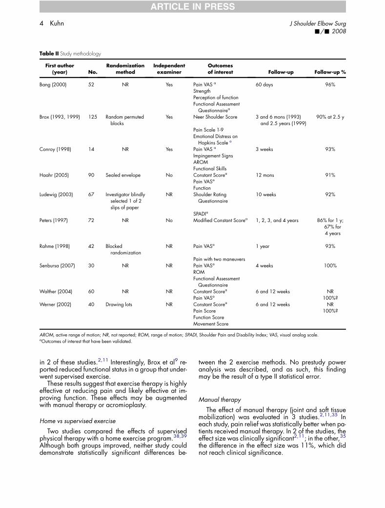
Table II Study methodology
First author(year) No.
Randomizationmethod
Independentexaminer
Outcomesof interest Follow-up Follow-up %
Bang (2000) 52 NR Yes Pain VAS a 60 days 96%StrengthPerception of functionFunctional Assessment
Questionnairea
Brox (1993, 1999) 125 Random permutedblocks
Yes Neer Shoulder Score 3 and 6 mons (1993)and 2.5 years (1999)
90% at 2.5 y
Pain Scale 1-9Emotional Distress on
Hopkins Scale a
Conroy (1998) 14 NR Yes Pain VAS a 3 weeks 93%Impingement SignsAROMFunctional Skills
Haahr (2005) 90 Sealed envelope No Constant Scorea 12 mons 91%Pain VASa
FunctionLudewig (2003) 67 Investigator blindly
selected 1 of 2slips of paper
NR Shoulder RatingQuestionnaire
10 weeks 92%
SPADIa
Peters (1997) 72 NR No Modified Constant Scorea 1, 2, 3, and 4 years 86% for 1 y;67% for4 years
Rahme (1998) 42 Blockedrandomization
NR Pain VASa 1 year 93%
Pain with two maneuversSenbursa (2007) 30 NR NR Pain VASa 4 weeks 100%
ROMFunctional Assessment
QuestionnaireWalther (2004) 60 NR NR Constant Scorea 6 and 12 weeks NR
Pain VASa 100%?Werner (2002) 40 Drawing lots NR Constant Scorea 6 and 12 weeks NR
Pain Score 100%?Function ScoreMovement Score
AROM, active range of motion; NR, not reported; ROM, range of motion; SPADI, Shoulder Pain and Disability Index; VAS, visual analog scale.aOutcomes of interest that have been validated.
4 Kuhn J Shoulder Elbow Surg-/- 2008
ARTICLE IN PRESS
in 2 of these studies.2,11 Interestingly, Brox et al9 re-ported reduced functional status in a group that under-went supervised exercise.
These results suggest that exercise therapy is highlyeffective at reducing pain and likely effective at im-proving function. These effects may be augmentedwith manual therapy or acromioplasty.
Home vs supervised exercise
Two studies compared the effects of supervisedphysical therapy with a home exercise program.38,39
Although both groups improved, neither study coulddemonstrate statistically significant differences be-
tween the 2 exercise methods. No prestudy poweranalysis was described, and as such, this findingmay be the result of a type II statistical error.
Manual therapy
The effect of manual therapy (joint and soft tissuemobilization) was evaluated in 3 studies.2,11,35 Ineach study, pain relief was statistically better when pa-tients received manual therapy. In 2 of the studies, theeffect size was clinically significant2,11; in the other,35
the difference in the effect size was 11%, which didnot reach clinical significance.
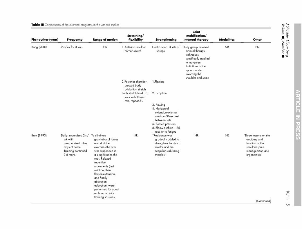
Table III Components of the exercise programs in the various studies
First author (year) Frequency Range of motionStretching/flexibility Strengthening
Jointmobilization/
manual therapy Modalities Other
Bang (2000) 2�/wk for 3 wks NR 1.Anterior shouldercorner stretch
Elastic band: 3 sets of10 reps
Study group receivedmanual therapytechniquesspecifically appliedto movementlimitations in theupper quarterinvolving theshoulder and spine
NR NR
2.Posterior shouldercrossed bodyadduction stretch
1.Flexion
Each stretch hold 30secs with 10-secrest, repeat 3�
2. Scaption
3. Rowing4. Horizontal
extension-externalrotation 60-sec restbetween sets
5. Seated press up6. Elbow push-up + 25
reps or to fatigueBrox (1993) Daily: supervised 2�/
wk withunsupervised otherdays at home.Training continued3-6 mons.
To eliminategravitational forcesand start theexercises the armwas suspended ina sling fixed to theroof. Relaxedrepetitivemovements (firstrotation, thenflexion-extension,and finallyabduction-adduction) wereperformed for aboutan hour in dailytraining sessions.
NR ‘‘Resistance wasgradually added tostrengthen the shortrotator and thescapular stabilizingmuscles’’
NR NR ‘‘Three lessons on theanatomy andfunction of theshoulder, painmanagement, andergonomics’’
(Continued )
JShoulder
ElbowSurg
Kuhn
5Volum
e-
,N
umber
-
AR
TIC
LE
INP
RE
SS
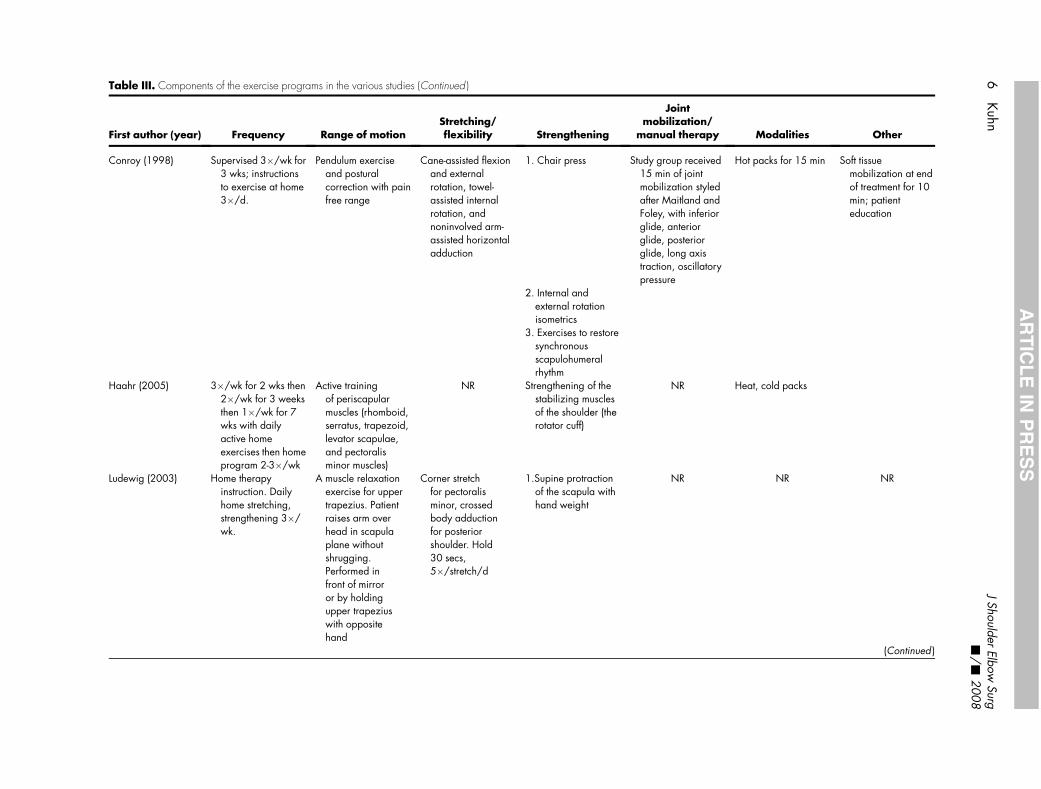
Table III. Components of the exercise programs in the various studies (Continued )
First author (year) Frequency Range of motionStretching/flexibility Strengthening
Jointmobilization/
manual therapy Modalities Other
Conroy (1998) Supervised 3�/wk for3 wks; instructionsto exercise at home3�/d.
Pendulum exerciseand posturalcorrection with painfree range
Cane-assisted flexionand externalrotation, towel-assisted internalrotation, andnoninvolved arm-assisted horizontaladduction
1. Chair press Study group received15 min of jointmobilization styledafter Maitland andFoley, with inferiorglide, anteriorglide, posteriorglide, long axistraction, oscillatorypressure
Hot packs for 15 min Soft tissuemobilization at endof treatment for 10min; patienteducation
2. Internal andexternal rotationisometrics
3. Exercises to restoresynchronousscapulohumeralrhythm
Haahr (2005) 3�/wk for 2 wks then2�/wk for 3 weeksthen 1�/wk for 7wks with dailyactive homeexercises then homeprogram 2-3�/wk
Active trainingof periscapularmuscles (rhomboid,serratus, trapezoid,levator scapulae,and pectoralisminor muscles)
NR Strengthening of thestabilizing musclesof the shoulder (therotator cuff)
NR Heat, cold packs
Ludewig (2003) Home therapyinstruction. Dailyhome stretching,strengthening 3�/wk.
A muscle relaxationexercise for uppertrapezius. Patientraises arm overhead in scapulaplane withoutshrugging.Performed infront of mirroror by holdingupper trapeziuswith oppositehand
Corner stretchfor pectoralisminor, crossedbody adductionfor posteriorshoulder. Hold30 secs,5�/stretch/d
1.Supine protractionof the scapula withhand weight
NR NR NR
(Continued )
6Kuhn
JShoulder
ElbowSurg
-/
-2008
AR
TIC
LE
INP
RE
SS
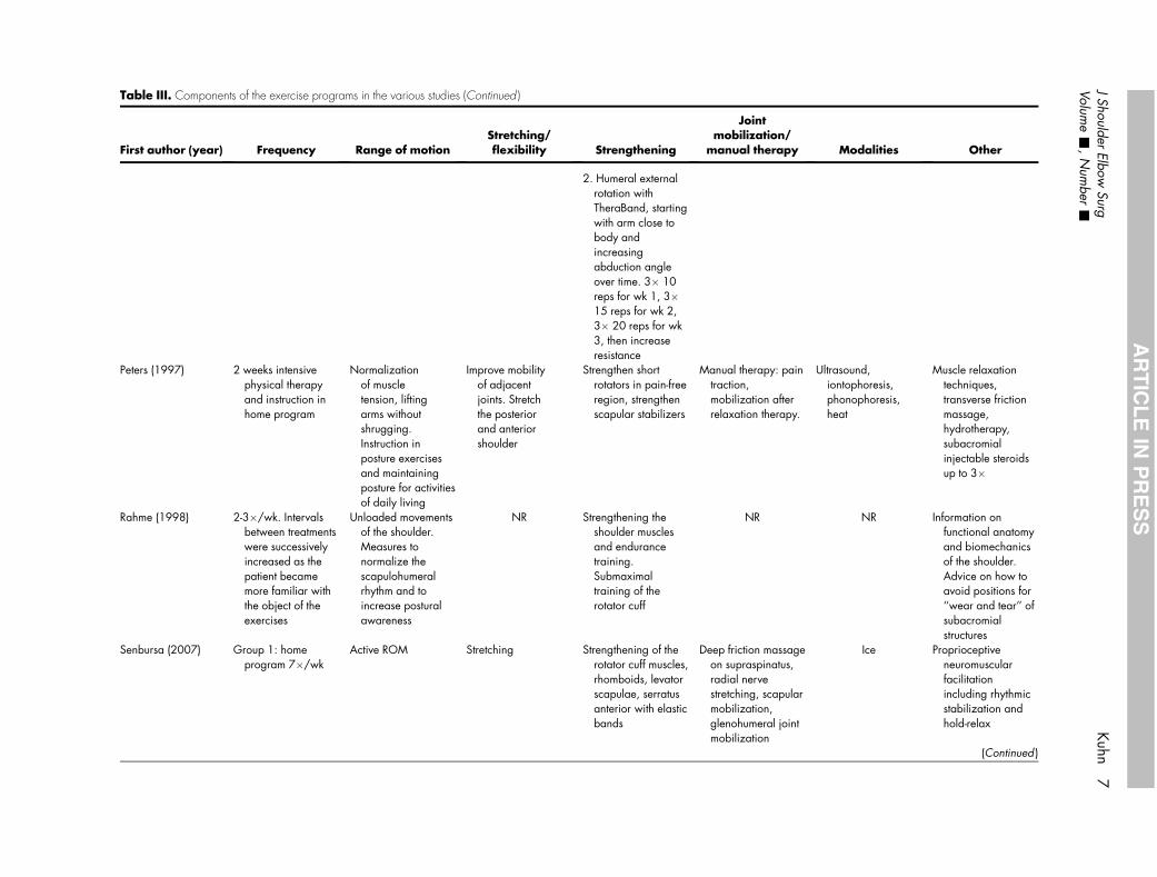
Table III. Components of the exercise programs in the various studies (Continued )
First author (year) Frequency Range of motionStretching/flexibility Strengthening
Jointmobilization/
manual therapy Modalities Other
2. Humeral externalrotation withTheraBand, startingwith arm close tobody andincreasingabduction angleover time. 3� 10reps for wk 1, 3�15 reps for wk 2,3� 20 reps for wk3, then increaseresistance
Peters (1997) 2 weeks intensivephysical therapyand instruction inhome program
Normalizationof muscletension, liftingarms withoutshrugging.Instruction inposture exercisesand maintainingposture for activitiesof daily living
Improve mobilityof adjacentjoints. Stretchthe posteriorand anteriorshoulder
Strengthen shortrotators in pain-freeregion, strengthenscapular stabilizers
Manual therapy: paintraction,mobilization afterrelaxation therapy.
Ultrasound,iontophoresis,phonophoresis,heat
Muscle relaxationtechniques,transverse frictionmassage,hydrotherapy,subacromialinjectable steroidsup to 3�
Rahme (1998) 2-3�/wk. Intervalsbetween treatmentswere successivelyincreased as thepatient becamemore familiar withthe object of theexercises
Unloaded movementsof the shoulder.Measures tonormalize thescapulohumeralrhythm and toincrease posturalawareness
NR Strengthening theshoulder musclesand endurancetraining.Submaximaltraining of therotator cuff
NR NR Information onfunctional anatomyand biomechanicsof the shoulder.Advice on how toavoid positions for‘‘wear and tear’’ ofsubacromialstructures
Senbursa (2007) Group 1: homeprogram 7�/wk
Active ROM Stretching Strengthening of therotator cuff muscles,rhomboids, levatorscapulae, serratusanterior with elasticbands
Deep friction massageon supraspinatus,radial nervestretching, scapularmobilization,glenohumeral jointmobilization
Ice Proprioceptiveneuromuscularfacilitationincluding rhythmicstabilization andhold-relax
(Continued )
JShoulder
ElbowSurg
Kuhn
7Volum
e-
,N
umber
-
AR
TIC
LE
INP
RE
SS
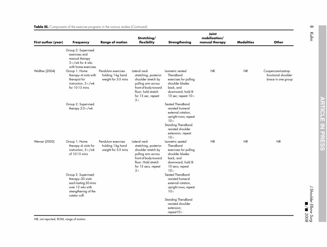
Table III. Components of the exercise programs in the various studies (Continued )
First author (year) Frequency Range of motionStretching/flexibility Strengthening
Jointmobilization/
manual therapy Modalities Other
Group 2: Supervisedexercises andmanual therapy3�/wk for 4 wkswith home exercises
Walther (2004) Group 1: Hometherapy–4 visits withtherapist forinstruction, 5�/wkfor 10-15 mins
Pendulum exercisesholding 1-kg handweight for 3-5 mins
Lateral neckstretching, posteriorshoulder stretch bypulling arm acrossfront of body towardfloor; hold stretchfor 15 sec, repeat3�
Isometric seatedTheraBandexercises for pullingshoulder bladesback, anddownward; hold 8-10 sec; repeat 10�
NR NR Coopercare-Lastrapfunctional shoulderbrace in one group
Group 2: Supervisedtherapy 2-3�/wk
Seated TheraBandresisted humeralexternal rotation,upright rows; repeat10�
Standing TheraBandresisted shoulderextension; repeat10�
Werner (2002) Group 1: Hometherapy–4 visits forinstruction, 5�/wkof 10-15 mins
Pendulum exercisesholding 1-kg handweight for 3-5 mins
Lateral neckstretching, posteriorshoulder stretch bypulling arm acrossfront of body towardfloor. Hold stretchfor 15 secs, repeat3�
Isometric seatedTheraBandexercises for pullingshoulder bladesback, anddownward; hold 8-10 secs; repeat10�
NR NR NR
Group 2: Supervisedtherapy–30 visitseach lasting 30 minsover 12 wks withstrengthening of therotator cuff.
Seated TheraBandresisted humeralexternal rotation,upright rows; repeat10�
Standing TheraBandresisted shoulderextension;repeat10�
NR, not reported; ROM, range of motion.
8Kuhn
JShoulder
ElbowSurg
-/
-2008
AR
TIC
LE
INP
RE
SS
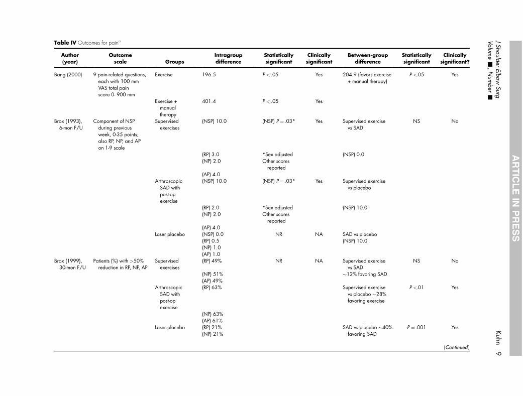
Table IV Outcomes for paina
Author(year)
Outcomescale Groups
Intragroupdifference
Statisticallysignificant
Clinicallysignificant
Between-groupdifference
Statisticallysignificant
Clinicallysignificant?
Bang (2000) 9 pain-related questions,each with 100 mmVAS total painscore 0- 900 mm
Exercise 196.5 P < .05 Yes 204.9 (favors exercise+ manual therapy)
P <.05 Yes
Exercise +manualtherapy
401.4 P < .05 Yes
Brox (1993),6-mon F/U
Component of NSPduring previousweek, 0-35 points;also RP, NP, and APon 1-9 scale
Supervisedexercises
(NSP) 10.0 (NSP) P ¼ .03* Yes Supervised exercisevs SAD
NS No
(RP) 3.0 *Sex adjusted (NSP) 0.0(NP) 2.0 Other scores
reported(AP) 4.0
ArthroscopicSAD withpost-opexercise
(NSP) 10.0 (NSP) P ¼ .03* Yes Supervised exercisevs placebo
(RP) 2.0 *Sex adjusted (NSP) 10.0(NP) 2.0 Other scores
reported(AP) 4.0
Laser placebo (NSP) 0.0 NR NA SAD vs placebo(RP) 0.5 (NSP) 10.0(NP) 1.0(AP) 1.0
Brox (1999),30-mon F/U
Patients (%) with >50%reduction in RP, NP, AP
Supervisedexercises
(RP) 49% NR NA Supervised exercisevs SAD
NS No
(NP) 51% �12% favoring SAD(AP) 49%
ArthroscopicSAD withpost-opexercise
(RP) 63% Supervised exercisevs placebo �28%favoring exercise
P <.01 Yes
(NP) 63%(AP) 61%
Laser placebo (RP) 21% SAD vs placebo �40%favoring SAD
P ¼ .001 Yes(NP) 21%
(Continued )
JShoulder
ElbowSurg
Kuhn
9Volum
e-
,N
umber
-
AR
TIC
LE
INP
RE
SS
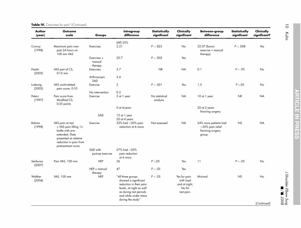
Table IV. Outcomes for paina (Continued )
Author(year)
Outcomescale Groups
Intragroupdifference
Statisticallysignificant
Clinicallysignificant
Between-groupdifference
Statisticallysignificant
Clinicallysignificant?
(AP) 25%Conroy
(1998)Maximum pain over
past 24 hours on100 mm VAS
Exercises 2.21 P ¼ .823 No 32.07 (favorsexercise + manualtherapy)
P ¼ .008 Yes
Exercises +manualtherapy
20.7 P ¼ .005 Yes
Haahr(2005)
VAS part of CS,0-15 mm
Exercises 3.7 NR NA 0.1 P ¼ .93 No
ArthroscopicSAD
3.6
Ludewig(2003)
VAS work-relatedpain score, 0-10
Exercise 2 P < .001 Yes 1.5 P <.05 No
No intervention 0.5Peters
(1997)Pain score from
Modified CS,0-35 points
Exercise 5 at 1 year No statisticalanalysis
NA 10 at 1 year NR NA
0 at 4-years 20 at 2 yearsfavoring surgery
SAD 15 at 1 year20 at 4 years
Rahme(1998)
VAS pain at rest+ VAS pain lifting 1-Lbottle with armextended. Datapresented as relativereduction in pain frompretreatment score
Exercise 33% had >50% painreduction at 6 mons
Not assessed NA 24% more patients had>50% pain relieffavoring surgerygroup.
NS NA
SAD withpost-op exercise
57% had >50%pain reductionat 6 mons
Senbursa(2007)
Pain VAS, 100 mm HEP 36 P <.05 Yes 11 P ¼ .05 No
HEP + manualtherapy
47 P < .05 Yes
Walther(2004)
VAS, 100 mm HEP ‘‘All three groupsshowed a significantreduction in their painlevels, at night as wellas during rest periodsand while under stressduring the study’’
P < .05 Yes for painwith load
and at night,No for
rest pain.
Minimal NS No
(Continued )
10
Kuhn
JShoulder
ElbowSurg
-/
-2008
AR
TIC
LE
INP
RE
SS
Table
IV.O
utco
mes
forp
aina
(Con
tinue
d)
Auth
or
(year)
Outc
om
esc
ale
Gro
ups
Intr
agro
up
dif
fere
nce
Sta
tist
ically
signifi
cant
Clinic
ally
signifi
cant
Betw
een-g
roup
dif
fere
nce
Sta
tist
ically
signifi
cant
Clinic
ally
signifi
cant?
Pain
atre
st,ni
ght
,lo
adre
cord
edSu
perv
ised
exer
cise
sBr
ace
Wer
ner
(2002)
Com
pone
ntof
CS,
0-3
5po
ints
HEP
App
rox
9-p
oint
impr
ovem
ent
No
stat
istic
alan
alys
isN
AM
inim
aldi
ffere
nces
betw
een
gro
ups
NRb
NA
Supe
rvis
edth
erap
yA
ppro
x8-p
oint
impr
ovem
ent
AP,
activ
itypa
in;C
S,C
onst
antS
core
,F/U
,fo
llow
-up;
HEP
,hom
eex
erci
sepr
ogra
m;N
A,n
otap
plic
able
;N
P,ni
ght
pain
;NR,
notr
epor
ted;
NS,
nots
igni
fican
t;N
SP,N
eersc
ale
forpa
in;R
P,re
stpa
in;S
AD
,sub
-ac
rom
iald
ecom
pres
sion
,VA
S,vi
sual
anal
ogsc
ale.
aA
utho
rsus
eda
variet
yof
scal
esto
mea
sure
pain
.In
tragro
updi
ffere
nces
dete
ctth
eef
fect
ofth
epa
rtic
ular
prot
ocol
over
time
and
com
pare
pret
reat
men
twith
stat
usat
follo
w-u
p.Be
twee
n-gro
updi
ffere
nces
are
repo
rted
fordi
ffere
nces
betw
een
the
diffe
rent
prot
ocol
s.C
linic
ally
signi
fican
tfind
ings
occu
rif
the
diffe
renc
esar
est
atis
tical
lysi
gni
fican
tand
the
mag
nitu
deof
the
diffe
renc
eis
20%
orm
ore.
b‘‘S
tatis
tical
lyth
edi
ffere
nces
betw
een
the
indi
vidu
alco
mpo
nent
s(o
fthe
Con
stan
tSco
re)w
ere
notd
iffer
entb
etw
een
the
two
gro
ups.
’’N
oP
valu
esgiv
en.
J Shoulder Elbow Surg Kuhn 11Volume -, Number -
ARTICLE IN PRESS
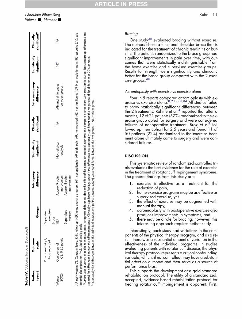
Bracing
One study38 evaluated bracing without exercise.The authors chose a functional shoulder brace that isindicated for the treatment of chronic tendinitis or bur-sitis. The patients randomized to the brace group hadsignificant improvements in pain over time, with out-comes that were statistically indistinguishable fromthe home exercise and supervised exercise groups.Results for strength were significantly and clinicallybetter for the brace group compared with the 2 exer-cise groups.38
Acromioplasty with exercise vs exercise alone
Four in 5 reports compared acromioplasty with ex-ercise vs exercise alone.8,9,17,33,34 All studies failedto show statistically significant differences betweenthe 2 treatments. Rahme et al34 reported that after 6months, 12 of 21 patients (57%) randomized to the ex-ercise group opted for surgery and were consideredfailures of nonoperative treatment. Brox et al8 fol-lowed up their cohort for 2.5 years and found 11 of50 patients (22%) randomized to the exercise treat-ment alone ultimately came to surgery and were con-sidered failures.
DISCUSSION
This systematic review of randomized controlled tri-als evaluates the best evidence for the role of exercisein the treatment of rotator cuff impingement syndrome.The general findings from this study are:
1. exercise is effective as a treatment for thereduction of pain,
2. home exercise programs may be as effective assupervised exercise, yet
3. the effect of exercise may be augmented withmanual therapy,
4. acromioplasty with postoperative exercise alsoproduces improvements in symptoms, and
5. there may be a role for bracing; however, thisinteresting approach requires further study.
Interestingly, each study had variations in the com-ponents of the physical therapy program, and as a re-sult, there was a substantial amount of variation in theeffectiveness of the individual programs. In studiesevaluating patients with rotator cuff disease, the phys-ical therapy protocol represents a critical confoundingvariable; which, if not controlled, may have a substan-tial effect on outcome and then serve as a source ofperformance bias.
This supports the development of a gold standardrehabilitation protocol. The utility of a standardized,accepted, evidence-based rehabilitation protocol fortreating rotator cuff impingement is apparent. First,
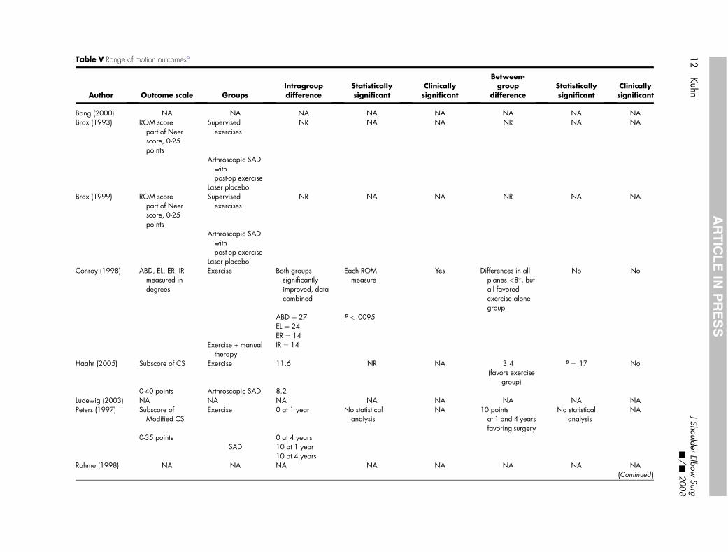
Table V Range of motion outcomesa
Author Outcome scale GroupsIntragroupdifference
Statisticallysignificant
Clinicallysignificant
Between-group
differenceStatisticallysignificant
Clinicallysignificant
Bang (2000) NA NA NA NA NA NA NA NABrox (1993) ROM score
part of Neerscore, 0-25points
Supervisedexercises
NR NA NA NR NA NA
Arthroscopic SADwithpost-op exercise
Laser placeboBrox (1999) ROM score
part of Neerscore, 0-25points
Supervisedexercises
NR NA NA NR NA NA
Arthroscopic SADwithpost-op exercise
Laser placeboConroy (1998) ABD, EL, ER, IR
measured indegrees
Exercise Both groupssignificantlyimproved, datacombined
Each ROMmeasure
Yes Differences in allplanes <8�, butall favoredexercise alonegroup
No No
ABD ¼ 27 P < .0095EL ¼ 24ER ¼ 14
Exercise + manualtherapy
IR ¼ 14
Haahr (2005) Subscore of CS Exercise 11.6 NR NA 3.4(favors exercise
group)
P ¼ .17 No
0-40 points Arthroscopic SAD 8.2Ludewig (2003) NA NA NA NA NA NA NA NAPeters (1997) Subscore of
Modified CSExercise 0 at 1 year No statistical
analysisNA 10 points
at 1 and 4 yearsfavoring surgery
No statisticalanalysis
NA
0-35 points 0 at 4 yearsSAD 10 at 1 year
10 at 4 yearsRahme (1998) NA NA NA NA NA NA NA NA
(Continued )
12
Kuhn
JShoulder
ElbowSurg
-/
-2008
AR
TIC
LE
INP
RE
SS
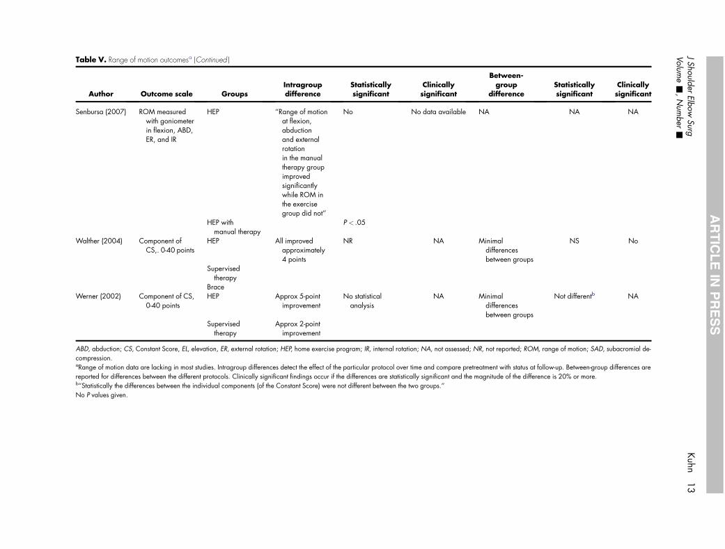
Table V. Range of motion outcomesa (Continued )
Author Outcome scale GroupsIntragroupdifference
Statisticallysignificant
Clinicallysignificant
Between-group
differenceStatisticallysignificant
Clinicallysignificant
Senbursa (2007) ROM measuredwith goniometerin flexion, ABD,ER, and IR
HEP ‘‘Range of motionat flexion,abductionand externalrotationin the manualtherapy groupimprovedsignificantlywhile ROM inthe exercisegroup did not’’
No No data available NA NA NA
HEP withmanual therapy
P < .05
Walther (2004) Component ofCS,. 0-40 points
HEP All improvedapproximately4 points
NR NA Minimaldifferencesbetween groups
NS No
Supervisedtherapy
BraceWerner (2002) Component of CS,
0-40 pointsHEP Approx 5-point
improvementNo statistical
analysisNA Minimal
differencesbetween groups
Not differentb NA
Supervisedtherapy
Approx 2-pointimprovement
ABD, abduction; CS, Constant Score, EL, elevation, ER, external rotation; HEP, home exercise program; IR, internal rotation; NA, not assessed; NR, not reported; ROM, range of motion; SAD, subacromial de-compression.aRange of motion data are lacking in most studies. Intragroup differences detect the effect of the particular protocol over time and compare pretreatment with status at follow-up. Between-group differences arereported for differences between the different protocols. Clinically significant findings occur if the differences are statistically significant and the magnitude of the difference is 20% or more.b‘‘Statistically the differences between the individual components (of the Constant Score) were not different between the two groups.’’No P values given.
JShoulder
ElbowSurg
Kuhn
13
Volume
-,N
umber
-
AR
TIC
LE
INP
RE
SS
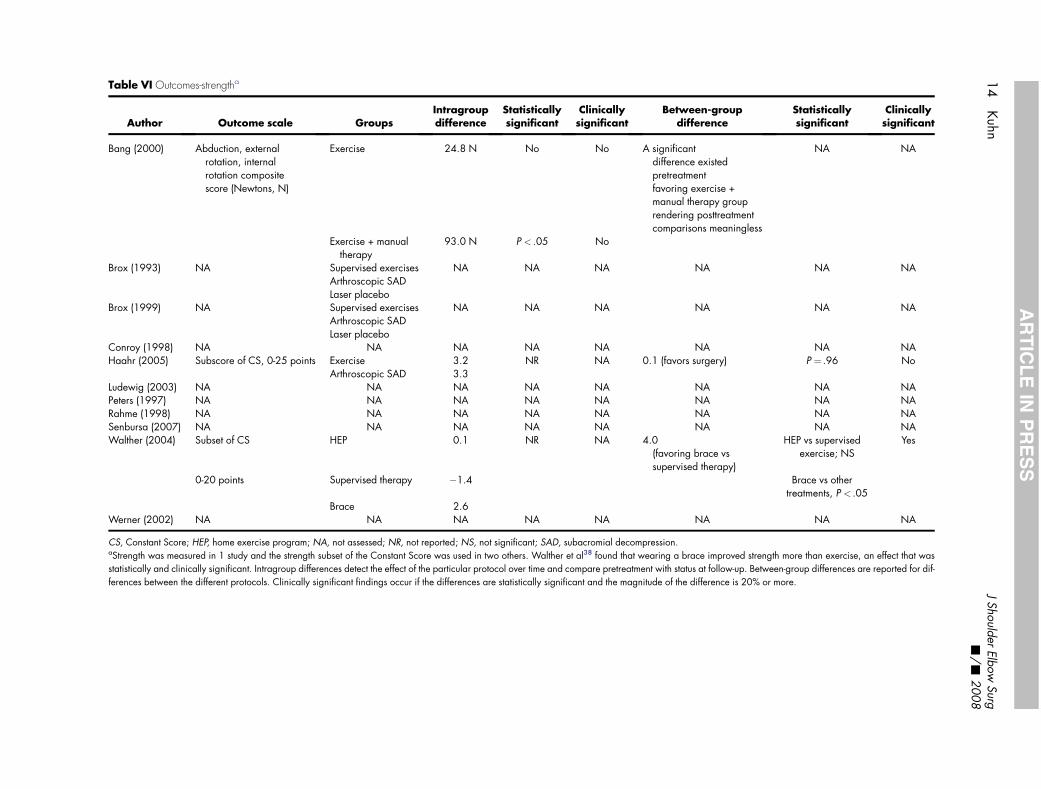
Table VI Outcomes-strengtha
Author Outcome scale GroupsIntragroupdifference
Statisticallysignificant
Clinicallysignificant
Between roupdiffer ce
Statisticallysignificant
Clinicallysignificant
Bang (2000) Abduction, externalrotation, internalrotation compositescore (Newtons, N)
Exercise 24.8 N No No A significantdifference ex tedpretreatmenfavoring exe ise +manual thera y grouprendering po treatmentcomparisons eaningless
NA NA
Exercise + manualtherapy
93.0 N P < .05 No
Brox (1993) NA Supervised exercises NA NA NA NA NA NAArthroscopic SADLaser placebo
Brox (1999) NA Supervised exercises NA NA NA NA NA NAArthroscopic SADLaser placebo
Conroy (1998) NA NA NA NA NA NA NA NAHaahr (2005) Subscore of CS, 0-25 points Exercise 3.2 NR NA 0.1 (favors sur ry) P ¼ .96 No
Arthroscopic SAD 3.3Ludewig (2003) NA NA NA NA NA NA NA NAPeters (1997) NA NA NA NA NA NA NA NARahme (1998) NA NA NA NA NA NA NA NASenbursa (2007) NA NA NA NA NA NA NA NAWalther (2004) Subset of CS HEP 0.1 NR NA 4.0
(favoring bra e vssupervised th rapy)
HEP vs supervisedexercise; NS
Yes
0-20 points Supervised therapy �1.4 Brace vs othertreatments, P < .05
Brace 2.6Werner (2002) NA NA NA NA NA NA NA NA
CS, Constant Score; HEP, home exercise program; NA, not assessed; NR, not reported; NS, not significant; SAD, subacromial decompression.aStrength was measured in 1 study and the strength subset of the Constant Score was used in two others. Walther et al38 found that wearing a brace proved strength more than exercise, an effect that wasstatistically and clinically significant. Intragroup differences detect the effect of the particular protocol over time and compare pretreatment with status a llow-up. Between-group differences are reported for dif-ferences between the different protocols. Clinically significant findings occur if the differences are statistically significant and the magnitude of the diffe nce is 20% or more.
14
Kuhn
JShoulder
ElbowSurg
-/
-2008
AR
TIC
LE
INP
RE
SS
-gen
istrcpstm
ge
ce
imt fore
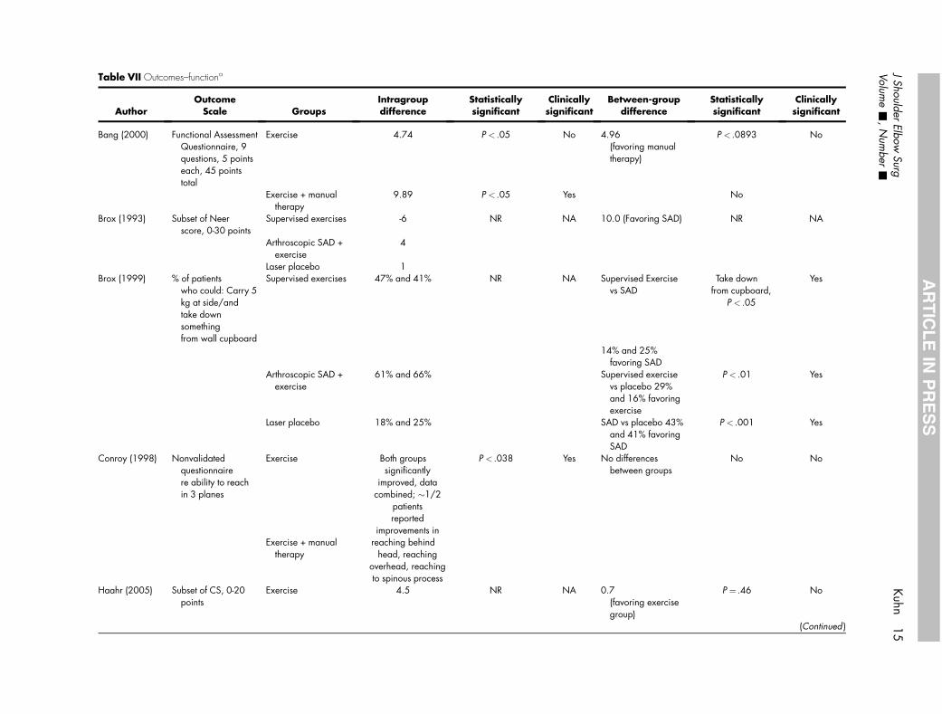
Table VII Outcomes–functiona
AuthorOutcome
Scale GroupsIntragroupdifference
Statisticallysignificant
Clinicallysignificant
Betwee -groupdiffe ence
Statisticallysignificant
Clinicallysignificant
Bang (2000) Functional AssessmentQuestionnaire, 9questions, 5 pointseach, 45 pointstotal
Exercise 4.74 P < .05 No 4.96(favorin manualtherapy
P < .0893 No
Exercise + manualtherapy
9.89 P < .05 Yes No
Brox (1993) Subset of Neerscore, 0-30 points
Supervised exercises -6 NR NA 10.0 (Fav ing SAD) NR NA
Arthroscopic SAD +exercise
4
Laser placebo 1Brox (1999) % of patients
who could: Carry 5kg at side/andtake downsomethingfrom wall cupboard
Supervised exercises 47% and 41% NR NA Supervise Exercisevs SAD
Take downfrom cupboard,
P < .05
Yes
14% and %favoring AD
Arthroscopic SAD +exercise
61% and 66% Supervise exercisevs place o 29%and 16 favoringexercise
P < .01 Yes
Laser placebo 18% and 25% SAD vs pl ebo 43%and 41 favoringSAD
P < .001 Yes
Conroy (1998) Nonvalidatedquestionnairere ability to reachin 3 planes
Exercise Both groupssignificantly
improved, datacombined; �1/2
patientsreported
improvements in
P < .038 Yes No differe cesbetwee groups
No No
Exercise + manualtherapy
reaching behindhead, reaching
overhead, reachingto spinous process
Haahr (2005) Subset of CS, 0-20points
Exercise 4.5 NR NA 0.7(favorin exercisegroup)
P ¼ .46 No
(Continued )
JShoulder
ElbowSurg
Kuhn
15
Volume
-,N
umber
-
AR
TIC
LE
INP
RE
SS
nr
g)
or
d
25S
db
%
ac%
nn
g
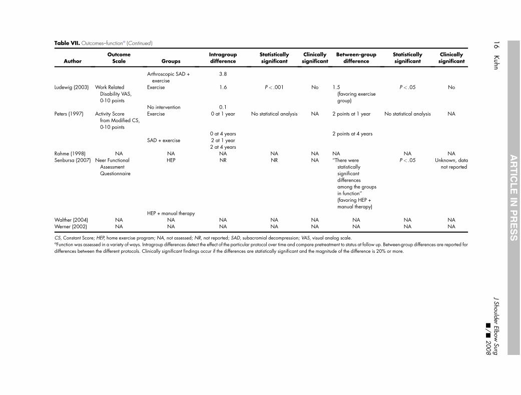
Table VII. Outcomes–functiona (Continued )
AuthorOutcome
Scale GroupsIntragroupdifference
Statisticallysignificant
Clinicallysignificant
Between-groupdifference
Statisticallysignificant
Clinicallysignificant
Arthroscopic SAD +exercise
3.8
Ludewig (2003) Work RelatedDisability VAS,0-10 points
Exercise 1.6 P < .001 No 1.5(favoring exercisegroup)
P < .05 No
No intervention 0.1Peters (1997) Activity Score
from Modified CS,0-10 points
Exercise 0 at 1 year No statistical analysis NA 2 points at 1 year No statistical analysis NA
0 at 4 years 2 points at 4 yearsSAD + exercise 2 at 1 year
2 at 4 yearsRahme (1998) NA NA NA NA NA NA NA NASenbursa (2007) Neer Functional
AssessmentQuestionnaire
HEP NR NR NA ‘‘There werestatisticallysignificantdifferencesamong the groupsin function’’(favoring HEP +manual therapy)
P < .05 Unknown, datanot reported
HEP + manual therapyWalther (2004) NA NA NA NA NA NA NA NAWerner (2002) NA NA NA NA NA NA NA NA
CS, Constant Score; HEP, home exercise program; NA, not assessed; NR, not reported; SAD, subacromial decompression; VAS, visual analog scale.aFunction was assessed in a variety of ways. Intragroup differences detect the effect of the particular protocol over time and compare pretreatment to status at follow up. Between-group differences are reported fordifferences between the different protocols. Clinically significant findings occur if the differences are statistically significant and the magnitude of the difference is 20% or more.
16
Kuhn
JShoulder
ElbowSurg
-/
-2008
AR
TIC
LE
INP
RE
SS
J Shoulder Elbow Surg Kuhn 17Volume -, Number -
ARTICLE IN PRESS
physicians and therapists will know that their patientsare receiving the best available rehabilitation pro-gram that has the greatest likelihood of improvingthe patient’s condition and avoiding surgery. Second,an accepted gold standard rehabilitation protocolwould reduce confounding variables and perfor-mance bias in research studies. This will allow com-parison of results between studies. A gold standardprotocol would also serve as a control allowing thestudy of modifications, such as modalities, adding ex-ercises or other treatments, eliminating specific com-ponents, and clarifying the effect of the investigatedtreatment. To assist with this, we synthesized the proto-cols described in these reviewed articles to developa standard rehabilitation protocol.
Data from the rehabilitation protocols used in thesearticles were compiled in table format (Table III). Infor-mation about specific components was extracted, in-cluding frequency, range of motion, flexibility,strengthening, manual therapy, and modalities, andthen synthesized into a comprehensive protocol(Appendix I).
Different authors had their patients perform exer-cises at different frequencies, ranging from twiceweekly2 to daily.8,9,24,35 Some authors used super-vised therapy with greater frequency early, progress-ing toward home exercises later.17,34
On the basis of this information, we suggest that pa-tients have supervised therapy 2 to 3 times each week,with the addition of manual therapy (see subsequenttext). Patients who no longer need manual therapyand have developed proficiency in the protocol canbe progressed to a home exercise program. Rangeof motion exercises and flexibility should be per-formed daily. Strengthening should be performed 3times weekly.
Range of motion exercises were described by mostauthors. Pendulum exercises were used in the cohortsof Conroy et al,11 Walther et al,38 and Werner et al.39
Postural exercises, such as shrugs, were used by Con-roy el al,11 Peters et al,33 and Rahme et al.34 Activeassisted range of motion was described witha cane,11 with the arm suspended,8,9 or with the otherarm.24 Brox et al8,9 recommended active assisted mo-tion with the arm suspended in a sling for rotation, flex-ion-extension, and abduction-adduction. Ludewig etal24 had patients stand before a mirror and work onshoulder elevation without shrugging. If a mirror wasnot available, they had the patient place the unin-volved hand on the contralateral trapezius to providefeedback, making sure the upper trapezius remainedrelaxed during elevation of the arm.24 Haahr et al17
described active training of the periscapular muscles(rhomboid, serratus, trapezoid, levator, and pectora-lis minor).
The conclusion from this information is that all pa-tients may begin their range of motion work with pos-
tural exercise, such as shrugs, and shoulder retraction.Glenohumeral motion should begin with pendulum ex-ercises, progress to active assisted motion, then to ac-tive motion as comfort dictates. Active assisted motionmay be performed with a cane, suspended with pul-leys, or with the uninvolved arm. Active motion maybe performed in front of a mirror or using the oppositehand on the trapezius to prevent hiking of the shoul-der, as described by Ludewig et al.24
Flexibility exercises generally were performed foranterior and posterior shoulder tightness.2,11,24,38,39
In addition, Conroy et al11 had patients performcane-assisted stretching in flexion and external rota-tion. A variety of techniques were described for poste-rior shoulder stretching, most commonly cross-bodyadduction.2,11,24,38,39 Interestingly, McClure et al27
conducted a randomized trial comparing 2 differenttechniques to stretch the posterior shoulder—thesleeper stretch and the cross-body stretch—and foundthat the cross-body stretch was most effective.27 Withregard to anterior shoulder stretching, Borstad et al5
performed a randomized trial of 3 stretches designedto stretch the pectoralis minor, consisting of unilateralself-stretch, supine manual stretch, and sitting manualstretch. Although all patients demonstrated gains inpectoralis minor length, they found the unilateral self-stretch (performed in a corner or on a door jamb) pro-duced the greatest effect.5 Most authors recommen-ded holding each stretch for 15 or 30 seconds andrepeating 3 to 5 times, with a 10-second rest betweeneach stretch.2,24,38,39
These data indicate that stretching should be per-formed daily and should include anterior shoulderstretching, performed by the patient in a corner ordoor jamb, and posterior shoulder stretching, usingthe cross-body adduction technique. Each stretchshould be held for 30 seconds and repeated 5 times,with a 10-second rest between each stretch. Canestretching in forward elevation and external rotationmay also be used in a similar fashion.
Some authors did not provide much detail regard-ing their programs for strengthening, other than re-porting that muscles of the rotator cuff and scapulastabilizers were involved.17,34,35 Others were morespecific in describing their exercise programs. For ex-ample, strengthening exercises include shoulder flex-ion,2 extension,38,39 scaption,2 rows,2,38,39 internalrotation of the adducted arm,2,11,24 and external rota-tion of the adducted arm.2,11,24,38,39
Most authors used elastic bands.2,24,35,38,39 Mostallowed joint movement for isotonic exercise2,24,35;others relied on static resistance with isometric musclecontraction.11,38,39
Each exercise was performed at 3 sets of 10 repe-titions with a 60-second rest between each set2 or 3sets of 10 the first week, followed by 3 sets of 15 thesecond week, followed by 3 sets of 20 the third
18 Kuhn J Shoulder Elbow Surg-/- 2008
ARTICLE IN PRESS
week; then increasing TheraBand (Hygenic Corp, Ak-ron, OH) resistance was used.24
Scapular stabilizing exercises included the seatedpress up2,11 and the elbow push-up plus2 and wereperformed on a chair or stable bench. Each was per-formed as 1 set of 25 repetitions.2 Supine push-upplus with a hand weight was used by Ludewig et al.24
The synthesis of these reports clearly shows thatstrengthening exercises should focus on the rotatorcuff and scapular stabilizing muscles. Rotator cuffstrengthening should involve the following exerciseswith the TheraBand: internal rotation with arm ad-ducted to side, external rotation with arm adductedto side, and scaption, if there is no pain associatedwith the exercise. Scapular stabilizer strengtheningshould include chair press, push-up plus (prone usingbody weight or supine with hand weight), and uprightrows using an elastic band. Combination strengthen-ing while standing using elastic bands should includeforward elevation and extension. Each exerciseshould be performed as 3 sets of 10 repetitions, withincreases in elastic resistance as strength improves.
Manual therapy has been shown to be effective ataugmenting the effect of exercise in relieving symp-toms of the impingement syndrome.2,11,35 Manualtherapy includes a variety of techniques, includingjoint mobilization, as described by Maitland25 andFoley et al,14 and soft tissue mobilization (effleurage,friction, and kneading techniques).11,17
Because the evidence favors the use of manual ther-apy, it should be included in a standard evidence-based protocol. Like exercise, the different varied as-pects of manual therapy are worthy of further studyto identify which components are effective in treat-ment. Manual therapy requires working with a physi-cal therapist. During the period that patients arereceiving manual therapy, they should be thoroughlyinstructed in the exercise program. Patients who nolonger need manual therapy should be progressedto a home exercise program.
Ultrasound as a therapeutic modality has beenevaluated by a number of studies. It is beyond thescope of this review to evaluate the effectiveness of ul-trasound; however, multiple systematic reviews statethat ultrasound is of little value in treating patientswith shoulder pain.16,28,37 Conroy et al11 and Haahret al17 both used heat in their protocols. Haahr et al17
and Senbursa et al35 used ice. There are no data for oragainst the use of cold or heat as a modality; thus, theiruse must be optional at this point. It is clear, however,that ultrasound has no value in a rehabilitation proto-col for the impingement syndrome.
With this information we offer a gold standard re-habilitation protocol (Appendix I). It is important to rec-ognize that this evidence-based protocol is not withoutlimitations. The protocol described is a collection offeatures that have demonstrated a reduction in symp-
toms for impingement syndrome in randomized con-trolled trials. Some components in these studies maybe unnecessary. Other features, which may be benefi-cial, may not be included. This may be reflective of an-other limitation of this study; namely, the diagnosis ofimpingement syndrome is based on a provocative testdesigned to produce pain in the subacromial space.32
The Neer impingement sign32 and the Hawkins im-pingement sign18 may be imperfect tools to diagnoserotator cuff disease because they both have relativelypoor specificities.19
It could be argued that impingement syndrome isnot a diagnosis at all; but rather, is the finding of a pro-vocative physical examination test that could be pro-duced by a variety of subacromial pathologies,including subacromial bursitis, bursal sided partial ro-tator cuff tears, biceps tendinitis, scapular dyskinesis,a tight posterior capsule, and postural abnormalities,among others. As a result, the protocol proposed inthis article may need modifications to make it specificto a particular patient’s anatomic diagnosis. For exam-ple, it may not be applicable to an athlete with rotatorcuff pain due to excessive laxity in the shoulder. In ad-dition, this protocol cannot be extrapolated to the post-operative state, where the clinicians may be interestedin protecting a healing rotator cuff.
Despite these limitations, this systematic review ofthe best available evidence for exercise in the treat-ment of impingement syndrome was able to generatea physical therapy protocol that has been shown to beeffective in level 1 and level 2 studies. This evidence-based protocol can be used by clinicians treatingimpingement syndrome and can serve as a gold stan-dard to reduce variables in future cohort and compar-ative studies to help find better treatments for patientswith this disorder.
Thanks to members of the Vanderbilt Sports MedicineJournal Club who assisted in reviewing the articles: KurtSpindler, Warren Dunn, Buddy Hannah, Andrew Gregory,Paul Rummo, Tara Holmes, Mick Koester, and KevinDoulens.
REFERENCES
1. Ainsworth R, Lewis JS. Exercise therapy for the conservative man-agement of full thickness tears of the rotator cuff: a systematicreview. Br J Sports Med 2007;41:200-10.
2. Bang M, Deyle G. Comparison of supervised exercise with andwithout manual physical therapy for patients with impingement syn-drome. J Ortho Sports Phys Ther 2000;30:126-37.
3. Bennell K, Coburn S, Wee E, et al. Efficacy and cost-effectivenessof a physiotherapy program for chronic rotator cuff pathology:a protocol for a randomised, double-blind, placebo-controlledtrial. BMC Musculoskelet Disord 2007;8:86.
4. Bohmer AS, Staff PH, Brox JI. Supervised exercises in relation to ro-tator cuff disease (impingement syndrome stages II and III): a treat-ment regimen and its rationale. Physiother Theory Pract 1998;14:93-105.
J Shoulder Elbow Surg Kuhn 19Volume -, Number -
ARTICLE IN PRESS
5. Borstad JD, Ludewig PM. Comparison of three stretches for the pec-toralis minor muscle. J Shoulder Elbow Surg 2006;15:324-30.
6. Brewster C, Schwab DR. Rehabilitation of the shoulder followingrotator cuff injury or surgery. J Orthop Sports Phys Ther 1993;18:422-6.
7. Browning DG, Desai MM. Rotator cuff injuries and treatment. PrimCare Clin Office Pract 2004;31:807-29.
8. Brox JI, Gjengedal E, Uppheim G, et al. Arthroscopic surgery ver-sus supervised exercises in patient with rotator cuff disease (stage IIimpingement syndrome): A prospective, randomised, controlledstudy in 125 patients with a 2 1⁄2 year follow-up. J Shoulder ElbowSurg 1997;8:102-11.
9. Brox J, Staff P, Ljunggren A, Brevik J. Arthroscopic surgery com-pared with supervised exercises in patients with rotator cuff dis-ease. BMJ 1993;307:899-903.
10. Cakmak A. Conservative treatment of subacromial impingementsyndrome. Acta Orthop Traumatol Turc 2003;37(Suppl 1):112-8.
11. Conroy DE, Hayes KW. The effect of mobilization as a componentof comprehensive treatment for primary shoulder impingement syn-drome. J Ortho Sports Phys Ther 1998;28:3-14.
12. Desmeules F, Cote CH, Fremont P. Therapeutic exercise and ortho-paedic manual therapy for impingement syndrome. A systematicreview. Clin J Sports Med 2003;13:176-82.
13. Ellenbecker TS, Derscheid GL. Rehabilitation of overuse injuries ofthe shoulder. Clin Sports Med 1989 Jul;8:583-604.
14. Foley R, Janos S, Johnson R, Petersen C. Active and passive move-ment testing of the extremities, spine, pelvis, and temporomandib-ular joint. In: Petersen C, editor. Teaching manual for physicaltherapy. Chicago: Northwestern University, Department of Physi-cal Therapy and Human Movement Sciences; 1994. p. 34–68.
15. Grant HJ, Arthur A, Pichora DR. Evaluation of interventions for rota-tor cuff pathology: a systematic review. J Hand Ther 2004;17:274-99.
16. Green S, Buchbinder R, Hetrick S. Physiotherapy interventions forshoulder pain. Cochrane Database Syst Rev 2003;2:CD004258.
17. Haahr JP, Ostergaard S, Dalsgaard J, Norup K, Frost P, Lausen S,et al. Exercises versus arthroscopic decompression in patients withsubacromial impingement: a randomised, controlled study in 90cases with a one year follow up. Ann Rheum Dis 2005;64:760-4.
18. Hawkins RJ, Kennedy JC. Impingement syndrome in athletes. Am JSports Med 1980;8:151-8.
19. Hegedus EJ, Goode A, Campbell S, et al. Physical examinationtests of the shoulder: a systematic review with meta-analysis of indi-vidual tests. Br J Sports Med 2008;42:80-92.
20. Jobe FW, Moynes DR. Delineation of diagnostic criteria and a re-habilitation program for rotator cuff injuries. Am J Sports Med1982;10:226-9.
21. Kibler WB, McMullen J, Uhl T. Shoulder rehabilitation strategies,guidelines, and practice. Orthop Clin North Am 2001;32:527-38.
22. Kibler WB. Rehabilitation of rotator cuff tendinopathy. Clin SportsMed 2003;22:837-47.
23. Krabak BJ, Sugar R, McFarland EG. Practical nonoperative man-agement of rotator cuff injuries. Clin J Sport Med 2003;13:102-5.
24. Ludewig PM, Borstad JD. Effects of a home exercise programme onshoulder pain and functional status in construction workers. OccupEnviron Med 2003;60:841-9.
25. Maitland G. Peripheral manipulation. London, UK: Butterworth-Heinmann Ltd; 1991: 47–52, 129-67.
26. Mantone JK, Burkhead WZ Jr, Noonan J Jr. Nonoperative treat-ment of rotator cuff tears. Orthop Clin North Am 2000;31:295-311.
27. McClure P, Balaicuis J, Heiland D, Broersma ME, Thorndike CK,Wood A. A randomized controlled comparison of stretching pro-cedures for posterior shoulder tightness. J Orthop Sports Phys Ther2007;37:108-14.
28. Michener LA, Walsworth MK, Burnet EN. Effectiveness of rehabil-itation for patients with subacromial impingement syndrome: a sys-tematic review. J Hand Ther 2004;17:152-64.
29. Millett PJ, Wilcox RB III, O’Holleran JD, Warner JJP. Rehabilitationof the rotator cuff: an evaluation-based approach. J Am AcadOrtho Surg 2006;14:599-609.
30. Morrison DS, Frogameni AD, Woodworth P. Conservative man-agement for subacromial impingement syndrome. J Bone JointSurg Am 1997;79:732-7.
31. Morrison DS, Greenbaum BS, Einhorn A. Shoulder impingement.Orthop Clin North Am 2000;31:285-93.
32. Neer CS 2nd. Impingement lesions. Clin Orthop Relat Res1983:70-7.
33. Peters G, Kohn D. Medium-tern clinical results after operative andnon-operative treatment of subacromial impingement. Unfallchir-urg 1997;100:623-9.
34. Rahme H, Solem-Bertoft E, Westerberg CE, Lundberg E,Sorensen S, Hilding S. The subacromial impingement syndrome.A study of results of treatment with special emphasis on predictivefactors and pain-generating mechanisms. Scand J Rehab Med1998;30:253-62.
35. Senbursa G, Baltaci G, Atay A. Comparison of conservativetreatment with and without manual physical therapy for patientswith shoulder impingement syndrome: a prospective, random-ized clinical trial. Knee Surg Sports Traumatol Arthrosc2007;15:915-21.
36. Spindler KP, Kuhn JE, Dunn W, Matthews CE, Harrell FE Jr,Dittus RS. Reading and reviewing the orthopaedic literature: a sys-tematic, evidence-based medicine approach. J Am Acad OrthopSurg 2005;13:220-9.
37. Van Der Heijden GJ. Physiotherapy for patients with soft tissue dis-orders: a systematic review of randomized clinical trials. BMJ1997;315:25-30.
38. Walther M, Werner A, Stahlschmidt T, Woeffel R, Gohlke F. Thesubacromial impingement syndrome of the shoulder treated byconventional physiotherapy, self-training, and a shoulder brace:results of a prospective, randomized study. J Shoulder ElbowSurg 2004;13:417-23.
39. Werner A, Walther M, Ilg A, Stahlschmidt T, Gohlke F. Self-train-ing versus conventional physiotherapy in subacromial impinge-ment syndrome [German]. Z Orthop Ihre Grenzgeb 2002;140:375-80.
Appendix I Evidence-based medicine exercise pro-tocol for impingement syndrome
General instructions: This physical therapy proto-col is based on the best evidence demonstrating a ben-eficial effect for exercise in the treatment of rotator cufftendonitis. It is largely unknown if adding or eliminat-ing exercises will affect the outcome. Range of motionand stretching exercises should be performed daily.Strengthening should be performed 3 times weekly.Modalities: Heat or cold, or both, may be used.Studies have demonstrated that the results of ultra-sound treatment are no better than results in control pa-tients, and it should not be used.Manual therapy: Joint and soft tissue mobilizationtechniques have been shown to augment the effect ofthe exercise program. Initially, supervised exerciseswith manual therapy are recommended. During thattime patients, should be instructed in a home program.Patients can move entirely to a home program whenthey no longer are in need of manual therapy.
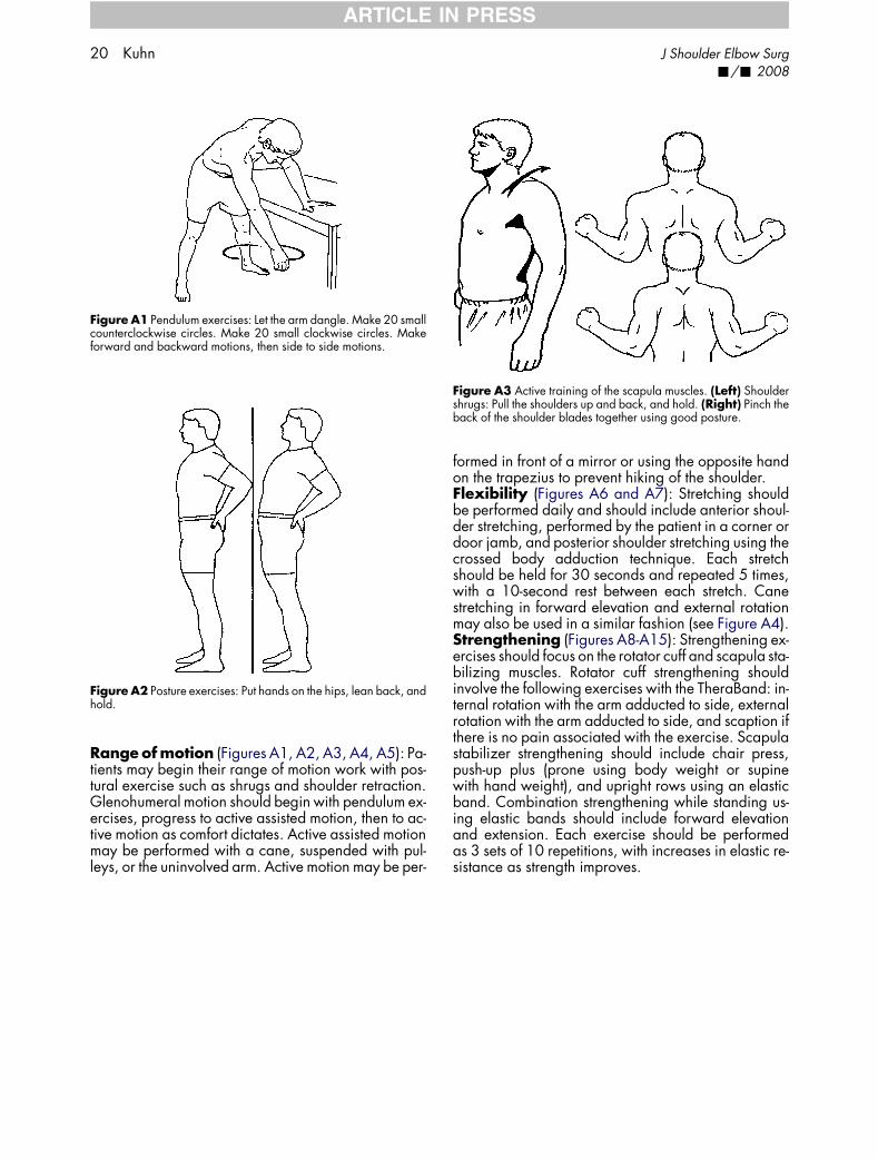
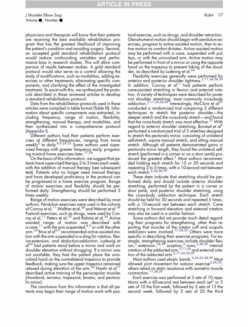
Figure A1 Pendulum exercises: Let the arm dangle. Make 20 smallcounterclockwise circles. Make 20 small clockwise circles. Makeforward and backward motions, then side to side motions.
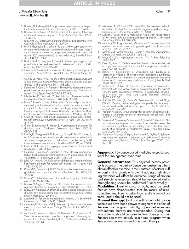
Figure A2 Posture exercises: Put hands on the hips, lean back, andhold.
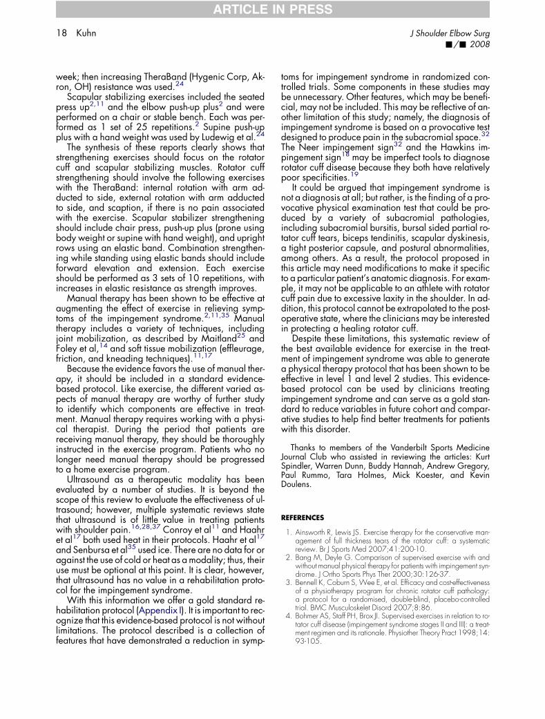
Figure A3 Active training of the scapula muscles. (Left) Shouldershrugs: Pull the shoulders up and back, and hold. (Right) Pinch theback of the shoulder blades together using good posture.
20 Kuhn J Shoulder Elbow Surg-/- 2008
ARTICLE IN PRESS
Range of motion (Figures A1, A2, A3, A4, A5): Pa-tients may begin their range of motion work with pos-tural exercise such as shrugs and shoulder retraction.Glenohumeral motion should begin with pendulum ex-ercises, progress to active assisted motion, then to ac-tive motion as comfort dictates. Active assisted motionmay be performed with a cane, suspended with pul-leys, or the uninvolved arm. Active motion may be per-
formed in front of a mirror or using the opposite handon the trapezius to prevent hiking of the shoulder.Flexibility (Figures A6 and A7): Stretching shouldbe performed daily and should include anterior shoul-der stretching, performed by the patient in a corner ordoor jamb, and posterior shoulder stretching using thecrossed body adduction technique. Each stretchshould be held for 30 seconds and repeated 5 times,with a 10-second rest between each stretch. Canestretching in forward elevation and external rotationmay also be used in a similar fashion (see Figure A4).Strengthening (Figures A8-A15): Strengthening ex-ercises should focus on the rotator cuff and scapula sta-bilizing muscles. Rotator cuff strengthening shouldinvolve the following exercises with the TheraBand: in-ternal rotation with the arm adducted to side, externalrotation with the arm adducted to side, and scaption ifthere is no pain associated with the exercise. Scapulastabilizer strengthening should include chair press,push-up plus (prone using body weight or supinewith hand weight), and upright rows using an elasticband. Combination strengthening while standing us-ing elastic bands should include forward elevationand extension. Each exercise should be performedas 3 sets of 10 repetitions, with increases in elastic re-sistance as strength improves.
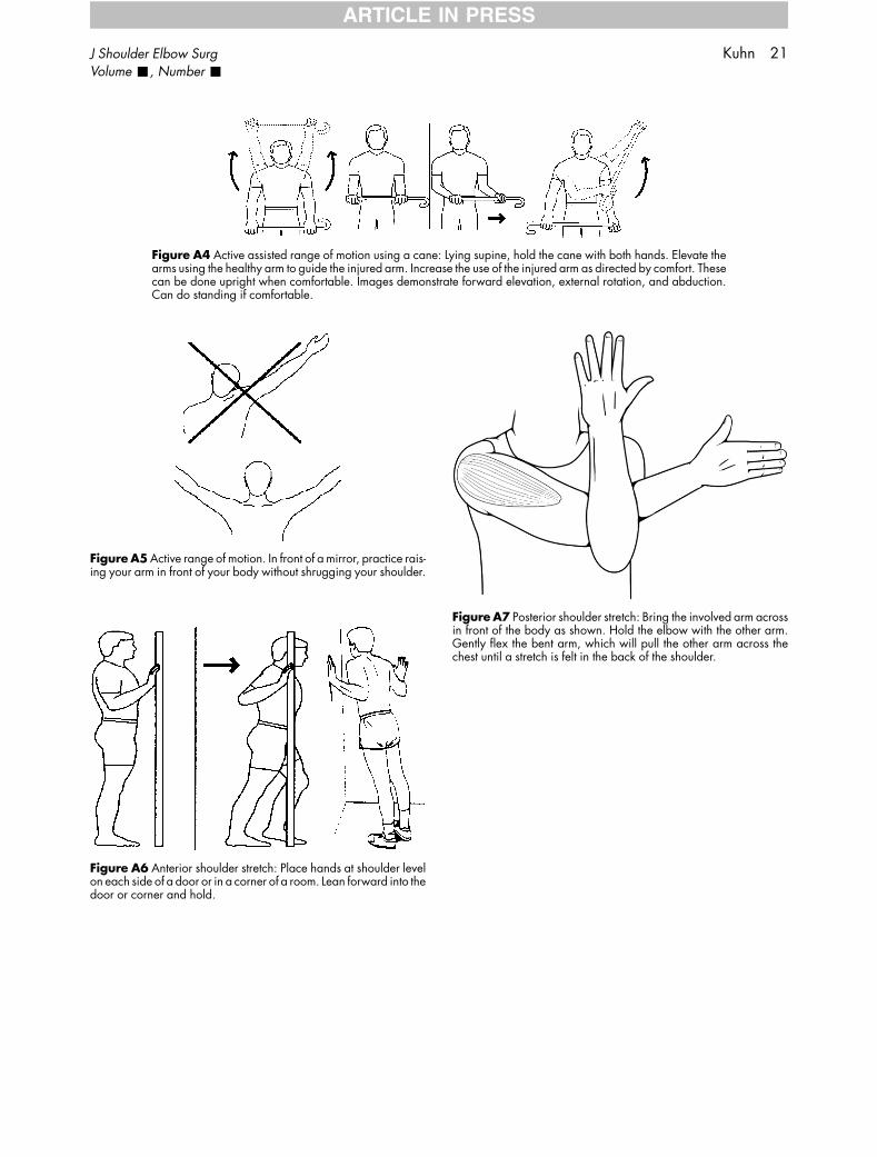
Figure A5 Active range of motion. In front of a mirror, practice rais-ing your arm in front of your body without shrugging your shoulder.
Figure A6 Anterior shoulder stretch: Place hands at shoulder levelon each side of a door or in a corner of a room. Lean forward into thedoor or corner and hold.
Figure A4 Active assisted range of motion using a cane: Lying supine, hold the cane with both hands. Elevate thearms using the healthy arm to guide the injured arm. Increase the use of the injured arm as directed by comfort. Thesecan be done upright when comfortable. Images demonstrate forward elevation, external rotation, and abduction.Can do standing if comfortable.
Figure A7 Posterior shoulder stretch: Bring the involved arm acrossin front of the body as shown. Hold the elbow with the other arm.Gently flex the bent arm, which will pull the other arm across thechest until a stretch is felt in the back of the shoulder.
J Shoulder Elbow Surg Kuhn 21Volume -, Number -
ARTICLE IN PRESS
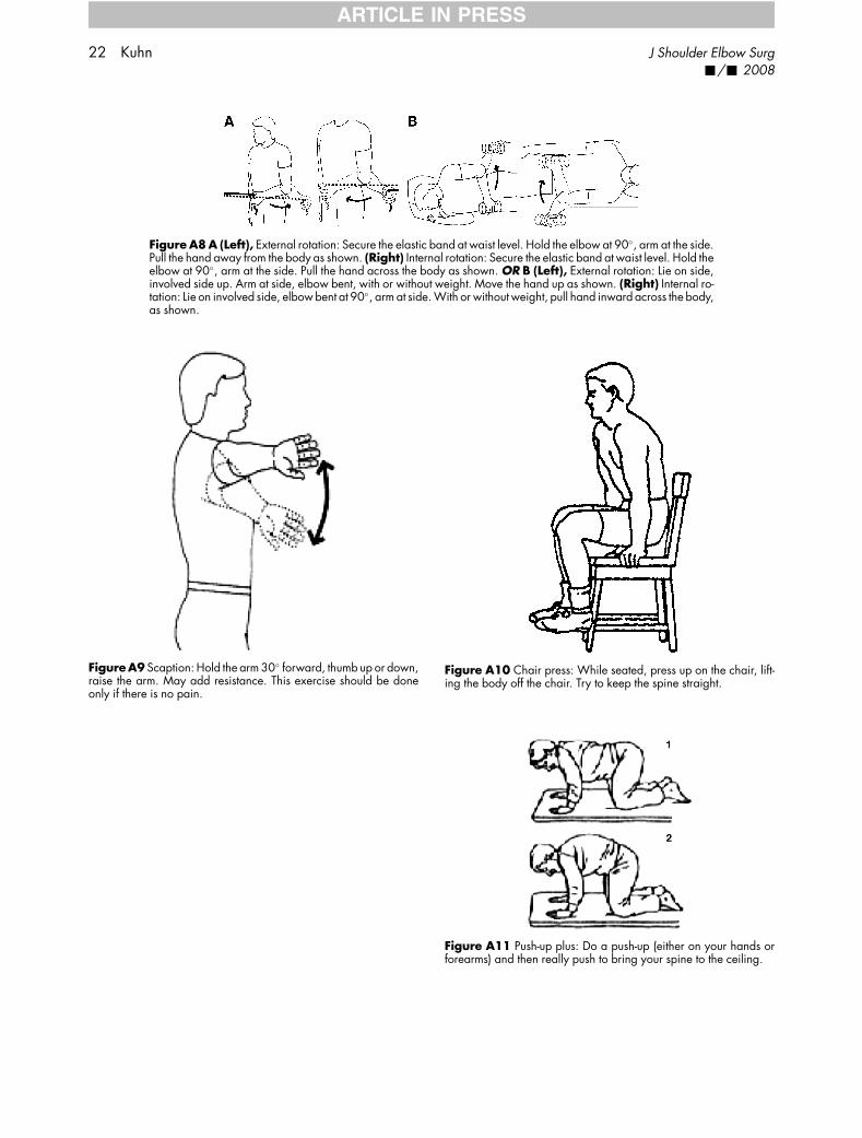
Figure A10 Chair press: While seated, press up on the chair, lift-ing the body off the chair. Try to keep the spine straight.
Figure A8 A (Left), External rotation: Secure the elastic band at waist level. Hold the elbow at 90�, arm at the side.Pull the hand away from the body as shown. (Right) Internal rotation: Secure the elastic band at waist level. Hold theelbow at 90�, arm at the side. Pull the hand across the body as shown. OR B (Left), External rotation: Lie on side,involved side up. Arm at side, elbow bent, with or without weight. Move the hand up as shown. (Right) Internal ro-tation: Lie on involved side, elbow bent at 90�, arm at side. With or without weight, pull hand inward across the body,as shown.
FigureA9 Scaption: Hold the arm 30� forward, thumb up or down,raise the arm. May add resistance. This exercise should be doneonly if there is no pain.
Figure A11 Push-up plus: Do a push-up (either on your hands orforearms) and then really push to bring your spine to the ceiling.
22 Kuhn J Shoulder Elbow Surg-/- 2008
ARTICLE IN PRESS
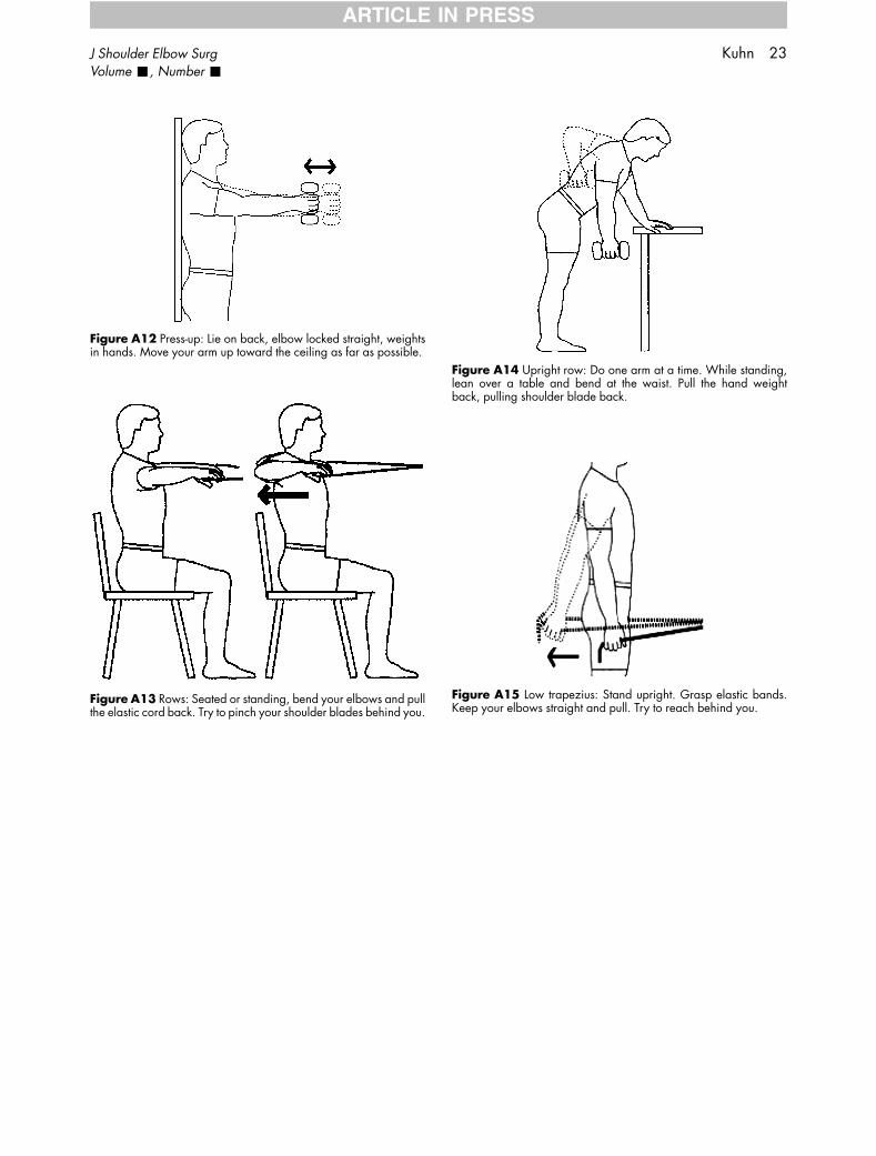
Figure A14 Upright row: Do one arm at a time. While standing,lean over a table and bend at the waist. Pull the hand weightback, pulling shoulder blade back.
Figure A15 Low trapezius: Stand upright. Grasp elastic bands.Keep your elbows straight and pull. Try to reach behind you.
Figure A12 Press-up: Lie on back, elbow locked straight, weightsin hands. Move your arm up toward the ceiling as far as possible.
Figure A13 Rows: Seated or standing, bend your elbows and pullthe elastic cord back. Try to pinch your shoulder blades behind you.
J Shoulder Elbow Surg Kuhn 23Volume -, Number -
ARTICLE IN PRESS