exertional rhabdomyolysis: physiological response or...
TRANSCRIPT
Exertional rhabdomyolysis:physiological response or manifestationof an underlying myopathy?
Renata S Scalco,1 Marc Snoeck,2 Ros Quinlivan,1 Susan Treves,3,4
Pascal Laforét,5 Heinz Jungbluth,6,7,8 Nicol C Voermans9
To cite: Scalco RS,Snoeck M, Quinlivan R, et al.Exertional rhabdomyolysis:physiological response ormanifestation of anunderlying myopathy?BMJ Open Sport Exerc Med2016;2:e000151.doi:10.1136/bmjsem-2016-000151
▸ Prepublication history forthis paper is available online.To view these files pleasevisit the journal online(http://dx.doi.org/10.1136/bmjsem-2016-000151).
HJ and NCV shared seniorauthorship.
Accepted 1 August 2016
For numbered affiliations seeend of article.
Correspondence toDr Renata Scalco; [email protected]
ABSTRACTExertional rhabdomyolysis is characterised by musclebreakdown associated with strenuous exercise ornormal exercise under extreme circumstances. Keyfeatures are severe muscle pain and sudden transientelevation of serum creatine kinase (CK) levels with orwithout associated myoglobinuria. Mild cases mayremain unnoticed or undiagnosed. Exertionalrhabdomyolysis is well described among athletes andmilitary personnel, but may occur in anybody exposedto unaccustomed exercise. In contrast, exertionalrhabdomyolysis may be the first manifestation of agenetic muscle disease that lowers the exercisethreshold for developing muscle breakdown. Repeatedepisodes of exertional rhabdomyolysis should raise thesuspicion of such an underlying disorder, in particularin individuals in whom the severity of therhabdomyolysis episodes exceeds the expectedresponse to the exercise performed. The present reviewaims to provide a practical guideline for the acutemanagement and postepisode counselling of patientswith exertional rhabdomyolysis, with a particularemphasis on when to suspect an underlying geneticdisorder. The pathophysiology and its clinical featuresare reviewed, emphasising four main stepwiseapproaches: (1) the clinical significance of an acuteepisode, (2) risks of renal impairment, (3) clinicalindicators of an underlying genetic disorders and (4)when and how to recommence sport activity followingan acute episode of rhabdomyolysis. Geneticbackgrounds that appear to be associated with bothenhanced athletic performance and increasedrhabdomyolysis risk are briefly reviewed.
INTRODUCTIONExertional rhabdomyolysis (ERM) is thegeneral term for muscle breakdown asso-ciated with strenuous exercise and is welldescribed among athletes and military per-sonnel. Although there is no universallyaccepted definition, ERM is often defined asa clinical syndrome associated with severemuscle pain, sudden elevation (and subse-quent fall) of serum creatine kinase (CK)levels with or without myoglobinuria.Individuals experiencing ERM present to a
variety of physicians, in particular to generalpractitioners and physicians working insports medicine, emergency and internalmedicine, neurology and the neuromuscularfield, and for the military. As a result of thediversity of medical personnel having to dealwith ERM, most physicians will have experi-ence with some but not necessarily allaspects of this important condition.ERM results in the entry of skeletal muscle
contents, in particular CK and myoglobin,into the systemic circulation. In most cases,the condition has a mild course charac-terised by postexertional myalgia withmild-to-moderate CK increases, mild pigmen-turia, and will often not even come tomedical attention. However, in a minority ofpatients the clinical course can be severe,resulting in marked hyperCKaemia, compart-ment syndrome, acute renal injury, dissemi-nated intravascular coagulation, cardiacarrhythmias secondary to electrolyte imbal-ances, and even cardiac arrest if leftuntreated. The annual rhabdomyolysis
What are the new findings
▪ Exertional rhabdomyolysis (ERM) may be thefirst manifestation of a genetic muscle diseasethat lowers the exercise threshold for developingmuscle breakdown.
▪ Consider a genetic cause of the ERM in case ofrecurrent episodes; high creatine kinase (CK)levels (>50×upper limit of normal) or persistinghyperCKaemia; absence of unaccustomed exer-cise; absence of (recreational or medical) drugs;or a positive family history of rhabdomyolysis orother exertional symptoms.
▪ Type 1 ryanodine receptor (RYR1) mutationshave been recently recognised to account for asubstantial proportion of patients presenting withERM.
▪ Participants with a genetic predisposition toERM need specific guidance when they recom-mence exercise and sporting activities.
Scalco RS, et al. BMJ Open Sport Exerc Med 2016;2:e000151. doi:10.1136/bmjsem-2016-000151 1
Open Access Reviewcopyright.
on 8 May 2018 by guest. P
rotected byhttp://bm
jopensem.bm
j.com/
BM
J Open S
port Exerc M
ed: first published as 10.1136/bmjsem
-2016-000151 on 7 Septem
ber 2016. Dow
nloaded from
prevalence has been reported as 26 000 cases in theUSA, with 47% meeting the diagnostic criteria of ERM.1
In other studies, a lower percentage of ERM has beensuggested,2 a discrepancy which reflects differences inthe cohorts studied.ERM most often represents a ‘physiological’ response
to extreme physical exercise or to normal exercise underextreme circumstances. In contrast, ERM might also bethe first manifestation of a genetic muscle disease, whichlowers the exercise threshold for developing musclebreakdown. A genetic cause should always be consideredin individuals with repeated episodes of ERM and inthose in whom the severity of rhabdomyolysis exceedsthe expected response to the exercise performed.The recognition of a patient with an underlying
genetic disorder is essential for acute management aswell as (and most importantly) for counselling after-wards. First, in type 1 ryanodine receptor (RYR1)-relatedcases of ERM,3 the specific RyR1 antagonist dantrolenecould be administered to limit further muscle break-down and CK increases and more severe, potentially life-threatening downstream medical complications. Patientswith RYR1-related ERM will have a higher recurrencerisk and will have to be specifically advised with regardto subsequent sporting activities. The same patients arealso at increased risk of malignant hyperthermia suscep-tibility (MHS), a pharmacogenetic response to volatileanaesthetics and/or the depolarising muscle relaxantsuccinylcholine, with important consequences foraffected individuals but possibly also for their relativescarrying the same RYR1 variants. Furthermore, certaingenetic myopathies carrying an increased rhabdomyoly-sis risk may also be associated with cardiac or other sys-temic symptoms that will have to be screened for. Finally,certain medications may be relatively contraindicatedand exercise advice should be individually adapted incase of an underlying genetic cause.The present review therefore aims to provide a prac-
tical guideline for the acute management and postepi-sode counselling of patients with ERM. The vast majorityof the relevant literature until now has focused either onthe physiological circumstances of ERM (particularly inpublications with an emphasis on sports or militarymedicine),4 5 on general causes of rhabdomyolysis,6 oron genetic myopathies which may predispose to rhabdo-myolysis (particularly in publications with an emphasison emergency medicine, genetics, neurology and neuro-muscular medicine).7–9 As a result, physicians workingin sports or military and emergency medicine are wellupdated on the symptoms and management of ERM,but may find it difficult to decide when to consider anunderlying genetic disorder. On the other hand, a neur-ologist may perform unnecessary muscle biopsies inhealthy individuals who have suffered from a singleepisode of ERM after extreme unaccustomed exercise.Therefore, a combined approach is necessary to recog-nise individuals who are at risk for recurrent rhabdo-myolysis and to advise patients with certain genetic
disorders on when and how to recommence exerciseand sporting activities.We will first discuss the pathophysiology of ERM and
its typical clinical features, and provide examples oftypical physiological ERM. Next, we will propose a struc-tured approach to patients with ERM, based on the fol-lowing four key questions: (1) is the ERM clinicallysignificant, requiring hospital admission and intravenousfluids administration? (2) What is the risk of developingacute renal failure (ARF)? (3) Is the ERM the (first)manifestation of a genetic disorder? And (4) when andhow to restart exercise and sporting activities? We expectthat such an approach will be of benefit for a widerange of physicians facing this severe and challengingcondition in different medical settings.
WHAT IS ERM?BackgroundIn muscle tissue, the process of neuronal signal transduc-tion leading to muscle contraction is called excitation-contraction coupling (ECC). During ECC, presynapticacetylcholine release induces postsynaptic depolarisationof the sarcolemma, resulting in activation of the voltage-gated L-type calcium channels (also known as dihydro-pyridine receptors (DHPRs)). Unlike in cardiac muscle,it is not this calcium influx itself but rather the directinteraction between DHPRs and RyR1s that causes theopening of RyR1 Ca2+ channels, leading to a rapidrelease of Ca2+ from the intracellular sarcoplasmic reticu-lum (SR) stores. The increased myoplasmic Ca2+ (fromabout 10-7 to 10-5 M) induces the effective interactionbetween thick and thin filaments by binding to troponinC, and this ultimately leads to muscle contraction. Aftercontraction, calcium needs to be restored. This happensthrough active transport facilitated by the Ca2+ATPase,an active process that on the molecular level takes 105
times longer than does RyR1-induced calcium releasebut is compensated for by the large number of Ca2+-ATPases on the surface of the SR membrane.10
Irrespective of its cause(s), the pathophysiologicalevents in rhabdomyolysis follow a common pathway.Normally, ion pumps and channels in the sarcolemmamaintain a low intracellular Na+ and Ca2+ and a highintracellular K+ concentration. Unaccustomed exercisemay cause direct injury to the sarcolemma (eg, in eccen-tric exercise) and/or lead to failure of energy pro-duction with subsequent pump dysfunction ofNa+/K+ATPase and Ca2+ATPase (eg, in prolonged train-ing beyond the limit of fatigue). Both processes lead toincreased cellular permeability to sodium ions and, con-sequently, increased intracellular calcium concentra-tions, with concordant muscle contraction increasingthe energy deficit. This increased intracellular calciumconcentration also enhances the activation of calcium-dependent proteases and phospholipases, which contri-butes to destruction of myofibrillar, cytoskeletal andmembrane proteins. Subsequently, large quantities of
2 Scalco RS, et al. BMJ Open Sport Exerc Med 2016;2:e000151. doi:10.1136/bmjsem-2016-000151
Open Accesscopyright.
on 8 May 2018 by guest. P
rotected byhttp://bm
jopensem.bm
j.com/
BM
J Open S
port Exerc M
ed: first published as 10.1136/bmjsem
-2016-000151 on 7 Septem
ber 2016. Dow
nloaded from
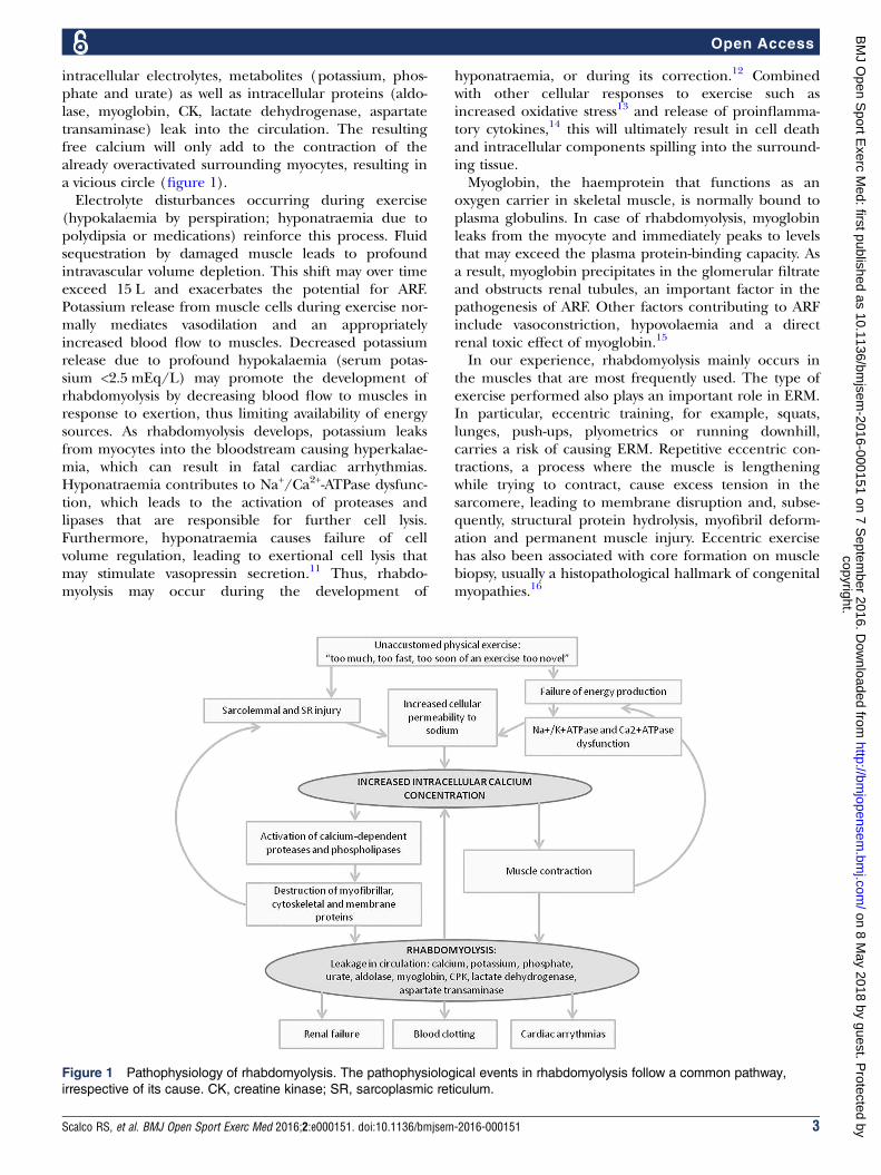
intracellular electrolytes, metabolites (potassium, phos-phate and urate) as well as intracellular proteins (aldo-lase, myoglobin, CK, lactate dehydrogenase, aspartatetransaminase) leak into the circulation. The resultingfree calcium will only add to the contraction of thealready overactivated surrounding myocytes, resulting ina vicious circle (figure 1).Electrolyte disturbances occurring during exercise
(hypokalaemia by perspiration; hyponatraemia due topolydipsia or medications) reinforce this process. Fluidsequestration by damaged muscle leads to profoundintravascular volume depletion. This shift may over timeexceed 15 L and exacerbates the potential for ARF.Potassium release from muscle cells during exercise nor-mally mediates vasodilation and an appropriatelyincreased blood flow to muscles. Decreased potassiumrelease due to profound hypokalaemia (serum potas-sium <2.5 mEq/L) may promote the development ofrhabdomyolysis by decreasing blood flow to muscles inresponse to exertion, thus limiting availability of energysources. As rhabdomyolysis develops, potassium leaksfrom myocytes into the bloodstream causing hyperkalae-mia, which can result in fatal cardiac arrhythmias.Hyponatraemia contributes to Na+/Ca2+-ATPase dysfunc-tion, which leads to the activation of proteases andlipases that are responsible for further cell lysis.Furthermore, hyponatraemia causes failure of cellvolume regulation, leading to exertional cell lysis thatmay stimulate vasopressin secretion.11 Thus, rhabdo-myolysis may occur during the development of
hyponatraemia, or during its correction.12 Combinedwith other cellular responses to exercise such asincreased oxidative stress13 and release of proinflamma-tory cytokines,14 this will ultimately result in cell deathand intracellular components spilling into the surround-ing tissue.Myoglobin, the haemprotein that functions as an
oxygen carrier in skeletal muscle, is normally bound toplasma globulins. In case of rhabdomyolysis, myoglobinleaks from the myocyte and immediately peaks to levelsthat may exceed the plasma protein-binding capacity. Asa result, myoglobin precipitates in the glomerular filtrateand obstructs renal tubules, an important factor in thepathogenesis of ARF. Other factors contributing to ARFinclude vasoconstriction, hypovolaemia and a directrenal toxic effect of myoglobin.15
In our experience, rhabdomyolysis mainly occurs inthe muscles that are most frequently used. The type ofexercise performed also plays an important role in ERM.In particular, eccentric training, for example, squats,lunges, push-ups, plyometrics or running downhill,carries a risk of causing ERM. Repetitive eccentric con-tractions, a process where the muscle is lengtheningwhile trying to contract, cause excess tension in thesarcomere, leading to membrane disruption and, subse-quently, structural protein hydrolysis, myofibril deform-ation and permanent muscle injury. Eccentric exercisehas also been associated with core formation on musclebiopsy, usually a histopathological hallmark of congenitalmyopathies.16
Figure 1 Pathophysiology of rhabdomyolysis. The pathophysiological events in rhabdomyolysis follow a common pathway,
irrespective of its cause. CK, creatine kinase; SR, sarcoplasmic reticulum.
Scalco RS, et al. BMJ Open Sport Exerc Med 2016;2:e000151. doi:10.1136/bmjsem-2016-000151 3
Open Accesscopyright.
on 8 May 2018 by guest. P
rotected byhttp://bm
jopensem.bm
j.com/
BM
J Open S
port Exerc M
ed: first published as 10.1136/bmjsem
-2016-000151 on 7 Septem
ber 2016. Dow
nloaded from
Definition of ERMWhat is generally agreed on are the following four keyfeatures of rhabdomyolysis:1. A CK elevation 12–36 hours after exercise, with a
maximum at 3–4 days, followed by normalisationwithin several weeks of rest;
2. The CK increase is preceded by exercise, usuallybeyond the limits of fatigue, also referred to as‘unaccustomed physical exertion’ or ‘involuntary exertion’;
3. The CK increase is symptomatic with any of the follow-ing features: myalgia (muscle soreness or tenderness;in general, rhabdomyolysis is very painful), swellingand/or weakness;
4. The presence of myoglobinaemia and/or myoglobinuria:either by inspection (pigmenturia) or by laboratorytesting. Since myoglobin testing in blood or urine isnot widely available, many experts consider the com-bination of the first three features diagnostic for ERM.
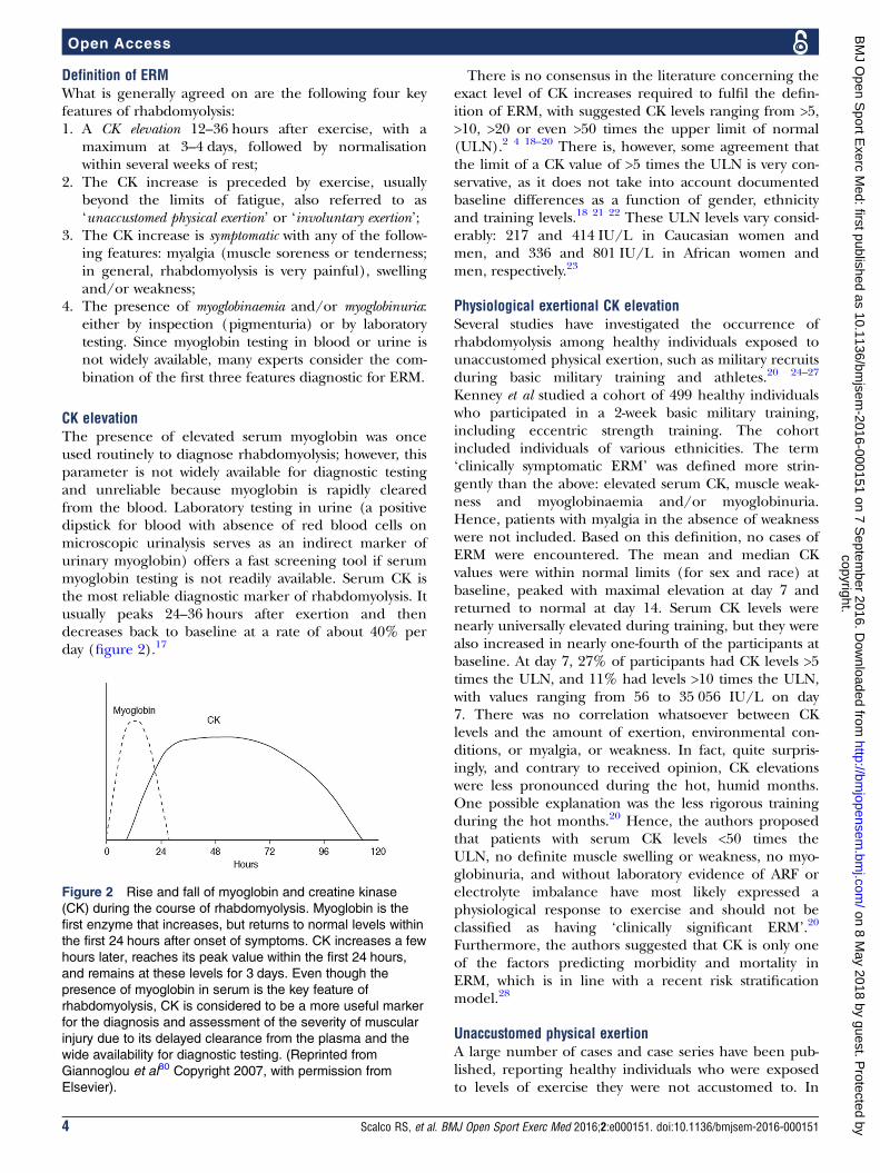
CK elevationThe presence of elevated serum myoglobin was onceused routinely to diagnose rhabdomyolysis; however, thisparameter is not widely available for diagnostic testingand unreliable because myoglobin is rapidly clearedfrom the blood. Laboratory testing in urine (a positivedipstick for blood with absence of red blood cells onmicroscopic urinalysis serves as an indirect marker ofurinary myoglobin) offers a fast screening tool if serummyoglobin testing is not readily available. Serum CK isthe most reliable diagnostic marker of rhabdomyolysis. Itusually peaks 24–36 hours after exertion and thendecreases back to baseline at a rate of about 40% perday (figure 2).17
There is no consensus in the literature concerning theexact level of CK increases required to fulfil the defin-ition of ERM, with suggested CK levels ranging from >5,>10, >20 or even >50 times the upper limit of normal(ULN).2 4 18–20 There is, however, some agreement thatthe limit of a CK value of >5 times the ULN is very con-servative, as it does not take into account documentedbaseline differences as a function of gender, ethnicityand training levels.18 21 22 These ULN levels vary consid-erably: 217 and 414 IU/L in Caucasian women andmen, and 336 and 801 IU/L in African women andmen, respectively.23
Physiological exertional CK elevationSeveral studies have investigated the occurrence ofrhabdomyolysis among healthy individuals exposed tounaccustomed physical exertion, such as military recruitsduring basic military training and athletes.20 24–27
Kenney et al studied a cohort of 499 healthy individualswho participated in a 2-week basic military training,including eccentric strength training. The cohortincluded individuals of various ethnicities. The term‘clinically symptomatic ERM’ was defined more strin-gently than the above: elevated serum CK, muscle weak-ness and myoglobinaemia and/or myoglobinuria.Hence, patients with myalgia in the absence of weaknesswere not included. Based on this definition, no cases ofERM were encountered. The mean and median CKvalues were within normal limits (for sex and race) atbaseline, peaked with maximal elevation at day 7 andreturned to normal at day 14. Serum CK levels werenearly universally elevated during training, but they werealso increased in nearly one-fourth of the participants atbaseline. At day 7, 27% of participants had CK levels >5times the ULN, and 11% had levels >10 times the ULN,with values ranging from 56 to 35 056 IU/L on day7. There was no correlation whatsoever between CKlevels and the amount of exertion, environmental con-ditions, or myalgia, or weakness. In fact, quite surpris-ingly, and contrary to received opinion, CK elevationswere less pronounced during the hot, humid months.One possible explanation was the less rigorous trainingduring the hot months.20 Hence, the authors proposedthat patients with serum CK levels <50 times theULN, no definite muscle swelling or weakness, no myo-globinuria, and without laboratory evidence of ARF orelectrolyte imbalance have most likely expressed aphysiological response to exercise and should not beclassified as having ‘clinically significant ERM’.20
Furthermore, the authors suggested that CK is only oneof the factors predicting morbidity and mortality inERM, which is in line with a recent risk stratificationmodel.28
Unaccustomed physical exertionA large number of cases and case series have been pub-lished, reporting healthy individuals who were exposedto levels of exercise they were not accustomed to. In
Figure 2 Rise and fall of myoglobin and creatine kinase
(CK) during the course of rhabdomyolysis. Myoglobin is the
first enzyme that increases, but returns to normal levels within
the first 24 hours after onset of symptoms. CK increases a few
hours later, reaches its peak value within the first 24 hours,
and remains at these levels for 3 days. Even though the
presence of myoglobin in serum is the key feature of
rhabdomyolysis, CK is considered to be a more useful marker
for the diagnosis and assessment of the severity of muscular
injury due to its delayed clearance from the plasma and the
wide availability for diagnostic testing. (Reprinted from
Giannoglou et al80 Copyright 2007, with permission from
Elsevier).
4 Scalco RS, et al. BMJ Open Sport Exerc Med 2016;2:e000151. doi:10.1136/bmjsem-2016-000151
Open Accesscopyright.
on 8 May 2018 by guest. P
rotected byhttp://bm
jopensem.bm
j.com/
BM
J Open S
port Exerc M
ed: first published as 10.1136/bmjsem
-2016-000151 on 7 Septem
ber 2016. Dow
nloaded from
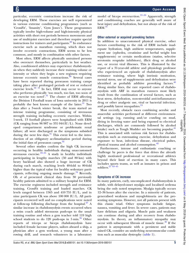
particular, eccentric contractions increase the risk ofdeveloping ERM. These exercises are well representedin various extreme conditioning programmes (such as‘CrossFit’, ‘Insanity’, ‘Gym Jones’). These programmestypically involve high-volume and high-intensity physicalactivities with short rest periods between movements anduse of multi-joint exercises and therefore require specialinstructions for trainers and participants.29 In enduranceexercise such as marathon running, which does notinvolve eccentric contractions, ERM seems to be lesscommon, and mostly in combination with other factors.Most often, ERM affects physically untrained persons
who overexert themselves, particularly in hot weather.Also, conditioned athletes may experience rhabdomyoly-sis when they suddenly increase their training volume orintensity or when they begin a new regimen requiringintense eccentric muscle contractions.30 Several caseshave been reported during preseason training campstaking place after prolonged periods of rest or reducedexercise levels.30 31 In fact, ERM may occur to anyonewho performs physically ‘too much, too fast, too soon ofan exercise too novel’.31 The cluster of ERM affectingthe Division I Football team of Iowa university in 2011 isprobably the best known example of the latter.31 Twodays after a 3-week winter break, the team started anintense training programme, consisting of strenuousstrength training including eccentric exercises. Within1-week, 13 football players were hospitalised with ERM(CK ranging from 96 987 to 331 044 IU/L). None devel-oped compartment syndrome or problematic renalfailure; all were discharged as the symptoms subsidedduring the next few days.31 This event led to the intro-duction of an obligatory acclimatisation period duringthe initial days of preseason camps.32
Several other studies confirm the high CK increaseoccurring in healthy individuals during unaccustomedstrenuous exercise.33–35 A study in healthy individualsparticipating in lengthy marches (50 and 80 km) withheavy backload also showed a huge increase of CKduring each march, reaching levels 40-fold to 80-foldhigher than the typical value for healthy sedentary parti-cipants, reflecting ongoing muscle damage.34 Recently,Oh et al presented clinical data from 30 previouslyhealthy patients admitted to a military hospital for ERM.The exercise regimens included strength and resistancetraining, CrossFit training and loaded marches. CKlevels ranged between 1565 and 410 755 IU/L, and innine participants CK was above 100 000 IU/L. All parti-cipants recovered well and no complications were notedat follow-up following discharge from the hospital.27 Asimilar increase in rhabdomyolysis events occurred whena swim coach added strenuous push-ups to the usualtraining routine and when a gym teacher told 119 highschool students to do 120 push-ups in 5 min.36 Otherreports of triceps or biceps rhabdomyolysis haveincluded female lacrosse players, sailors aboard a ship, aphysician after a gym workout, a young man after arowing drill, and research volunteers in a laboratory
model of biceps overexertion.37–39 Apparently, strengthand conditioning coaches are generally well aware ofheat injury and dehydration, but not always of the risk ofERM.31
Other external or acquired provoking factorsIn addition to unaccustomed physical exercise, otherfactors contributing to the risk of ERM include inad-equate hydration, high ambient temperatures, supple-ment use (ephedra, creatinine and herbal weight losssupplements) or certain medications (statins, selectiveserotonin reuptake inhibitors), illicit drug or alcoholuse, or recent viral illnesses. This is illustrated by thereport of a 31-year-old athlete and physiology professorwho experienced an episode of rhabdomyolysis afterresistance training, where high intrinsic motivation,mental stress, use of supplements and dehydration wereidentified as probably additive provoking factors.40
Along similar lines, the rare reported cases of rhabdo-myolysis with ARF in marathon runners most likelyresult from the combination of several factors, such asheat stress, dehydration, non-steroidal anti-inflammatorydrug or other analgesic use, viral or bacterial infection,and possibly latent myopathies.24
Most recently, obstacle races combining aerobic andanaerobic activities performed at different environmen-tal settings (eg, running and/or crawling on mud,diving in freezing water and being exposed to electricaldischarges, which are commonly followed by alcoholintake) such as Tough Mudder are becoming popular.41
This is associated with various risk factors for rhabdo-myolysis such as extenuating exercise, dehydration andexposure to heat, infectious disease, electrical pulses,physical trauma and alcohol consumption.42
Furthermore, intense and enthusiastic coaching orchallenge by peers is the force that drives the alreadyhighly motivated professional or recreational athletesbeyond their limit of exertion in many cases. Thisincludes sports teams, as well as inmates in prison andsoldiers.27 43–45
Symptoms of CK increaseIn many patients, early, uncomplicated rhabdomyolysis issubtle, with delayed-onset myalgia and localised oedemabeing the only noted symptoms. Myalgia typically occurs12–36 hours after the exercise. In a minority of patients,generalised weakness and myoglobinuria are the pre-senting symptoms. However, not all patients present withthis classic triad. Other symptoms include fatigue,nausea, vomiting and fever. In severe cases, patients mayhave delirium and oliguria. Muscle pain and weaknesscan continue during and after recovery from rhabdo-myolysis. In theory, an inflammatory myopathy mayoccur with subsequent fibrosis of muscle tissue.46 If thepatient is asymptomatic with a persistent and stableraised CK, consider an underlying neuromuscular condi-tion such as muscular dystrophy (table 1).
Scalco RS, et al. BMJ Open Sport Exerc Med 2016;2:e000151. doi:10.1136/bmjsem-2016-000151 5
Open Accesscopyright.
on 8 May 2018 by guest. P
rotected byhttp://bm
jopensem.bm
j.com/
BM
J Open S
port Exerc M
ed: first published as 10.1136/bmjsem
-2016-000151 on 7 Septem
ber 2016. Dow
nloaded from
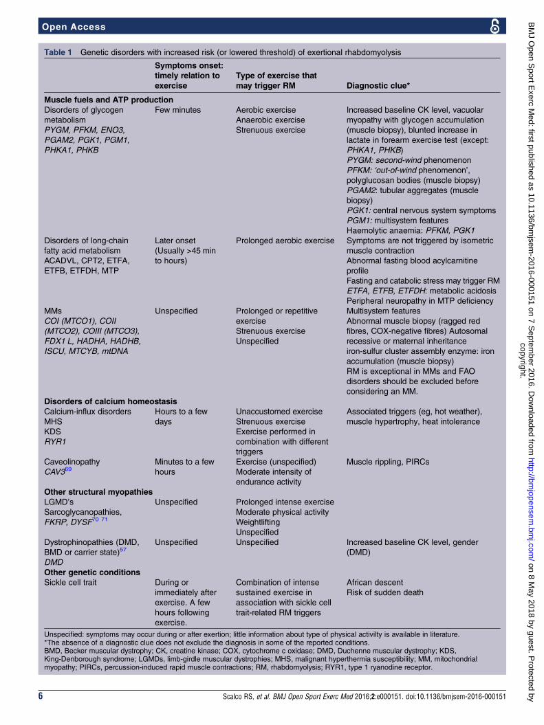
Table 1 Genetic disorders with increased risk (or lowered threshold) of exertional rhabdomyolysis
Symptoms onset:
timely relation to
exercise
Type of exercise that
may trigger RM Diagnostic clue*
Muscle fuels and ATP production
Disorders of glycogen
metabolism
PYGM, PFKM, ENO3,
PGAM2, PGK1, PGM1,
PHKA1, PHKB
Few minutes Aerobic exercise
Anaerobic exercise
Strenuous exercise
Increased baseline CK level, vacuolar
myopathy with glycogen accumulation
(muscle biopsy), blunted increase in
lactate in forearm exercise test (except:
PHKA1, PHKB)
PYGM: second-wind phenomenon
PFKM: ‘out-of-wind phenomenon’,
polyglucosan bodies (muscle biopsy)
PGAM2: tubular aggregates (muscle
biopsy)
PGK1: central nervous system symptoms
PGM1: multisystem features
Haemolytic anaemia: PFKM, PGK1
Disorders of long-chain
fatty acid metabolism
ACADVL, CPT2, ETFA,
ETFB, ETFDH, MTP
Later onset
(Usually >45 min
to hours)
Prolonged aerobic exercise Symptoms are not triggered by isometric
muscle contraction
Abnormal fasting blood acylcarnitine
profile
Fasting and catabolic stress may trigger RM
ETFA, ETFB, ETFDH: metabolic acidosis
Peripheral neuropathy in MTP deficiency
MMs
COI (MTCO1), COII
(MTCO2), COIII (MTCO3),
FDX1 L, HADHA, HADHB,
ISCU, MTCYB, mtDNA
Unspecified Prolonged or repetitive
exercise
Strenuous exercise
Unspecified
Multisystem features
Abnormal muscle biopsy (ragged red
fibres, COX-negative fibres) Autosomal
recessive or maternal inheritance
iron-sulfur cluster assembly enzyme: iron
accumulation (muscle biopsy)
RM is exceptional in MMs and FAO
disorders should be excluded before
considering an MM.
Disorders of calcium homeostasis
Calcium-influx disorders
MHS
KDS
RYR1
Hours to a few
days
Unaccustomed exercise
Strenuous exercise
Exercise performed in
combination with different
triggers
Associated triggers (eg, hot weather),
muscle hypertrophy, heat intolerance
Caveolinopathy
CAV369Minutes to a few
hours
Exercise (unspecified)
Moderate intensity of
endurance activity
Muscle rippling, PIRCs
Other structural myopathies
LGMD’s
Sarcoglycanopathies,
FKRP, DYSF70 71
Unspecified Prolonged intense exercise
Moderate physical activity
Weightlifting
Unspecified
Dystrophinopathies (DMD,
BMD or carrier state)57
DMD
Unspecified Unspecified Increased baseline CK level, gender
(DMD)
Other genetic conditions
Sickle cell trait During or
immediately after
exercise. A few
hours following
exercise.
Combination of intense
sustained exercise in
association with sickle cell
trait-related RM triggers
African descent
Risk of sudden death
Unspecified: symptoms may occur during or after exertion; little information about type of physical activilty is available in literature.*The absence of a diagnostic clue does not exclude the diagnosis in some of the reported conditions.BMD, Becker muscular dystrophy; CK, creatine kinase; COX, cytochrome c oxidase; DMD, Duchenne muscular dystrophy; KDS,King-Denborough syndrome; LGMDs, limb-girdle muscular dystrophies; MHS, malignant hyperthermia susceptibility; MM, mitochondrialmyopathy; PIRCs, percussion-induced rapid muscle contractions; RM, rhabdomyolysis; RYR1, type 1 ryanodine receptor.
6 Scalco RS, et al. BMJ Open Sport Exerc Med 2016;2:e000151. doi:10.1136/bmjsem-2016-000151
Open Accesscopyright.
on 8 May 2018 by guest. P
rotected byhttp://bm
jopensem.bm
j.com/
BM
J Open S
port Exerc M
ed: first published as 10.1136/bmjsem
-2016-000151 on 7 Septem
ber 2016. Dow
nloaded from
Myoglobinuria and ARFMyoglobinuria is defined as the presence of myoglobinin the urine, usually associated with rhabdomyolysis ortraumatic muscle injury. Myoglobin is the first enzymethat increases in plasma, but, because under physio-logical circumstances it is readily filtered by the glom-erulus and rapidly cleared from the serum into theurine, it normally returns to normal levels within thefirst 24 hours after symptom onset. However, when largeamounts of myoglobin enter the renal tubule lumen, itinteracts with the Tamm-Horsfall protein and precipi-tates, a process enhanced in the presence of acidicurine. Visible myoglobinuria (tea-coloured or cola-coloured urine) occurs when urinary myoglobin exceeds250 μg/mL (normal <5 ng/mL), corresponding to thedestruction of more than 100 g of muscle.47 Tubuleobstruction principally occurs at the level of the distaltubule. In addition, reactive oxygen species generated bydamage to both muscle and kidney epithelial cellspromote the oxidation of ferrous oxide to ferric oxide,thus generating a hydroxyl radical. Both the heme moi-eties and the free iron-driven hydroxyl radicals may becritical mediators of direct tubule toxicity, which mainlyoccurs in the proximal tubule. A higher volume of urineflow and a higher urine alkalinity prevent myoglobinfrom precipitating as readily as it would otherwise do.Myoglobinuria causes little or no morbidity or mortal-
ity unless it is associated with the secondary complica-tions of rhabdomyolysis, including hyperkalaemia,hypocalcaemia and acute kidney injury. In adults, thereported ARF incidence in patients with rhabdomyolysiswas reported to vary from 4.7% to 94% in a recentreview. This variation is likely to be at least partly due tothe differing definitions of rhabdomyolysis and ARF, andto the differences in number and types of patients evalu-ated.48 In the paediatric age group, although previoussmall case series reported ARF rates of 40–50%, a largeretrospective review indicates that only about 5% of par-ticipants with rhabdomyolysis develop acute kidneyinjury.49 CK level is only one of the many determinantsof renal failure, including comorbidities, use of certainmedications, alcohol or illicit drugs and concomitantelectrolyte disturbances.28
Natural course of ERMAmong young, physically active patients, the incidenceof ERM is low, as is the risk of recurrence. Furthermore,ERM is associated with lower complication rates com-pared with other causes of rhabdomyolysis, probablybecause participants do not have other associated dis-eases or do not take potentially myotoxic treatments.This was shown in a retrospective case series, includingpopulation-based and cohort-based analyses in a cohortof 177 patient with rhabdomyolysis at a US army medicalcentre.25 There were 44 cases of ERM from a populationof 198 399 total military trainees over the study period,or 22.2 cases per 100 000 per year. This was one-third ofall cases of rhabdomyolysis in this period; other causes
included trauma, use of toxins, infection and heatillness. During the follow-up period (ranging from 20 to60 months, with a mean of 31.2 months) with return toprior levels of physical activity, there was one recurrenceof rhabdomyolysis (at 1-month, following exertion).25
Hence, long-term risk of recurrence is low. Otherreports, including that concerning the football teamcited above, also show that the prognosis of ERMwithout systemic complications is very good.20 Ingeneral, in the young, healthy population performingsports at an intensity that may cause rhabdomyolysis,complication rate is expected to be low.28
Genetic factors with increased risk for ERMIn addition, several genetic disorders increase the risk ofdeveloping ERM. These include metabolic myopathies,disorders of calcium homoeostasis and other structuralmyopathies, and the sickle cell trait (table 1). RYR1mutations have been recently recognised to account fora substantial proportion of patients presenting withERM.3 Mutations in this gene are a common cause ofneuromuscular disease, ranging from various congenitalmyopathies to the MHS trait without associated weak-ness. Associated clinicopathological features inRYR1-associated ERM may be subtle and require a highdegree of suspicion. Additional family studies are para-mount in order to identify potentially MH-susceptiblerelatives.3 50 51–53
Remarkably, some genetic disorders lower the thresh-old for developing rhabdomyolysis and may also be asso-ciated with greater athletic abilities. This is likely to bethe case for RYR1 mutations, in addition to what hasbeen reported regarding variants/polymorphisms in theACE and α-actinin-3 (ACTN3) genes.54–56 In other raresituations, a combination of two genetic disorders mayalleviate exercise-related phenotypes such as the coexist-ence of α-thalassaemia and sickle cell trait, lowering riskfor exercise collapse associated with the sickle cell trait(ECAST).57
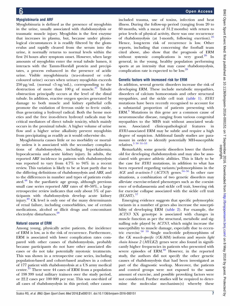
Emerging evidence suggests that specific polymorphicvariants in a number of genes also increase the suscepti-bility of developing ERM (table 2). For example, theACTN3 XX genotype is associated with changes inmuscle function as per the structural, metabolic and sig-nalling role played by ACTN3, which might increase thesusceptibility to muscle damage, especially due to eccen-tric exercise.58 59 Single nucleotide polymorphisms ofthe CK muscle-specific (CK-MM) isoform and myosin lightchain kinase 2 (MYLK2) genes were also found in signifi-cantly higher frequencies in patients who presented withsevere episodes of ERM.60 However, in the reportedstudy, the authors did not specify the other geneticcauses of rhabdomyolysis that had been investigated aspart of the diagnostic workup. Moreover, the patientsand control groups were not exposed to the sameamount of exercise, and possible provoking factors werenot considered. Further studies will be required to deter-mine the molecular mechanism(s) whereby these
Scalco RS, et al. BMJ Open Sport Exerc Med 2016;2:e000151. doi:10.1136/bmjsem-2016-000151 7
Open Accesscopyright.
on 8 May 2018 by guest. P
rotected byhttp://bm
jopensem.bm
j.com/
BM
J Open S
port Exerc M
ed: first published as 10.1136/bmjsem
-2016-000151 on 7 Septem
ber 2016. Dow
nloaded from
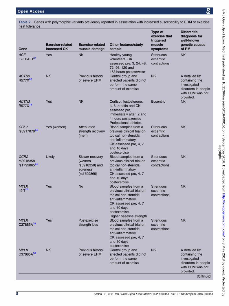
Table 2 Genes with polymorphic variants previously reported in association with increased susceptibility to ERM or exercise
heat tolerance
Gene
Exercise-related
increased CK
Exercise-related
muscle damage
Other features/study
sample
Type of
exercise that
triggered
muscle
symptoms
Differential
diagnosis for
well-known
genetic causes
of RM
ACE
II>ID>DD72Yes NK Healthy young
volunteers; CK
assessed pre, 3, 24, 48,
72, 96, 120 and
168 hours postexercise
Strenuous
eccentric
contractions
NK
ACTN3
R577X60
NK Previous history
of severe ERM
Control group and
affected patients did not
perform the same
amount of exercise
NK A detailed list
containing the
investigated
disorders in people
with ERM was not
provided.
ACTN3
R577X73
Yes NK Cortisol, testosterone,
IL-6, α-actin and CK
assessed pre,
immediately after, 2 and
4 hours postexercise
Professional athletes
Eccentric NK
CCL2
rs391787874Yes (women) Attenuated
strength recovery
(men)
Blood samples from a
previous clinical trial on
topical non-steroidal
anti-inflammatory
CK assessed pre, 4, 7
and 10 days
postexercise
Strenuous
eccentric
contractions
NK
CCR2
rs3918358
rs179986574
Likely Slower recovery
(women—
rs3918358) and
soreness
(rs1799865)
Blood samples from a
previous clinical trial on
topical non-steroidal
anti-inflammatory
CK assessed pre, 4, 7
and 10 days
postexercise
Strenuous
eccentric
contractions
NK
MYLK
49 T75Yes No Blood samples from a
previous clinical trial on
topical non-steroidal
anti-inflammatory
CK assessed pre, 4, 7
and 10 days
postexercise
Higher baseline strength
Strenuous
eccentric
contractions
NK
MYLK
C37885A75
Yes Postexercise
strength loss
Blood samples from a
previous clinical trial on
topical non-steroidal
anti-inflammatory
CK assessed pre, 4, 7
and 10 days
postexercise
Strenuous
eccentric
contractions
NK
MYLK
C37885A60
NK Previous history
of severe ERM
Control group and
affected patients did not
perform the same
amount of exercise
NK A detailed list
containing the
investigated
disorders in people
with ERM was not
provided.
Continued
8 Scalco RS, et al. BMJ Open Sport Exerc Med 2016;2:e000151. doi:10.1136/bmjsem-2016-000151
Open Accesscopyright.
on 8 May 2018 by guest. P
rotected byhttp://bm
jopensem.bm
j.com/
BM
J Open S
port Exerc M
ed: first published as 10.1136/bmjsem
-2016-000151 on 7 Septem
ber 2016. Dow
nloaded from
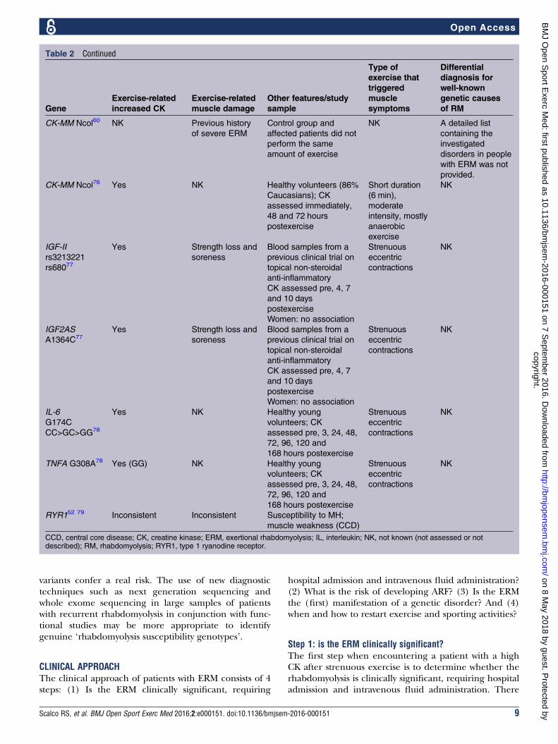
variants confer a real risk. The use of new diagnostictechniques such as next generation sequencing andwhole exome sequencing in large samples of patientswith recurrent rhabdomyolysis in conjunction with func-tional studies may be more appropriate to identifygenuine ‘rhabdomyolysis susceptibility genotypes’.
CLINICAL APPROACHThe clinical approach of patients with ERM consists of 4steps: (1) Is the ERM clinically significant, requiring
hospital admission and intravenous fluid administration?(2) What is the risk of developing ARF? (3) Is the ERMthe (first) manifestation of a genetic disorder? And (4)when and how to restart exercise and sporting activities?
Step 1: is the ERM clinically significant?The first step when encountering a patient with a highCK after strenuous exercise is to determine whether therhabdomyolysis is clinically significant, requiring hospitaladmission and intravenous fluid administration. There
Table 2 Continued
Gene
Exercise-related
increased CK
Exercise-related
muscle damage
Other features/study
sample
Type of
exercise that
triggered
muscle
symptoms
Differential
diagnosis for
well-known
genetic causes
of RM
CK-MM Ncol60 NK Previous history
of severe ERM
Control group and
affected patients did not
perform the same
amount of exercise
NK A detailed list
containing the
investigated
disorders in people
with ERM was not
provided.
CK-MM Ncol76 Yes NK Healthy volunteers (86%
Caucasians); CK
assessed immediately,
48 and 72 hours
postexercise
Short duration
(6 min),
moderate
intensity, mostly
anaerobic
exercise
NK
IGF-II
rs3213221
rs68077
Yes Strength loss and
soreness
Blood samples from a
previous clinical trial on
topical non-steroidal
anti-inflammatory
CK assessed pre, 4, 7
and 10 days
postexercise
Women: no association
Strenuous
eccentric
contractions
NK
IGF2AS
A1364C77
Yes Strength loss and
soreness
Blood samples from a
previous clinical trial on
topical non-steroidal
anti-inflammatory
CK assessed pre, 4, 7
and 10 days
postexercise
Women: no association
Strenuous
eccentric
contractions
NK
IL-6
G174C
CC>GC>GG78
Yes NK Healthy young
volunteers; CK
assessed pre, 3, 24, 48,
72, 96, 120 and
168 hours postexercise
Strenuous
eccentric
contractions
NK
TNFA G308A78 Yes (GG) NK Healthy young
volunteers; CK
assessed pre, 3, 24, 48,
72, 96, 120 and
168 hours postexercise
Strenuous
eccentric
contractions
NK
RYR152 79 Inconsistent Inconsistent Susceptibility to MH;
muscle weakness (CCD)
CCD, central core disease; CK, creatine kinase; ERM, exertional rhabdomyolysis; IL, interleukin; NK, not known (not assessed or notdescribed); RM, rhabdomyolysis; RYR1, type 1 ryanodine receptor.
Scalco RS, et al. BMJ Open Sport Exerc Med 2016;2:e000151. doi:10.1136/bmjsem-2016-000151 9
Open Accesscopyright.
on 8 May 2018 by guest. P
rotected byhttp://bm
jopensem.bm
j.com/
BM
J Open S
port Exerc M
ed: first published as 10.1136/bmjsem
-2016-000151 on 7 Septem
ber 2016. Dow
nloaded from
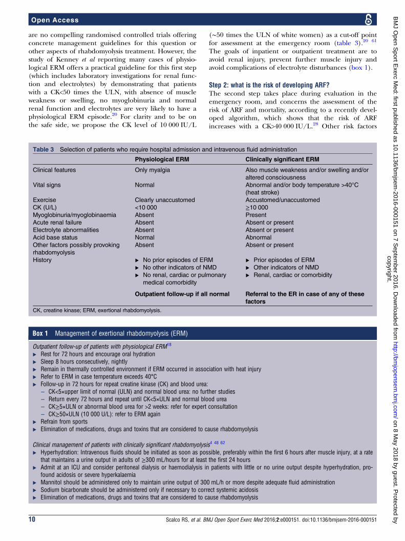
are no compelling randomised controlled trials offeringconcrete management guidelines for this question orother aspects of rhabdomyolysis treatment. However, thestudy of Kenney et al reporting many cases of physio-logical ERM offers a practical guideline for this first step(which includes laboratory investigations for renal func-tion and electrolytes) by demonstrating that patientswith a CK<50 times the ULN, with absence of muscleweakness or swelling, no myoglobinuria and normalrenal function and electrolytes are very likely to have aphysiological ERM episode.20 For clarity and to be onthe safe side, we propose the CK level of 10 000 IU/L
(∼50 times the ULN of white women) as a cut-off pointfor assessment at the emergency room (table 3).20 61
The goals of inpatient or outpatient treatment are toavoid renal injury, prevent further muscle injury andavoid complications of electrolyte disturbances (box 1).
Step 2: what is the risk of developing ARF?The second step takes place during evaluation in theemergency room, and concerns the assessment of therisk of ARF and mortality, according to a recently devel-oped algorithm, which shows that the risk of ARFincreases with a CK>40 000 IU/L.28 Other risk factors
Table 3 Selection of patients who require hospital admission and intravenous fluid administration
Physiological ERM Clinically significant ERM
Clinical features Only myalgia Also muscle weakness and/or swelling and/or
altered consciousness
Vital signs Normal Abnormal and/or body temperature >40°C
(heat stroke)
Exercise Clearly unaccustomed Accustomed/unaccustomed
CK (U/L) <10 000 ≥10 000
Myoglobinuria/myoglobinaemia Absent Present
Acute renal failure Absent Absent or present
Electrolyte abnormalities Absent Absent or present
Acid base status Normal Abnormal
Other factors possibly provoking
rhabdomyolysis
Absent Absent or present
History ▸ No prior episodes of ERM
▸ No other indicators of NMD
▸ No renal, cardiac or pulmonary
medical comorbidity
▸ Prior episodes of ERM
▸ Other indicators of NMD
▸ Renal, cardiac or comorbidity
Outpatient follow-up if all normal Referral to the ER in case of any of these
factors
CK, creatine kinase; ERM, exertional rhabdomyolysis.
Box 1 Management of exertional rhabdomyolysis (ERM)
Outpatient follow-up of patients with physiological ERM18
▸ Rest for 72 hours and encourage oral hydration▸ Sleep 8 hours consecutively, nightly▸ Remain in thermally controlled environment if ERM occurred in association with heat injury▸ Refer to ERM in case temperature exceeds 40°C▸ Follow-up in 72 hours for repeat creatine kinase (CK) and blood urea:
– CK<5×upper limit of normal (ULN) and normal blood urea: no further studies– Return every 72 hours and repeat until CK<5×ULN and normal blood urea– CK≥5×ULN or abnormal blood urea for >2 weeks: refer for expert consultation– CK≥50×ULN (10 000 U/L): refer to ERM again
▸ Refrain from sports▸ Elimination of medications, drugs and toxins that are considered to cause rhabdomyolysis
Clinical management of patients with clinically significant rhabdomyolysis4 48 62
▸ Hyperhydration: Intravenous fluids should be initiated as soon as possible, preferably within the first 6 hours after muscle injury, at a ratethat maintains a urine output in adults of ≥300 mL/hours for at least the first 24 hours
▸ Admit at an ICU and consider peritoneal dialysis or haemodialysis in patients with little or no urine output despite hyperhydration, pro-found acidosis or severe hyperkalaemia
▸ Mannitol should be administered only to maintain urine output of 300 mL/h or more despite adequate fluid administration▸ Sodium bicarbonate should be administered only if necessary to correct systemic acidosis▸ Elimination of medications, drugs and toxins that are considered to cause rhabdomyolysis
10 Scalco RS, et al. BMJ Open Sport Exerc Med 2016;2:e000151. doi:10.1136/bmjsem-2016-000151
Open Accesscopyright.
on 8 May 2018 by guest. P
rotected byhttp://bm
jopensem.bm
j.com/
BM
J Open S
port Exerc M
ed: first published as 10.1136/bmjsem
-2016-000151 on 7 Septem
ber 2016. Dow
nloaded from
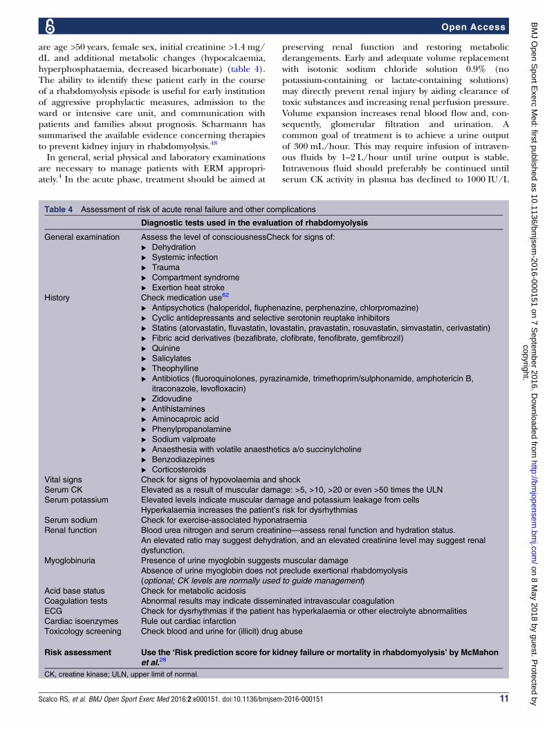
are age >50 years, female sex, initial creatinine >1.4 mg/dL and additional metabolic changes (hypocalcaemia,hyperphosphataemia, decreased bicarbonate) (table 4).The ability to identify these patient early in the courseof a rhabdomyolysis episode is useful for early institutionof aggressive prophylactic measures, admission to theward or intensive care unit, and communication withpatients and families about prognosis. Scharmann hassummarised the available evidence concerning therapiesto prevent kidney injury in rhabdomyolysis.48
In general, serial physical and laboratory examinationsare necessary to manage patients with ERM appropri-ately.4 In the acute phase, treatment should be aimed at
preserving renal function and restoring metabolicderangements. Early and adequate volume replacementwith isotonic sodium chloride solution 0.9% (nopotassium-containing or lactate-containing solutions)may directly prevent renal injury by aiding clearance oftoxic substances and increasing renal perfusion pressure.Volume expansion increases renal blood flow and, con-sequently, glomerular filtration and urination. Acommon goal of treatment is to achieve a urine outputof 300 mL/hour. This may require infusion of intraven-ous fluids by 1–2 L/hour until urine output is stable.Intravenous fluid should preferably be continued untilserum CK activity in plasma has declined to 1000 IU/L
Table 4 Assessment of risk of acute renal failure and other complications
Diagnostic tests used in the evaluation of rhabdomyolysis
General examination Assess the level of consciousnessCheck for signs of:
▸ Dehydration
▸ Systemic infection
▸ Trauma
▸ Compartment syndrome
▸ Exertion heat stroke
History Check medication use62
▸ Antipsychotics (haloperidol, fluphenazine, perphenazine, chlorpromazine)
▸ Cyclic antidepressants and selective serotonin reuptake inhibitors
▸ Statins (atorvastatin, fluvastatin, lovastatin, pravastatin, rosuvastatin, simvastatin, cerivastatin)
▸ Fibric acid derivatives (bezafibrate, clofibrate, fenofibrate, gemfibrozil)
▸ Quinine
▸ Salicylates
▸ Theophylline
▸ Antibiotics (fluoroquinolones, pyrazinamide, trimethoprim/sulphonamide, amphotericin B,
itraconazole, levofloxacin)
▸ Zidovudine
▸ Antihistamines
▸ Aminocaproic acid
▸ Phenylpropanolamine
▸ Sodium valproate
▸ Anaesthesia with volatile anaesthetics a/o succinylcholine
▸ Benzodiazepines
▸ Corticosteroids
Vital signs Check for signs of hypovolaemia and shock
Serum CK Elevated as a result of muscular damage: >5, >10, >20 or even >50 times the ULN
Serum potassium Elevated levels indicate muscular damage and potassium leakage from cells
Hyperkalaemia increases the patient’s risk for dysrhythmias
Serum sodium Check for exercise-associated hyponatraemia
Renal function Blood urea nitrogen and serum creatinine—assess renal function and hydration status.
An elevated ratio may suggest dehydration, and an elevated creatinine level may suggest renal
dysfunction.
Myoglobinuria Presence of urine myoglobin suggests muscular damage
Absence of urine myoglobin does not preclude exertional rhabdomyolysis
(optional; CK levels are normally used to guide management)
Acid base status Check for metabolic acidosis
Coagulation tests Abnormal results may indicate disseminated intravascular coagulation
ECG Check for dysrhythmias if the patient has hyperkalaemia or other electrolyte abnormalities
Cardiac isoenzymes Rule out cardiac infarction
Toxicology screening Check blood and urine for (illicit) drug abuse
Risk assessment Use the ‘Risk prediction score for kidney failure or mortality in rhabdomyolysis’ by McMahon
et al.28
CK, creatine kinase; ULN, upper limit of normal.
Scalco RS, et al. BMJ Open Sport Exerc Med 2016;2:e000151. doi:10.1136/bmjsem-2016-000151 11
Open Accesscopyright.
on 8 May 2018 by guest. P
rotected byhttp://bm
jopensem.bm
j.com/
BM
J Open S
port Exerc M
ed: first published as 10.1136/bmjsem
-2016-000151 on 7 Septem
ber 2016. Dow
nloaded from
or below. In patients with little or no urine outputdespite aggressive hydration, profound acidosis or severehyperkalaemia, consider peritoneal dialysis or haemodi-alysis and consult a nephrologist. Aggressive hydrationmay not be appropriate in patients with other comorbid-ities such as heart failure. Furthermore, if kidney injuryis already established, overly aggressive hydration in apatient with already established renal failure may lead tovolume overload and pulmonary oedema.Mannitol leads to an increase in blood flow and glom-
erular filtration, which reduces the obstruction by myo-globin casts. It must only be given after volumereplacement and must be avoided in patients with oli-guria. It is not routinely recommended, but may beadministered if the target urine output cannot beachieved by other means. Loop diuretics have also beeninvestigated, but published data are too limited tosupport recommending them routinely. Although notproven by randomised control trials, some experts rec-ommend the addition of bicarbonate. Alkalisation of theurine (target pH 6.5) promotes myoglobulin washoutand corrects the metabolic acidosis and hyperkalemia. Arecent systematic review has shown that sodium bicar-bonate should only be administered to patients with sys-temic acidosis.48
Monitoring for and treating rhabdomyolysis-relatedelectrolyte abnormalities is an important part of man-aging patients with ERM. Hyperkalaemia, hypocalcae-mia, hyperphosphataemia and hyperuricaemia arecommon in patients with rhabdomyolysis, and should betreated with standard therapy. Although hypocalcaemiais common in early rhabdomyolysis, hypercalcaemia canoccur later. Calcium supplementation should only beprescribed for patients with symptomatic hypocalcaemiaor severe hyperkalaemia. More research is needed todetermine whether allopurinol may be useful for pre-venting ERM and acute renal damage.13
Compartment syndrome requires surgical consultation.If the compartment pressure measured by an intramus-cular needle exceeds 50 mm Hg, or if pressure persistsbetween 30 and 50 mm Hg for longer than 6 hours,decompressive fasciotomy must be considered.63
Haemorrhagic complications in patients with dissemi-nated intravascular coagulation (DIC) and rhabdomyoly-sis require careful management. Drugs and toxinsshould be eliminated (eg, haemodialysis/antidotes) ifpossible. Anaesthetic precautions (such as avoidance ofdepolarising muscle relaxants and volatile anaesthetics)should be taken in patients with an ongoing rhabdo-myolysis. Anaesthetic precautions are also suggested forundiagnosed patients with strong evidence of an under-lying RYR1-related rhabdomyolysis episode and of coursefor MHS patients and their relatives.
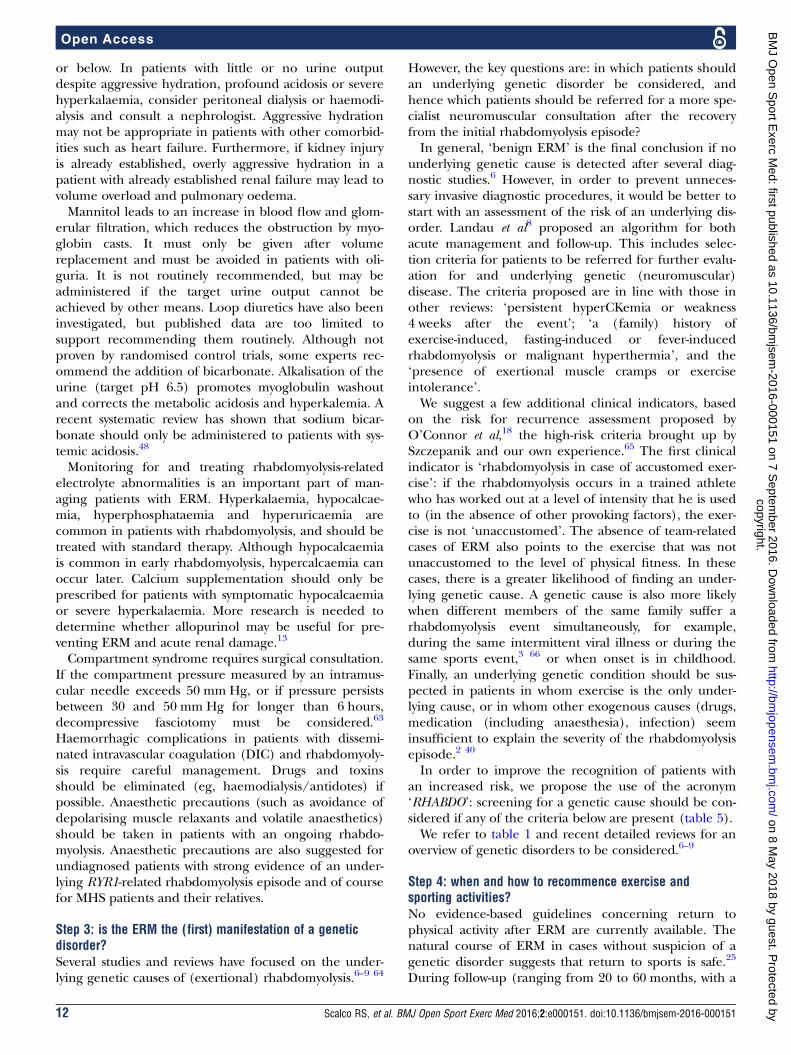
Step 3: is the ERM the (first) manifestation of a geneticdisorder?Several studies and reviews have focused on the under-lying genetic causes of (exertional) rhabdomyolysis.6–9 64
However, the key questions are: in which patients shouldan underlying genetic disorder be considered, andhence which patients should be referred for a more spe-cialist neuromuscular consultation after the recoveryfrom the initial rhabdomyolysis episode?In general, ‘benign ERM’ is the final conclusion if no
underlying genetic cause is detected after several diag-nostic studies.6 However, in order to prevent unneces-sary invasive diagnostic procedures, it would be better tostart with an assessment of the risk of an underlying dis-order. Landau et al8 proposed an algorithm for bothacute management and follow-up. This includes selec-tion criteria for patients to be referred for further evalu-ation for and underlying genetic (neuromuscular)disease. The criteria proposed are in line with those inother reviews: ‘persistent hyperCKemia or weakness4 weeks after the event’; ‘a (family) history ofexercise-induced, fasting-induced or fever-inducedrhabdomyolysis or malignant hyperthermia’, and the‘presence of exertional muscle cramps or exerciseintolerance’.We suggest a few additional clinical indicators, based
on the risk for recurrence assessment proposed byO’Connor et al,18 the high-risk criteria brought up bySzczepanik and our own experience.65 The first clinicalindicator is ‘rhabdomyolysis in case of accustomed exer-cise’: if the rhabdomyolysis occurs in a trained athletewho has worked out at a level of intensity that he is usedto (in the absence of other provoking factors), the exer-cise is not ‘unaccustomed’. The absence of team-relatedcases of ERM also points to the exercise that was notunaccustomed to the level of physical fitness. In thesecases, there is a greater likelihood of finding an under-lying genetic cause. A genetic cause is also more likelywhen different members of the same family suffer arhabdomyolysis event simultaneously, for example,during the same intermittent viral illness or during thesame sports event,3 66 or when onset is in childhood.Finally, an underlying genetic condition should be sus-pected in patients in whom exercise is the only under-lying cause, or in whom other exogenous causes (drugs,medication (including anaesthesia), infection) seeminsufficient to explain the severity of the rhabdomyolysisepisode.2 40
In order to improve the recognition of patients withan increased risk, we propose the use of the acronym‘RHABDO’: screening for a genetic cause should be con-sidered if any of the criteria below are present (table 5).We refer to table 1 and recent detailed reviews for an
overview of genetic disorders to be considered.6–9
Step 4: when and how to recommence exercise andsporting activities?No evidence-based guidelines concerning return tophysical activity after ERM are currently available. Thenatural course of ERM in cases without suspicion of agenetic disorder suggests that return to sports is safe.25
During follow-up (ranging from 20 to 60 months, with a
12 Scalco RS, et al. BMJ Open Sport Exerc Med 2016;2:e000151. doi:10.1136/bmjsem-2016-000151
Open Accesscopyright.
on 8 May 2018 by guest. P
rotected byhttp://bm
jopensem.bm
j.com/
BM
J Open S
port Exerc M
ed: first published as 10.1136/bmjsem
-2016-000151 on 7 Septem
ber 2016. Dow
nloaded from
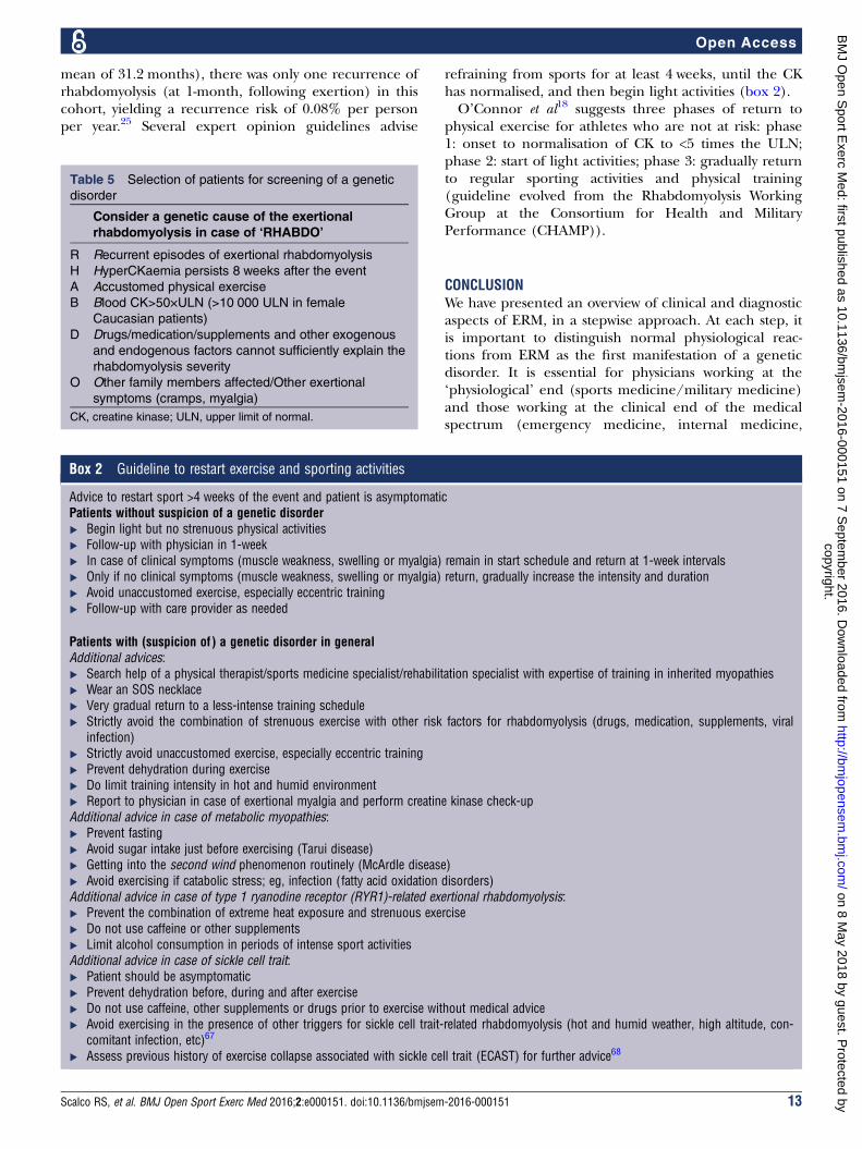
mean of 31.2 months), there was only one recurrence ofrhabdomyolysis (at 1-month, following exertion) in thiscohort, yielding a recurrence risk of 0.08% per personper year.25 Several expert opinion guidelines advise
refraining from sports for at least 4 weeks, until the CKhas normalised, and then begin light activities (box 2).O’Connor et al18 suggests three phases of return to
physical exercise for athletes who are not at risk: phase1: onset to normalisation of CK to <5 times the ULN;phase 2: start of light activities; phase 3: gradually returnto regular sporting activities and physical training(guideline evolved from the Rhabdomyolysis WorkingGroup at the Consortium for Health and MilitaryPerformance (CHAMP)).
CONCLUSIONWe have presented an overview of clinical and diagnosticaspects of ERM, in a stepwise approach. At each step, itis important to distinguish normal physiological reac-tions from ERM as the first manifestation of a geneticdisorder. It is essential for physicians working at the‘physiological’ end (sports medicine/military medicine)and those working at the clinical end of the medicalspectrum (emergency medicine, internal medicine,
Box 2 Guideline to restart exercise and sporting activities
Advice to restart sport >4 weeks of the event and patient is asymptomaticPatients without suspicion of a genetic disorder▸ Begin light but no strenuous physical activities▸ Follow-up with physician in 1-week▸ In case of clinical symptoms (muscle weakness, swelling or myalgia) remain in start schedule and return at 1-week intervals▸ Only if no clinical symptoms (muscle weakness, swelling or myalgia) return, gradually increase the intensity and duration▸ Avoid unaccustomed exercise, especially eccentric training▸ Follow-up with care provider as needed
Patients with (suspicion of ) a genetic disorder in generalAdditional advices:▸ Search help of a physical therapist/sports medicine specialist/rehabilitation specialist with expertise of training in inherited myopathies▸ Wear an SOS necklace▸ Very gradual return to a less-intense training schedule▸ Strictly avoid the combination of strenuous exercise with other risk factors for rhabdomyolysis (drugs, medication, supplements, viral
infection)▸ Strictly avoid unaccustomed exercise, especially eccentric training▸ Prevent dehydration during exercise▸ Do limit training intensity in hot and humid environment▸ Report to physician in case of exertional myalgia and perform creatine kinase check-upAdditional advice in case of metabolic myopathies:▸ Prevent fasting▸ Avoid sugar intake just before exercising (Tarui disease)▸ Getting into the second wind phenomenon routinely (McArdle disease)▸ Avoid exercising if catabolic stress; eg, infection (fatty acid oxidation disorders)Additional advice in case of type 1 ryanodine receptor (RYR1)-related exertional rhabdomyolysis:▸ Prevent the combination of extreme heat exposure and strenuous exercise▸ Do not use caffeine or other supplements▸ Limit alcohol consumption in periods of intense sport activitiesAdditional advice in case of sickle cell trait:▸ Patient should be asymptomatic▸ Prevent dehydration before, during and after exercise▸ Do not use caffeine, other supplements or drugs prior to exercise without medical advice▸ Avoid exercising in the presence of other triggers for sickle cell trait-related rhabdomyolysis (hot and humid weather, high altitude, con-
comitant infection, etc)67
▸ Assess previous history of exercise collapse associated with sickle cell trait (ECAST) for further advice68
Table 5 Selection of patients for screening of a genetic
disorder
Consider a genetic cause of the exertional
rhabdomyolysis in case of ‘RHABDO’
R Recurrent episodes of exertional rhabdomyolysis
H HyperCKaemia persists 8 weeks after the event
A Accustomed physical exercise
B Blood CK>50×ULN (>10 000 ULN in female
Caucasian patients)
D Drugs/medication/supplements and other exogenous
and endogenous factors cannot sufficiently explain the
rhabdomyolysis severity
O Other family members affected/Other exertional
symptoms (cramps, myalgia)
CK, creatine kinase; ULN, upper limit of normal.
Scalco RS, et al. BMJ Open Sport Exerc Med 2016;2:e000151. doi:10.1136/bmjsem-2016-000151 13
Open Accesscopyright.
on 8 May 2018 by guest. P
rotected byhttp://bm
jopensem.bm
j.com/
BM
J Open S
port Exerc M
ed: first published as 10.1136/bmjsem
-2016-000151 on 7 Septem
ber 2016. Dow
nloaded from
neurology) to consider that ERM can happen to anyonewho is exposed to unaccustomed exercise. The thresh-old, however, is much lower in patients with an under-lying genetic disorder. In addition, several otherexogenous and endogenous factors also lower thisthreshold. The most important question is whether thelevel and type of exercise in combination with otherexogenous and endogenous factors is sufficient toexplain the rhabdomyolysis episode. If not, screening foran underlying genetic (neuromuscular) disorder needsto be performed.Future research should focus on defining specific,
evidence-based training and exercise advice for patientswith various myopathies, in order to prevent recurrentepisodes of rhabdomyolysis while enabling patients tooptimise their physical condition or even improve theirstrength. New treatments to reduce the risk of ERMshould be prospectively tested in various cohorts ofpatients with genetic risk factors for rhabdomyolysis.13
Author affiliations1MRC Centre for Neuromuscular Diseases, Institute of Neurology, UniversityCollege London, London, UK2MH-investigation Unit, Department of Anesthesia, Canisius-WilhelminaHospital, Nijmegen, The Netherlands3Departments of Anesthesia and of Biomedicine, Basel University Hospital,Basel, Switzerland4Department of Life Sciences, General Pathology Section, University ofFerrara, Ferrara, Italy5Institut de Myologie, Hôpital Pitié-Salpêtrière, Paris, France6Department of Paediatric Neurology—Neuromuscular Service, EvelinaChildren’s Hospital, Guy’s & St Thomas’ NHS Foundation Trust, London, UK7Randall Division of Cell and Molecular Biophysics, Muscle Signalling Section,London, UK8Department of Basic and Clinical Neuroscience, Institute of Psychiatry,Psychology and Neuroscience (IoPPN), King’s College London, London, UK9Department of Neurology, Radboud University Medical Centre, Nijmegen,The Netherlands
Competing interests None declared.
Provenance and peer review Not commissioned; externally peer reviewed.
Open Access This is an Open Access article distributed in accordance withthe Creative Commons Attribution Non Commercial (CC BY-NC 4.0) license,which permits others to distribute, remix, adapt, build upon this work non-commercially, and license their derivative works on different terms, providedthe original work is properly cited and the use is non-commercial. See: http://creativecommons.org/licenses/by-nc/4.0/
REFERENCES1. Sinert R, Kohl L, Rainone T, et al. Exercise-induced rhabdomyolysis.
Ann Emerg Med 1994;23:1301–6.2. Melli G, Chaudhry V, Cornblath DR. Rhabdomyolysis: an evaluation
of 475 hospitalized patients. Medicine 2005;84:377–85.3. Dlamini N, Voermans NC, Lillis S, et al. Mutations in RYR1 are a
common cause of exertional myalgia and rhabdomyolysis.Neuromuscul Disord 2013;23:540–8.
4. Furman J. When exercise causes exertional rhabdomyolysis. JAAPA2015;28:38–43.
5. Goldberg J, Magruder KM, Forsberg CW, et al. Prevalence ofpost-traumatic stress disorder in aging Vietnam-era veterans:veterans administration cooperative study 569: course andconsequences of post-traumatic stress disorder in Vietnam-eraveteran twins. Am J Geriatr Psychiatry 2016;24:181–91.
6. Nance JR, Mammen AL. Diagnostic evaluation of rhabdomyolysis.Muscle Nerve 2015;51:793–810.
7. Barca E, Emmanuele V, DiMauro SB. Metabolic myoglobinuria. CurrNeurol Neurosci Rep 2015;15:69.
8. Landau ME, Kenney K, Deuster P, et al. Exertional rhabdomyolysis:a clinical review with a focus on genetic influences. J ClinNeuromuscul Dis 2012;13:122–36.
9. Quinlivan R, Jungbluth H. Myopathic causes of exercise intolerancewith rhabdomyolysis. Dev Med Child Neurol 2012;54:886–91.
10. Rebbeck RT, Willemse H, Groom L, et al. Regions of ryanodinereceptors that influence activation by the dihydropyridine receptorβ1a subunit. Skelet Muscle 2015;5:23.
11. Ellis C, Cuthill J, Hew-Butler T, et al. Case report:exercise-associated hyponatremia with rhabdomyolysis duringendurance exercise. Phys Sportsmed 2009;37:126–32.
12. Katsarou A, Singh S. Hyponatraemia associated rhabdomyolysisfollowing water intoxication. BMJ Case Rep 2010;2010:pii:bcr0220102720.
13. Sanchis-Gomar F, Pareja-Galeano H, Perez-Quilis C, et alEffects ofallopurinol on exercise-induced muscle damage: new therapeuticapproaches? Cell Stress Chaperones 2015;20:3–13.
14. Hamel Y, Mamoune A, Mauvais FX, et al. Acute rhabdomyolysis andinflammation. J Inherit Metab Dis 2015;38:621–8.
15. Bosch X, Poch E, Grau JM. Rhabdomyolysis and acute kidneyinjury. N Engl J Med 2009;361:62–72.
16. Nurenberg P, Giddings CJ, Stray-Gundersen J, et al. MRimaging-guided muscle biopsy for correlation of increased signalintensity with ultrastructural change and delayed-onset musclesoreness after exercise. Radiology 1992;184:865–9.
17. Brown AS, Davis JM, Murphy EA, et al. Gender differences in viralinfection after repeated exercise stress. Med Sci Sports Exerc2004;36:1290–5.
18. O’Connor FG, Brennan FH Jr, Campbell W, et al. Return to physicalactivity after exertional rhabdomyolysis. Curr Sports Med Rep2008;7:328–31.
19. Joy TR, Hegele RA. Narrative review: statin-related myopathy.Ann Intern Med 2009;150:858–68.
20. Kenney K, Landau ME, Gonzalez RS, et al. Serum creatine kinaseafter exercise: drawing the line between physiological response andexertional rhabdomyolysis. Muscle Nerve 2012;45:356–62.
21. Brewster LM, Stronks K, Zwinderman AH, et al. Creatine kinase andthe correlates of blood pressure in a random population sample.Hypertension 2008;51:e4–5.
22. Neal RC, Ferdinand KC, Ycas J, et al. Relationship of ethnic origin,gender, and age to blood creatine kinase levels. Am J Med2009;122:73–8.
23. Kyriakides T, Angelini C, Schaefer J, et al. EFNS guidelines on thediagnostic approach to pauci- or asymptomatic hyperCKemia.Eur J Neurol 2010;17:767–73.
24. Clarkson PM. Exertional rhabdomyolysis and acute renal failure inmarathon runners. Sports Med 2007;37:361–3.
25. Alpers JP, Jones LK Jr. Natural history of exertionalrhabdomyolysis: a population-based analysis. Muscle Nerve2010;42:487–91.
26. Smoot MK, Amendola A, Cramer E, et al. A cluster of exertionalrhabdomyolysis affecting a Division I Football team. Clin J SportMed 2013;23:365–72.
27. Oh RC, Arter JL, Tiglao SM, et al. Exertional rhabdomyolysis:a case series of 30 hospitalized patients. Mil Med 2015;180:201–7.
28. McMahon GM, Zeng X, Waikar SS. A risk prediction score for kidneyfailure or mortality in rhabdomyolysis. JAMA Intern Med2013;173:1821–8.
29. Knapik JJ. Extreme conditioning programs: potential benefits andpotential risks. J Spec Oper Med 2015;15:108–13.
30. Cleary MA, Sadowski KA, Lee SY, et al. Exertional rhabdomyolysisin an adolescent athlete during preseason conditioning: a perfectstorm. J Strength Cond Res 2011;25:3506–13.
31. Eichner ER. An outbreak of muscle breakdown: a morality play infour acts. Curr Sports Med Rep 2010;9:325–6.
32. Eichner ER. Ramifications of rhabdomyolysis. Curr Sports Med Rep2014;13:135–6.
33. Olerud JE, Homer LD, Carroll HW. Incidence of acute exertionalrhabdomyolysis. Serum myoglobin and enzyme levels as indicatorsof muscle injury. Arch Intern Med 1976;136:692–7.
34. Chevion S, Moran DS, Heled Y, et al. Plasma antioxidant status andcell injury after severe physical exercise. Proc Natl Acad Sci USA2003;100:5119–23.
35. Clarkson PM. Case report of exertional rhabdomyolysis in a12-year-old boy. Med Sci Sports Exerc 2006;38:197–200.
36. Lin H, Chie W, Lien H. Epidemiological analysis of factorsinfluencing an episode of exertional rhabdomyolysis in high schoolstudents. Am J Sports Med 2006;34:481–6.
14 Scalco RS, et al. BMJ Open Sport Exerc Med 2016;2:e000151. doi:10.1136/bmjsem-2016-000151
Open Accesscopyright.
on 8 May 2018 by guest. P
rotected byhttp://bm
jopensem.bm
j.com/
BM
J Open S
port Exerc M
ed: first published as 10.1136/bmjsem
-2016-000151 on 7 Septem
ber 2016. Dow
nloaded from
37. Brown JA, Elliott MJ, Sray WA. Exercise-induced upper extremityrhabdomyolysis and myoglobinuria in shipboard military personnel.Mil Med 1994;159:473–5.
38. Sayers SP, Clarkson PM, Rouzier PA, et al. Adverse eventsassociated with eccentric exercise protocols: six case studies.Med Sci Sports Exerc 1999;31:1697–702.
39. Springer BL, Clarkson PM. Two cases of exertional rhabdomyolysisprecipitated by personal trainers. Med Sci Sports Exerc2003;35:1499–502.
40. Pearcey GE, Bradbury-Squires DJ, Power KE, et al. Exertionalrhabdomyolysis in an acutely detrained athlete/exercise physiologyprofessor. Clin J Sport Med 2013;23:496–8.
41. Eichner ER. Tough Mudder injuries, Triathlon Drownings, and TeamRhabdomyolysis in the Navy. Curr Sports Med Rep 2014;13:66–7.
42. Greenberg MR, Kim PH, Duprey RT, et al. Unique obstacle raceinjuries at an extreme sports event: a case series. Ann Emerg Med2014;63:361–6.
43. Juray RM. Exertional rhabdomyolysis in unsupervised exercises in acorrectional setting: a case study. Urol Nurs 2005;25:117–19.
44. Norquist C, LoVecchio F, Young MR. The card game: outcomesafter exercise-induced rhabdomyolysis in prisoners. Am J EmergMed 2009;27:115–16.
45. MacDonald R, Rosner Z, Venters H. Case series ofexercise-induced rhabdomyolysis in the New York City jail system.Am J Emerg Med 2014;32:466–7.
46. Atias D, Druyan A, Heled Y. Recurrent exertional rhabdomyolysis:coincidence, syndrome, or acquired myopathy? Curr Sports MedRep 2013;12:365–9.
47. Warren JD, Blumbergs PC, Thompson PD. Rhabdomyolysis: areview. Muscle Nerve 2002;25:332–47.
48. Scharman EJ, Troutman WG. Prevention of kidney injury followingrhabdomyolysis: a systematic review. Ann Pharmacother2013;47:90–105.
49. Mannix R, Tan ML, Wright R, et al. Acute pediatric rhabdomyolysis:causes and rates of renal failure. Pediatrics 2006;118:2119–25.
50. Snoeck M, van Engelen BG, Küsters B, et al. RYR1-relatedmyopathies: a wide spectrum of phenotypes throughout life.Eur J Neurol 2015;22:1094–112.
51. Wappler F. Malignant hyperthermia. Eur J Anaesthesiol2001;18:632–52.
52. Davis M, Brown R, Dickson A, et al. Malignant hyperthermiaassociated with exercise-induced rhabdomyolysis or congenitalabnormalities and a novel RYR1 mutation in New Zealand andAustralian pedigrees. Br J Anaesth 2002;88:508–15.
53. Vladutiu GD, Isackson PJ, Kaufman K, et al. Genetic risk formalignant hyperthermia in non-anesthesia-induced myopathies.Mol Genet Metab 2011;104:167–73.
54. Maffulli N, Margiotti K, Longo UG, et al. The genetics of sportsinjuries and athletic performance. Muscles Ligaments Tendons J2013;3:173–89.
55. Papadimitriou ID, Lucia A, Pitsiladis YP, et al. ACTN3 R577X andACE I/D gene variants influence performance in elite sprinters: amulti-cohort study. BMC Genomics 2016;17:285.
56. Garton FC, North KN. The effect of heterozygosity for the ACTN3null allele on human muscle performance. Med Sci Sports Exerc2016;48:509–20.
57. Quattrone RD, Eichner ER, Beutler A, et al. Exercise collapseassociated with sickle cell trait (ECAST): case report and literaturereview. Curr Sports Med Rep 2015;14:110–16.
58. Seto JT, Lek M, Quinlan KG, et al. Deficiency of alpha-actinin-3 isassociated with increased susceptibility to contraction-induceddamage and skeletal muscle remodeling. Hum Mol Genet2011;20:2914–27.
59. Lee FX, Houweling PJ, North KN, et al. How does alpha-actinin-3deficiency alter muscle function? Mechanistic insights into
ACTN3, the ‘gene for speed’. Biochim Biophys Acta2016;1863:686–93.
60. Deuster PA, Contreras-Sesvold CL, O’Connor FG, et al. Geneticpolymorphisms associated with exertional rhabdomyolysis.Eur J Appl Physiol 2013;113:1997–2004.
61. Delaney KA, Givens ML, Vohra RB. Use of RIFLE criteria to predictthe severity and prognosis of acute kidney injury in emergencydepartment patients with rhabdomyolysis. J Emerg Med2012;42:521–8.
62. Zutt R, van der Kooi AJ, Linthorst GE, et al. Rhabdomyolysis: reviewof the literature. Neuromuscul Disord 2014;24:651–9.
63. von Keudell AG, Weaver MJ, Appleton PT, et al. Diagnosis andtreatment of acute extremity compartment syndrome. Lancet2015;386:1299–310.
64. Lee E, Chahin N. A patient with mutation in the SCN4A p.M1592vpresenting with fixed weakness, rhabdomyolysis, and episodicworsening of weakness. Muscle Nerve 2013;48:306–7.
65. Szczepanik ME, Heled Y, Capacchione J, et al. Exertionalrhabdomyolysis: identification and evaluation of the athlete at risk forrecurrence. Curr Sports Med Rep 2014;13:113–19.
66. Smith R, Jones N, Martin D, et al. ‘Too much of a coincidence’:identical twins with exertional heatstroke in the same race.BMJ Case Rep 2016;2016: pii:bcr2015212592.
67. Harrelson GL, Fincher AL, Robinson JB. Acute exertionalrhabdomyolysis and its relationship to sickle cell trait. J Athl Train1995;30:309–12.
68. Asplund CA, O’Connor FG. Challenging return to play decisions:heat stroke, exertional rhabdomyolysis, and exertional collapseassociated with sickle cell trait. Sports health. 2016;8:117–25.
69. Scalco RS, Gardiner AR, Pitceathly RD, et al. CAV3 mutationscausing exercise intolerance, myalgia and rhabdomyolysis:expanding the phenotypic spectrum of caveolinopathies.Neuromuscul Disord 2016;26:504–10. Epub 2016 May 11.
70. Lindberg C, Sixt C, Oldfors A. Episodes of exercise-induced darkurine and myalgia in LGMD 2I. Acta Neurol Scand 2012;125:285–7.
71. Lahoria R, Milone M. Rhabdomyolysis featuring musculardystrophies. J Neurol Sci 2016;361:29–33.
72. Yamin C, Amir O, Sagiv M, et al. ACE ID genotype affects bloodcreatine kinase response to eccentric exercise. J Appl Physiol(1985) 2007;103:2057–61.
73. Pimenta EM, Coelho DB, Cruz IR, et alThe ACTN3 genotype insoccer players in response to acute eccentric training. Eur J ApplPhysiol 2012;112:1495–503.
74. Hubal MJ, Devaney JM, Hoffman EP, et al. CCL2 and CCR2polymorphisms are associated with markers of exercise-inducedskeletal muscle damage. J Appl Physiol (1985) 2010;108:1651–8.
75. Clarkson PM, Hoffman EP, Zambraski E, et al. ACTN3 and MLCKgenotype associations with exertional muscle damage. J ApplPhysiol (1985) 2005;99:564–9.
76. Heled Y, Bloom MS, Wu TJ, et al. CK-MM and ACE genotypes andphysiological prediction of the creatine kinase response to exercise.J Appl Physiol (1985) 2007;103:504–10.
77. Devaney JM, Hoffman EP, Gordish-Dressman H, et al. IGF-II generegion polymorphisms related to exertional muscle damage. J ApplPhysiol (1985) 2007;102:1815–23.
78. Yamin C, Duarte JA, Oliveira JM, et al. IL6 (-174) and TNFA (-308)promoter polymorphisms are associated with systemic creatinekinase response to eccentric exercise. Eur J Appl Physiol2008;104:579–86.
79. Wappler F, Fiege M, Steinfath M, et al. Evidence for susceptibility tomalignant hyperthermia in patients with exercise-inducedrhabdomyolysis. Anesthesiology 2001;94:95–100.
80. Giannoglou GD, Chatzizisis YS, Misirli G. The syndrome ofrhabdomyolysis: pathophysiology and diagnosis. Eur J Intern Med2007;18:90–100.
Scalco RS, et al. BMJ Open Sport Exerc Med 2016;2:e000151. doi:10.1136/bmjsem-2016-000151 15
Open Accesscopyright.
on 8 May 2018 by guest. P
rotected byhttp://bm
jopensem.bm
j.com/
BM
J Open S
port Exerc M
ed: first published as 10.1136/bmjsem
-2016-000151 on 7 Septem
ber 2016. Dow
nloaded from