exocytosis and endocytosis - unam · introduction exocytosis is a general term used to denote...
TRANSCRIPT

The Plant Cell, Vol. 11, 643–659, April 1999, www.plantcell.org © 1999 American Society of Plant Physiologists
Exocytosis and Endocytosis
Nicholas H. Battey,
a,1
Nicola C. James,
a
Andrew J. Greenland,
b
and Colin Brownlee
c
a
Department of Horticulture, School of Plant Sciences, University of Reading, Whiteknights, Reading RG6 6AS,United Kingdom
b
ZENECA Agrochemicals, Jealott’s Hill Research Station, Bracknell RG42 6ET, United Kingdom
c
Marine Biological Association, Citadel Hill, Plymouth PL1 2PB, United Kingdom
INTRODUCTION
Exocytosis is a general term used to denote vesicle fusion atthe plasma membrane, and it is the final step in the secre-tory pathway that typically begins in the endoplasmic reticu-lum (ER), passes through the Golgi apparatus, and ends atthe outside of the cell. Endocytosis refers to the recovery ofvesicles from the plasma membrane. Exocytotic vesicle fu-sion involves the coalescence of vesicle and plasma mem-branes and allows the so-called fusion pore to form. Thefusion pore is a channel that passes through the vesicle andplasma membranes and allows delivery of the vesicle con-tents to the extracellular compartment. Docking is the pro-cess by which the exocytotic vesicle is fixed beneath theplasma membrane before fusion. It is generally believed toinvolve molecular recognition between vesicle and plasmamembrane and is therefore one aspect of vesicle targeting.Another kind of targeting can be provided by the cytoskele-tal proteins that move vesicles around the cell. Sorting is aterm that can be applied to vesicles, in which case it simplydescribes the consequences of targeting. Sorting has amore useful and distinct meaning when applied to vesiclecontents: these contents vary according to the destinationof the vesicle and the state of differentiation of the cell. Sort-ing of contents can occur in the ER or Golgi or post-Golgicompartments, as can processing, in which polysaccharidesor proteins are modified enzymatically into their matureform, ready for delivery.
The problem in discussing exocytosis and endocytosis isdeciding where to start and where to finish: it is difficult toconsider vesicle fusion without docking, docking withouttargeting, and targeting without sorting. Similarly, to stop atthe point of vesicle retrieval (perhaps only microseconds af-ter its fusion) is arbitrary, if only because the subsequentjourney can involve recycling (see, e.g., Murthy and Stevens,1998), so that the same vesicle may visit the plasma mem-brane many times. Furthermore, for a full explanation of exo-cytosis and endocytosis, we must consider the myriad uses
to which these processes are put in the full diversity of plantcell types; vesicle trafficking pathways are likely to vary ac-cording to the needs of the cell and its stage of develop-ment. Figure 1 illustrates the range of secretory vesicle/plasma membrane dynamics that require mechanistic expla-nations.
In this review, we follow the secretory vesicle on its jour-ney from the Golgi apparatus to the plasma membrane anddiscuss the factors that control its docking, fusion, and re-cycling there. We emphasize the relatively new results fromplant cells but place these against a background of datafrom animal cells and yeast, in which more is known. One in-teresting feature of exocytosis in plants is its Ca
2
1
sensitiv-ity, and we discuss the significance of this in relation to theknown importance of Ca
2
1
as an intracellular regulator. Fi-nally, we consider key areas for future study that will lead toa more complete understanding of the role that the regula-tion of exocytosis and endocytosis plays in plant develop-ment.
EXOCYTOSIS
From the Golgi Apparatus to the Plasma Membrane Docking Zone
A recent review indicated that there are at least three parallelpathways from the Golgi apparatus to the cell surface inmammals and that these pathways operate to varying ex-tents in different cell types (Keller and Simons, 1997). Thereis no reason to suppose that in plants the situation is anyless complex. Indeed, the older, morphological literatureabounds with possible pathways and vesicle types (see Batteyand Blackbourn, 1993). It cannot be determined by inspec-tion whether vesicles are exocytotic, endocytotic, or boundfor destinations other than the plasma membrane. In manycases, however, particularly in green algae (Domozych,1991), it seems clear that there is more than one type of ves-icle delivered to the plasma membrane. Electrophysiologicaldata similarly indicate at least two vesicle populations in
1
To whom correspondence should be addressed. E-mail [email protected]; fax 44-118-9750630.
644 The Plant Cell
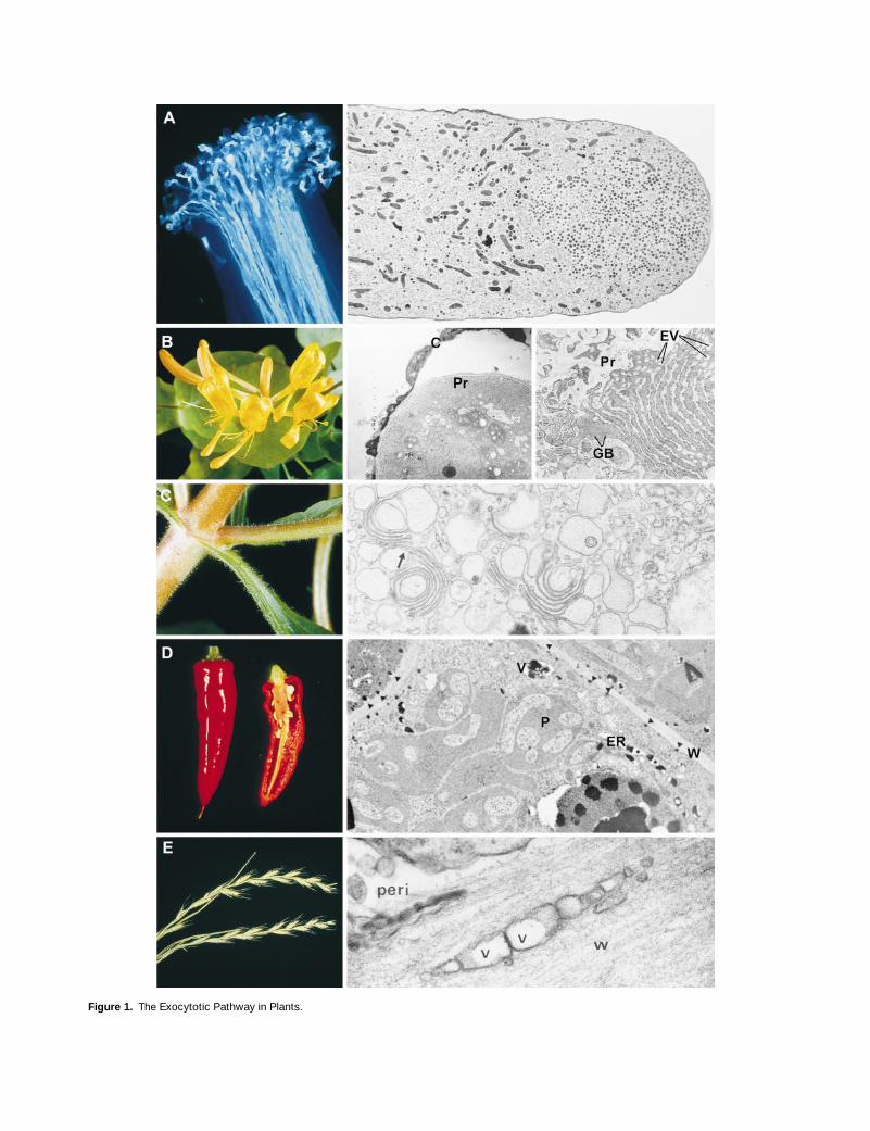
Figure 1.
The Exocytotic Pathway in Plants.

Exocytosis and Endocytosis 645
barley aleurone protoplasts (Homann and Tester, 1997). Wemust nevertheless await future classification of the factorsthat sort vesicle contents and regulate the delivery of differ-ent vesicle types to the plasma membrane. A major techni-cal obstacle is the lack of good assays of the kind that havebeen used so widely and effectively in mammalian and yeastcells for analysis of post-Golgi transport (reviewed in Batteyet al., 1996).
The commonly held view is that secretory vesicles are de-livered to their target membrane by the cytoskeleton. In ani-mal cells, this delivery role is effected by microtubules (Coleand Lippincott-Schwartz, 1995), and vesicles are distributedfrom a centrally located Golgi apparatus to the plasmamembrane. In plants, there are many Golgi apparatuses ineach cell, and microfilaments play the major role in vesicledelivery, as is evidenced in highly polarized cells such as thepollen tube, where microfilaments must transport vesiclesconsiderable distances (Figure 1A; Taylor and Hepler, 1997;see also Franklin-Tong, 1999, in this issue). Microtubules arealso present but appear to be more concerned with nuclearmigration and cytoplasmic zonation than vesicle transport(Cai et al., 1997). Even in cells from the coleoptile and root,which exhibit less polarized growth, current evidence sug-
gests a mainly microfilament-based vesicle transport mech-anism (see Battey and Blackbourn, 1993).
Although the cytoskeletal proteins provide a mechanismfor vesicle delivery, it cannot be assumed that they deter-mine where vesicles dock and fuse; that information, ac-cording to a hypothesis with strong support in animals andyeast, resides on the vesicle and target membranes them-selves (Rothman, 1994). This view is strikingly illustrated bythe behavior of secretory vesicles visualized with green fluo-rescent protein in mammalian Vero cells, in which vesiclemovement appears random, suggesting that microtubulesmaintain vesicles in motion so as to increase the chances ofvesicle–plasma membrane contact (Wacker et al., 1997). Onthe other hand, a key role for the actin cytoskeleton in local-izing secretion is indicated by cytoskeletal mutants of yeast(
Saccharomyces cerevisiae
) in which polarized growth andvesicle distribution are disrupted concomitantly (Winsor andSchiebel, 1997). Such data may relate directly to cell plateformation in plant cells, in which there is evidence for bothmicrofilament- and microtubule-based transport of secre-tory vesicles (Schopfer and Hepler, 1991; Samuels et al.,1995). It also may be relevant that prospore membrane for-mation in yeast is similar to cell plate formation in that a
Figure 1.
(continued).
(A)
The pollen tube. At left, aniline-blue staining of pollen tubes growing from the stigmatic surface down the style of
Capsicum
. Photographcourtesy of Dr. Naci Onus (Department of Agricultural Botany, University of Reading). At right, directional exocytosis maintains polar growth inthe lily pollen tube. Golgi vesicles accumulate at the tip, where they undergo exocytosis to supply membrane and wall material for growth.
3
3240. Reprinted from Lancelle et al. (1997), by permission of the authors and Springer-Verlag, Vienna.
(B)
The honeysuckle nectary. At left, honeysuckle flowers. The floral tube is lined with hairs, which secrete sugary nectar. Photograph by DaveMaw (Jealott’s Hill Research Station). Middle, the one-celled hairs develop elaborate wall protuberances (Pr) and a ribbed cuticle (C), which de-taches from the wall early in development. The wall structure is believed to result from fusion of Golgi-derived vesicles.
3
1310. At right, later,when the flower opens, sugar secretion is at a maximum and is associated with massive swelling of the ER. The relatively insignificant Golgibodies (GB), coupled with the elaborate ER and associated vesicles (EV), suggest that the ER plays a major role in secretion by direct traffickingto the plasma membrane.
3
7840. Reprinted from Fahn and Rachmilevitz (1970), by permission of the authors and Academic Press, London; andfrom Fahn (1988), by permission of the author and the New Phytologist Trust, Lancaster, UK.
(C)
Secretion of polysaccharide slime by
Mimulus
. At left, leaf, branch, and stem of
Mimulus
, showing a thick covering of secretory epidermalhairs. The hairs actively secrete polysaccharide slime from their head cells. Photograph by Dave Maw (Jealott’s Hill Research Station). At right,the head cells have Golgi apparatuses with hypertrophied cisternae, which give rise to very large vesicles (arrow), often directly as a result ofmaturation of the
trans
-most cisterna.
3
30,150. Reprinted from Schnepf and Busch (1976), by permission of the authors and Gustav FischerVerlag, Jena.
(D)
Capsaicin secretion from the placenta of chili pepper. At left, chili peppers. The placenta of hot pepper has a glandular epidermis that syn-thesizes the capsaicinoids responsible for the “hot” characteristic. Photograph courtesy of Dr. Barbara Pickersgill (Department of AgriculturalBotany, University of Reading). At right, placental epidermal cells of chili pepper have enlarged ER that contain and give rise to vesicles (V) con-taining electron-dense capsaicinoid droplets that are deposited outside the plasma membrane (arrowheads) by a currently undefined exocytoticmechanism. P, plastid; W, cell wall.
3
9600. Reprinted from Zamski et al. (1987), by permission of the authors and the University of ChicagoPress.
(E)
Secretion from the ligule of darnel grass (
Lolium temulentum
). At left, darnel. Photograph courtesy of Dr. Bob Froud-Williams (Department ofAgricultural Botany, University of Reading). In darnel, as well as in other plants such as
Selaginella
(Bilderback and Sloane, 1987), polysaccha-ride is secreted from the ligule (Chaffey, 1985). The adaxial epidermal cells have hypertrophied Golgi bodies, consistent with a function inpolysaccharide production. Because the primary function of exocytosis is secretion, not growth, plasma membrane recycling is presumably veryactive. At right, the presence of vesicular “paramural bodies” (V) in the cell wall (W), outside the plasma membrane and periplasmic space (peri),may reflect passage of vesicles through the plasma membrane (as with mucilage secretion in
Plantago
[Hyde, 1970]) or extrusions derived fromthe plasma membrane. It is possible that the vesicular membrane is a functionally important element of the secreted material.
3
51,770. Re-printed from Chaffey (1995), by permission of the author and Academic Press, London.

646 The Plant Cell
branch of the secretory pathway operates to direct newplasma membrane growth during sporulation (Neiman,1998).
Maturation and Processing in Secretory Vesicles
It is known that polysaccharides and secreted proteinsreach the cell wall after their production or modification inthe Golgi apparatus (Driouich et al., 1993). Therefore, it isassumed that secretory vesicles in plants contain cell wallpolysaccharide precursors and proteins, and both biochem-ical (e.g., Van der Woude et al., 1971) and electron micro-scopic (e.g., Moore et al., 1991; Lynch and Staehelin, 1992,1995) analyses of the polysaccharide content of secretoryvesicles have been undertaken (for a more detailed discus-sion, see Battey et al., 1996). Apart from these studies, thecomposition of the cargo and membrane of secretory vesi-cles is largely unknown, and the extent of processing andmaturation that takes place within vesicles is therefore un-clear. Our ignorance of these issues is partly due to the diffi-culties associated with isolating and identifying the vesicles(Hohl et al., 1996).
Protein Processing
The identification in plants of subtilases, a superfamily ofserine proteases that predominantly cleave dibasic residues(Siezen and Leunissen, 1997), is circumstantial evidencethat cleavage of plant proproteins may occur in post-Golgivesicles. In prokaryotes and lower eukaryotes, these en-zymes have low specificity and are secreted to function ex-tracellularly in growth or defense (Siezen and Leunissen,1997). In yeast, however, the subtilase Kex2 is responsiblefor the maturation of the
a
-mating factor, and in mammalsthere are at least four classes of subtilase, including prohor-mone convertases and furin (Barr, 1991). These enzymesplay an important role in post-Golgi processing of proteinsthat are released by exocytosis. In plants, at least six puta-tive subtilases have been identified (Siezen and Leunissen,1997), although only the evidence from a study of a tobaccosubtilase related to Kex2 of yeast directly argues for a pro-cessing role for subtilases in plant secretory vesicles. Spe-cifically, this Kex2p-like activity in tobacco processes thepreprotoxin KP6 of
Ustilago maydis
(Kinal et al., 1995). Pre-sumably, this occurs late in the secretory pathway, becauseno active toxin can be detected intracellularly.
Other plant subtilases, such as AG12 from
Alnus glutinosa
(Ribeiro et al., 1995) and the systemin binding proteinSBP50 from tomato (Schaller and Ryan, 1994), may be in-volved in processing foreign proteins and signaling peptidesfrom other parts of the plant. SBP50, located in the tomatoplasma membrane, binds to the defense signaling peptidesystemin at the recognition site required by furin and has fu-rin-like proteolytic activity (Schaller and Ryan, 1994). Stud-
ies of the biosynthesis of plant signaling peptides, includingENOD40 and systemin, indicate that they are produced cy-tosolically and therefore would not undergo processing inthe secretory pathway (Schaller and Ryan, 1995; Cohn et al.,1998).
Proteins that have a putative protective function often ex-emplify post-Golgi processing and maturation (Broekaert etal., 1997). In many cases, these proteins accumulate withinvacuoles and presumably are released by holocrine secre-tion, a crude type of exocytosis inasmuch as it is character-ized by the release of vacuolar contents coupled with celldegeneration. For example, a proteinase inhibitor that is be-lieved to act against insects (Heath et al., 1997) is expressedin stigmatic cells of
Nicotiana alata
as a pentameric proteinthat is thought to undergo conversion to its active formwithin the vacuole (Atkinson et al., 1993). On the assumptionthat the protein precursor is inactive, it has been suggestedthat such post-Golgi activation may provide a self-protec-tion mechanism (Heath et al., 1995). Proteins with putativeantimicrobial activity, such as 2S albumins (Terras et al.,1992), thionins (Bohlmann and Apel, 1991), and chitin bind-ing proteins (Raikhel et al., 1993), also accumulate withinvacuoles, and their release may simply involve nonspecificcell breakdown. However, the release of vacuolar contentsin response to invasion and other triggering processes maybe more regulated. Fusion of the mammalian lysosome withthe plasma membrane, for example, is regulated by Ca
2
1
ina manner that suggests an analogy with regulated exocyto-sis of vesicles (Rodriguez et al., 1997).
Polysaccharide Processing
A model system for the study of polysaccharide biosynthe-sis is the hypersecretory cells of the maize root cap(Rougier, 1981; Battey and Blackbourn, 1993). All root capcells secrete primitive cell wall polymers, but those in the hy-persecretory region additionally secrete a fibrillar polysac-charide mucilage (Rougier, 1981). In maize, the mucilagepolymers in secretory vesicles undergo a morphologicaltransition from a granular, Golgi-like appearance to a fibrillarappearance. The mechanisms governing this maturation pro-cess, which is termed fibrillogenesis, are unknown but seemto coincide with vesicle membrane maturation (Volkman,1981). A similar pattern has been observed in pea (Vian andRoland, 1974).
Perhaps the most impressive examples of post-Golgimaturation of polysaccharides are found in the Prasino-phycean algae. Figure 2 shows that in these plants, thescales that ultimately coat the plasma and flagellar mem-branes are synthesized in the Golgi apparatus as macromo-lecular structures and then delivered to the cell surface byexocytosis. In
Pyramimonas
, the scale reservoir is a post-Golgi compartment that contains a range of different scaletypes (Figure 2) and appears to sort and layer the scales inpreparation for their release at the plasma membrane. The

Exocytosis and Endocytosis 647
scale reticulum in
Scherffelia
is believed to play a similarrole, sorting the pentagonal and rod scales that will coatthe flagellar surface into separate vesicles for exocytosis(McFadden and Melkonian, 1986; Melkonian et al., 1986). Incontrast, the wall scales of
Scherffelia
are not sorted in thisway; instead, whole, mature Golgi cisternae fuse directlywith the plasma membrane to release the scales (McFaddenet al., 1986).
Arrival: Docking at the Plasma Membrane
As schematized in Figure 3, the distinction between thedocking and fusion of vesicles is clear: when a vesicle docksat the plasma membrane, it is tethered there, probably re-versibly (Steyer et al., 1997). Fusion is a separate, later eventthat involves the coalescence of vesicle and plasma mem-brane and formation of the fusion pore, resulting in the re-lease of vesicle contents into the extracellular matrix (seeThiel and Battey, 1998). Much less clear is whether someproteins are specifically concerned with docking and otherproteins are needed for fusion or whether both docking andfusion events are mediated by a common set of proteins.Current models, based on work in animals and yeast, pro-pose that the correct docking of vesicles is mediated bycomplexes formed between v-SNARE (soluble NSF-attach-ment protein receptor) proteins located on the external sur-face of vesicles, and t-SNAREs, partner proteins located onthe cytosolic face of the target membrane (the plasma mem-brane in the case of exocytosis) (Hay and Scheller, 1997).Specific v- and t-SNAREs are associated with exocytosis;
different combinations of SNAREs are characteristic of otherdocking events (see Sanderfoot and Raikhel, 1999, this is-sue). Whereas the base model for docking predicts that adimer formed between a t- and a v-SNARE is sufficient, it ismore likely that multimeric complexes are formed with oneor more SNAREs contributed from each of the two fusingmembranes (Hay and Scheller, 1997). In presynaptic nerveterminals, for example, the core complex is trimeric, com-prised of syntaxin and SNAP-25 (both t-SNAREs; in this in-stance, SNAP stands for synaptosome-associated proteinof 25 kD) and synaptobrevin (a v-SNARE) (Söllner et al.,1993; Hayashi et al., 1994).
Work on the product of the Arabidopsis
KNOLLE
geneprovides evidence in plants that docking at the plasmamembrane also is dependent on the formation of SNAREcomplexes. Mutations in
KNOLLE
affect early developmentof the Arabidopsis seedling so that the radial patterning oftissue layers is disrupted (Lukowitz et al., 1996). In
knolle
embryos, cytokinesis is impaired, cross walls fail to form, andthe enlarged cells that result contain polyploid nuclei. Cloningof
KNOLLE
has revealed that the protein it encodes is relatedto the syntaxin group of t-SNAREs (Lukowitz et al., 1996),and like other proteins in this class, it contains a conservedC-terminal domain, a variable N-terminal domain, and aputative membrane anchor. It is thought that KNOLLE func-tions specifically in vesicle docking during cell plate for-mation, a conclusion supported by the observations thatvesicle trafficking to the cell plate is not impaired in
knolle
embryos (Lauber et al., 1997) and that other exocytoticevents are not affected.
Although the work with KNOLLE indicates that SNARE func-tion in exocytosis is conserved during cell plate formation in
Figure 2. Post-Golgi Maturation in Algae.
(A) The scaly green alga Tetraselmis. Cell wall precursors assemble in the Golgi and condense at the trans face into scale precursors (arrow).These are exocytosed at the cell surface, where they self-assemble into the rigid wall, or theca. 313,300.(B) The prasinophyte alga Pyramimonas. Wall scales are synthesized in the Golgi apparatus (G) and arranged in the scale reservoir (SR) beforeexocytosis. 311,800.(C) Pyramimonas. The scale reservoir is multichambered and contains a range of different scale types. 310,910.Original negatives kindly supplied by Dr. David Domozych (Department of Biology, Skidmore College, Saratoga Springs, NY).

648 The Plant Cell
plants, a number of questions remain. A pressing issue is toidentify the v-SNAREs (and potentially other t-SNAREs) thatare required for vesicle docking at the cell plate so as toconfirm that the animal and yeast docking models apply inthis process. Based on a mutant phenotype, the product ofthe
KEULE
gene has been proposed as a candidate for av-SNARE (Lauber et al., 1997), but KEULE easily could beanother component of the docking machinery. It is also nec-essary to establish the extent of SNARE involvement in othertypes of exocytosis in plants. Interestingly, cell expansionand tip growth in root hairs and pollen tubes, developmentalprocesses that are all dependent on trafficking of membraneand wall materials to the cell surface, are unaffected in
knolle
mutants (Lukowitz et al., 1996). This suggests eitherthat a completely different docking machinery is involved ormore likely, and in common with other organisms, that dif-ferent sets of t-SNAREs (and v-SNARES) may be employed.Table 1 shows that candidate plant genes for both SNAP-25and synaptobrevin can be found in the databases; in time,their involvement in exocytosis may be unraveled.
Two other types of protein, NSF (
N
-ethylmaleimide–sensi-tive factor) and SNAPs (soluble NSF-attachment proteins),are important in docking and fusion of vesicles in animal andyeast cells (Goda and Südhof, 1997), although their exactroles are less clear than is the case for the SNAREs. Origi-nally proposed as a regulator of the final fusion event(Söllner et al., 1993), the function of NSF is now a subject ofcontroversy: it may act to prime SNAREs before docking(Ungermann et al., 1998) or to recycle SNAREs after fusion(Hay and Scheller, 1997). Other recent data are consistentwith a role immediately after docking but before fusion(Schweizer et al., 1998). As with the SNAREs, genes encod-ing proteins related to NSF and SNAP have been isolatedfrom plants (Table 1). Although there has been no confirma-tion of the biological role of these homologs in plant cells, itmay be significant that in
Capsicum
, a 72-kD protein withsome similarity to NSF is involved in plastid membrane bio-genesis (Hugueney et al., 1995).
GTPases from the Ras superfamily, termed Ypt in yeastand Rab in mammals, are proteins that play a crucial regula-
Figure 3. Sequence of Exocytosis and Endocytosis: Transport, Docking, Fusion, Content Release, and Recycling.
Shown is a speculative diagram that incorporates the suggested pathways to and from the plasma membrane in plants. CCP, clathrin-coatedpit; CCV, clathrin-coated vesicle; ER, endoplasmic reticulum; MVB, multivesicular body; PCR, partially coated reticulum.

Exocytosis and Endocytosis 649
tory role in the docking/fusion process. Sec4p is the Yptmember that regulates vesicle fusion at the plasma mem-brane of
S. cerevisiae
(Goud et al., 1988). Its homolog in
Schizosaccharomyces pombe
, Ypt2p, is also required fortransport between the Golgi apparatus and plasma mem-brane (Craighead et al., 1993), and the mammalian homolog,Rab8, plays a role in basolateral plasma membrane traffick-ing (Huber et al., 1993). Plant homologs are known and aremore similar to mammalian Rab8 and
S. pombe
Ypt2p thanto Sec4p of
S. cerevisiae
. This indirect evidence suggeststhe plant Rab8 homologs may function at the ultimate, exo-cytotic step in the secretory pathway. The way in which Ypt/Rab GTPases regulate exocytosis is unclear, but data forRab5, a GTPase that controls vesicle trafficking through theendocytotic pathway, indicate that GTP hydrolysis is not di-rectly coupled to membrane fusion; rather, the complete cy-cle of GDP/GTP binding and release, together with GTPhydrolysis, regulates the kinetics of docking, possibly by af-fecting the activation or stability of SNAREs on vesicle andtarget membranes (Rybin et al., 1996).
Rho-type GTPases are another group in the Ras super-family. They have functions in a range of cell biological pro-cesses, but of particular relevance are the roles of the Rho-related Cdc42p and the Ras-related Bud1p in polarizedgrowth in
S. cerevisiae
(Chant, 1996). Rop1, a plant RhoGTPase, is localized at the apical cortex of pollen tubes (Linet al., 1996) such that microinjected antibodies to Rop1 in-hibit pollen tube growth (Lin and Yang, 1997). These datahave led to the proposal that Rop1 regulates polarized exo-cytosis at the pollen tube tip (Lin and Yang, 1997). It is un-clear whether this GTPase controls docking or some otheraspect of vesicle targeting.
The Ca
2
1
Connection: Controlling ExocytoticMembrane Fusion
In animal cells, Ca
2
1
seems to be a key regulator of exocyto-sis. This turns out to be true even in some cells that havebeen conventionally regarded as showing “constitutive”
Table 1.
Plant Proteins with Sequence Similarity to Components of the Docking/Fusion Machinery in Other Organisms
TypeProtein Name
EMBL AccessionNumber Species
a
References Related to
b
EMBL AccessionNumber Species
c
Identity with Plant Sequence
d
References
t-SNARE KNOLLE U39451 At Lukowitz et al. (1996) Syntaxin 2 D14582 Hs 46 (39/84) Hirai (1993)SNAP25A X92419 At X. Gansel and
L. Sticher (unpublished data)
SNAP-25 U85806 Hm 39 (25/64) Bruns et al.(1997)
v-SNARE SAR1 M90418 At M. Schena andR.W. Davies (unpublished data)
Synaptobrevin X96737 Mm 44 (93/211) M. D’Espositoet al. (unpub-lished data)
e
Unnamed 023429
f
At M. Bevan et al. (unpublished data)
g
Synaptobrevin X92396 Hs 59 (20/39) D’Esposito et al. (1996)
SNAP Unnamed AB001375 Vv S. Matsumoto, I.B. Dry, and M. Thomas (unpublished data)
b
-SNAP P81126
h
Bt 48 (122/256) Whiteheart et al. (1993)
NSF Unnamed D86506 Nt Sato et al., 1997 NSF X15652 Cg 62 (190/302) Wilson et al.(1989)
Unnamed D25240
i
Os H. Uchimiya (unpublished data)
NSF AF006826 Dd 66 (71/106) Weidenhaupt et al. (1998)
a
Plant species: At,
Arabidopsis thaliana
; Vv,
Vitis vinifera
; Nt,
Nicotiana tabacum
; Os,
Oryza sativa
.
b
The protein showing the highest BLAST score with the plant sequence.
c
Animal/fungal species: Hs,
Homo sapiens
; Hm,
Hirudo medicinalis
; Mm,
Mus musculus
; Bt,
Bos taurus
; Cg,
Cricetulus griseus
; Dd,
Dictyostel-ium discoideum
.
d
Percentage of identity and, in parentheses, the number of identical amino acids over the longest matching region.
e
Full contributors are M. D’Esposito, M.R. Matarazzo, A. Ciccodicola, R. Mazzarella, N. Quaderi, M. Rocchi, N. Archidiacono, D. Schlessinger,and M. D’Urso.
f
ORF from contig fragment 4, chromosome 4, EMBL accession number Z97339.
g
Full contributors are M. Bevan, W. Stiekema, G. Murphy, R. Wambutt, T. Pohl, N. Terryn, M. Kreis, T. Kavanagh, K.D. Entian, M. Rieger, R.James, P. Puigdomenech, P. Hatzopoulos, B. Obermaier, A. Duesterhoft, J.D.G. Jones, K. Palme, W. Ansorge, M. Delseny, I. Bancroft, H.W.Mewes, C. Schueller, and N. Chalwatzis.
h
SwissProt entry.
i
Partial sequence.

650 The Plant Cell
secretion (Morimoto et al., 1995; Chavez et al., 1996;Coorssen et al., 1996). In plants, all cells studied so far showpronounced Ca
2
1
regulation of exocytosis (Zorec and Tester,1992, 1993; Thiel et al., 1994; Homann and Tester, 1997;Carroll et al., 1998). Yeast, on the other hand, does not showCa
2
1
regulation of exocytosis. Similarly, although there arereports of a role for Ca
2
1
at earlier stages in the secretorypathway (e.g., Beckers and Balch, 1989), in animal cells,Ca
2
1
dependence is most clearly associated with exocytosisand the subsequent retrieval of vesicles via the endosomalroute (Gruenberg and Maxfield, 1995). This information sug-gests that many plant and animal cells may have a specificrequirement for Ca
2
1
at the late stages of the secretorypathway. This requirement links exocytosis closely to theneeds of the cell, because Ca
2
1
is a signaling elementwhose level is rapidly regulated in the local vicinity of theplasma membrane.
Because fusion of membranes can only occur once theyhave been brought into close proximity, the first potentialrole for Ca
2
1
is the regulation of the process that draws thedocked vesicle membrane into immediate apposition withthe plasma membrane. Although the mechanism that under-lies such membrane juxtaposition is not fully understood,work on the mode of membrane fusion that promotes viralinfection provides the clearest pointer. The influenza virus isa very efficient machine for cell invasion, an essential com-ponent of which is the ability to cross the plasma mem-brane. The viral hemagglutinin protein forms a scaffold thatdraws the virus and host cell membranes together and pro-motes their fusion (White, 1996). Membrane fusion involvestransient domains that lead ultimately to formation of the fu-sion pore (Blumenthal et al., 1995).
Evidence from a variety of sources suggests that in animalcells, membrane fusion may involve a similar scaffold mech-anism (Monck and Fernandez, 1996). Recent data suggestthat synaptobrevin in association with syntaxin and SNAP-25can cause membrane fusion when incorporated into sepa-rate populations of artificial liposomes, and it has been pro-posed that the v-/ t-SNARE complex achieves this by amechanism analagous to that used by the viral proteins(Weber et al., 1998). The Ca
2
1
binding proteins that mightregulate this process in vivo have not been defined, but syn-aptotagmin has become a strong candidate in synapses(Goda and Südhof, 1997), where it exhibits Ca
2
1
-dependentbinding to syntaxin, SNAP-25, and phospholipid. It has fur-ther been suggested that synaptotagmin binding could beinvolved in formation of the fusion pore (Südhof and Rizo,1996). However, synaptotagmin homologs have not beendetected in neutrophils, where analysis of exocytosis indi-cates that different vesicle populations have specific Ca
2
1
sensitivities and that sophisticated Ca
2
1
regulation is there-fore at work (Nüsse et al., 1998).
Ca
2
1
-dependent activator protein for secretion (CAPS) isrequired for Ca
2
1
-triggered exocytosis from permeabilizedanimal neuroendocrine PC12 cells (Walent et al., 1992).CAPS may activate exocytosis by Ca
2
1
-dependent binding
to phospholipids, in particular phosphatidylinositol (4,5)-bis-phosphate, whose presence is a prerequisite for fusion (re-viewed in Martin, 1997). It is interesting that the failure toidentify CAPS homologs in yeast, where exocytosis is Ca
2
1
-independent, is taken to reflect a key role for CAPS in regu-lated (Ca
2
1
-dependent) exocytosis (Martin, 1997). Exactlythe same argument could be applied to another group ofCa
21-dependent phospholipid binding proteins, the annex-ins, which are also absent from yeast. Some members ofthis family aggregate vesicles and lead to membrane defor-mation (Swairjo et al., 1994; see Thiel and Battey, 1998).Recent work has further suggested a role for annexins inexocytosis from plant cells (Carroll et al., 1998). The ques-tion thus arises as to how annexins might be related toSNAREs. The latter may facilitate targeting and membranespecificity, whereas the former may promote membrane fu-sion and pore formation (see also Jahn and Hansen, 1998).
Recently, the sequence of a tomato gene (CLB1) waspublished (Kiyosue and Ryan, 1997) that contains a Ca21-dependent lipid binding domain similar to that found in theC2 domain proteins, such as synaptotagmin, that regulatemembrane traffic in neuronal cells (Südhof and Rizo, 1996).An Arabidopsis gene (CaLB; accession number X96598; un-published data) very similar to CLB1 is present in the se-quence databases. Also reported in the databases is an openreading frame (TREMBL accession number O23480), identi-fied in the Arabidopsis genome sequencing project (Bevanet al., 1998), that shows similarity to frequenin, an EF-handCa21 binding protein also thought to play a role in mem-brane trafficking (Pongs et al., 1993). Although the existenceof these genes further suggests that exocytosis may be reg-ulated in a Ca21-dependent manner by using proteins con-served between plants and animals, hard proof that theseproteins are involved awaits biochemical and genetic tests.
Once the fusion pore has formed, vesicular contents canbe released to the extracellular matrix. In animal cells, fusionpore opening can be transient, with content release followedvery rapidly by pore closure (Figure 3; Alvarez de Toledo etal., 1993). Alternatively, pore formation is followed by fullpore opening with concomitant incorporation of the vesiclemembrane into the plasma membrane (Spruce et al., 1990).Both transient and permanent fusion can be observed at dif-ferent times in the same animal (Oberhauser et al., 1992)and, indeed, plant cell (Thiel and Battey, 1998; Thiel et al.,1998). The frequency of transient relative to permanent fu-sion is a very important issue because it determines whetherthe vesicle matrix (to which the vesicle cargo is bound) islost or recycled and whether vesicle membrane becomes in-corporated into the plasma membrane (Figure 3). Figure 4indicates the potential significance of regulation of transientversus permanent vesicle fusion for the physiology of theplant cell. Clearly, in nonexpanding cells, a mechanism forcargo discharge without membrane incorporation would beuseful, especially in view of the energy costs of generatingendocytotic vesicles in the presence of high turgor pressure(Cram, 1980; Gradmann and Robinson, 1989; see below).

Exocytosis and Endocytosis 651
More detailed discussion of this question can be found inThiel and Battey (1998).
ENDOCYTOSIS
Recovering Excess Membrane
Evidence from several plant systems indicates that the rate atwhich new membrane is incorporated into the plasma mem-brane during cell elongation and growth far exceeds the netincrease in cell surface area (Samuels and Bisalputra, 1990;Low and Chandra, 1994). This finding bespeaks the opera-tion of significant membrane recycling. In nongrowing secre-tory cells, delivery of vesicles to the plasma membrane must be
balanced by the equivalent recycling of membrane from thecell surface. In animal and S. cerevisiae cells, another majorfunction of endocytosis is internalization and degradation ofmembrane proteins such as receptor/ligand complexes (Fig-ure 3). Furthermore, plant cells often undergo volume fluctu-ations in response to changes in the external osmoticenvironment. Because any fixed amount of plasma mem-brane is essentially inexpandable (Wolfe and Steponkus,1983), significant cell volume increase must be accom -panied by increased exocytosis. Similarly, osmoregulatoryvolume decrease after hypoosmotic treatment is likely to beaccompanied by increased recycling of plasma membrane(Taylor et al., 1996; see also Thiel and Battey, 1998).
Despite the evidence that membrane recycling occurs inplants, there has been considerable doubt until recentlywhether the underlying mechanisms involve vesicle endocy-tosis. These uncertainties arise mainly from consideration of
Figure 4. Transient or Permanent Fusion: Consequences for the Plant Cell?
The diagram depicts the potential effects of transient and permanent vesicle fusion on plant cell growth. Although events believed to correspondto transient and permanent vesicle fusion have been detected in plants (Thiel and Battey, 1998; Thiel et al., 1998), the extent to which either orboth predominate or are regulated at all remains unknown. V, vacuole.

652 The Plant Cell
the energetics of vesicle recycling in the turgid cell (e.g.,Cram, 1980), whereby two factors must be considered: themetabolic energy needed to sustain endocytosis againstpositive hydrostatic pressure and the free energy barrier tovesicle formation. It is likely that the total metabolic energyfor endocytosis is well within the cell’s metabolic energybudget (Raven, 1987). The free energy requirements for ves-icle formation are lower for smaller vesicles (Saxton andBreidenbach, 1988) and crucially depend on the widely vari-able value of turgor (Gradmann and Robinson, 1989). There-fore, it can be argued that the energetics of endocytosis arelikely to be favorable for a range of conditions and vesicletypes. In addition, there may be a role for molecular recy-cling of membrane lipids (Craig and Staehelin, 1988), forwhich some evidence has been obtained using fluorescentlylabeled lipids to measure trafficking from the plasma mem-brane to the ER (Grabski et al., 1993).
Evidence for Endocytosis in Plants
Direct visualization of endocytosis in plant cells comes fromobservations of internalization of membrane-impermeantmolecules. Although earlier interpretation of Lucifer Yellowuptake into plant cells has become problematic as a resultof the appreciation that this dye can be transported acrossmembranes (e.g., O’Driscoll et al., 1991), internalization of arange of other impermeant molecules has now been re-ported, including heavy metals (Hubner et al., 1985; Lazzaroand Thomson, 1992), biotinylated molecules (e.g., Horn etal., 1990), gold-conjugated proteins and lectin (Hillmer et al.,1986; Villanueva et al., 1993), and cationic ferritin (Tanchaket al., 1984). The use of lipophilic styryl (FM) dyes such asFM1-43, which fluorescently label the plasma membrane ofintact cells, is beginning to provide additional evidence formembrane recycling (e.g., Smith and Betz, 1996). Figure 5shows how fluorescence internalization thus can be readilyvisualized with confocal microscopy, giving a direct indica-tion of membrane recycling. The related dye FM4-64 hasbeen used to visualize endocytosis and vacuolar membranedynamics in yeast (Vida and Emr, 1995), and more recently,the use of FM1-43 has revealed significant membrane inter-nalization over a time scale of minutes in secretory maizeroot cap cells (Carroll et al., 1998). In zygotes of the marinealga Fucus, FM1-43 and FM4-64 internalization occurs pre-dominantly at the growing rhizoid apex and at the develop-ing cell plate (Belanger and Quatrano, 1998; Brownlee et al.,1998).
In animal cells and yeast, endocytosis occurs via clathrin-coated vesicles (CCVs) that act in plasma membrane recov-ery and in cycling of vesicles in the endomembrane system.CCVs are characterized by the presence of protrusions ofclathrin on the cytoplasmic surface (Low and Chandra,1994; Robinson, 1996). Clathrin-coated pits in the plasmamembrane and CCVs have been described widely in plantcells (e.g., Fowke et al., 1991; Robinson, 1996) and are es-
pecially abundant in actively growing cells (e.g., Samuelsand Bisalputra, 1990). Other structures known to be in-volved in endocytosis in animal cells that are also observedin plants and algae include Golgi bodies, multivesicular bod-ies, and partially coated reticulum (Record and Griffing,1988; Domozych, 1991; Low and Chandra, 1994; see alsoFigure 3). Multivesicular bodies are involved in transport tovacuoles and appear to contain degradative enzymes. Par-tially coated reticulum may be either equivalent to the animaltrans-Golgi network or an early endosomal compartment(Robinson, 1996).
Coated pits and CCVs have a well-conserved complex ofpolypeptides specific to their cytoplasmic surface. Clathrinpolypeptides form the outer (cytosolic-facing) layer of thecoat and comprise three heavy chains and three light chainsthat assemble to form triskelions (reviewed in Robinson, 1996).Plant clathrin heavy chains have a number of well-conservedregions in common with animal and yeast cells (Blackbournand Jackson, 1996). Less information is available for clathrinlight chains in plants, although these have been well charac-terized in animals (33- and 36-kD polypeptides) and yeast(38-kD polypeptide) and possess Ca21-calmodulin bindingproperties (Acton and Brodsky, 1990; Nathke et al., 1992).CCVs can be either Golgi- or plasma membrane–derived(Blackbourn and Jackson, 1996; Robinson, 1996). Recentwork has concentrated mainly on CCVs in trafficking in thetrans-Golgi network, although the study by Blackbourn andJackson (1996) clearly implicates CCVs in plasma mem-brane recycling during pollen tube elongation, becauseassembled clathrin is present at the pollen tube tip, corre-sponding to the site of uptake of fluorescein isothiocyanate–dextran (O’Driscoll et al., 1991). Blackbourn and Jackson(1996) further present evidence for selective membrane cy-cling in the pollen tube tip via an early endosomal compart-ment and suggest that two previously described vesiclepopulations with diameters of z300 and 50 nm could repre-sent secretory vesicles and re-internalized membrane, re-spectively.
Another essential element of the inner region of the vesi-cle coat in CCVs is the adaptins. In animals and yeast, theseheterotetrameric adaptor proteins are involved in attachingto the membrane and can interact with membrane-spanningreceptors. Both Golgi- and plasma membrane–specificadaptor complexes exist in animals and yeast. Plant coun-terparts are considerably less well characterized, but currentevidence supports a role for g- and b-type adaptins inplasma membrane recycling (Drucker et al., 1995; Keon etal., 1995). Another feature of animal adaptins is the posses-sion of autophosphorylating and casein kinase activity,which recently has been shown in adaptin-like polypeptidesfrom zucchini (Drucker et al., 1998).
A variety of other molecules is associated with the en-docytotic pathway in animals and yeast, and some of thesemolecules are beginning to be described in plant systems.The GTP-binding protein dynamin is required for rapid en-docytosis coupled to exocytosis in adrenal chromaffin cells

Exocytosis and Endocytosis 653
(Artalejo et al., 1995). Although this particular endocytoticsystem is independent of clathrin, dynamin is involvedelsewhere in clathrin-mediated endocytosis, for instance, inthe mammalian COS-7 cell line (Herskovits et al., 1993).One plant homolog of dynamin has been isolated in Arabi-dopsis with significant similarity to yeast Vps1p, rat dy-namin, and murine Mx1 protein (Dombrowski and Raikhel,1995). Another dynamin homolog, phragmoplastin, hasbeen found at the cell plate in soybean (Gu and Verma,1996). It appears that polymerized dynamin acts to“squeeze” vesicles from the plasma membrane during en-docytosis (Takei et al., 1995), and so it is tempting to specu-late that phragmoplastin acts in a similar way to allow theexocytotic release of vesicle contents during growth of thecell plate. Finally, an uncoating ATPase facilitates the re-moval of the clathrin coat from CCVs (Hannan et al., 1998),
and preliminary evidence exists for such ATPases in plants(Beevers, 1996).
Receptor-Mediated Endocytosis
Major functions of clathrin and associated proteins in ani-mals and yeast are the internalization of membrane recep-tors and proteins and the correct targeting of CCVs to theendosomal lysosome, late-Golgi compartments, or vacuole.Such receptor-mediated endocytosis is an essential processin signal transduction whereby receptor–ligand complexesare internalized and processed (e.g., Egner and Kuchler,1996). This process further involves the ubiquitination of tar-get molecules before endocytosis and degradation (Egnerand Kuchler, 1996; Hicke and Riezman, 1996), and it also
Figure 5. Confocal Images of the Pattern of Internalization of the Fluorescent Membrane Dye FM1-43 in a Rhizoid Cell of Fucus serratus.
Embryos were incubated in 1 mM FM1-43 for the duration of the experiment. Hypoosmotic treatment (from sea water to 50% sea water) wasgiven at time (t) 0. Little or no dye internalization was observed for at least 390 sec before the hypoosmotic shock. However, dye internalizationwas apparent within 20 sec of the shock and continued for the duration of the recording (240 sec).

654 The Plant Cell
requires myosin and actin (Geli and Riezman, 1996). Evi-dence for receptor-mediated endocytosis in plants is cur-rently very limited. Endocytotic internalization of Rhizobiuminvolving lectin binding occurs in pea root hairs (Kijne et al.,1988), and receptor-mediated uptake of elicitor molecules (Lowand Chandra, 1994) and auxin binding protein (Robinson,1996) has been proposed.
Quantification of Endocytosis
Electrophysiological experiments allow direct measure-ments of protoplast membrane surface area and provide fur-ther evidence for membrane cycling, and although changesdetected in cell capacitance reflect only the net result ofexocytosis and endocytosis (Battey et al., 1996), such mea-surements with barley aleurone and maize root protoplastsreveal significant membrane internalization upon intracellu-lar perfusion with solutions of very low [Ca21] (Zorec andTester, 1992; Carroll et al., 1998). Rapid increases in barleyaleurone protoplast surface area, induced by increasing in-ternal pressure, are followed by a decline to resting values,suggesting the presence of a pressure-responsive exo-endocytosis cycle (Zorec and Tester, 1993). Furthermore,hypoosmotic treatment of stomatal guard cell protoplastscauses a reversible increase in plasma membrane area thatis quantifiable as an increase in cell volume (Homann, 1998).This observation is of particular interest because the guardcell is generally regarded as nonsecretory and nongrowing;the vesicles involved in this response, therefore, may have aspecific role in osmoregulation. In the marine alga Pelvetia,freeze-fracture studies have similarly identified a class ofvesicles that is exocytosed in response to hypoosmotictreatment (Gilkey and Staehelin, 1989). Hypoosmotically in-duced cell expansion in Fucus zygotes is followed by vol-ume regulation that is demonstrable by a dramatic increasein the internalization of FM1-43 at the growing rhizoid apex(Brownlee et al., 1998; Figure 5).
Taken together, the above evidence amply suggests thatmembrane recycling can occur rapidly (within seconds tominutes) during osmotically induced cell volume changes.This rate of recycling may be faster than during normalgrowth and secretion, suggesting that the usual membranecycling pathway may be bypassed. Observation of vesicularmaterial extruded from the plasma membrane after pollentube plasmolysis (Kroh and Knuiman, 1985) and in osmoti-cally regulating protoplasts (Steponkus, 1991) also suggeststhat atypical membrane structures may be an importantmechanism to allow volume regulation (see also Thiel andBattey, 1998).
The simultaneous monitoring of exocytosis and endocyto-sis through measurements of cell capacitance and internal-ization of FM1-43, respectively, has been achieved withadrenal chromaffin cells (Smith and Betz, 1996). This studyshowed that after short exocytotic stimuli, endocytosis oc-curred after exocytosis had ceased, whereas prolonged
exocytotic stimuli resulted in endocytosis that eventuallybalanced the rate of exocytosis. The potential of FM dyes forquantification of endocytosis also has been shown in hip-pocampal neurons, where unitary endocytotic events havebeen visualized and quantified (Ryan et al., 1997), and suchhigh-resolution imaging could potentially provide unique quan-titative information on both protoplasts and whole plant cells.
CONCLUSIONS AND FUTURE PROSPECTS
Our initial interest in exocytosis was triggered by the need tofind mechanisms that underlie the pronounced effects ofCa21 on plant cell development (Hepler and Wayne, 1985)and the circumstantial evidence that Ca21 might controlthe exocytotic/endocytotic balance in plants (Steer, 1988;Battey and Blackbourn, 1993). Ca21 has subsequently beenshown to be needed for a sustained increase in the plasmamembrane capacitance of plant protoplasts, reflecting theactivation of exocytosis relative to endocytosis. This evi-dence supports the hypothesis that regulation of vesicle fu-sion at the plasma membrane is a critical control point forCa21. It remains to be demonstrated that these effects ofCa21 lead to developmental changes; indeed, effects on se-cretion (polysaccharide or protein) in addition to membranegrowth have not been shown. Therefore, the electrophysio-logical measurement of membrane capacitance has enhancedour knowledge of the factors that influence exocytosis/endocytosis in plants, but the need to use protoplasts andthe resultant lack of necessary relevance to plant physiologyand development are serious limitations to progress. Never-theless, the recent paper by Homann (1998), which relateschanges in capacitance to guard cell osmotic regulation, isan important step in the right direction. Similarly, the abilityof the dye FM1-43 to reveal membrane cycling in plants(Carroll et al., 1998) offers the opportunity, in conjuction withcapacitance measurements, to monitor exocytosis and en-docytosis independently. At a still finer level of detail, thereis much potential in the ability of the attached-patch methodto distinguish transient fusion from full fusion (Thiel et al.,1998). A major technical challenge that remains is to mea-sure exocytosis/endocytosis in walled cells to relate thesemeasurements to cell and tissue changes.
Those cells of most interest for understanding exocytosisand endocytosis are often highly differentiated and difficultsubjects for biochemical analysis (e.g., the honeysucklenectary or Mimulus glandular hairs; see Figure 1). The avail-ability of a range of (labeled) protein probes whose traffick-ing pathways are known would provide the means tounderstand plasma membrane dynamics in such specializedcells, as is indicated by success in studies on bone-resorb-ing osteoclasts in animals, where the plasma membrane isdivided into domains that are maintained by directed vesicletrafficking along the endocytotic pathway (Salo et al., 1996;Palokangas et al., 1997). In plants, probes that allow this

Exocytosis and Endocytosis 655
kind of analysis are unavailable at present, but the develop-ment of green fluorescent protein tagging offers potential forthe future. An equivalent tool with which to probe polysac-charide distribution would be of great value in plant cells. Itis already possible to visualize directly the transport of sec-ondary metabolites in an exocytotic pathway that leads tothe deposition of defense-related products in the cell wall(Stewart and Mansfield, 1985). This provides a useful meansto understand this novel exocytotic pathway as well as tostudy an important aspect of the response of plants to fun-gal invasion.
The full range of SNAREs and other proteins that regulatevesicle docking and fusion at the plant plasma membraneneeds to be identified. Confirmation that SNAREs are impor-tant in exocytosis may be obtainable by using the clostridialtoxins, such as tetanus and botulinum toxins, in patch–clamp experiments. These toxins are specific proteases thatcleave syntaxin and SNAP-25 and synaptobrevin in humannerve terminals (Huttner, 1993; Hayashi et al., 1994), therebypreventing neurotransmitter release. It is apparently notknown whether these toxins have similar effects on exocyto-sis in plant cells, although one line of evidence implicating asyntaxin in guard cell responses to abscisic acid is a spe-cific inhibition by botulinum C toxin (Leyman et al., 1999). Arelated question is whether plant cells with pronounced po-larity of exocytosis, such as the hypersecretory cells of themaize root cap (Roy and Vian, 1991), seed coat cells ofPlantago (Hyde, 1970), or pollen tubes (Taylor and Hepler,1997), achieve this polarity by using SNARE mechanisms,Ca21 gradients, cytoskeletal targeting, or a combination ofall three.
Interestingly, it seems that secreted polysaccharide mayplay an important role in perpetuating polarized growth.Thus, the inhibitor of Golgi secretion brefeldin A has beenshown to disrupt polarized secretion of a rhizoid-specificpolysaccharide in polarizing Fucus zygotes (Shaw andQuatrano, 1996). This treatment prevents fixation of the po-lar axis but is without effect on actin localization or cell wallsynthesis associated with cell division, and it implies that lo-calized exocytosis of cell wall components is an early step inpolar axis fixation such that the cell wall is a source of posi-tion-dependent information. In Arabidopsis, mutations of theGNOM gene, which has homology with the yeast SEC7gene, similarly show disrupted apical–basal polarity and theabsence of specific organs (Mayer et al., 1993; Shevell et al.,1994), further suggesting that vesicle-mediated trafficking ofpositional information to the cell wall compartment is a fun-damental step determining embryonic polarity.
ACKNOWLEDGMENTS
We thank Ian Moore (Oxford University, UK) for Rab guidance and forcomments on the manuscript; Gerhard Thiel (Göttingen University,Germany) for comments on the manuscript; James Carew and Paul
Le Mière (Department of Horticulture, University of Reading) for helpwith Figure 1 and for drawing the diagrams in Figures 3 and 4; JeanWhiterow (Department of Horticulture, University of Reading) for herinvaluable help in preparing the manuscript; and the United KingdomBiotechnology and Biological Sciences Research Council (BBSRC),the Leverhulme Trust, the University of Reading Research Endow-ment Trust Fund, the Marine Biological Association, UK, andZENECA Agrochemicals for financial support of the authors’ labs.N.C.J. acknowledges the support of an Industrial CASE studentshipfrom the BBSRC.
REFERENCES
Acton, S.L., and Brodsky, F.M. (1990). Predominance of clathrinlight chain LCb correlates with the presence of a regulated secre-tory pathway. J. Cell Biol. 111, 1419–1426.
Alvarez de Toledo, G., Fernandez-Chacùn, R., and Fernandez,J.M. (1993). Release of secretory products during transient vesi-cle fusion. Nature 363, 554–558.
Artalejo, C.R., Henley, J.R., McNiven, M.A., and Palfrey, H.C.(1995). Rapid endocytosis coupled to exocytosis in adrenal chro-maffin cells involves Ca21, GTP and dynamin but not clathrin.Proc. Natl. Acad. Sci. USA 92, 8328–8332.
Atkinson, A.H., Heath, R.L., Simpson, R.J., Clarke, A.E., andAnderson, M.A. (1993). Proteinase inhibitors in Nicotiana alatastigmas are derived from a precursor protein which is processedinto five homologous inhibitors. Plant Cell 5, 203–213.
Barr, P.J. (1991). Mammalian subtilisins: The long-sought dibasicprocessing endoproteases. Cell 66, 1–3.
Battey, N.H., and Blackbourn, H.D. (1993). The control of exocyto-sis in plant cells. New Phytol. 125, 307–338.
Battey, N.H., Carroll, A., van Kesteren, P., Taylor, A., and Brownlee,C. (1996). The measurement of exocytosis in plant cells. J. Exp.Bot. 47, 717–728.
Beckers, C.J.M., and Balch, W.E. (1989). Calcium and GTP: Essen-tial components in vesicular trafficking between the endoplasmicreticulum and the Golgi apparatus. J. Cell Biol. 108, 1245–1256.
Beevers, L. (1996). Clathrin-coated vesicles in plants. Int. Rev.Cytol. 167, 1–35.
Belanger, K.D., and Quatrano, R.S. (1998). Characterisation ofmembrane populations in Fucus distichus using the vital dyeFM4-64. Keystone Symp. Plant Cell Biol. (abstr.).
Bevan, M., et al. (1998). Analysis of 1.9 Mb of contiguous sequencefrom chromosome 4 of Arabidopsis thaliana. Nature 391, 485–488.
Bilderback, D.E., and Sloane, J.H. (1987). The ultrastructure of thesecreting ligule of Selaginella kraussiana. Bot. Gaz. 148, 413–419.
Blackbourn, H.D., and Jackson, A.P. (1996). Plant clathrin heavychain: Sequence analysis and restricted localisation in growingpollen tubes. J. Cell Sci. 109, 777–787.
Blumenthal, R., Pak, C.C., Raviv, Y., Krumbiegel, M., Bergelson,L.D., Morris, S.J., and Lowry, R.J. (1995). Transient domainsinduced by influenza haemagglutinin during membrane fusion.Mol. Membr. Biol. 12, 135–142.
Bohlmann, H., and Apel, K. (1991). Thionins. Annu. Rev. PlantPhysiol. Plant Mol. Biol. 42, 227–240.

656 The Plant Cell
Broekaert, W.F., Cammue, B.P.A., De Bolle, M.F.C., Thevissen,K., De Samblanx, G.W., and Osborn, R.W. (1997). Antimicrobialpeptides from plants. Crit. Rev. Plant Sci. 16, 297–323.
Brownlee, C., Manison, N.F.H., and Anning, R. (1998). Calcium,polarity and osmoregulation in Fucus embryos: One messenger,multiple messages. Exp. Biol. OnLine 3, 11.
Bruns, D., Engers, S., Yang, C., Ossig, R., Jeromin, A., and Jahn,R. (1997). Inhibition of transmitter release correlates with the pro-teolytic activity of tetanus toxin and botulinum toxin A in individualcultured synapses of Hirudo medicinalis. J. Neurosci. 17, 1898–1910.
Cai, G., Moscatelli, A., and Cresti, M. (1997). Cytoskeletal organi-zation and pollen tube growth. Trends Plant Sci. 2, 86–91.
Carroll, A.D., Moyen, C., van Kesteren, W.J.P., Tooke, F., Battey,N.H., and Brownlee, C. (1998). Ca21, annexins, and GTP modu-late exocytosis from maize root cap protoplasts. Plant Cell 10,1267–1276.
Chaffey, N.J. (1985). Structure and function in the grass ligule: Opti-cal and electron microscopy of the membranous ligule of Loliumtemulentum L. (Poaceae). Ann. Bot. 55, 65–75.
Chaffey, N.J. (1995). Structure and function in the grass ligule: Theendomembrane system of the adaxial epidermis of the membranousligule of Lolium temulentum L. (Poaceae). Ann. Bot. 76, 103–112.
Chant, J. (1996). Generation of cell polarity in yeast. Curr. Opin. CellBiol. 8, 557–565.
Chavez, R.A., Miller, S.G., and Moore, H.-P.H. (1996). A biosyn-thetic regulated secretory pathway in constitutive secretory cells.J. Cell Biol. 133, 1177–1191.
Cohn, J., Day, R.B., and Stacey, G. (1998). Legume nodule organo-genesis. Trends Plant Sci. 3, 105–110.
Cole, N.B., and Lippincott-Schwartz, J. (1995). Organization oforganelles and membrane traffic by microtubules. Curr. Opin. CellBiol. 7, 55–64.
Coorssen, J.R., Schmitt, H., and Almers, W. (1996). Ca21-trig-gered massive exocytosis in Chinese hamster ovary cell. EMBO J.15, 3787–3791.
Craig, S., and Staehelin, L.A. (1988). High pressure freezing ofintact plant tissues: Evaluation and characterization of novel fea-tures of the endoplasmic reticulum and associated membranesystems. Eur. J. Cell Biol. 46, 80–93.
Craighead, M.W., Bowden, S., Watson, R., and Armstrong, J.(1993). Function of the ypt2 gene in the exocytic pathway ofSchizosaccharomyces pombe. Mol. Biol. Cell 4, 1069–1076.
Cram, W.J. (1980). Pinocytosis in plants. New Phytol. 84, 1–17.
D’Esposito, M., Ciccodicola, A., Gianfrancesco, F., Esposito, T.,Flagiello, L., Mazzarella, R., Schlessinger, D., and D’Urso, M.(1996). A synaptobrevin-like gene in the Xq28 pseudoautosomalregion undergoes X inactivation. Nature Genet. 13, 227–229.
Dombrowski, J.E., and Raikhel, N.V. (1995). Isolation of a cDNAencoding a novel GTP-binding protein of Arabidopsis thaliana.Plant Mol. Biol. 28, 1121–1126.
Domozych, D.S. (1991). The Golgi apparatus and membrane traf-ficking in green algae. Int. Rev. Cytol. 131, 213–253.
Driouich, A., Faye, L., and Staehelin, A. (1993). The plant Golgiapparatus: A factory for complex polysaccharides and glycopro-teins. Trends Biochem. Sci. 18, 210–214.
Drucker, M., Herkt, B., and Robinson, D.G. (1995). Demonstrationof a b-type adaptin at the plant plasma membrane. Cell Biol. Int.19, 191–201.
Drucker, M., Happel, N., and Robinson, D.G. (1998). Localisationand properties of kinase activities in clathrin coated vesicles fromzucchini hypocotyls. Eur. J. Biochem. 240, 570–575.
Egner, R., and Kuchler, K. (1996). The yeast multidrug transporterPDR5 of the plasma membrane is ubiquitinated prior to endocyto-sis and degradation in the vacuole. FEBS Lett. 378, 177–181.
Fahn, A. (1988). Secretory tissues in vascular plants. New Phytol.108, 229–257.
Fahn, A., and Rachmilevitz, T. (1970). Ultrastructure and nectarsecretion in Lonicera japonica. In New Research in Plant Anat-omy, N.K.B. Robson, D.F. Cutler, and M. Gregory, eds (London:Academic Press), pp. 51–56.
Fowke, L.C., Tanchak, M.A., and Galway, M.E. (1991). Ultrastruc-tural cytology of the endocytotic pathway in plants. In Endocyto-sis, Exocytosis and Vesicle Traffic in Plants, C.R. Hawes, J.O.D.Coleman, and D.E. Evans, eds (Cambridge, UK: Cambridge Uni-versity Press), pp. 15–40.
Franklin-Tong, V.E. (1999). Signaling and the modulation of pollentube growth. Plant Cell 11, 727–738.
Geli, M.I., and Riezman, H. (1996). Role of type-I myosins in recep-tor-mediated endocytosis in yeast. Science 272, 533–535.
Gilkey, J.C., and Staehelin, L.A. (1989). A new organelle related toosmoregulation in ultrarapidly frozen Pelvetia embryos. Planta178, 425–435.
Goda, Y., and Südhof, T.C. (1997). Calcium regulation of neu-rotransmitter release: Reliably unreliable? Curr. Opin. Cell Biol. 9,513–518.
Goud, B., Salminen, A., Walworth, N.C., and Novick, P.J. (1988).A GTP-binding protein required for secretion rapidly associateswith secretory vesicles and the plasma membrane in yeast. Cell53, 753–768.
Grabski, S., de Feijter, A.W., and Schindler, M. (1993). Endoplas-mic reticulum forms a dynamic continuum for lipid diffusionbetween contiguous soybean root cells. Plant Cell 5, 25–38.
Gradmann, D., and Robinson, D.G. (1989). Does turgor preventendocytosis in plant cells? Plant Cell Environ. 12, 151–154.
Gruenberg, J., and Maxfield, F.R. (1995). Membrane transport inthe endocytic pathway. Curr. Opin. Cell Biol. 7, 552–563.
Gu, X., and Verma, D.P.S. (1996). Phragmoplastin, a dynamin-likeprotein associated with cell plate formation in plants. EMBO J. 15,695–704.
Hannan, L.A., Newmyer, S.L., and Schmidt, L.A. (1998). ATP- andcytosol-dependent release of adaptor proteins from clathrin-coated vesicles. Mol. Biol. Cell 9, 2217–2229.
Hay, J.C., and Scheller, R.H. (1997). SNAREs and NSF in targetedmembrane fusion. Curr. Opin. Cell Biol. 9, 505–512.
Hayashi, T., McMahon, H., Yamasaki, S., Binz, T., Hata, Y., Südhof,T.C., and Niemann, H. (1994). Synaptic vesicle membrane fusioncomplex: Action of clostridial neurotoxins on assembly. EMBO J.13, 5051–5061.
Heath, R.L., Barton, P.A., Simpson, R.J., Reid, G.E., Lim, G., andAnderson, M.A. (1995). Characterization of the protease process-ing sites in a multidomain proteinase inhibitor precursor from Nic-otiana alata. Eur. J. Biochem. 230, 250–257.

Exocytosis and Endocytosis 657
Heath, R.L., McDonald, G., Christeller, J.T., Lee, M., Bateman,K., West, J., van Heeswijck, R., and Anderson, M.A. (1997).Proteinase inhibitors from Nicotiana alata enhance plant resis-tance to insect pests. J. Insect Physiol. 43, 833–842.
Hepler, P.K., and Wayne, R.O. (1985). Calcium and plant develop-ment. Annu. Rev. Plant Physiol. 36, 397–439.
Herskovits, J.S., Burgess, C.C., Obar, R.A., and Vallee, R.B.(1993). Effects of mutant rat dynamin on endocytosis. J. Cell Biol.122, 565–578.
Hicke, L., and Riezman, H. (1996). Ubiquitination of a yeast plasmamembrane receptor signals its ligand-stimulated endocytosis. Cell84, 277–287.
Hillmer, S., Depta, H., and Robinson, D.G. (1986). Confirmation ofendocytosis in higher plant protoplasts using lectin–gold conju-gates. Eur. J. Cell Biol. 41, 142–149.
Hirai, Y. (1993). Molecular cloning of human epimorphin: Identifica-tion of isoforms and their unique properties. Biochem. Biophys.Res. Commun. 191, 1332–1337.
Hohl, I., Robinson, D.G., Chrispeels, M.J., and Hinz, G. (1996).Transport of storage proteins to the vacuole is mediated by vesi-cles without a clathrin coat. J. Cell Sci. 109, 2539–2550.
Homann, U. (1998). Osmotically-induced excursions in the surfacearea of guard cell protoplasts. Planta 206, 329–333.
Homann, U., and Tester, M. (1997). Ca21-independent and Ca21/GTP-binding protein–controlled exocytosis in a plant cell. Proc.Natl. Acad. Sci. USA 94, 6565–6570.
Horn, M.A., Heinstein, P.F., and Low, P.S. (1990). Biotin-mediateddelivery of exogenous molecules into soybean cells. Plant Physiol.93, 1492–1496.
Huber, L.A., Pimplikar, S., Parton, R.G., Virta, H., Zerial, M., andSimons, K. (1993). Rab8, a small GTPase involved in vesiculartraffic between the TGN and the basolateral plasma membrane. J.Cell Biol. 123, 35–45.
Hubner, R., Depta, H., and Robinson, D.G. (1985). Endocytosis inmaize root cap cells: Evidence obtained using heavy metal saltsolutions. Protoplasma 129, 214–222.
Hugueney, P., Bouvier, F., Badillo, A., D’Harlingue, A., Kuntz, M.,and Camara, B. (1995). Identification of a plastid protein involvedin vesicle fusion and/or membrane protein translocation. Proc.Natl. Acad. Sci. USA 92, 5630–5634.
Huttner, W.B. (1993). Snappy exocytoxins. Nature 365, 104–105.
Hyde, B.B. (1970). Mucilage-producing cells in the seed coat ofPlantago ovata: Developmental fine structure. Am. J. Bot. 57,1197–1206.
Jahn, R., and Hansen, P.I. (1998). SNAREs line up in new environ-ment. Nature 393, 14–15.
Keller, P., and Simons, K. (1997). Post-Golgi biosynthetic traffick-ing. J. Cell Sci. 110, 3001–3009.
Keon, J.P.R., Jewitt, S., and Hargreaves, J.A. (1995). A geneencoding g-adaptin is required for apical extension growth in Usti-lago maydis. Gene 162, 141–145.
Kijne, J.W., Smit, G., Diaz, C.L., and Lugtenberg, B.J.J. (1988).Lectin enhanced accumulation of manganese-limited Rhizobiumleguminosarum cells on pea root hair tips. J. Bacteriol. 170, 2994–3000.
Kinal, H., Park, C.-M., Berry, J.O., Koltin, Y., and Bruenn, J.A.(1995). Processing and secretion of a virally encoded antifungaltoxin in transgenic tobacco plants: Evidence for a Kex2p pathwayin plants. Plant Cell 7, 677–688.
Kiyosue, T., and Ryan, C.A. (1997). A novel gene of tomato prefer-entially expressed in tomato fruit encodes a protein with a Ca21-dependent lipid-binding domain. Plant Mol. Biol. 35, 969–972.
Kroh, M., and Knuiman, B. (1985). Exocytosis in non-plasmolysedand plasmolysed tobacco pollen tubes. Planta 166, 287–299.
Lancelle, S.A., Cresti, M., and Hepler, P.K. (1997). Growth inhibi-tion and recovery in freeze-substituted Lilium longiflorum pollentubes: Structural effects of caffeine. Protoplasma 196, 21–33.
Lauber, M.H., Waizenegger, I., Steinmann, T., Schwarz, H.,Mayer, U., Hwang, I., Lukowitz, W., and Jürgens, G. (1997). TheArabidopsis KNOLLE protein is a cytokinesis-specific syntaxin. J.Cell Biol. 139, 1485–1493.
Lazzaro, M.D., and Thomson, W.W. (1992). Endocytosis of lantha-num nitrate in the organic acid–secreting trichomes of chickpea(Cicer arietinum). Am. J. Bot. 79, 1113–1118.
Leyman, B., Geelan, D., Quintero, F.J., and Blatt, M.R. (1999). Atobacco syntaxin with a role in hormonal control of guard cell ionchannels. Science 283, 537–540.
Lin, Y., and Yang, Z. (1997). Inhibition of pollen tube elongation bymicroinjected anti-Rop1Ps antibodies suggests a crucial role forRho-type GTPases in the control of tip growth. Plant Cell 9, 1647–1659.
Lin, Y., Wang, Y., Zhu, J.-K., and Yang, Z. (1996). Localization of aRho GTPase implies a role in tip growth and movement of thegenerative cell in pollen tubes. Plant Cell 8, 293–303.
Low, P.S., and Chandra, S. (1994). Endocytosis in plants. Annu.Rev. Plant Physiol. Plant Mol. Biol. 43, 609–631.
Lukowitz, W., Mayer, U., and Jürgens, G. (1996). Cytokinesis inthe Arabidopsis embryo involves the syntaxin-related KNOLLEgene product. Cell 84, 61–71.
Lynch, M.A., and Staehelin, L.A. (1992). Domain-specific and cell-type specific localization of two types of cell wall matrix polysac-charides in the clover root tip. J. Cell Biol. 118, 467–480.
Lynch, M.A., and Staehelin, L.A. (1995). Immunocytochemicallocalization of cell wall polysaccharides in the root tip of Avenasativa. Protoplasma 188, 115–127.
Martin, T.F.J. (1997). Stages of regulated exocytosis. Trends CellBiol. 7, 271–276.
Mayer, U., Buttner, G., and Jürgens, G. (1993) Apical–basal pat-tern formation in the Arabidopsis embryo: Studies on the role ofthe GNOM gene. Development 117, 149–162.
McFadden, G.I., and Melkonian, M. (1986). Golgi apparatus activityand membrane flow during scale biogenesis in the green flagellateScherffelia dubia (Prasinophyceae). I. Flagellar regeneration. Pro-toplasma 130, 186–198.
McFadden, G.I., Preisig, H.R., and Melkonian, M. (1986). Golgiapparatus activity and membrane flow during scale biogenesis inthe green flagellate Scherffelia dubia (Prasinophyceae). II. Cell wallsecretion and assembly. Protoplasma 131, 174–184.
Melkonian, M., McFadden, G.I., Reize, I.B., and Becker, D. (1986).Secretion of organic scales in green algae: Secretory products aretransported through the Golgi apparatus by cisternal progression.Ber. Deutsch. Bot. Ges. 99, 263–280.

658 The Plant Cell
Monck, J.R., and Fernandez, J.M. (1996). The fusion pore andmechanisms of biological membrane fusion. Curr. Opin. Cell Biol.8, 524–533.
Moore, P.J., Swords, K.M.M., Lynch, M.A., and Staehelin, L.A.(1991). Spatial organization of the assembly pathways of glyco-proteins and complex polysaccharides in the Golgi apparatus ofplants. J. Cell Biol. 112, 589–602.
Morimoto, T., Popov, S., Buckley, K.M., and Poo, M.-M. (1995).Calcium-dependent transmitter secretion from fibroblasts: Modu-lation by synaptotagmin I. Neuron 15, 689–696.
Murthy, V.N., and Stevens, C.F. (1998). Synaptic vesicles retaintheir identity through the endocytic cycle. Nature 392, 497–501.
Nathke, I.S., Heuser, J., Lupas, A., Stock, J., Turck, C.W., andBrodsky, F.M. (1992). Folding and trimerization of clathrin sub-units at the triskelion hub. Cell 68, 899–910.
Neiman, A.M. (1998). Prospore membrane formation defines adevelopmentally regulated branch of the secretory pathway inyeast. J. Cell Biol. 140, 29–37.
Nüsse, O., Serrander, L., Lew, D.P., and Krause, K.-H. (1998).Ca21-induced exocytosis in individual human neutrophils: High-and low-affinity granule populations and submaximal responses.EMBO J. 17, 1279–1288.
Oberhauser, A.F., Monck, J.R., and Fernandez, J.M. (1992).Events leading to the opening and closing of the exocytotic fusionpore have markedly different temperature dependencies: Kineticanalysis of single fusion events in patch-clamped mouse mastcells. Biophys. J. 61, 800–809.
O’Driscoll, D., Wolson, G., and Steer, M.W. (1991). Lucifer yellowand fluorescein isothiocyanate uptake by cells of Morinda citrifoliain suspension cultures is not confined to the endocytotic path-way. J. Cell Sci. 100, 237–241.
Palokangas, H., Mulari, M., and Väänänen, H.K. (1997). Endocyticpathway from the basal plasma membrane to the ruffled bordermembrane in bone-resorbing osteoclasts. J. Cell Sci. 110, 1767–1780.
Pongs, O., Lindemeier, J., Zhu, X.R., Theil, T., Engelkamp, D.,Krah-Jentgengs, I., Lambrecht, H.G., Koch, K.W., Schwemer,J., Rivosecchi, R., Mallart, A., Galceran, J., Canal, I., Barbas,J.A., and Ferras, A. (1993). Frequenin—A novel calcium-bindingprotein that modulates synaptic efficacy in the Drosophila nervoussystem. Neuron 11, 15–28.
Raikhel, N.V., Lee, H.-I., and Broekaert, W.F. (1993). Structure andfunctions of chitin-binding proteins. Annu. Rev. Plant Physiol.Plant Mol. Biol. 44, 591–615.
Raven, J.A. (1987). The role of vacuoles. New Phytol. 196, 357–422.
Record, R.D., and Griffing, L.R. (1988). Convergence of endocyto-sis and lysosomal pathways in soybean protoplasts. Planta 176,425–432.
Ribeiro, A., Akkermans, A.D.L., van Kammen, A., Bisseling, T.,and Pawlowski, K. (1995). A nodule-specific gene encoding asubtilisin-like protease is expressed in early stages of actinorhizalnodule development. Plant Cell 7, 785–794.
Robinson, D.G. (1996). Clathrin-mediated trafficking. Trends PlantSci. 1, 323–361.
Rodriguez, A., Webster, P., Ortego, J., and Andrews, N.W. (1997).Lysosomes behave as Ca21-regulated exocytic vesicles in fibro-blasts and epithelial cells. J. Cell Biol. 137, 93–104.
Rothman, J.E. (1994). Mechanisms of intracellular protein transport.Nature 372, 55–63.
Rougier, M. (1981). Secretory activity of the root cap. In Encyclo-paedia of Plant Physiology, Vol. 13B, Plant Carbohydrates. II.Extracellular Carbohydrates, W. Tanner and F.A. Loewus, eds(Berlin: Springer-Verlag), pp. 542–574.
Roy, S., and Vian, B. (1991). Transmural exocytosis in maize rootcap: Visualization by simultaneous use of a cellulose-probe and afucose-probe. Protoplasma 161, 181–191.
Ryan, T.A., Reuter, H., and Smith, S.J. (1997). Optical detection ofa quantal presynaptic membrane turnover. Nature 388, 478–482.
Rybin, V., Ullrich, O., Rubino, M., Alexandrov, K., Simon, I.,Seabra, M.C., Goody, R., and Zerial, M. (1996). GTPase activityof Rab5 acts as a timer for endocytic membrane fusion. Nature383, 266–269.
Salo, J., Metsikkö, K., Palokangas, H., Lehenkari, P., andVäänänen, H.K. (1996). Bone-resorbing osteoclasts reveal adynamic division of basal plasma membrane into two differentdomains. J. Cell Sci. 109, 301–307.
Samuels, A.L., and Bisalputra, T. (1990). Endocytosis in elongatingroot cells. J. Cell Sci. 97, 157–165.
Samuels, A.L., Giddings, T.H., Jr, and Staehelin, L.A. (1995).Cytokinesis in tobacco BY-2 and root tip cells: A new model ofcell plate formation in higher plants. J. Cell Biol. 130, 1345–1357.
Sanderfoot, A.A., and Raikhel, N.V. (1999). The specificity of vesi-cle trafficking: Coat proteins and SNAREs. Plant Cell 11, 629–641.
Sato, Y., Matsuoka, K., and Nakamura, K. (1997). A tobacco cDNAencoding a homologue of N-ethylmaleimide–sensitive fusion pro-tein (NSF) (Accession No. D86506) (PGR97-063). Plant Physiol.113, 1464.
Saxton, M.J., and Breidenbach, R.W. (1988). Receptor-mediatedendocytosis in plants is energetically possible. Plant Physiol. 86,993–995.
Schaller, A., and Ryan, C.A. (1994). Identification of a 50-kDa sys-temin-binding protein in tomato plasma membranes havingKex2p-like properties. Proc. Natl. Acad. Sci. USA 91, 11802–11806.
Schaller, A., and Ryan, C.A. (1995). Systemin—A polypeptidedefense signal in plants. Bioessays 18, 27–33.
Schnepf, E., and Busch, J. (1976). Morphology and kinetics ofslime secretion in glands of Mimulus tilingii. Z. Pflanzenphysiol.79, 62–71.
Schopfer, C.R., and Hepler, P.K. (1991). Distribution of membranesand the cytoskeleton during cell plate formation in pollen mothercells of Tradescantia. J. Cell Sci. 100, 717–728.
Schweizer, F.E., Dresbach, T., DeBello, W.M., O’Connor, V.,Augustine, G.J., and Betz, H. (1998). Regulation of neurotrans-mitter release kinetics by NSF. Science 279, 1203–1206.
Shaw, S.L., and Quatrano, R.S. (1996). The role of targetted secre-tion in the establishment of cell polarity and the orientation of thedivision plane in Fucus zygotes. Development 122, 2623–2630.
Shevell, D.E., Leu, W.-M., Gillmor, C.S., Xia, G., Feldmann, K.A.,and Chua, N.-H. (1994). EMB30 is essential for normal cell divi-sion, cell expansion and cell adhesion in Arabidopsis andencodes a protein that has similarity to Sec7. Cell 77, 1051–1062.
Siezen, R.J., and Leunissen, J.A.M. (1997). Subtilases: The super-family of subtilisin-like serine proteases. Protein Sci. 6, 501–523.

Exocytosis and Endocytosis 659
Smith, C.B., and Betz, W.J. (1996). Simultaneous independentmeasurement of endocytosis and exocytosis. Nature 380, 531–534.
Söllner, T., Bennett, M.K., Whiteheart, S.W., Scheller, R.H., andRothman, J.E. (1993). A protein assembly–disassembly pathwayin vitro that may correspond to sequential steps of synaptic vesi-cle docking, activation, and fusion. Cell 75, 409–418.
Spruce, A.E., Breckenridge, L.J., Lee, A.K., and Almers, W.(1990). Properties of the fusion pore that forms during exocytosisof a mast cell secretory vesicle. Neuron 4, 643–654.
Steer, M.W. (1988). The role of calcium in exocytosis and endocyto-sis in plant cells. Physiol. Plant. 72, 213–220.
Steponkus, P.L. (1991). Behaviour of the plasma membrane duringosmotic excursions. In Endocytosis, Exocytosis and Vesicle Traf-fic in Plants, C.R. Hawes, J.O.D. Coleman, and D.E. Evans, eds(Cambridge, UK: Cambridge University Press), pp. 103–128.
Stewart, A., and Mansfield, J.W. (1985). The composition of wallalterations and appositions (reaction material) and their role in theresistance of onion bulb scale epidermis to colonization by Botry-tis allii. Plant Pathol. 34, 25–37.
Steyer, J.A., Horstmann, H., and Almers, W. (1997). Transport,docking and exocytosis of single secretory granules in live chro-maffin cells. Nature 388, 474–478.
Südhof, T.C., and Rizo, J. (1996). Synaptotagmins: C2-domain pro-teins that regulate membrane traffic. Neuron 17, 379–388.
Swairjo, M.A., Roberts, M.F., Campos, M.-B., Dedman, J.R., andSeaton, B.A. (1994). Annexin V binding to the outer leaflet ofsmall unilamellar vesicles leads to altered inner-leaflet properties:31P and 1H-NMR studies. Biochemistry 33, 10944–10950.
Takei, K., McPherson, P.S., Schmid, S., and Camilli, P.D. (1995).Tubular membrane invaginations coated by dynamin rings areinduced by GTP-gS in nerve terminals. Nature 374, 186–190.
Tanchak, M.A., Griffing, L.R., Mersey, B.G., and Fowke, L.C.(1984). Endocytosis of cationised ferritin by coated vesicles ofsoybean protoplasts. Planta 162, 481–486.
Taylor, A.R., Manison, N.F.H., Fernandez, C., Wood, J.W., andBrownlee, C. (1996). Spatial organization of calcium signalinginvolved in cell volume control in the Fucus rhizoid. Plant Cell 8,2015–2031.
Taylor, L.P., and Hepler, P.K. (1997). Pollen germination and tubegrowth. Annu. Rev. Plant Physiol. Plant Mol. Biol. 48, 461–491.
Terras, F.R.G., Schoofs, H.M.E., De Bolle, M.F.C., Van Leuven, F.,Rees, S.B., Vanderleyden, J., Cammue, B.P.A., and Broekaert,W.F. (1992). Analysis of two novel classes of plant antifungal pro-teins from radish (Raphanus sativa L.) seeds. J. Biol. Chem. 267,15301–15309.
Thiel, G., and Battey, N.H. (1998). Exocytosis in plants. Plant Mol.Biol. 38, 111–125.
Thiel, G., Rupnik, M., and Zorec, R. (1994). Raising the cytosolicCa21 concentration increases the membrane capacitance ofmaize coleoptile protoplasts: Evidence for Ca21-stimulated exo-cytosis. Planta 195, 305–308.
Thiel, G., Kreft, M., and Zorec, R. (1998). Unitary exocytotic andendocytotic events in Zea mays coleoptile protoplasts. Plant J.13, 101–104.
Ungermann, C., Nichols, B.J., Pelham, H.R.B., and Wickner, W.T.(1998). A vacuolar v–t-SNARE complex, the predominant form in
vivo and on isolated vacuoles, is disassembled and activated fordocking and fusion. J. Cell Biol. 140, 61–69.
Van der Woude, W.J., Morré, D.J., and Bracker, C.E. (1971). Isola-tion and characterization of secretory vesicles in germinated pol-len of Lilium longiflorum. J. Cell Sci. 8, 331–351.
Vian, B., and Roland, J.-C. (1974). Cytochemical and ultrastructuralobservations on polysaccharides during secretion and exocytosis.Portulaca. Acta Biol. 14, 475–482.
Vida, T.A., and Emr, S.D. (1995). A new vital stain for visualizingvacuolar membrane dynamics and endocytosis in yeast. J. CellBiol. 128, 779–792.
Villanueva, M.A., Taylor, J., Sui, X., and Griffing, L.R. (1993).Endocytosis in plant protoplasts: Visualisation and quantitation offluid-phase endocytosis using silver-enhanced bovine serumalbumin-gold. J. Exp. Bot. 44, 275–281.
Volkman, D. (1981). Structural differentiation of membranesinvolved in the secretion of polysaccharide slime by root cap cellsof cress (Lepidium sativum L.). Planta 151, 180–188.
Wacker, I., Kaether, C., Kröner, A., Migala, A., Almers, W., andGerdes, H.-H. (1997). Microtubule-dependent transport of secre-tory vesicles visualized in real time with a GFP-tagged secretoryprotein. J. Cell Sci. 110, 1453–1463.
Walent, J.H., Porter, B.W., and Martin, T.F.J. (1992). A novel 145-kDbrain cytosolic protein reconstitutes Ca21-regulated secretion inpermeable neuroendocrine cells. Cell 70, 765–775.
Weber, T., Zemelman, B.V., McNew, J.A., Westermann, B.,Gmachl, M., Parlati, F., Söllner, T.H., and Rothman, J.E. (1998).SNAREpins: Minimal machinery for membrane fusion. Cell 92,759–772.
Weidenhaupt, M., Bruckert, F., and Satre, M. (1998). Identificationof the Dictyostelium discoideum homolog of the N-ethylmalei-mide–sensitive fusion protein. Gene 207, 53–60.
White, J.M. (1996). Membrane fusion: The influenza paradigm. ColdSpring Harbor Symp. Quant. Biol. 60, 581–588.
Whiteheart, S.W., Griff, I.C., Brunner, M., Clary, D.O., Mayer, T.,Buhrow, S.A., and Rothman, J.E. (1993). SNAP family of NSFattachment proteins includes a brain-specific isoform. Nature362, 353–355.
Wilson, D.W., Wilcox, C.A., Flynn, G.C., Chen, E., Kuang, W.J.,Henzel, W.J., Block, M.R., Ullrich, A., and Rothman, J.E.(1989). A fusion protein required for vesicle-mediated transport inboth mammalian cells and yeast. Nature 339, 355–359.
Winsor, B., and Schiebel, E. (1997). Review: An overview of theSaccharomyces cerevisiae microtubule and microfilament cyto-skeleton. Yeast 13, 399–434.
Wolfe, J., and Steponkus, P.L. (1983). Mechanical properties of theplasma membrane of isolated plant protoplasts. Plant Physiol. 71,276–285.
Zamski, E., Shoham, O., Palevitch, D., and Levy, A. (1987). Ultra-structure of capsaicinoid-secreting cells in pungent and nonpun-gent red pepper (Capsicum annuum L.) cultivars. Bot. Gaz. 148, 1–6.
Zorec, R., and Tester, M. (1992). Cytoplasmic Ca21 stimulates exo-cytosis in a plant secretory cell. Biophys. J. 63, 864–867.
Zorec, R., and Tester, M. (1993). Rapid pressure driven exocytosis–endocytosis cycle in a single plant cell. FEBS Lett. 333, 283–286.