exogenous pdgf-d is a potent mesangial cell mitogen and
TRANSCRIPT

Exogenous PDGF-D Is a Potent Mesangial Cell Mitogen andCauses a Severe Mesangial Proliferative Glomerulopathy
KELLY L. HUDKINS,* DEBRA G. GILBERTSON,† MATTHEW CARLING,*SEKIKO TANEDA,* STEVEN D. HUGHES,† MATTHEW S. HOLDREN,†
THOMAS E. PALMER,† STAVROS TOPOUZIS,† AARON C. HARAN,†
ANDREW L. FELDHAUS,† and CHARLES E. ALPERS**University of Washington, Seattle, Washington; and †Zymogenetics, Inc., Seattle, Washington
Abstract. The PDGF family consists of at least four members,PDGF-A, -B, -C, and -D. All of the PDGF isoforms bind andsignal through two known receptors, PDGF receptor-� andPDGF receptor-�, which are constitutively expressed in thekidney and are upregulated in specific diseases. It is wellestablished that PDGF-B plays a pivotal role in the mediationof glomerular mesangial cell proliferation. However, little isknown of the roles of the recently discovered PDGF-C and -Din mediating renal injury. In this study, adenovirus constructsencoding PDGF-B, -C, and -D were injected into mice. Micewith high circulating levels of PDGF-D developed a severemesangial proliferative glomerulopathy, characterized by en-
larged glomeruli and a striking increase in glomerular cellu-larity. The PDGF-B–overexpressing mice had a milder prolif-erative glomerulopathy, whereas the mice overexpressingPDGF-C and those that received adenovirus alone showed nomeasurable response. Mitogenicity of PDGF-D and -B formesangial cells was confirmed in vitro. These findings empha-size the importance of engagement of PDGF receptor-� intransducing mesangial cell proliferation and demonstrate thatPDGF-D is a major mediator of mesangial cell proliferation.Finally, this approach has resulted in a unique and potentiallyvaluable model of mesangial proliferative glomerulopathy andits resolution.
Proliferation of mesangial cells and accumulation of extracel-lular matrix characterize a wide variety of progressive renaldiseases. The best characterized mediators of mesangial cellproliferation are the members of the PDGF family of growthfactors (1–5).
The PDGF family consists of at least four members,PDGF-A, -B, -C, and -D. Of these, PDGF-A and -B have beenstudied extensively in the context of renal disease, but little isknown of the recently discovered family members PDGF-Cand -D. The original members of the PDGF family are pro-cessed intracellularly and secreted as disulfide bonded homo-or heterodimers (PDGF-AA, -BB, and -AB). In contrast,PDGF-C and -D both have a unique two-domain structure,with an amino terminal CUB domain and a C-terminal growthfactor domain (GFD) (6–8), and are secreted in a latent full-length form (designated PDGF-C and -D). Both of these pep-tides require extracellular proteolytic cleavage to release thebiologically active GFD (designated PDGF-CC and -DD). TheGFD of PDGF-C and -D share significant homology with one
another (approximately 50%) and with the previously de-scribed PDGF (20 to 23%) (7, 8).
There are two known tyrosine kinase receptors for PDGF,PDGF receptor-� (PDGFR-�) and PDGF receptor-� (PDGFR-�). These receptors are bivalent and can exist in any of threepossible combinations, PDGF receptor-�/�, -�/�, or -�/�. Thereceptor subunits require PDGF-mediated dimerization for li-gand binding and signaling (9). The accepted paradigm for thelast two decades has been that PDGF-A binds and signals onlythrough PDGFR-��, and PDGF-B can bind both PDGFR-��and PDGFR-�/� (10–12). It has now been shown that acti-vated PDGF-CC can bind to both PDGFR-� and -�/� (6, 8),whereas PDGF-DD is the only known PDGFR-�–specific li-gand (7).
Increased expression of PDGF-A and -B and PDGF recep-tors has been shown in a wide variety of human and experi-mental renal diseases (13). It is well established that PDGF-B,signaling through PDGFR-�, plays a pivotal role in the medi-ation of glomerular mesangial cell proliferation and interstitialfibrosis (3, 14–18). Recent studies have shown that PDGF-C isexpressed constitutively in rat and human kidney and that it isupregulated in mesangial cells, visceral epithelial cells, andinterstitial cells in injury states (19, 20). PDGF-C is a mitogenfor cells of mesenchymal origin in vitro, including rat mesan-gial cells (19). Although it was shown recently that PDGF-D isexpressed constitutively in human kidney by visceral epithelialcells and vascular smooth muscle cells, no information isavailable on its potential function in the kidney (21). On thebasis of its known binding properties to PDGFR-�, we soughtto investigate whether PDGF-D could mediate mesangial pro-
Received May 19, 2003. Accepted October 31, 2003.Dr. Roger Wiggins served as Guest Editor and supervised the review and finaldisposition of this manuscript.Correspondence to Dr. Charles E. Alpers, University of Washington, Depart-ment of Pathology, Box 357470, Seattle, WA 98195. Phone: 206-598-6409;Fax: 206-543-3644; E-mail: [email protected]
1046-6673/1502-0286Journal of the American Society of NephrologyCopyright © 2004 by the American Society of Nephrology
DOI: 10.1097/01.ASN.0000108522.79652.63
J Am Soc Nephrol 15: 286–298, 2004

liferative injury. Using adenovirus constructs containing thecoding regions of PDGF-B, -C, and -D to produce elevatedsystemic levels of these peptides in mice, we were surprised tofind that PDGF-D might be the most potent stimulus formurine mesangial cell proliferation identified to date. Theeffects of PDGF-D extended beyond mesangial cell prolifera-tion and resulted in a mesangial proliferative glomerulonephri-tis characterized by a large glomerular macrophage influx,collagen IV–positive matrix accumulation, and activation ofmesangial cells as demonstrated by increased �-smooth muscleactin (�-SMA) expression. Accordingly, we demonstrate animportant biologic activity in the glomerulus for PDGF-D,localize its constitutive pattern of expression in normal murinekidney, which differs from its pattern of expression in humans,and characterize a murine model of mesangial proliferativeglomerulonephritis resulting from systemic overexpression ofthis peptide.
Materials and MethodsAdenoviral Constructs
Adenoviral constructs were generated as previously published (6).Briefly, the protein coding regions of PDGF-B, -C, and -D werecloned into a modified pAdTrack-CMV construct containing thegreen fluorescence protein (GFP) marker gene in which the CMVpromoter was replaced with the SV40 promoter and the SV40 poly-adenylation signal was replaced with the human growth hormonepolyadenylation signal site. Recombinant adenovirus constructs werethen propagated in 293A cells (Quantum Biotechnologies [Qbiogene],Carlsbad, CA) and purified on cesium chloride gradients. Viral par-ticle numbers were determined by spectrophotometry, and infectiousparticle numbers were determined by TCID 50 assay (QuantumBiotechnologies).
Animal StudiesIn a pilot study, female C57Bl/6 mice were given a single intrave-
nous injection of 1.0 � 1011 particles of adenovirus-GFP (Ad) alone(n � 8), Ad-PDGF-B (n � 8), Ad-PDGF-C (n � 8), or Ad-PDGF-D(n � 8) or left untreated (n � 9). The mice were killed at 3 wk afterinjection. At necropsy, kidneys were collected in neutral-bufferedformalin, processed, and embedded according to standard protocols.Serum was collected at necropsy for determination of serum levels ofPDGF by ELISA assay.
A time-course study involved the serial killing of mice at 2-wkintervals after injection with the adenovirus constructs as describedabove in the pilot study. At each time point (2, 4, 6, and 8 wk), fourmice in the Ad, Ad-PDGF-B, Ad-PDGF-C, and Ad-PDGF-D groupsand two untreated control mice were killed. Kidney tissue was col-lected in neutral-buffered formalin and methyl Carnoy’s solution(60% methanol, 30% acetic acid, and 10% chloroform), then pro-cessed and embedded in paraffin by standard histologic methods.Small pieces of kidney tissue were snap-frozen for use either for RNAisolation or for immunofluorescence studies. Tissue was also collectedin half-strength Karnovsky’s fixative for electron microscopic exam-ination. Blood was collected for routine renal function analysis andmeasurements of circulating PDGF levels. For routine pathologicexamination, the formalin-fixed tissue was stained with hematoxylinand eosin (H&E), the periodic acid-Schiff reagent, and modified silvermethenamine.
Laboratory DataSerum creatinine and blood urea nitrogen levels were measured using
a standard clinical chemistry analyzer (Hitachi AJTICHI 747; Roche,Indianapolis, IN). ELISA were developed to detect and quantifyPDGF-CC and -DD, which used monoclonal anti-PDGF antibodies. Theantibodies were tested for cross-reactivity to other PDGF isoforms beforetheir use in the ELISA assay. The mAb against PDGF-CC reacted onlywith the GFD of PDGF-C, whereas the antibody against PDGF-DDrecognized both the full-length and the GFD of PDGF-D. The primarymAb was coated onto 96-well microtiter plates at 1 �g/ml in 0.1 mol/LNa2HCO3 (pH 9.6) and incubated overnight at 4°C. The plates werewashed with PBS containing 0.05% Tween 20, then blocked with PBScontaining 0.1% BSA and 0.05% Tween 20 for 2 h at 37°C. Test samplesof known PDGF standards were diluted in blocking buffer and incubatedfor 1 h at 37°C. For PDGF-CC and -DD ELISA, plates were washed andincubated with a ligand-specific biotinylated secondary mAb at 0.5 �g/mlto wells for 1 h at 37°C. The plates were washed, then incubated withstreptavidin conjugated to horseradish peroxidase (Pierce, Rockford, IL)at 0.5 �g/ml diluted in blocking buffer. For PDGF-BB ELISA, a well-characterized rabbit anti–B chain polyclonal antibody was added at 1�g/ml to wells for 1 h at 37°C. The plates were washed, then incubatedwith goat anti-rabbit IgG conjugated to horseradish peroxidase (Bio-source, Camarillo, CA). After a wash, all plates were incubated with OPDsubstrate (12.5 ml 0.1 mol/L Na citrate [pH 5.0], 5 mg o-phenylenedi-amine [Pierce], 10 �l of 30% H2O2) for 10 min at ambient temperature.The reaction was stopped by the addition of 1 N H2SO4, and the plateswere read at an absorbance of 490 nm in a SpectraMax 340 ELISA platereader (Molecular Devices, Sunnyvale, CA).
ImmunohistochemistryThe protocol used for immunohistochemistry has previously been
described in detail (22–24). Antibodies used in this study are listed inTable 1.
Sections of either formalin-fixed or methyl Carnoy’s–fixed, paraf-fin-embedded tissue were used for single-label immunohistochemistryfor most reagents, as noted in Table 1. The CD31 antibody was usedon frozen tissue sections postfixed in acetone.
Double immunohistochemistry was performed on methyl Car-noy’s–fixed tissue sections using antibodies to Ki-67, �-SMA, andMac-2. The slides were first immunostained with the Ki-67 antibody,using the Animal Research Kit (Dako, Carpinteria, CA) according tothe manufacturer’s instructions and diaminobenzidine without nickelenhancement, resulting in a brown reaction product. The slides werethen incubated sequentially with either rat anti–Mac-2 (Cedarlane,Hornby, Ontario, Canada) or mouse anti–�-SMA (Dako), the appro-priate biotinylated secondary antibody (see Table 1), ABC-Elite (Vec-tor, Burlingame, CA), and Vector VIP Substrate to yield a purplereaction product.
TUNELTo detect DNA fragments characteristic of apoptosis by TUNEL,
we used the TdT-FragEL DNA Fragmentation Detection kit (Onco-gene Research Products, Boston, MA), as described previously (25).
Morphometric AnalysisMorphometry was performed on H&E- and silver methenamine–
stained slides, as well as on slides immunostained for collagen IVaccumulations as a principal component of the glomerular matrix byan observer who was blinded to the origin of the sections. From eachstained histologic section, 15 consecutive glomerular cross-sectionswere photographed using an Olympus DP11 digital camera (Olympus
J Am Soc Nephrol 15: 286–298, 2004 PDGF-D and Mesangial Proliferative Glomerulopathy 287
America, Melville, NY), and the images were imported into theImage-Pro Plus (Media Cybernetics, Silver Spring, MD) software.The following parameters were measured: (1) the number of glomer-ular nuclei and the total glomerular tuft cross-sectional area (gcsa) onH&E-stained slides (pilot study only), (2) the area of glomerularhematoxylin-positive nuclei and the total gcsa on H&E-stained slides,(3) the area of glomerular matrix and the total gcsa on silver methe-namine–stained slides (pilot study only), and (4) the area of glomer-ular collagen IV matrix staining and the total gcsa. Results wereexpressed as the cell number per glomerular cross-section (gcs), thecell number per gcsa, the nuclear area per gcs, the percentage ofnuclear area (area occupied by nuclei per gcsa), the area of collagenIV–positive matrix per gcs, and the percentage of collagen IV–positive matrix (area of collagen IV–positive matrix per gcsa).
The number of Mac-2–positive cells was counted in a minimum of50 gcs. The expression of �-SMA was scored in a minimum of 50 gcsusing a semiquantitative scale as described previously, with 0 � nostaining, 1 � trace staining, 2 � �25% of the glomerular tuft positive,3 � 25 to 75% of the glomerular tuft positive, and 4 � �75% of thetuft positive, and an overall average score was generated (26).
Electron MicroscopyTissue fixed in half-strength Karnovsky’s solution was processed,
embedded, stained, and examined according to standard protocols aspreviously published (27, 28).
Mitogenic AssayThe mitogenic activity of purified PDGF-AA, -BB, -CC, and
-DD was assessed by the ability to stimulate incorporation of[3H]thymidine into normal human mesangial cells, as describedpreviously (6). Cells were plated at a density of 2000 cells/well in96-well culture plates and grown for approximately 72 h in DMEMcontaining 10% FCS. After incubation for 20 to 24 h in serum-freeDMEM/Ham’s F-12 medium containing insulin (5 �g/ml), trans-ferrin (20 �g/ml), and selenium (16 pg/ml; ITS), medium wasremoved and test samples were added to the wells in triplicate. Testsamples included 1 �g/ml, 100 ng/ml, 10 ng/ml, 1 ng/ml, 0.1ng/ml, and 0.01 ng/ml of the following growth factors: PDGF-AA,PDGF-BB, PDGF-CC, and PDGF-DD, or appropriate buffer con-trols. For measurement of [3H]thymidine incorporation, 20 �l of a50-�Ci/ml stock in DMEM was added directly to the cells, for afinal activity of 1 �Ci per well. After an additional 24-h incuba-tion, mitogenic activity was assessed by measuring the uptake of[3H]thymidine. Medium was removed, and cells were incubatedwith 0.1 ml of trypsin until cells detached. Cells were harvestedonto 96-well filter plates using a sample harvester (FilterMateHarvester; Packard Instrument Co., Meriden, CT). The plates werethen dried at 65°C for 15 min, sealed after adding 40 �l per wellof scintillation fluid (Microscint O; Packard Instrument Co.), andcounted on a microplate scintillation counter (Topcount; PackardInstrument Co.).
Table 1. Antibodies used for immunohistochemistry
Antibody Clone Host Source Secondary(Source)
TissueFixative Reference
Mac-2 M3/38 Rat CedarLane (Hornby,Ontario, Canada)
Bio-anti-rat(Vector)
NBF or MC (37–39)
CD45, commonleukocyte marker
30F11 Rat Pharmingen (SanDiego, CA)
Bio-anti-rat(Vector)
MC (39)
CD31, endothelialmarker
MEC 13.3 Rat Pharmingen Bio- anti-rat(Vector)
Acetone(frozen)
(40, 41)
Collagen IV Goat SouthernBiotechnology(Birmingham, AL)
Bio-anti-goat(Vector)
MC (26)
�-Smooth muscleactin
1A4 Mouse Dako (Carpinteria,CA)
Bio-anti-mouseIgG2a(Pharmingen)
MC (23, 26, 29, 42)
Ki-67 B56 Mouse Pharmingen ARK Kit (Dako) NBF (43)PDGF-B (Ab-1) Rabbit Oncogene Research
(La Jolla, CA)Bio-anti-rabbit
(Vector)MC (44)
PDGFR-� (958) Rabbit Santa Cruz (SantaCruz, CA)
Bio-anti-rabbit(Vector)
MC (45–47)
PDGF-D Rabbit ZymoGenetics(Seattle, WA)
Bio-anti-rabbit(Vector)
MC (21)
T cell CD3-2 Rat Pharmingen Bio-anti- rat(Vector)
MC (39)
Neutrophil 7/4 Rat Accurate (Westbury,NY)
Bio-anti-rat(Vector)
MC (48)
WT-1 Rabbit Santa Cruz Bio-anti-rabbit(Vector)
MC (35, 49)
Bio, biotinylated; ARK, Animal Research Kit; NBF, neutral-buffered formalin; MC, methyl Carnoy’s
288 Journal of the American Society of Nephrology J Am Soc Nephrol 15: 286–298, 2004

Statistical AnalysesStatistical analyses were performed using the SPSS program (Ver-
sion 11.0 for Windows; SPSS, Inc., Chicago, IL). The means from thepilot study were compared using one-way ANOVA and Levene’s testfor homogeneity of variances. When variances were equal, the Tukeypost hoc test was applied to determine significance. When varianceswere significantly different between the study groups, Tamhane posthoc test for significance was used. The small number of animals ineach group of the time-course study (n � 2 or 4 at each time point)precluded statistical analysis of this portion of the overall set ofstudies. P � 0.05 was considered to be statistically significant. Grapherror bars represent the SEM.
ResultsPilot Study
Mice overexpressing PDGF-D as a result of adenovirus-mediated gene transfer (Ad-D mice) developed a severe mes-angial proliferative glomerulopathy characterized by a largeincrease in glomerular size and cellularity, as well as accumu-lation of extracellular matrix (Figure 1, D and H). The micethat received the Ad-PDGF-B construct (Ad-B mice) demon-strated a mild response, with slightly enlarged glomeruli andsome accumulation of extracellular matrix (Figure 1, B and F).The mice that received the Ad-PDGF-C construct (Ad-C mice)and adenovirus-GFP construct (Ad control mice) showed noapparent pathology on histologic examination (Figure 1, A, C,E, and G).
Besides the kidney, other organs were affected by the cir-culating PDGF proteins. In mice that received the PDGF-B andPDGF-D adenoviruses, significant histopathologic changeswere observed in the liver, bone, and lung. These includedhepatic stellate cell proliferation and fibrosis in the liver,endosteal bone proliferation in long bones including the femurand humerus, and perivascular lymphoid cell infiltrates in thelung. The most severely affected organ in the mice that wereexposed to the PDGF-C adenovirus was the liver, where he-patic stellate cell proliferation and fibrosis were observed. Adetailed analysis of the morphologic alterations in liver will bepresented in a separate article (in preparation).
Computer-aided morphometry was done to quantify changeswithin the kidney, including the average gcsa, average area ofthe glomerular tuft occupied by cell nuclei, the percentage ofthe glomerular tuft stained black with the silver methenaminestain, and the percentage of the glomerular tuft occupied bycollagen IV–positive matrix in all study groups. The averagegcsa in the Ad-D mice was 4632.5 (versus 2307.8 and 3578.5�m2 in untreated and adenovirus control animals, respectively;Table 2). The average number of cells per glomerular tuft was108 in the Ad-D animals (versus 45 and 48 in the untreated andadenovirus control animals, respectively). As can be seen inFigure 1D, it is difficult to count the number of cells present inthe glomeruli of the Ad-D mice because of the marked numer-ical increase and subsequent crowding of cells; as a conse-quence, the morphometry software used often counted groupsof five to 10 nuclei as one cell because of their close proximityto one other. Generating accurate glomerular cell counts re-quired a great deal of subjective manual manipulation within
the software program to separate the clumps into single cells.To determine a more objective measure of glomerular cellu-larity, we repeated the morphometric measurements on all ofthe mice by measuring the total nuclear area (hematoxylin-positive area) per gcsa (Table 2). Comparison of the twomeasurements demonstrated a significant correlation betweenthe cell number counted and the nuclear area measurement (r2
� 0.86). Thus, we used this approach to quantify glomerularcellularity in the subsequent time-course study.
The average percentage of the glomerular tuft stained bysilver methenamine in the pilot study Ad-D mice was 13.75%(versus 10.8 and 10.2% in the untreated and adenovirus controlanimals, respectively), and the percentage of the glomerular
Figure 1. PDGF-B and PDGF-D cause mesangial proliferative glo-merulopathy. Hematoxylin and eosin–stained glomeruli from repre-sentative mice (A through D), as well as representative glomeruli fromtissue sections immunostained for collagen IV (E through H), Ki67 (Ithrough L), Mac-2 (M through P), and �-smooth muscle actin (�-SMA; Q through T). (A, E, I, M, and Q) Glomeruli from an adeno-virus alone control mouse shows normal histology with a well-definedmesangial stalk apparent in the collagen IV–immunostained section(B), few proliferating Ki-67–positive cells (I) or macrophages (M),and no glomerular actin expression (Q). (B, F, J, N, and R) Glomerulifrom mice that received Ad-B have enlarged glomeruli and an in-crease in mesangial matrix, shown by the increase in dark stainingmaterial in the collagen IV–immunostained section (F). These micealso had an influx of glomerular macrophages (N) and an increase in�-SMA staining (R). (C, G, K, O, and S) Mice that received Ad-Cseem essentially normal, with no increase in glomerular size orcollagen IV accumulation. (D, H, L, P, and T) Mice that receivedAd-D have extremely enlarged and hypercellular glomeruli (D) andhave an accumulation of collagen IV matrix (H). The Ad-D mice alsohad significant increases in numbers of glomerular proliferating cells(L) and macrophages (P), as well as increased �-SMA expression (T).Magnification, �400.
J Am Soc Nephrol 15: 286–298, 2004 PDGF-D and Mesangial Proliferative Glomerulopathy 289

tuft occupied by collagen IV–positive matrix was 33.52%(versus 20.2 and 17.3% in the untreated and adenovirus controlanimals, respectively; Figures 1H and 2B and not shown, Table2). For technical reasons, either as a result of processing or thenature of the glomerular lesions, silver methenamine stainingwas inconsistent among the samples and did not adequatelydemonstrate the degree of matrix accumulation in the Ad-Band Ad-D animals, so we chose to use the production andaccumulation of collagen IV within the mesangial matrix formorphometric measurements in the time-course study. For allof the histologic parameters measured (glomerular size, cellu-larity, and matrix accumulation), the mice that received theadenovirus PDGF-D construct were significantly differentfrom all other groups except the PDGF-B mice (P � 0.05;Figure 2, A and B, Table 2).
The Ad-B mice in the pilot study had a mild increase inglomerular size (4343.5 �m2), number of cells per glomerulartuft (78), and matrix accumulation (15.7% silver methenamineand 26.0% collagen IV; Figures 1, B and F, and 2, A and B,Table 2), and the increase in glomerular size and cellularitywas statistically significant when compared with the mice thatreceived the PDGF-C construct as well as the untreated andempty adenovirus controls (P � 0.05). The Ad-B mice hadsignificant increases in mesangial collagen IV accumulation
when compared with the adenovirus controls (P � 0.05; Figure2B).
Mice that received the PDGF-C construct (Ad-C mice) hadno significant increase in glomerular size, cell number, ormatrix accumulation, but the glomerular cellularity (percentageof glomerular area occupied by nuclei) was increased whencompared with the adenovirus control (P � 0.05; Figures 1, Cand G, and 2, A and B, Table 2).
Control mice that received adenovirus alone (Ad mice)demonstrated no obvious histopathologic abnormalities, andalthough their glomeruli were slightly enlarged (3274.5 versus2821.1 �m2 in the untreated mice), the difference is not sta-tistically significant, and there was no significant increase inglomerular cellularity or matrix accumulation when comparedwith the untreated mice (Figures 1, A and E, and 2, A and B,Table 2).
For better characterization of the identity of the cells presentin the glomeruli of the Ad-D animals, tissue sections from allof the study animals were stained with cellular markers formacrophages (Mac-2), leukocytes (pan-leukocyte markerCD45), activated mesangial cells (�-SMA), and proliferatingcells (Ki-67). In the Ad-D mice, there was a significant in-crease of both glomerular proliferating cells (P � 0.05 versusuntreated, adenovirus alone and Ad-C; Figures 1L and 2C) and
Table 2. Systemic levels of PDGF-B and PDGF-D result in enlarged, hypercellular glomerulia
Pilot Study
Group Glomerular Cross-Sectional Area(�m2 � SEM)
Nuclear Area(�m2 � SEM)
Number of Cells PerGlomerular Cross-Section
Untreated 2821.1 � 99.0 885.6 � 30.6 45.3 � 1.6Adeno 3274.5 � 125.9 1202.4 � 77.6d 47.9 � 2.4Ad-B 4472.4 � 206.5b 1706.9 � 77.1c 77.9 � 3.2b
Ad-C 3256.8 � 129.4 1087.9 � 46.8d 59.6 � 2.4c
Ad-D 5765.5 � 424.2b 2479.1 � 156.9c 108.2 � 8.5b
Time-Course Study-Mean Glomerular Cross-Sectional Area (�m2 � SEM)
Group 2 Weeks 4 Weeks 6 Weeks 8 Weeks
Untreated 2633.7 � 184.9 2610.5 � 406.1 2990.2 � 98.2 2831.7 � 56.9Adeno 2820.2 � 178.6 2454.7 � 113.3 3042.2 � 123.2 2975.2 � 59.3Ad-B 3501.7 � 161.7 3080.1 � 137.9 3278.2 � 178.6 3257.0 � 278.0Ad-C 3259.4 � 289.8 3213.6 � 144.0 2807.7 � 207.1 3332.7 � 253.5Ad-D 5820.4 � 139.9 4224.6 � 283.8 3835.9 � 166.0 4509.2 � 228.2
Time-Course Study-Mean Glomerular Nuclear Area (�m2 � SEM)
Group 2 Weeks 4 Weeks 6 Weeks 8 Weeks
Untreated 788.0 � 128.7 784.7 � 163.8 1032.0 � 94.4 834.5 � 13.3Adeno 870.6 � 87.8 686.0 � 48.1 1033.4 � 51.1 893.2 � 11.0Ad-B 1188.5 � 126.4 1101.8 � 114.3 1229.4 � 103.6 1139.5 � 89.2Ad-C 1108.0 � 230.6 858.9 � 55.8 891.1 � 75.2 983.7 � 112.7Ad-D 2350.5 � 173.7 2283.7 � 179.4 1752.9 � 65.5 2012.9 � 230.5
a Morphometry results of pilot and time-course study for glomerular area, glomerular nuclear area, and cells per glomerulus. bP � 0.05versus untreated, adenovirus alone (Adeno), and Ad-C groups; cP � 0.05 versus all other groups; dP � 0.05 versus untreated.
290 Journal of the American Society of Nephrology J Am Soc Nephrol 15: 286–298, 2004

glomerular macrophages (P � 0.05 versus all other groups;Figures 1P and 2D). These increases are not due simply to theincreased glomerular size, as the numbers remain statisticallysignificant when expressed as positive cells per average glo-merular area (data not shown). There was not a substantialidentifiable population of infiltrating leukocytes other thanMac-2–expressing monocyte/macrophages in the glomeruli ofthe Ad-D mice, as the immunostaining with the pan-leukocyte
marker CD45 matched the pattern of Mac-2 staining (data notshown). The Ad-D mice also had significantly more �-SMAexpressed in the glomerular tuft (P � 0.05 versus all othergroups; Figures 1T and 2E). The Ad-B and Ad-C mice hadslight increases in numbers of glomerular macrophages andproliferating cells, as well as glomerular actin expression, butthese did not reach statistical significance (Figures 1, J, K, N,O, R, and S, and 2, C through E).
Figure 2. (A) Glomerular cellularity is increased in animals that received Ad-D. Morphometric analysis of glomerular cellularity (thepercentage of the glomerular cross-sectional area [gcsa] occupied by hematoxylin-positive nuclei) demonstrates that the mice with high levelsof circulating PDGF-D have an increase in overall glomerular cellularity. Shown are data from both the pilot study and the time-course study(*P � 0.01 versus adenovirus control [Ad]; **P � 0.01 versus Ad and Ad-D; #P � 0.01 versus all other groups; bars represent SEM). (B)Mice that received Ad-B and Ad-D have an increase in mesangial collagen IV–positive matrix. Morphometric analysis of the percentage of gcsaoccupied by collagen IV. Data include the pilot study and the time-course study. The Ad-B and Ad-D mice have a significant increase incollagen IV matrix accumulation (*P � 0.05 versus Ad; **P � 0.05 versus all other groups; bars represent SEM). (C) Mice that received Ad-Dhave an increase in proliferating cells localized within glomeruli. Morphometric analysis of the average number of Ki-67–positive proliferatingcells per glomerular cross-section (gcs). Data include the pilot study and the time-course study. The Ad-D mice have a significant increase inthe number of proliferating cells per glomerulus, which decreases to normal levels over time in the 8-wk study (*P � 0.05 versus untreated,Ad, and Ad-C; bars represent SEM). (D) Mice that received Ad-B and Ad-D have an influx of glomerular macrophages. Morphometric analysisof the average number of Mac-2–positive macrophages per gcs. Data include the pilot study and the time-course study. The Ad-B and Ad-Dmice have a large increase in glomerular macrophages, which decreases over time in the 8-wk study (*P � 0.05 versus all other groups; barsrepresent SEM). (E) Mice that received Ad-B and Ad-D have an increase in mesangial �-SMA expression. Analysis of the average glomerularactin score. Data include the pilot study and the time-course study. The Ad-B and Ad-D mice have a significant increase in �-SMA expressionwithin glomeruli (*P � 0.05 versus all other groups; bars represent SEM).
J Am Soc Nephrol 15: 286–298, 2004 PDGF-D and Mesangial Proliferative Glomerulopathy 291

The total amount of circulating PDGF varied widely depend-ing on both the construct injected and the individual mouse(Table 3). The circulating levels of PDGF-BB achieved by theadenovirus gene transfer was at least 10-fold less than thatobtained with the PDGF-C and PDGF-D constructs. SerumPDGF levels in the control mice were below the limits ofdetection. Despite the wide range of serum PDGF-D levels(range, 13.4 to 565.3 ng/ml), the pathology seen in the micethat received Ad-D construct was uniform, with no significantdifference in glomerular size or cellularity between the micewith low (13.4 ng/ml) and high (565.4 ng/ml) serum levels ofPDGF-D.
Time-Course StudyThe mice that received the Ad-D construct showed the same
extreme glomerular proliferative response as was seen in thepilot study, with maximal pathology seen at the 2-wk timepoint. The Ad-B mice again demonstrated a mild response,with slightly enlarged glomeruli and some accumulation ofextracellular matrix. The Ad-C and adenovirus control miceshowed no apparent pathology on histologic examination.
Computer-aided morphometry was done to quantify the av-erage gcsa, average area of the glomerular tuft occupied by cellnuclei, and the percentage of the glomerular tuft occupied bycollagen IV–positive matrix in all study groups. Glomeruli inthe 2-wk Ad-D group were enlarged (average, 5820 versus2634 �m2 in untreated control mice) and so hypercellular thatvirtually no patent capillary loops could be seen. Immunostain-ing of collagen IV demonstrated a marked accumulation ofextracellular matrix in the Ad-B and Ad-D mice (Figure 2B).
In the Ad-D mice, glomerular size, glomerular nuclear area,and matrix accumulation were maximal at 2 wk after injectionand then decreased somewhat during the subsequent 6-wk timecourse (Figure 2, A and B, Table 2). As in the pilot study, theAd-B mice had a milder response, with a slight to moderateincrease in glomerular size, glomerular cellularity, and colla-gen IV accumulation (Figure 2, A and B, Table 2). The Ad-Cmice also had a slight increase in glomerular size and cellu-larity at the 2-wk time point compared with untreated controls;however, these parameters returned to normal at the subse-quent time points (Figure 2A, Table 2). Mice that received
Figure 3. Double immunohistochemistry demonstrates proliferationby activated mesangial cells. (A and C) A mouse that received theAd-D construct (2-wk time point) demonstrates association of Ki-67–positive proliferating cells (brown nuclei) with immunostaining for�-SMA (purple). (B and D) A sequential tissue section shows thatproliferating cells (brown nuclei) are generally distinct from Mac-2–positive macrophages (purple). A and B and C and D represent twodifferent Ad-D mice at 2-wk time point. Magnification, � 400.
Table 3. Serum levels of PDGF-BB, PDGF-CC, and PDGF-DD are elevated in mice that received adenovirusconstructsa
Ad-B Mice BBng/ml SEM Range
Ad-B Pilot 20.60 3.82 3.6–34.1Ad-B 2 wk 8.05 4.80 0.4–22.1Ad-B 4 wk 2.08 0.25 1.5–2.7Ad-B 6 wk 1.98 0.17 1.6–2.3Ad-B 8 wk 1.43 0.29 0.7–2.1
Ad-C Mice CCng/ml SEM Range
Ad-C Pilot 316.40 92.92 71–790Ad-C 2 wk 122.90 33.50 80–200.1Ad-C 4 wk 29.50 9.84 7.5–53.5Ad-C 6 wk 5.95 2.56 1.9–13.4Ad-C 8 wk 14.70 7.48 2.9–35.6
Ad-D Mice DDng/ml SEM Range
Ad-D Pilot 216.10 70.85 13.4–565.3Ad-D 2 wk 1153.43 140.02 925.7–1554.9Ad-D 4 wk 145.70 78.16 38–375Ad-D 6 wk 23.75 3.03 17–31.4Ad-D 8 wk 8.48 2.84 3.6–16.4
Ad Mice BBng/ml
CCng/ml DD ng/ml
Ad 2 wk 2.2 14 4Ad 4 wk 3.2 0.0 1.6Ad 6 wk 2.1 0.0 5.8Ad 8 wk 2.5 1.5 2.8
Untreated Mice BBng/ml
CCng/ml DD ng/ml
Untreated 2 wk nd 5.3 0.8Untreated 4 wk 0.0 0.0 ndUntreated 6 wk nd 0.0 2.5Untreated 8 wk 2.5 0.5 nd
a Serum levels of PDGF isoforms from mice in pilot study andtime-course study as determined by ELISA assay.
292 Journal of the American Society of Nephrology J Am Soc Nephrol 15: 286–298, 2004

adenovirus alone showed no significant increase in glomerularsize, cellularity, or collagen IV accumulation when comparedwith the untreated controls (Figure 2, A and B, Table 2).
Immunohistochemistry revealed that the Ad-D mice had alarge glomerular macrophage influx at the 2-wk time pointwith an average of 4.85 � 1.18 macrophages per glomerulus(Figure 2D). The number of macrophages per glomerulusdecreased during the subsequent time course of the study butremained elevated at 8 wk (2.4 � 0.25) when compared withthe adenovirus (1.2 � 0.2) or untreated (0.7 � 0.08) controlmice at 8 wk. The Ad-B mice, Ad-C mice, and Ad control micealso had a mild glomerular macrophage influx at the 2-wk timepoint (2.4 � 0.6, 1.8 � 0.1, and 2.0 � 0.3 macrophages perglomerulus, respectively), and by the end of the study period,both of these groups had macrophage numbers similar to thoseof the untreated control mice. As in the pilot study, CD45immunostaining revealed that macrophages constituted the vastmajority of the infiltrating leukocytes, as the number of CD45-positive cells was essentially equivalent to the number ofMac-2–positive cells within the glomeruli of all mice (data notshown). There was no significant influx of neutrophils or Tcells into the kidneys of any of the groups (data not shown).
Similar to the pilot study, the Ad-D mice had an increase inthe number of proliferating (Ki-67 positive) cells localizedwithin the glomerulus at the 2-wk time point when comparedwith all other groups. (Figure 2C). The number of glomerularproliferating cells decreased rapidly and were at control levelsat 4, 6, and 8 wk. The Ad-B, Ad-C, and adenovirus controlmice also had increased numbers of proliferating cells at the2-wk time point, but the numbers also decreased to controllevels during the subsequent time course.
Expression of �-SMA is generally acknowledged to be amarker of mesangial cell activation. In the time-course study,both the Ad-D and -BB mice showed increases in mesangialSMA staining, whereas the Ad-C, Ad, and control mice all hadminimal glomerular actin expression (Figure 2E). Although theexpression of �-SMA remained elevated in the Ad-B and -Dmice during the 8-wk time course, there were large varianceswithin these groups.
There was no difference in the expression patterns of WT-1,a marker of visceral epithelial cells, and CD31, a marker ofendothelial cells, among the various groups (data not shown),demonstrating that these cell types were not contributing to thehypercellularity seen in the Ad-D mice.
Double immunohistochemistry was done to identify the pro-liferating cells within the glomeruli of the Ad-D mice. Theproliferating, Ki-67–positive nuclei often co-localized with�-SMA and were mostly distinct from the Mac-2–positivemacrophages within glomeruli (Figure 3), indicating that themajority of the identifiable proliferating cells within the glo-merular tuft were mesangial cells and not circulating mono-cyte/macrophages. There were also a number of Ki-67–posi-tive cells that did not co-localize with either �-SMA or Mac-2,and thus their identity remains unknown.
Immunohistochemistry was performed with antibodies di-rected against PDGF-B, PDGF-D, and PDGFR-�. PDGF-Dwas expressed constitutively by mesangial cells and vascular
smooth muscle cells in all mice (Figure 4, A and E, and datanot shown). The expression or accumulation of PDGF-D wasincreased in glomeruli of both the Ad-B and -D mice, consis-tent with a mesangial pattern, although we cannot be certainthat all of the staining was within the mesangium (Figure 4A).PDGF-B and PDGFR-� were also localized in a mesangialpattern in all of the mice and seemed to be increased in both theAd-B and Ad-D mice (Figure 4, B and C). The glomerularexpression pattern of PDGF-D matched the expression patternof PDGF-B, PDGFR-�, and collagen IV (Figure 4, A throughD).
TUNEL staining was performed on tissue sections from theAd-D mice, as well as from the Ad and untreated control mice.The Ad-D and control mice had a similar number of TUNEL-positive cells within the glomerular tuft at the 2-wk time point(average, 0.025 � 0.013 versus 0.028 � 0.02 and 0.0 in theadenovirus and untreated control mice). At the 4-wk time
Figure 4. PDGF-D protein localizes in a predominantly mesangialpattern and co-localizes with PDGF-B and PDGFR-�. (A) Immuno-localization of PDGF-D in a mouse that received the Ad-D constructdemonstrates increased glomerular expression of PDGF-D (comparewith E). (B) PDGFR-� co-localizes with PDGF-D in a sequentialtissue section. (C) PDGF-B is also localized in a similar pattern in asequential section. (D) Collagen IV immunostaining in subsequenttissue section shows localization of mesangial matrix in a patternidentical to those of PDGF-D, PDGF-B, and PDGFR-�. (E) Immu-nolocalization of PDGF-D in an adenovirus-only control mouseshows PDGF-D expression in the mesangial stalk and vascularsmooth muscle cells. Magnification, �400.
J Am Soc Nephrol 15: 286–298, 2004 PDGF-D and Mesangial Proliferative Glomerulopathy 293

point, the Ad-D mice had an average of 0.06 � 0.015 TUNEL-positive cells per glomerulus (versus 0.01 in both adenovirusand untreated control mice). At 6 wk, the Ad-D mice had 0.022� 0.017 TUNEL-positive cells per glomerulus (versus 0.028 �0.022 and 0.012 � 0.010 in the adenovirus and untreatedcontrol mice, respectively). At the 8-wk time point, the Ad-Dmice had 0.047 � 0.022 TUNEL-positive cells per glomerulus(versus 0.041 � 0.023 and 0.025 � 0.013 in the adenovirusand untreated control mice, respectively). When all of the datafrom all four time points are averaged, the Ad-D mice had 0.04� 0.022 TUNEL-positive cells per glomerulus, whereas the Adcontrol mice had 0.027 � 0.022 and the untreated control micehad 0.01 � 0.012 TUNEL-positive cells per glomerulus. Whenadjusted for differences in glomerular size, there was no sig-nificant difference between the groups (data not shown).
Electron microscopy was performed on two of the Ad-Dmice (2-wk and 8-wk time points) and one Ad control mouse(2-wk time point). At the 2-wk time point, the glomeruli of thePDGF-D mice had a greatly expanded mesangium, with accu-mulation of both mesangial cells and matrix (Figure 5A), withencroachment of the expanded mesangium into adjacent cap-illary loops. There was little evidence of immune complexdeposition in either the mesangium or the capillary basementmembranes (Figure 5B), although rare, small, ill-defined elec-tron densities in mesangial regions were identified. There wasalso focal effacement of visceral epithelial cell foot processes(Figure 5B, inset). At 8 wk, the Ad-D mice (Figure 5C) stillhad an expanded mesangium when compared with the adeno-
virus control (Figure 5D) and reduction of the extent of mes-angial expansion compared with animals from the 2-wk timepoint, in concordance with the histologic findings.
As in the pilot study, the circulating serum levels of PDGFvaried widely among the various isoforms and also from ani-mal to animal (Table 3). The serum levels of PDGF-BB wererelatively low at 2 wk in the Ad-B mice (average, 8.05 ng/ml;range, 0.4 to 22.1 ng/ml) and dropped off to levels equivalentto the untreated mice by the end of the study (average, 1.4ng/ml; range, 0.7 to 2.1 ng/ml). The Ad-C mice had circulatingPDGF-C levels averaging 123 ng/ml at 2 wk (range, 80 to200.1 ng/ml), which dropped to an average of 14.7 ng/ml at 8wk (range, 2.9 to 35.6 ng/ml). At 2 wk, the Ad-D mice hadvery high levels of serum PDGF-D (average, 1153.4 ng/ml;range, 925.7 to 1554.9 ng/ml), dropping to an average of 8.5ng/ml by 8 wk (range, 3.6 to 16.4 ng/ml). The average serumlevels of PDGF-B, -C, and -D in the adenovirus and untreatedcontrol mice are shown in Table 3, with serum levels of allPDGF isoforms generally �5 ng/ml in the control mice.
Blood chemistry was analyzed at the time that the mice werekilled, and serum creatinine, blood urea nitrogen, albumin, andglobulin levels demonstrated that despite the histopathologyobserved in the Ad-B and Ad-D mice, there was no significantdecrease in renal function in any of the study groups (Table 4and data not shown).
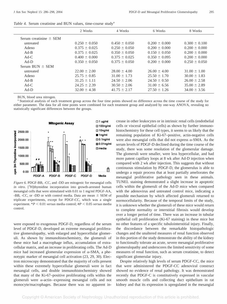
In Vitro StudiesThe mitogenic activity of purified PDGF-AA, -BB, -CC, and
-DD was assessed by their ability to stimulate incorporation of[3H]thymidine into growth-arrested human mesangial cells. Asshown in Figure 6, PDGF-BB, -CC, and -DD all led to signif-icant induction of mesangial cell proliferation at 0.01 ng/mlthat resulted in an approximate twofold increase of tritiatedthymidine incorporation versus media alone. All three growthfactor isoforms demonstrated very similar dose-dependent ef-fects, which plateaued at 10 to 100 ng/ml, with an increasedtritium incorporation of sevenfold (PDGF-BB and -DD) to10-fold (PDGF-CC) at these higher concentrations. In thisexperiment, PDGF-AA resulted in a weak mitogenic responsein the mesangial cells at all of the concentrations tested (0.01to 1 �g/ml) and was statistically significant at 1 and 10 ng/ml(P � 0.05 versus media alone).
DiscussionThe recent discovery of two new members of the PDGF
family, PDGF-C and -D, has provided us the opportunity toexplore the role of these growth factors in renal disease. Thereis some evidence that PDGF-C expression is upregulated inboth rodent models of injury and in human disease and hencemay play a role in mesangial injury and repair (19, 20), but todate, nothing is known about a potential role for PDGF-D. Inthis study, adenovirus constructs were used to introduce thecoding regions of PDGF-BB, -C, and -D into mice, whichresulted in an increase in circulating serum levels of each ofthese growth factors, providing a means to determine the renalresponse to exogenous PDGF.
The major finding of this study is that all of the mice that
Figure 5. Electron microscopy demonstrates mesangial proliferationin mice that received PDGF-D adenovirus construct. (A) Low-powerview of a glomerulus from a PDGF-D mouse at the 2-wk time pointshows accumulation of mesangial cells and matrix. (B) Higher-powerview of A. Inset shows effacement of visceral epithelial cells. (C) APDGF-D mouse at 8 wk shows a reduction in the mesangial expansioncompared with the 2-wk time point. (D) Low-power view of a mousethat received adenovirus alone at 2 wk after injection shows normalglomerular histology. Magnifications: �1600 in A, C, and D; �3000in B; �10,400 in inset.
294 Journal of the American Society of Nephrology J Am Soc Nephrol 15: 286–298, 2004
were exposed to exogenous PDGF-D, regardless of the serumlevel of PDGF-D, developed an extreme mesangial prolifera-tive glomerulopathy, with enlarged and hypercellular glomer-uli. As shown by immunohistochemistry, the glomeruli ofthese mice had a macrophage influx, accumulation of extra-cellular matrix, and an increase in proliferating cells. The Ad-Dmice had increased glomerular expression of �-SMA, a phe-notypic marker of mesangial cell activation (23, 29, 30). Elec-tron microscopy demonstrated that the majority of cells presentwithin these extremely hypercellular glomeruli were in factmesangial cells, and double immunohistochemistry showedthat many of the Ki-67–positive proliferating cells within theglomeruli were �-actin–expressing mesangial cells and notmonocyte/macrophages. Because there was no apparent in-
crease in other leukocytes or in intrinsic renal cells (endothelialcells or visceral epithelial cells) as shown by further immuno-histochemistry for these cell types, it seems to us likely that theremaining population of Ki-67–positive, actin-negative cellswere also mesangial cells that did not express �-SMA. As theserum levels of PDGF-D declined during the time course of thestudy, there was some resolution of the glomerular damage.The glomeruli were smaller, were less hypercellular, and hadmore patent capillary loops at 8 wk after Ad-D injection whencompared with 2 wk after injection. This suggests that withoutcontinuous stimulation by PDGF-D, the glomerulus is able toundergo a repair process that at least partially ameliorates themesangial proliferative pathology seen in these animals.TUNEL staining demonstrated a slight increase in apoptoticcells within the glomeruli of the Ad-D mice when comparedwith the adenovirus and untreated control mice, indicating apossible mechanism by which affected glomeruli returned tonormocellularity. Because of the temporal limits of the study,it is unknown whether the glomeruli of these mice would returnto complete normality or interstitial fibrosis would developover a longer period of time. There was an increase in tubularepithelial cell proliferation (Ki-67 staining) in these mice butno other features of a specific tubulointerstitial injury. Finally,the discordance between the remarkable histopathologicchanges and the unaltered measures of renal function observedin this portion of the study demonstrate the ability of the kidneyto functionally tolerate an acute, severe mesangial proliferativeglomerulopathy and underscores the limited sensitivity of somemeasures of renal function, such as serum creatinine, to detectsignificant glomerular injury.
Despite relatively high levels of serum PDGF-CC, the micethat were administered the PDGF-CC adenoviral constructshowed no evidence of renal pathology. It was demonstratedrecently that PDGF-C is constitutively expressed in vascularsmooth muscle cells and collecting duct epithelium in ratkidney and that its expression is upregulated in the mesangial
Figure 6. PDGF-BB, -CC, and -DD are mitogenic for mesangial cellsin vitro. [3H]thymidine incorporation into growth-arrested humanmesangial cells that were stimulated with 0.01 to 1 mg/ml PDGF-AA,-BB, -CC, or -DD or with control media. Data are mean � SEM oftriplicate experiments, except for PDGF-CC, which was a singleexperiment. *P � 0.01 versus media control; #P � 0.05 versus mediacontrol.
Table 4. Serum creatinine and BUN values, time-course studya
2 Weeks 4 Weeks 6 Weeks 8 Weeks
Serum creatinine � SEMuntreated 0.250 � 0.050 0.450 � 0.050 0.200 � 0.000 0.300 � 0.100Adeno 0.375 � 0.025 0.250 � 0.050 0.200 � 0.000 0.200 � 0.000Ad-B 0.375 � 0.025 0.350 � 0.050 0.150 � 0.050 0.200 � 0.000Ad-C 0.400 � 0.000 0.375 � 0.025 0.350 � 0.095 0.200 � 0.000Ad-D 0.350 � 0.050 0.375 � 0.050 0.200 � 0.000 0.250 � 0.050
Serum BUN � SEMuntreated 22.00 � 2.00 28.00 � 4.00 26.00 � 4.00 31.00 � 1.00Adeno 25.75 � 0.85 31.00 � 1.73 25.50 � 1.70 30.00 � 1.83Ad-B 31.25 � 1.11 24.50 � 2.06 24.50 � 0.50 26.00 � 2.58Ad-C 24.25 � 2.39 30.50 � 2.06 31.00 � 6.56 35.00 � 2.89Ad-D 32.00 � 4.38 41.75 � 2.17 27.50 � 1.26 34.00 � 3.56
BUN, blood urea nitrogen.a Statistical analysis of each treatment group across the four time points showed no difference across the time course of the study for
either parameter. The data for all time points were combined for each treatment group and analyzed by one-way ANOVA, revealing nostatistically significant differences between the groups.
J Am Soc Nephrol 15: 286–298, 2004 PDGF-D and Mesangial Proliferative Glomerulopathy 295

cells of rats in the Thy1.1 model of mesangioproliferativeglomerulonephritis (19). In concordance with results reportedin that study, we demonstrate that PDGF-C is a potent mesan-gial cell mitogen in vitro. It is therefore puzzling (but unex-plained) that the Ad-C mice in this study lacked any significantrenal pathology.
The mice that received the PDGF-BB adenovirus vector didnot attain the high circulating serum levels seen in the Ad-Cand Ad-D mice. Despite lower levels of serum PDGF-B, theglomeruli of these mice demonstrated a mesangial proliferativeresponse, with a significant increase in glomerular size andmatrix accumulation as well as increased numbers of glomer-ular proliferating cells, macrophage influx, and increased mes-angial actin expression. These observations are consistent withother studies that demonstrated mesangial cell proliferation andmatrix accumulation in rats that were infused with PDGF-BB(15) or rats that overexpressed PDGF-BB in their kidney afterin vivo transfection (31). We recognize that observed differ-ences in renal pathology between Ad-D and Ad-B mice mayindicate a true biologic difference but also may be attributableto differences in the systemic levels of overexpressed peptidesthat were achieved and perhaps to differences in bioavailabilityas a result of binding to other circulating plasma proteins.Hence, differences in the apparent efficacy of PDGF-BB and-DD in mediating mesangial proliferation in vivo in this studyneed to be interpreted with caution, especially given the sim-ilarity of mesangial proliferative responses to these two iso-forms in vitro.
A potential cause of the differences seen in systemic levelsof the various PDGF isoforms may be a consequence of theadenovirus delivery system that was used. Adenoviruses areone of several viruses that have been used as gene deliveryvectors in experimental and clinical gene therapy (32) and asone of the few strategies available for specifically targeting thekidney (33, 34). As seen in the present study, the time periodof expression of the transfected gene is generally limited toweeks or months, as the adenovirus does not integrate into thehost genome. The adenovirus vectors used in this study werenot specifically targeted to the kidney but were introducedintravenously, and the majority of gene expression was seen inthe liver. The renal effects thus were due primarily to circu-lating PDGF and not to direct expression of PDGF-B, -C, or -Dby intrinsic renal cells. It is not clear why there was such awide variation in the serum levels achieved with the differentvectors and even among different mice that received the samevector. One of the intrinsic problems with using adenovirusvectors is their ability to elicit an immune response in the hostanimal. It is possible that local immune/inflammatory re-sponses at the site of vector localization and gene expressionmay be the cause of diminished or extinguished gene activity.Thus, the variations in serum level seen among animals thatreceived the same vector may reflect differences in specific andhumoral immune responses that mediate clearance of adeno-virus-infected cells. Another possibility is variation in bindingto circulating plasma proteins such as albumin.
An important finding of this study was a discrepancy be-tween sites of renal expression of PDGF-D in humans versus
mice. In the human kidney, PDGF-D is expressed constitu-tively by visceral epithelial cells and vascular smooth musclecells but not in mesangial cells (21). In contrast, immunostain-ing of mouse kidney performed with the same antibody used inthe human studies demonstrated constitutive expression ofPDGF-D by mesangial cells and not glomerular epithelial cells.Constitutive expression of PDGF-D by vascular smooth mus-cle cells was similar to that seen in humans. We previouslyidentified molecules with constitutive expression in the glo-merulus in which the principal cell type expressing the mole-cule alternated between mesangial cells in rodent species andvisceral epithelial cells in humans, and vice versa (e.g., p75nerve growth factor receptor (35) [unpublished results in ro-dents] and PTPRQ, a newly discovered phosphatase (36)). Inthe proliferative lesions of the Ad-B and Ad-D mice, immu-nolocalization of PDGF-D was increased in the mesangium inconcert with an increase in PDGF-B and PDGFR-� expression.Thus, in the mouse, PDGF-D may act in concert with orcompete with PDGF-B in autocrine signaling within themesangium.
In conclusion, the principal findings of this study are in vivoand in vitro evidence that PDGF-D alone is a potent mitogenfor mesangial cells but not other cell types in the kidney andthat PDGF-D can initiate events that lead to a mesangialproliferative glomerulonephritis, including influx of monocyte/macrophages and production of extracellular matrix. The pre-dictable and florid features of the induced glomerulonephritisand relatively short interval between introduction of the ad-enoviral construct and development of morphologic diseasemake this a particularly attractive model for studies of mesan-gial proliferative injury and its repair. Comparative studiesyielded the somewhat surprising finding that overexpression ofPDGF-C did not result in any significant injury, despite itsability to cause mesangial cell proliferation in vitro. In con-junction with the finding that overexpression of PDGF-B alsois sufficient to produce a mesangioproliferative injury, ourstudy emphasizes a key role for PDGFR-�, through which bothPDGF-B and -D signal, in mediating mesangial proliferativediseases.
AcknowledgmentsSupport for this study was provided by a National Institutes of
Health–funded O’Brien Kidney Center of Research Excellence grant(DK 47659) and by a grant from ZymoGenetics, Inc. A portion of thiswork was presented as an abstract at the meeting of the AmericanSociety of Nephrology, Philadelphia, 2002.
We thank Barbara Gutierrez and Heather Day for the adenovirusconstructs.
References1. Johnson R, Iida H, Yoshimura A, Floege J, Bowen-Pope DF:
Platelet-derived growth factor: A potentially important cytokinein glomerular disease. Kidney Int 41: 590–594, 1992
2. Johnson RJ, Floege J, Couser WG, Alpers CE: Role of platelet-derived growth factor in glomerular disease. J Am Soc Nephrol 4:119–128, 1993
296 Journal of the American Society of Nephrology J Am Soc Nephrol 15: 286–298, 2004

3. Alpers CE, Johnson RJ: Growth factors and the glomerulus:Relationships between development and injury. Adv NephrolNecker Hosp 24: 33–52, 1995
4. Floege J, Topley N, Resch K: Regulation of mesangial cellproliferation. Am J Kidney Dis 17: 673–676, 1991
5. Floege J, Topley N, Hoppe J, Barrett TB, Resch K: Mitogeniceffect of platelet-derived growth factor in human glomerularmesangial cells: Modulation and/or suppression by inflammatorycytokines. Clin Exp Immunol 86: 334–341, 1991
6. Gilbertson DG, Duff ME, West JW, Kelly JD, Sheppard PO,Hofstrand PD, Gao Z, Shoemaker K, Bukowski TR, Moore M,Feldhaus AL, Humes JM, Palmer TE, Hart CE: Platelet-derivedgrowth factor C (PDGF-C), a novel growth factor that binds toPDGF alpha and beta receptor. J Biol Chem 276: 27406–27414,2001
7. LaRochelle WJ, Jeffers M, McDonald WF, Chillakuru RA, GieseNA, Lokker NA, Sullivan C, Boldog FL, Yang M, Vernet C,Burgess CE, Fernandes E, Deegler LL, Rittman B, Shimkets J,Shimkets RA, Rothberg JM, Lichenstein HS: PDGF-D, a newprotease-activated growth factor. Nat Cell Biol 3: 517–521, 2001
8. Li X, Ponten A, Aase K, Karlsson L, Abramsson A, Uutela M,Backstrom G, Hellstrom M, Bostrom H, Li H, Soriano P, Bet-sholtz C, Heldin CH, Alitalo K, Ostman A, Eriksson U: PDGF-Cis a new protease-activated ligand for the PDGF alpha-receptor.Nat Cell Biol 2: 302–309, 2000
9. Heldin CH, Westermark B: Mechanism of action and in vivo roleof platelet-derived growth factor. Physiol Rev 79: 1283–1316,1999
10. Hart CE, Forstrom JW, Kelly JD, Seifert RA, Smith RA, Ross R,Murray MJ, Bowen-Pope DF: Two classes of PDGF receptorrecognize different isoforms of PDGF. Science 240: 1529–1531,1988
11. Seifert RA, Hart CE, Phillips PE, Forstrom JW, Ross R, MurrayMJ, Bowen-Pope DF: Two different subunits associate to createisoform-specific platelet-derived growth factor receptors. J BiolChem 264: 8771–8778, 1989
12. Claesson-Welsh L, Eriksson A, Moren A, Severinsson L, Ek B,Ostman A, Betsholtz C, Heldin CH: cDNA cloning and expres-sion of a human platelet-derived growth factor (PDGF) receptorspecific for B-chain-containing PDGF molecules. Mol Cell Biol8: 3476–3486, 1988
13. Floege J, Ostendorf T, Wolf G: Growth factors and cytokines. In:Immunologic Renal Diseases, edited by Neilson EG, CouserWG, 2nd Ed., Baltimore, Lippincott Williams & Wilkins,2001
14. Johnson RJ, Floege J, Couser WG, Alpers CE: Role of platelet-derived growth factor in glomerular disease [Editorial]. J Am SocNephrol 4: 119–128, 1993 [published erratum appears in J AmSoc Nephrol 4: 1237, 1993]
15. Floege J, Eng E, Young BA, Alpers CE, Barrett TB, Bowen-Pope DF, Johnson RJ: Infusion of platelet-derived growth factoror basic fibroblast growth factor induces selective glomerularmesangial cell proliferation and matrix accumulation in rats.J Clin Invest 92: 2952–2962, 1993
16. Floege J, Ostendorf T, Janssen U, Burg M, Radeke HH, VargeeseC, Gill SC, Green LS, Janjic N: Novel approach to specificgrowth factor inhibition in vivo: Antagonism of platelet-derivedgrowth factor in glomerulonephritis by aptamers. Am J Pathol154: 169–179, 1999
17. Iida H, Seifert R, Alpers CE, Gronwald RG, Phillips PE, Pritzl P,Gordon K, Gown AM, Ross R, Bowen-Pope DF, et al.: Platelet-
derived growth factor (PDGF) and PDGF receptor are induced inmesangial proliferative nephritis in the rat. Proc Natl Acad Sci US A 88: 6560–6564, 1991
18. Ostendorf T, Kunter U, Grone HJ, Bahlmann F, Kawachi H,Shimizu F, Koch KM, Janjic N, Floege J: Specific antagonism ofPDGF prevents renal scarring in experimental glomerulonephri-tis. J Am Soc Nephrol 12: 909–918, 2001
19. Eitner F, Ostendorf T, Van Roeyen C, Kitahara M, Li X, Aase K,Grone HJ, Eriksson U, Floege J: Expression of a novel PDGFisoform, PDGF-C, in normal and diseased rat kidney. J Am SocNephrol 13: 910–917, 2002
20. Eitner F, Ostendorf T, Kretzler M, Cohen CD, Eriksson U, GroneHJ, Floege J: PDGF-C Expression in the developing and normaladult human kidney and in glomerular diseases. J Am Soc Neph-rol 14: 1145–1153, 2003
21. Changsirikulchai S, Hudkins KL, Goodpaster TA, Volpone J,Topouzis S, Gilbertson DG, Alpers CE: Platelet-derived growthfactor-D expression in developing and mature human kidneys.Kidney Int 62: 2043–2054, 2002
22. Hudkins KL, Giachelli CM, Cui Y, Couser WG, Johnson RJ,Alpers CE: Osteopontin expression in fetal and mature humankidney. J Am Soc Nephrol 10: 444–457, 1999
23. Alpers CE, Hudkins KL, Gown AM, Johnson RJ: Enhancedexpression of “muscle-specific” actin in glomerulonephritis. Kid-ney Int 41: 1134–1142, 1992
24. Alpers CE, Seifert RA, Hudkins KL, Johnson RJ, Bowen-PopeDF: Developmental patterns of PDGF B-chain, PDGF-receptor,and alpha-actin expression in human glomerulogenesis. KidneyInt 42: 390–399, 1992
25. Segerer S, Eitner F, Cui Y, Hudkins KL, Alpers CE: Cellularinjury associated with renal thrombotic microangiopathy in hu-man immunodeficiency virus-infected macaques. J Am SocNephrol 13: 370–378, 2002
26. Taneda S, Pippin JW, Sage EH, Hudkins KL, Takeuchi Y,Couser WG, Alpers CE: Amelioration of diabetic nephropathy inSPARC-null mice. J Am Soc Nephrol 14: 968–980, 2003
27. Alpers CE, Hudkins KL, Pritzl P, Johnson RJ: Mechanisms ofclearance of immune complexes from peritubular capillaries inthe rat. Am J Pathol 139: 855–867, 1991
28. Eitner F, Cui Y, Hudkins KL, Schmidt A, Birkebak T, Agy MB,Hu SL, Morton WR, Anderson DM, Alpers CE: Thromboticmicroangiopathy in the HIV-2-infected macaque. Am J Pathol155: 649–661, 1999
29. Johnson RJ, Floege J, Yoshimura A, Iida H, Couser WG, AlpersCE: The activated mesangial cell: a glomerular “myofibroblast”?J Am Soc Nephrol 2[Suppl]: S190–S197, 1992
30. Johnson RJ, Iida H, Alpers CE, Majesky MW, Schwartz SM,Pritzi P, Gordon K, Gown AM: Expression of smooth musclecell phenotype by rat mesangial cells in immune complex ne-phritis. Alpha-smooth muscle actin is a marker of mesangial cellproliferation. J Clin Invest 87: 847–858, 1991
31. Isaka Y, Fujiwara Y, Ueda N, Kaneda Y, Kamada T, Imai E:Glomerulosclerosis induced by in vivo transfection of transform-ing growth factor-beta or platelet-derived growth factor gene intothe rat kidney. J Clin Invest 92: 2597–2601, 1993
32. Lotze MT, Kost TA: Viruses as gene delivery vectors: Applica-tion to gene function, target validation, and assay development.Cancer Gene Ther 9: 692–699, 2002
33. McDonald GA: Targeted adenoviral gene transfer to the kidney.Kidney Int Suppl 61[Suppl 1]: 42–46, 2002
J Am Soc Nephrol 15: 286–298, 2004 PDGF-D and Mesangial Proliferative Glomerulopathy 297

34. Imai E: Gene therapy approach in renal disease in the 21stcentury. Nephrol Dial Transplant 16[Suppl 5]: 26–34, 2001
35. Seifert RA, Coats SA, Oganesian A, Wright MB, Dishmon M,Booth CJ, Johnson RJ, Alpers CE, Bowen-Pope DF: PTPRQ isa novel phosphatidylinositol phosphatase that can be expressedas a cytoplasmic protein or as a subcellularly localized receptor-like protein. Exp Cell Res 287: 374–386, 2003
36. Rosenberg I, Cherayil BJ, Isselbacher KJ, Pillai S: Mac-2-bind-ing glycoproteins. Putative ligands for a cytosolic beta-galacto-side lectin. J Biol Chem 266: 18731–18736, 1991
37. Segerer S, Hudkins KL, Taneda S, Wen M, Cui Y, Segerer M,Farr AG, Alpers CE: Oral interferon-alpha treatment of micewith cryoglobulinemic glomerulonephritis. Am J Kidney Dis 39:876–888, 2002
38. Taneda S, Segerer S, Hudkins KL, Cui Y, Wen M, Segerer M,Wener MH, Khairallah CG, Farr AG, Alpers CE: Cryoglobu-linemic glomerulonephritis in thymic stromal lymphopoietintransgenic mice. Am J Pathol 159: 2355–2369, 2001
39. Vanzulli S, Gazzaniga S, Braidot MF, Vecchi A, Mantovani A,Wainstok de Calmanovici R: Detection of endothelial cells byMEC 13.3 monoclonal antibody in mice mammary tumors. Bio-cell 21: 39–46, 1997
40. Vecchi A, Garlanda C, Lampugnani MG, Resnati M, Matteucci C,Stoppacciaro A, Schnurch H, Risau W, Ruco L, Mantovani A, et al.:Monoclonal antibodies specific for endothelial cells of mouse bloodvessels. Their application in the identification of adult and embry-onic endothelium. Eur J Cell Biol 63: 247–254, 1994
41. Skalli O, Ropraz P, Trzeciak A, Benzonana G, Gillessen D,Gabbiani G: A monoclonal antibody against alpha-smooth mus-cle actin: A new probe for smooth muscle differentiation. J CellBiol 103: 2787–2796, 1986
42. Kubbutat MH, Key G, Duchrow M, Schluter C, Flad HD, GerdesJ: Epitope analysis of antibodies recognising the cell prolifera-tion associated nuclear antigen previously defined by the anti-body Ki-67 (Ki-67 protein). J Clin Pathol 47: 524–528, 1994
43. LaRochelle WJ, Robbins KC, Aaronson SA: Immunochemicallocalization of the epitope for a monoclonal antibody that neu-tralizes human platelet-derived growth factor mitogenic activity.Mol Cell Biol 9: 3538–3542, 1989
44. Hu Y, Schett G, Zou Y, Dietrich H, Xu Q: Abundance ofplatelet-derived growth factors (PDGFs), PDGF receptors andactivation of mitogen-activated protein kinases in brain declinewith age. Brain Res Mol Brain Res 53: 252–259, 1998
45. Hu Y, Zou Y, Dietrich H, Wick G, Xu Q: Inhibition of neointimahyperplasia of mouse vein grafts by locally applied suramin.Circulation 100: 861–868, 1999
46. Matsumoto K, Hiraiwa N, Yoshiki A, Ohnishi M, Kusakabe M:PDGF receptor-alpha deficiency in glomerular mesangial cells oftenascin-C knockout mice. Biochem Biophys Res Commun 290:1220–1227, 2002
47. Hirsch S, Gordon S: Polymorphic expression of a neutrophildifferentiation antigen revealed by monoclonal antibody 7/4.Immunogenetics 18: 229–239, 1983
48. Alpers CE, Hudkins KL, Ferguson M, Johnson RJ, Rutledge JC:Platelet-derived growth factor A-chain expression in developingand mature human kidneys and in Wilms’ tumor. Kidney Int 48:146–154, 1995
49. Nakagawa H, Sasahara M, Haneda M, Koya D, Hazama F,Kikkawa R: Immunohistochemical characterization of glomeru-lar PDGF B-chain and PDGF beta-receptor expression in dia-betic rats. Diabetes Res Clin Pract 48: 87–98, 2000
298 Journal of the American Society of Nephrology J Am Soc Nephrol 15: 286–298, 2004