exosomes derived from mir-92a-3p-overexpressing human
TRANSCRIPT
RESEARCH Open Access
Exosomes derived from miR-92a-3p-overexpressing human mesenchymal stemcells enhance chondrogenesis and suppresscartilage degradation via targeting WNT5AGuping Mao†, Ziji Zhang†, Shu Hu†, Zhiqi Zhang, Zongkun Chang, Zhiyu Huang, Weiming Liao* and Yan Kang*
Abstract
Background: WNT5A is known to be involved in the pathogenesis of osteoarthritis. This study investigated themolecular mechanism of exosomal miR-92a-3p and WNT5A in chondrogenesis and cartilage degeneration.
Methods: Exosomal miR-92a-3p expression was assessed in vitro in a human mesenchymal stem cell (MSC) modelof chondrogenesis and in normal and OA primary human chondrocytes (PHCs). MSCs and PHCs were treated withexosomes derived from MSC-miR-92a-3p (MSC-miR-92a-3p-Exos) or its antisense inhibitor (MSC-anti-miR-92a-3p-Exos), respectively. Small interfering RNAs (siRNAs) and luciferase reporter assay were used to reveal the molecularrole of exosomal miR-92a-3p and WNT5A in chondrogenesis. The protective effect of exosomes in vivo wasmeasured using Safranin-O and Fast Green staining and immunohistochemical staining.
Results: Exosomal miR-92a-3p expression was elevated in the MSC chondrogenic exosome, while it wassignificantly reduced in the OA chondrocyte-secreted exosome compared with normal cartilage. Treatment withMSC-miR-92a-3p-Exos promoted cartilage proliferation and matrix genes expression in MSCs and PHCs, respectively.In contrast, treatment with MSC-anti-miR-92a-3p-Exos repressed chondrogenic differentiation and reduced cartilagematrix synthesis by enhancing the expression of WNT5A. Luciferase reporter assay demonstrated that miR-92a-3psuppressed the activity of a reporter construct containing the 3’-UTR and inhibited WNT5A expression in both MSCsand PHCs. MSC-miR-92a-3p-Exos inhibit cartilage degradation in the OA mice model.
Conclusions: Our results suggest that exosomal miR-92a-3p regulates cartilage development and homeostasis bydirectly targeting WNT5A. This indicates that exosomal miR-92a-3p may act as a Wnt inhibitor and exhibits potentialas a disease-modifying osteoarthritis drug.
Keywords: Exosomes, Human mesenchymal stem cells, WNT5A, miRNA-92a-3p, Osteoarthritis, Chondrocyte
BackgroundOsteoarthritis (OA) is strongly associated with joint de-generative diseases, and leads to chronic pain, disability,and economic burden [1]. However, the molecularmechanisms of OA have not yet been fully elucidated.OA is characterized by the loss of extracellular matrix(ECM) and cartilage destruction. It is widely acceptedthat the development of OA is associated with
pro-inflammatory cytokines, which increases the activityof matrix metalloproteinase (MMP) and a disintegrinand metalloproteinase with thrombospondin motifs(ADAMTS) [2, 3].Recently, increasing evidence has suggested that mesen-
chymal stem cells (MSCs)-derived exosomes play a vitalrole in modulating cell migration, proliferation, differenti-ation, and matrix synthesis [4–6]. In 2010, it was first re-ported that the exosome was the active agent secreted byMSCs in response to myocardial ischemia reperfusion (I/R) injury [7]. It has also been reported that MSC exo-somes mediate cartilage repair and regeneration by
* Correspondence: [email protected]; [email protected]†Guping Mao, Ziji Zhang and Shu Hu contributed equally to this work.Department of Joint Surgery, First Affiliated Hospital of Sun Yat-senUniversity, #58 Zhongshan 2nd Road, Guangzhou 510080, China
© The Author(s). 2018 Open Access This article is distributed under the terms of the Creative Commons Attribution 4.0International License (http://creativecommons.org/licenses/by/4.0/), which permits unrestricted use, distribution, andreproduction in any medium, provided you give appropriate credit to the original author(s) and the source, provide a link tothe Creative Commons license, and indicate if changes were made. The Creative Commons Public Domain Dedication waiver(http://creativecommons.org/publicdomain/zero/1.0/) applies to the data made available in this article, unless otherwise stated.
Mao et al. Stem Cell Research & Therapy (2018) 9:247 https://doi.org/10.1186/s13287-018-1004-0
enhancing proliferation, attenuating apoptosis, and modu-lating immune reactivity [8–10]. Exosomes are small, se-creted bilipid membrane vesicles of about 50–150 nm,and are thought to act as intercellular communication ve-hicles. Bioactive lipids, nucleic acids (mRNAs and micro-RNAs), and proteins are transferred by exosome and playbiological responses in cells and recipient cells [7]. Evi-dence has shown that exosomes have no obvious im-munogenicity or tumorigenicity adverse effects [11, 12].To our knowledge, however, the specific molecular mech-anism that causes MSC exosomes to promote cartilagechondrogenesis has not been investigated.MicroRNAs (miRNAs) are an important component of
the exosome and have attracted attention for many years.They bind to the 3′-untranslated regions (3′-UTRs) of tar-get genes and suppress target gene expression [13]. Whileexosomal miRNAs are widely thought to mediate intercel-lular communication and gene regulation [14, 15], thespecific molecular mechanism of MSC-derived exosomalmiRNAs responsible for chondrogenic differentiation anddegeneration remain unclear.WNT5A is considered a non-canonical Wnt protein.
Increasing evidence has shown that the development ofarticular joints, including cartilage, bone, and joint cav-ities, is highly dependent on Wnt signaling [16–20].WNT5A has a dual function, acting in both chondro-genic differentiation and cartilage degradation [21]. Inthe early stage of cartilage formation, it activates theproliferation and inhibits the differentiation of chondro-cytes [22–24]. In addition, WNT5A can activate matrixmetalloproteinases (MMPs) and reduce cartilage forma-tion and the synthesis of cartilage ECM in the late stageof chondrogenesis and in mature chondrocytes [21, 25].WNT5A plays a key role in cartilage destruction anddegradation in the pathogenesis of OA. Huang et al.showed that WNT5A can promote chondrocyte cata-bolic activity via non-canonical Wnt signaling in humanOA cartilage [25]. Moreover, Ge et al. showed thatWNT5A plays an essential role in interleukin (IL)-1β--mediated cartilage destruction via the NF-κB pathway[26, 27], and Shi et al. demonstrated that the silencing ofWNT5A prevents interleukin (IL)-1β-induced collagentype II degradation in rat chondrocytes [28].In order to elucidate the specific MSC-derived exoso-
mal miRNAs responsible for MSC-mediated chondro-genic differentiation, in the present study, we used amiRNA microarray to determine miRNA expressionprofiles in exosomes during the chondrogenic differenti-ation of human MSCs. We observed significant upregu-lation of the exosomal miR-92a-3p during MSC-inducedchondrogenesis. Previous studies we have demonstratedmiR-92a-3p plays a key role in chondrogenesis and car-tilage degradation via directly targeting noggin3,HDAC2,ADAMTS4, and ADAMTS5 [29–31]. Given the
potential role of MSC-derived exosomal miRNAs inregulating cartilage homeostasis and cartilage develop-ment, we hypothesized that exosomal miR-92a-3p mayplay a role in both chondrogenic differentiation and OApathogenesis. Thus, our study further aimed to deter-mine whether MSC-derived exosomal miR-92a-3p en-hances cartilage development and prevents degradationby targeting WNT5A.
MethodsHuman mesenchymal stem cell isolation, culture, andinduction of chondrogenesis in micromass cultureBone marrow samples were obtained from the First Af-filiated Hospital of Sun Yat-Sen University. MSCs wereisolated as described previously [32]. Bone marrow sam-ples were obtained by iliac crest aspiration from six nor-mal human donors (mean age: 35 years; range: 32–38 years; male: 3, female: 3). Human MSCs were cul-tured in alpha-modified Eagle’s medium (α-MEM)(Gibco, Grand Island, NY, USA) supplemented with 10%fetal bovine serum (FBS; Gibco), 100 IU/mL penicillin,and 100 μg/mL streptomycin. Cells were cultured at 37 °C in a 5% CO2 atmosphere. Culture media were chan-ged every 3 days. When cultures neared 80% confluence,cells were detached by treatment with 0.05% trypsin/eth-ylenediaminetetraacetic acid (EDTA) and passaged inculture. All hMSCs were used at passage 3 to inducehMSC chondrogenesis by micromass culture, as previ-ously described [16]. Briefly, hMSCs were resuspendedat 2 × 10⁷ cells/mL in incomplete chondrogenic medium[97 mL human mesenchymal stem cell chondrogenicdifferentiation basal medium, 10 μL dexamethasone,300 μL ascorbate, 1 mL of ITS (insulin, transferrin, sel-enium) supplement, 100 μL sodium pyruvate, and100 μL proline; Cyagen Biosciences, Guangzhou, China].Droplets of resuspended cells (12.5 μL) were carefullytransferred to individual wells of a 24-well plate and in-cubated at 37 °C for 90 min to stimulate the adherenceof cells to the plate. Droplets were divided into twogroups: the first group was cultured in 500 μL incom-plete chondrogenic medium per well; the second groupwas cultured in 500 μL complete chondrogenic induc-tion medium, prepared by the addition of 10 μL trans-forming growth factor (TGF)-β 3 to 1 mL incompletechondrogenic medium (Cyagen Biosciences). Sampleswere collected for experiments at selected time points.
Characterization of MSCs and flow cytometry analysisTo identify the multiple differentiation potential ofMSCs, cells were induced to differentiate in osteogenic,adipogenic, or chondrogenic differentiation medium. Weused Alizarin Red, Oil Red O, and Alcian Blue stainingto identify the three cell types, respectively. Flow cytom-etry was used to identify the surface antigens of hMSCs.
Mao et al. Stem Cell Research & Therapy (2018) 9:247 Page 2 of 13
CD11b, CD19, CD34, CD45, CD73, CD90, CD105, andHLA-DR (eBioscience, Inc., San Diego, CA, USA) mono-clonal antibodies were used for detection. Mouse IgGmonoclonal antibody was used as a negative control.
Isolation and identification of exosomesExosome isolation was carried out using ultracentrifuga-tion [33]. In brief, MSC culture supernatants were sub-jected to successive centrifugations at 3000 × g (30 min)and 10,000 × g (30 min). Exosomes were then pelleted at64,000 × g for 110 min using an SW28 rotor (BeckmanCoulter, Brea, CA, USA). Exosome pellets were resus-pended in 0.32 M sucrose and centrifuged at 100,000 × gfor 1 h (SW60Ti rotor, Beckman Coulter). The exosomepellet was then resuspended in phosphate-buffered sa-line (PBS). Nanosight 2000 analysis and transmissionelectron microscopy (TEM) were used to identify exo-somes. RNA and proteins were extracted from exosomesusing a Total Exosome RNA & Protein Isolation Kit(Invitrogen, Carlsbad, CA, USA) for further analysis.
Primary chondrocyte collection, isolation, and cell cultureDegraded joint cartilage samples were obtained from pa-tients [n = 6; mean age: 60.24 years; male: 3, female: 3]with OA knee joints during total knee replacement oper-ations. Normal cartilage samples were taken from pa-tients (n = 6; mean age: 54.46 years; male: 3, female: 3)with no previous history of OA or rheumatoid arthritis,who underwent total hip replacement surgery because offemoral neck fractures. The cartilages were dissectedaway from the subchondral bone and then digested by4 mg/mL protease and 0.25 mg/mL collagenase P as de-scribed previously [30]. Cells were cultured in DMEM/F-12 (Gibco Life Technologies, Grand Island, NY, USA)containing 5% fetal bovine serum (FBS; Gibco Life Tech-nologies), 1% penicillin and streptomycin (Gibco LifeTechnologies). The chondrocytes were used our in ex-periments within 3–7 days and without passaging toavoid dedifferentiation.
Chondrocyte migration and proliferation assayA scratch wound assay was used to analyze the effect of exo-somes secreted by mesenchymal stem cells (MSC-Exos) andexosomes secreted by miR-92a-3p-overexpressing mesen-chymal stem cells (MSC-miR-92a-3p-Exos) on the migra-tion of chondrocytes, as described previously [34]. Theimages were obtained at the same position before and afterincubation. Scratched areas were measured using Image-ProPlus 6.0 software (Media Cybernetics, Bethesda, MD, USA).The effect of MSC-Exos and MSC-miR-92a-3p-Exos on theproliferation of human chondrocytes was evaluated usingthe Cell Counting Kit-8 (CCK-8; Dojindo, Kyushu Island,Japan) as described previously [34]. Cell proliferation curveswere constructed by measuring the amount of formazan
dye generated by cellular dehydrogenase activity with a mi-croplate reader at a wave length of 450 nm.
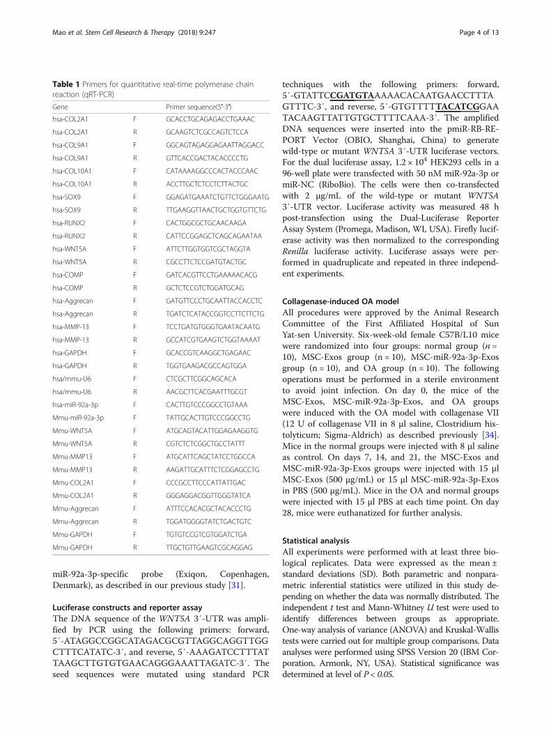
RNA extraction, reverse transcription, and quantitativereal-time polymerase chain reaction (qRT-PCR)RNA extraction and reverse transcription were performedas described previously [31]. Transcript levels were nor-malized to that of the housekeeping gene glyceraldehyde3-phosphate dehydrogenase (GAPDH; for mRNA) or thesmall U6 RNA (for miRNA). The specific primers used forthese analyses are listed in Table 1. Gene expression wascalculated using the 2 − ΔΔCt method, and each experi-ment was performed in triplicate.
TransfectionThe MSCs were transfected with miR-92a-3p mimic orinhibitor (RiboBio, Guangzhou, China) at a concentra-tion of 50 nM; they were also transfected with WNT5AsiRNA or NC (RiboBio). Lipofectamine® 2000 Transfec-tion Reagent (Gibco Life Technologies) was used totransfect cells according to the manufacturer’s instruc-tions. Cells were then harvested after 48 h for quantita-tive real-time reverse transcription-polymerase chainreaction (qRT-PCR), or after 72 h for western blotanalysis.
Western blot analysisWestern blot analysis was carried out as described previ-ously [31]. Membranes were incubated with primaryantibodies against WNT5A and RUNX2 (1:1000 dilu-tion, Cell Signaling Technology); GAPDH (1:3000, CellSignaling Technology); aggrecan, COL2A1, and MMP13(1:1000, Abcam, Cambridge, MA, USA); and SOX9(1:2000, EMD Millipore, Burlington, MA, USA). Theblots were then incubated with appropriate secondaryantibodies (1:3000 dilution, Cell Signaling Technology)at 4 °C overnight, after which they were developed withan ECL chemiluminescence Kit (EMD Millipore).
Immunohistochemical analysis, histology staining, and insitu hybridizationImmunohistochemical analysis was performed as de-scribed previously [31]. Briefly, samples were fixed in 4%paraformaldehyde (Sigma-Aldrich, St. Louis, MO, USA),decalcified, embedded in paraffin, and cut into 5-μmsections that were deparaffinized, rehydrated, and thenstained with Safranin O/Fast Green. COL2A1, aggrecan,MMP13, and WNT5A expression was analyzed by im-munohistochemistry. For in situ hybridization ofmiR-92a-3p expression, tissues were subsequently dehy-drated with a graded series of ethanol, embedded in par-affin, and cut into 5-μm-thick sections. Sections weresubjected to in situ hybridization analysis using
Mao et al. Stem Cell Research & Therapy (2018) 9:247 Page 3 of 13
miR-92a-3p-specific probe (Exiqon, Copenhagen,Denmark), as described in our previous study [31].
Luciferase constructs and reporter assayThe DNA sequence of the WNT5A 3′-UTR was ampli-fied by PCR using the following primers: forward,5′-ATAGGCCGGCATAGACGCGTTAGGCAGGTTGGCTTTCATATC-3′, and reverse, 5′-AAAGATCCTTTATTAAGCTTGTGTGAACAGGGAAATTAGATC-3′. Theseed sequences were mutated using standard PCR
techniques with the following primers: forward,5′-GTATTCCGATGTAAAAACACAATGAACCTTTAGTTTC-3′, and reverse, 5′-GTGTTTTTACATCGGAATACAAGTTATTGTGCTTTTCAAA-3′. The amplifiedDNA sequences were inserted into the pmiR-RB-RE-PORT Vector (OBIO, Shanghai, China) to generatewild-type or mutant WNT5A 3′-UTR luciferase vectors.For the dual luciferase assay, 1.2 × 10⁴ HEK293 cells in a96-well plate were transfected with 50 nM miR-92a-3p ormiR-NC (RiboBio). The cells were then co-transfectedwith 2 μg/mL of the wild-type or mutant WNT5A3′-UTR vector. Luciferase activity was measured 48 hpost-transfection using the Dual-Luciferase ReporterAssay System (Promega, Madison, WI, USA). Firefly lucif-erase activity was then normalized to the correspondingRenilla luciferase activity. Luciferase assays were per-formed in quadruplicate and repeated in three independ-ent experiments.
Collagenase-induced OA modelAll procedures were approved by the Animal ResearchCommittee of the First Affiliated Hospital of SunYat-sen University. Six-week-old female C57B/L10 micewere randomized into four groups: normal group (n =10), MSC-Exos group (n = 10), MSC-miR-92a-3p-Exosgroup (n = 10), and OA group (n = 10). The followingoperations must be performed in a sterile environmentto avoid joint infection. On day 0, the mice of theMSC-Exos, MSC-miR-92a-3p-Exos, and OA groupswere induced with the OA model with collagenase VII(12 U of collagenase VII in 8 μl saline, Clostridium his-tolyticum; Sigma-Aldrich) as described previously [34].Mice in the normal groups were injected with 8 μl salineas control. On days 7, 14, and 21, the MSC-Exos andMSC-miR-92a-3p-Exos groups were injected with 15 μlMSC-Exos (500 μg/mL) or 15 μl MSC-miR-92a-3p-Exosin PBS (500 μg/mL). Mice in the OA and normal groupswere injected with 15 μl PBS at each time point. On day28, mice were euthanatized for further analysis.
Statistical analysisAll experiments were performed with at least three bio-logical replicates. Data were expressed as the mean ±standard deviations (SD). Both parametric and nonpara-metric inferential statistics were utilized in this study de-pending on whether the data was normally distributed. Theindependent t test and Mann-Whitney U test were used toidentify differences between groups as appropriate.One-way analysis of variance (ANOVA) and Kruskal-Wallistests were carried out for multiple group comparisons. Dataanalyses were performed using SPSS Version 20 (IBM Cor-poration, Armonk, NY, USA). Statistical significance wasdetermined at level of P < 0.05.
Table 1 Primers for quantitative real-time polymerase chainreaction (qRT-PCR)
Gene Primer sequence(5′-3′)
hsa-COL2A1 F GCACCTGCAGAGACCTGAAAC
hsa-COL2A1 R GCAAGTCTCGCCAGTCTCCA
hsa-COL9A1 F GGCAGTAGAGGAGAATTAGGACC
hsa-COL9A1 R GTTCACCGACTACACCCCTG
hsa-COL10A1 F CATAAAAGGCCCACTACCCAAC
hsa-COL10A1 R ACCTTGCTCTCCTCTTACTGC
hsa-SOX9 F GGAGATGAAATCTGTTCTGGGAATG
hsa-SOX9 R TTGAAGGTTAACTGCTGGTGTTCTG
hsa-RUNX2 F CACTGGCGCTGCAACAAGA
hsa-RUNX2 R CATTCCGGAGCTCAGCAGAATAA
hsa-WNT5A F ATTCTTGGTGGTCGCTAGGTA
hsa-WNT5A R CGCCTTCTCCGATGTACTGC
hsa-COMP F GATCACGTTCCTGAAAAACACG
hsa-COMP R GCTCTCCGTCTGGATGCAG
hsa-Aggrecan F GATGTTCCCTGCAATTACCACCTC
hsa-Aggrecan R TGATCTCATACCGGTCCTTCTTCTG
hsa-MMP-13 F TCCTGATGTGGGTGAATACAATG
hsa-MMP-13 R GCCATCGTGAAGTCTGGTAAAAT
hsa-GAPDH F GCACCGTCAAGGCTGAGAAC
hsa-GAPDH R TGGTGAAGACGCCAGTGGA
hsa/mmu-U6 F CTCGCTTCGGCAGCACA
hsa/mmu-U6 R AACGCTTCACGAATTTGCGT
hsa-miR-92a-3p F CACTTGTCCCGGCCTGTAAA
Mmu-miR-92a-3p F TATTGCACTTGTCCCGGCCTG
Mmu-WNT5A F ATGCAGTACATTGGAGAAGGTG
Mmu-WNT5A R CGTCTCTCGGCTGCCTATTT
Mmu-MMP13 F ATGCATTCAGCTATCCTGGCCA
Mmu-MMP13 R AAGATTGCATTTCTCGGAGCCTG
Mmu-COL2A1 F CCCGCCTTCCCATTATTGAC
Mmu-COL2A1 R GGGAGGACGGTTGGGTATCA
Mmu-Aggrecan F ATTTCCACACGCTACACCCTG
Mmu-Aggrecan R TGGATGGGGTATCTGACTGTC
Mmu-GAPDH F TGTGTCCGTCGTGGATCTGA
Mmu-GAPDH R TTGCTGTTGAAGTCGCAGGAG
Mao et al. Stem Cell Research & Therapy (2018) 9:247 Page 4 of 13
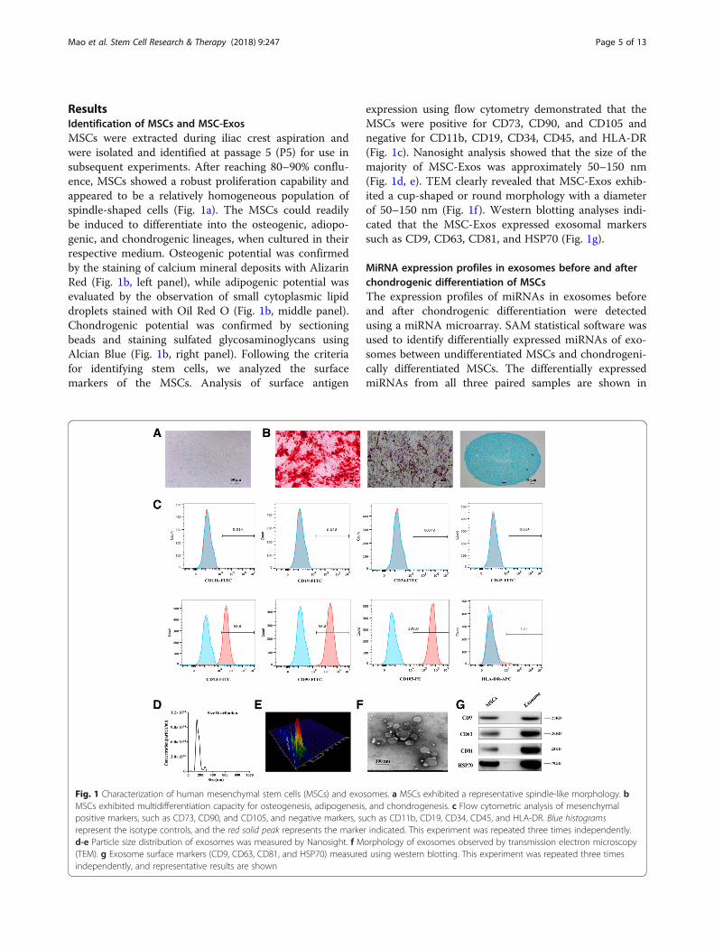
ResultsIdentification of MSCs and MSC-ExosMSCs were extracted during iliac crest aspiration andwere isolated and identified at passage 5 (P5) for use insubsequent experiments. After reaching 80–90% conflu-ence, MSCs showed a robust proliferation capability andappeared to be a relatively homogeneous population ofspindle-shaped cells (Fig. 1a). The MSCs could readilybe induced to differentiate into the osteogenic, adiopo-genic, and chondrogenic lineages, when cultured in theirrespective medium. Osteogenic potential was confirmedby the staining of calcium mineral deposits with AlizarinRed (Fig. 1b, left panel), while adipogenic potential wasevaluated by the observation of small cytoplasmic lipiddroplets stained with Oil Red O (Fig. 1b, middle panel).Chondrogenic potential was confirmed by sectioningbeads and staining sulfated glycosaminoglycans usingAlcian Blue (Fig. 1b, right panel). Following the criteriafor identifying stem cells, we analyzed the surfacemarkers of the MSCs. Analysis of surface antigen
expression using flow cytometry demonstrated that theMSCs were positive for CD73, CD90, and CD105 andnegative for CD11b, CD19, CD34, CD45, and HLA-DR(Fig. 1c). Nanosight analysis showed that the size of themajority of MSC-Exos was approximately 50–150 nm(Fig. 1d, e). TEM clearly revealed that MSC-Exos exhib-ited a cup-shaped or round morphology with a diameterof 50–150 nm (Fig. 1f ). Western blotting analyses indi-cated that the MSC-Exos expressed exosomal markerssuch as CD9, CD63, CD81, and HSP70 (Fig. 1g).
MiRNA expression profiles in exosomes before and afterchondrogenic differentiation of MSCsThe expression profiles of miRNAs in exosomes beforeand after chondrogenic differentiation were detectedusing a miRNA microarray. SAM statistical software wasused to identify differentially expressed miRNAs of exo-somes between undifferentiated MSCs and chondrogeni-cally differentiated MSCs. The differentially expressedmiRNAs from all three paired samples are shown in
Fig. 1 Characterization of human mesenchymal stem cells (MSCs) and exosomes. a MSCs exhibited a representative spindle-like morphology. bMSCs exhibited multidifferentiation capacity for osteogenesis, adipogenesis, and chondrogenesis. c Flow cytometric analysis of mesenchymalpositive markers, such as CD73, CD90, and CD105, and negative markers, such as CD11b, CD19, CD34, CD45, and HLA-DR. Blue histogramsrepresent the isotype controls, and the red solid peak represents the marker indicated. This experiment was repeated three times independently.d-e Particle size distribution of exosomes was measured by Nanosight. f Morphology of exosomes observed by transmission electron microscopy(TEM). g Exosome surface markers (CD9, CD63, CD81, and HSP70) measured using western blotting. This experiment was repeated three timesindependently, and representative results are shown
Mao et al. Stem Cell Research & Therapy (2018) 9:247 Page 5 of 13
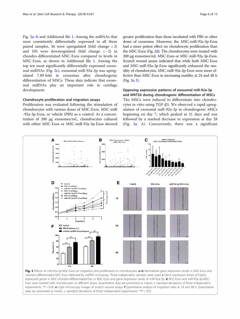
Fig. 2a–b and Additional file 1. Among the miRNAs thatwere consistently differentially expressed in all threepaired samples, 36 were upregulated (fold change > 2)and 105 were downregulated (fold change <− 2) inchondro-differentiated MSC-Exos compared to levels inMSC-Exos, as shown in Additional file 1. Among thetop ten most significantly differentially expressed exoso-mal miRNAs (Fig. 2c), exosomal miR-92a-3p was upreg-ulated 7.89-fold in exosomes after chondrogenicdifferentiation of MSCs. These data indicate that exoso-mal miRNAs play an important role in cartilagedevelopment.
Chondrocyte proliferation and migration assaysProliferation was evaluated following the stimulation ofchondrocytes with various doses of MSC-Exos, MSC-miR-92a-3p-Exos, or vehicle (PBS) as a control. At a concen-tration of 200 μg exosomes/mL, chondrocytes culturedwith either MSC-Exos or MSC-miR-92a-3p-Exos showed
greater proliferation than those incubated with PBS or otherdoses of exosomes. Moreover, the MSC-miR-92a-3p-Exoshad a more potent effect on chondrocyte proliferation thanthe MSC-Exos (Fig. 2d). The chondrocytes were treated with200 μg exosomes/mL MSC-Exos or MSC-miR-92a-3p-Exos.Scratch wound assays indicated that while both MSC-Exosand MSC-miR-92a-3p-Exos significantly enhanced the mo-tility of chondrocytes, MSC-miR-92a-3p-Exos were more ef-fective than MSC-Exos in increasing motility at 24 and 48 h(Fig. 2e, f).
Opposing expression patterns of exosomal miR-92a-3pand WNT5A during chondrogenic differentiation of MSCsThe MSCs were induced to differentiate into chondro-cytes in vitro using TGF-β3. We observed a rapid upreg-ulation of exosomal miR-92a-3p in chondrogenic MSCsbeginning on day 7, which peaked at 21 days and wasfollowed by a marked decrease in expression at day 28(Fig. 3a. A). Concurrently, there was a significant
Fig. 2 Effects of miR-92a-3p-MSC-Exos on migration and proliferation in chondrocytes. a–b Normalized gene expression levels in MSC-Exos andchondro-differentiated MSC-Exos detected by miRNA microarray. Three independent samples were used. c Gene expression levels of highlyexpressed genes in MSC-chondro-differentiated-Exo vs MSC-Exos and gene expression levels of miR-92a-3p. d MSC-Exos and miR-92a-3p-MSC-Exos were treated with chondrocytes at different doses. Quantitative data are presented as means ± standard deviations of three independentexperiments. *P < 0.05. e Light microscopy images of scratch wound assays. f Quantitative analysis of migration rates at 24 and 48 h. Quantitativedata are presented as means ± standard deviations of three independent experiments. **P < 0.01
Mao et al. Stem Cell Research & Therapy (2018) 9:247 Page 6 of 13
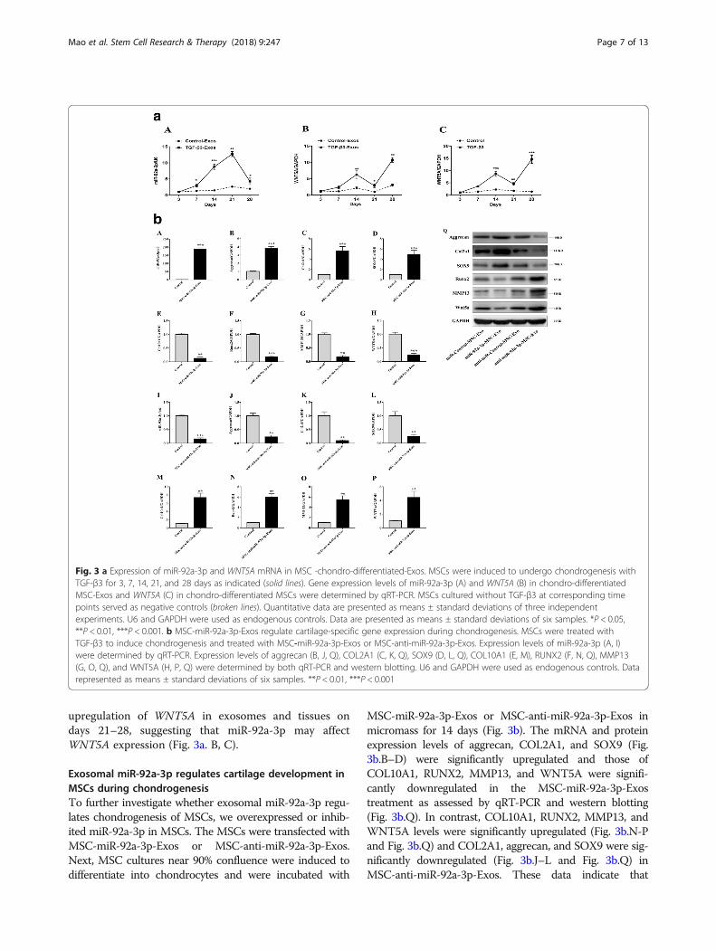
upregulation of WNT5A in exosomes and tissues ondays 21–28, suggesting that miR-92a-3p may affectWNT5A expression (Fig. 3a. B, C).
Exosomal miR-92a-3p regulates cartilage development inMSCs during chondrogenesisTo further investigate whether exosomal miR-92a-3p regu-lates chondrogenesis of MSCs, we overexpressed or inhib-ited miR-92a-3p in MSCs. The MSCs were transfected withMSC-miR-92a-3p-Exos or MSC-anti-miR-92a-3p-Exos.Next, MSC cultures near 90% confluence were induced todifferentiate into chondrocytes and were incubated with
MSC-miR-92a-3p-Exos or MSC-anti-miR-92a-3p-Exos inmicromass for 14 days (Fig. 3b). The mRNA and proteinexpression levels of aggrecan, COL2A1, and SOX9 (Fig.3b.B–D) were significantly upregulated and those ofCOL10A1, RUNX2, MMP13, and WNT5A were signifi-cantly downregulated in the MSC-miR-92a-3p-Exostreatment as assessed by qRT-PCR and western blotting(Fig. 3b.Q). In contrast, COL10A1, RUNX2, MMP13, andWNT5A levels were significantly upregulated (Fig. 3b.N-Pand Fig. 3b.Q) and COL2A1, aggrecan, and SOX9 were sig-nificantly downregulated (Fig. 3b.J–L and Fig. 3b.Q) inMSC-anti-miR-92a-3p-Exos. These data indicate that
Fig. 3 a Expression of miR-92a-3p and WNT5A mRNA in MSC -chondro-differentiated-Exos. MSCs were induced to undergo chondrogenesis withTGF-β3 for 3, 7, 14, 21, and 28 days as indicated (solid lines). Gene expression levels of miR-92a-3p (A) and WNT5A (B) in chondro-differentiatedMSC-Exos and WNT5A (C) in chondro-differentiated MSCs were determined by qRT-PCR. MSCs cultured without TGF-β3 at corresponding timepoints served as negative controls (broken lines). Quantitative data are presented as means ± standard deviations of three independentexperiments. U6 and GAPDH were used as endogenous controls. Data are presented as means ± standard deviations of six samples. *P < 0.05,**P < 0.01, ***P < 0.001. b MSC-miR-92a-3p-Exos regulate cartilage-specific gene expression during chondrogenesis. MSCs were treated withTGF-β3 to induce chondrogenesis and treated with MSC-miR-92a-3p-Exos or MSC-anti-miR-92a-3p-Exos. Expression levels of miR-92a-3p (A, I)were determined by qRT-PCR. Expression levels of aggrecan (B, J, Q), COL2A1 (C, K, Q), SOX9 (D, L, Q), COL10A1 (E, M), RUNX2 (F, N, Q), MMP13(G, O, Q), and WNT5A (H, P, Q) were determined by both qRT-PCR and western blotting. U6 and GAPDH were used as endogenous controls. Datarepresented as means ± standard deviations of six samples. **P < 0.01, ***P < 0.001
Mao et al. Stem Cell Research & Therapy (2018) 9:247 Page 7 of 13
miR-92a-3p may target WNT5A to promote SOX9 andaggrecan expression and enhance cartilage development.
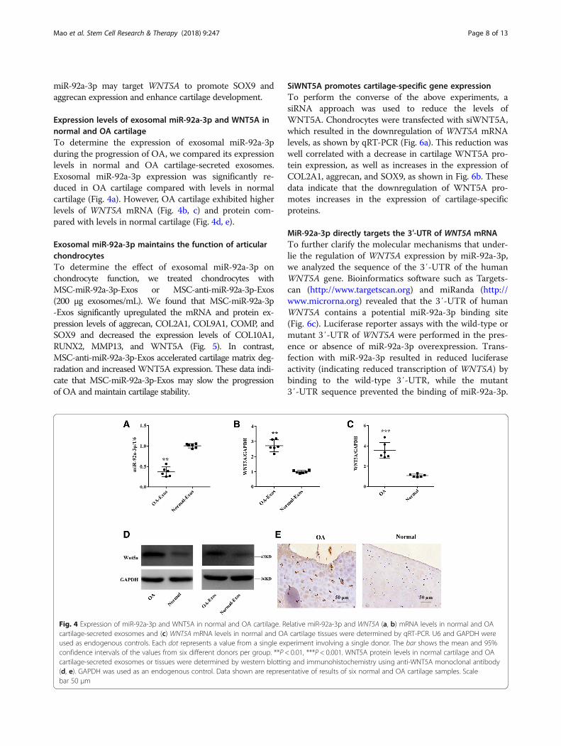
Expression levels of exosomal miR-92a-3p and WNT5A innormal and OA cartilageTo determine the expression of exosomal miR-92a-3pduring the progression of OA, we compared its expressionlevels in normal and OA cartilage-secreted exosomes.Exosomal miR-92a-3p expression was significantly re-duced in OA cartilage compared with levels in normalcartilage (Fig. 4a). However, OA cartilage exhibited higherlevels of WNT5A mRNA (Fig. 4b, c) and protein com-pared with levels in normal cartilage (Fig. 4d, e).
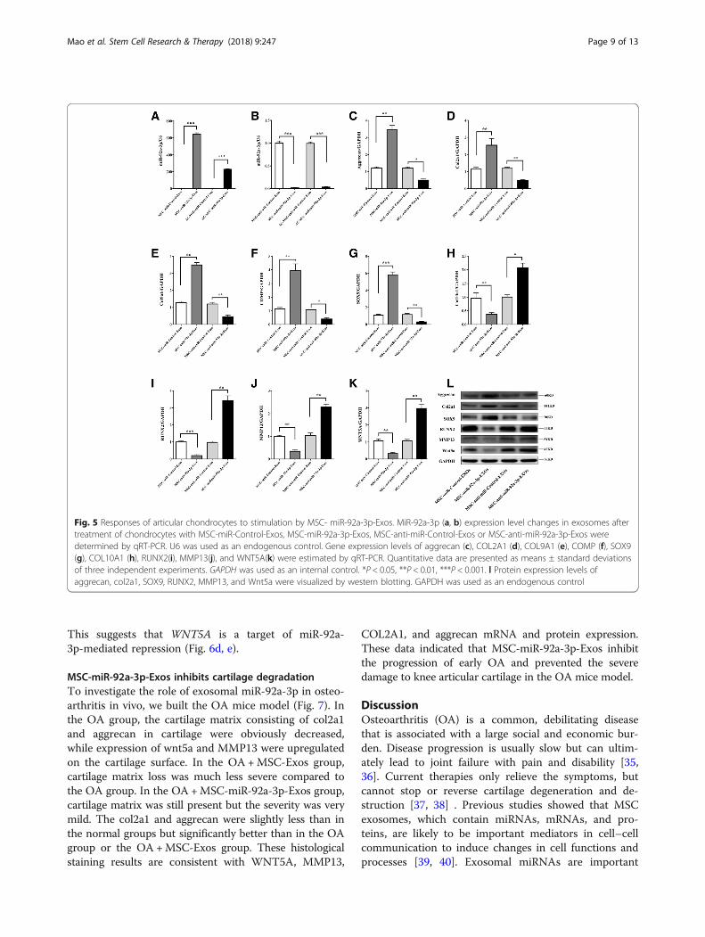
Exosomal miR-92a-3p maintains the function of articularchondrocytesTo determine the effect of exosomal miR-92a-3p onchondrocyte function, we treated chondrocytes withMSC-miR-92a-3p-Exos or MSC-anti-miR-92a-3p-Exos(200 μg exosomes/mL). We found that MSC-miR-92a-3p-Exos significantly upregulated the mRNA and protein ex-pression levels of aggrecan, COL2A1, COL9A1, COMP, andSOX9 and decreased the expression levels of COL10A1,RUNX2, MMP13, and WNT5A (Fig. 5). In contrast,MSC-anti-miR-92a-3p-Exos accelerated cartilage matrix deg-radation and increased WNT5A expression. These data indi-cate that MSC-miR-92a-3p-Exos may slow the progressionof OA and maintain cartilage stability.
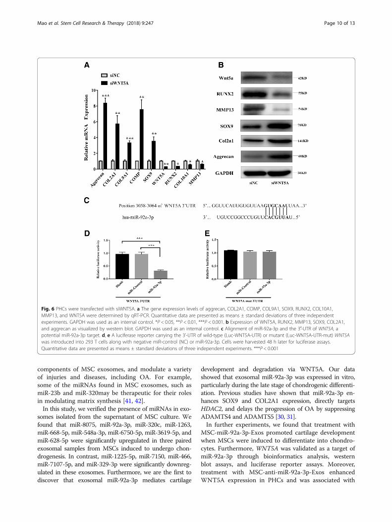
SiWNT5A promotes cartilage-specific gene expressionTo perform the converse of the above experiments, asiRNA approach was used to reduce the levels ofWNT5A. Chondrocytes were transfected with siWNT5A,which resulted in the downregulation of WNT5A mRNAlevels, as shown by qRT-PCR (Fig. 6a). This reduction waswell correlated with a decrease in cartilage WNT5A pro-tein expression, as well as increases in the expression ofCOL2A1, aggrecan, and SOX9, as shown in Fig. 6b. Thesedata indicate that the downregulation of WNT5A pro-motes increases in the expression of cartilage-specificproteins.
MiR-92a-3p directly targets the 3′-UTR of WNT5A mRNATo further clarify the molecular mechanisms that under-lie the regulation of WNT5A expression by miR-92a-3p,we analyzed the sequence of the 3′-UTR of the humanWNT5A gene. Bioinformatics software such as Targets-can (http://www.targetscan.org) and miRanda (http://www.microrna.org) revealed that the 3′-UTR of humanWNT5A contains a potential miR-92a-3p binding site(Fig. 6c). Luciferase reporter assays with the wild-type ormutant 3′-UTR of WNT5A were performed in the pres-ence or absence of miR-92a-3p overexpression. Trans-fection with miR-92a-3p resulted in reduced luciferaseactivity (indicating reduced transcription of WNT5A) bybinding to the wild-type 3′-UTR, while the mutant3′-UTR sequence prevented the binding of miR-92a-3p.
Fig. 4 Expression of miR-92a-3p and WNT5A in normal and OA cartilage. Relative miR-92a-3p and WNT5A (a, b) mRNA levels in normal and OAcartilage-secreted exosomes and (c) WNT5A mRNA levels in normal and OA cartilage tissues were determined by qRT-PCR. U6 and GAPDH wereused as endogenous controls. Each dot represents a value from a single experiment involving a single donor. The bar shows the mean and 95%confidence intervals of the values from six different donors per group. **P < 0.01, ***P < 0.001. WNT5A protein levels in normal cartilage and OAcartilage-secreted exosomes or tissues were determined by western blotting and immunohistochemistry using anti-WNT5A monoclonal antibody(d, e). GAPDH was used as an endogenous control. Data shown are representative of results of six normal and OA cartilage samples. Scalebar 50 μm
Mao et al. Stem Cell Research & Therapy (2018) 9:247 Page 8 of 13
This suggests that WNT5A is a target of miR-92a-3p-mediated repression (Fig. 6d, e).
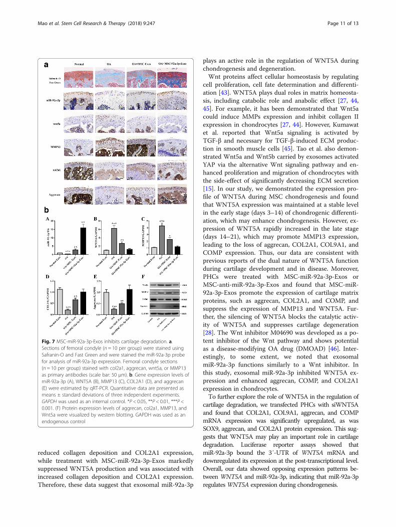
MSC-miR-92a-3p-Exos inhibits cartilage degradationTo investigate the role of exosomal miR-92a-3p in osteo-arthritis in vivo, we built the OA mice model (Fig. 7). Inthe OA group, the cartilage matrix consisting of col2a1and aggrecan in cartilage were obviously decreased,while expression of wnt5a and MMP13 were upregulatedon the cartilage surface. In the OA +MSC-Exos group,cartilage matrix loss was much less severe compared tothe OA group. In the OA +MSC-miR-92a-3p-Exos group,cartilage matrix was still present but the severity was verymild. The col2a1 and aggrecan were slightly less than inthe normal groups but significantly better than in the OAgroup or the OA +MSC-Exos group. These histologicalstaining results are consistent with WNT5A, MMP13,
COL2A1, and aggrecan mRNA and protein expression.These data indicated that MSC-miR-92a-3p-Exos inhibitthe progression of early OA and prevented the severedamage to knee articular cartilage in the OA mice model.
DiscussionOsteoarthritis (OA) is a common, debilitating diseasethat is associated with a large social and economic bur-den. Disease progression is usually slow but can ultim-ately lead to joint failure with pain and disability [35,36]. Current therapies only relieve the symptoms, butcannot stop or reverse cartilage degeneration and de-struction [37, 38] . Previous studies showed that MSCexosomes, which contain miRNAs, mRNAs, and pro-teins, are likely to be important mediators in cell–cellcommunication to induce changes in cell functions andprocesses [39, 40]. Exosomal miRNAs are important
Fig. 5 Responses of articular chondrocytes to stimulation by MSC- miR-92a-3p-Exos. MiR-92a-3p (a, b) expression level changes in exosomes aftertreatment of chondrocytes with MSC-miR-Control-Exos, MSC-miR-92a-3p-Exos, MSC-anti-miR-Control-Exos or MSC-anti-miR-92a-3p-Exos weredetermined by qRT-PCR. U6 was used as an endogenous control. Gene expression levels of aggrecan (c), COL2A1 (d), COL9A1 (e), COMP (f), SOX9(g), COL10A1 (h), RUNX2(i), MMP13(j), and WNT5A(k) were estimated by qRT-PCR. Quantitative data are presented as means ± standard deviationsof three independent experiments. GAPDH was used as an internal control. *P < 0.05, **P < 0.01, ***P < 0.001. l Protein expression levels ofaggrecan, col2a1, SOX9, RUNX2, MMP13, and Wnt5a were visualized by western blotting. GAPDH was used as an endogenous control
Mao et al. Stem Cell Research & Therapy (2018) 9:247 Page 9 of 13
components of MSC exosomes, and modulate a varietyof injuries and diseases, including OA. For example,some of the miRNAs found in MSC exosomes, such asmiR-23b and miR-320may be therapeutic for their rolesin modulating matrix synthesis [41, 42].In this study, we verified the presence of miRNAs in exo-
somes isolated from the supernatant of MSC culture. Wefound that miR-8075, miR-92a-3p, miR-320c, miR-1263,miR-668-5p, miR-548a-3p, miR-6750-5p, miR-3619-5p, andmiR-628-5p were significantly upregulated in three pairedexosomal samples from MSCs induced to undergo chon-drogenesis. In contrast, miR-1225-5p, miR-7150, miR-466,miR-7107-5p, and miR-329-3p were significantly downreg-ulated in these exosomes. Furthermore, we are the first todiscover that exosomal miR-92a-3p mediates cartilage
development and degradation via WNT5A. Our datashowed that exosomal miR-92a-3p was expressed in vitro,particularly during the late stage of chondrogenic differenti-ation. Previous studies have shown that miR-92a-3p en-hances SOX9 and COL2A1 expression, directly targetsHDAC2, and delays the progression of OA by suppressingADAMTS4 and ADAMTS5 [30, 31].In further experiments, we found that treatment with
MSC-miR-92a-3p-Exos promoted cartilage developmentwhen MSCs were induced to differentiate into chondro-cytes. Furthermore, WNT5A was validated as a target ofmiR-92a-3p through bioinformatics analysis, westernblot assays, and luciferase reporter assays. Moreover,treatment with MSC-anti-miR-92a-3p-Exos enhancedWNT5A expression in PHCs and was associated with
Fig. 6 PHCs were transfected with siWNT5A. a The gene expression levels of aggrecan, COL2A1, COMP, COL9A1, SOX9, RUNX2, COL10A1,MMP13, and WNT5A were determined by qRT-PCR. Quantitative data are presented as means ± standard deviations of three independentexperiments. GAPDH was used as an internal control. *P < 0.05, **P < 0.01, ***P < 0.001. b Expression of WNT5A, RUNX2, MMP13, SOX9, COL2A1,and aggrecan as visualized by western blot. GAPDH was used as an internal control. c Alignment of miR-92a-3p and the 3′-UTR of WNT5A, apotential miR-92a-3p target. d, e A luciferase reporter carrying the 3’-UTR of wild-type (Luc-WNT5A-UTR) or mutant (Luc-WNT5A-UTR-mut) WNT5Awas introduced into 293 T cells along with negative miR-control (NC) or miR-92a-3p. Cells were harvested 48 h later for luciferase assays.Quantitative data are presented as means ± standard deviations of three independent experiments. ***P < 0.001
Mao et al. Stem Cell Research & Therapy (2018) 9:247 Page 10 of 13
reduced collagen deposition and COL2A1 expression,while treatment with MSC-miR-92a-3p-Exos markedlysuppressed WNT5A production and was associated withincreased collagen deposition and COL2A1 expression.Therefore, these data suggest that exosomal miR-92a-3p
plays an active role in the regulation of WNT5A duringchondrogenesis and degeneration.Wnt proteins affect cellular homeostasis by regulating
cell proliferation, cell fate determination and differenti-ation [43]. WNT5A plays dual roles in matrix homeosta-sis, including catabolic role and anabolic effect [27, 44,45]. For example, it has been demonstrated that Wnt5acould induce MMPs expression and inhibit collagen IIexpression in chondrocytes [27, 44]. However, Kumawatet al. reported that Wnt5a signaling is activated byTGF-β and necessary for TGF-β-induced ECM produc-tion in smooth muscle cells [45]. Tao et al. also demon-strated Wnt5a and Wnt5b carried by exosomes activatedYAP via the alternative Wnt signaling pathway and en-hanced proliferation and migration of chondrocytes withthe side-effect of significantly decreasing ECM secretion[15]. In our study, we demonstrated the expression pro-file of WNT5A during MSC chondrogenesis and foundthat WNT5A expression was maintained at a stable levelin the early stage (days 3–14) of chondrogenic differenti-ation, which may enhance chondrogenesis. However, ex-pression of WNT5A rapidly increased in the late stage(days 14–21), which may promote MMP13 expression,leading to the loss of aggrecan, COL2A1, COL9A1, andCOMP expression. Thus, our data are consistent withprevious reports of the dual nature of WNT5A functionduring cartilage development and in disease. Moreover,PHCs were treated with MSC-miR-92a-3p-Exos orMSC-anti-miR-92a-3p-Exos and found that MSC-miR-92a-3p-Exos promote the expression of cartilage matrixproteins, such as aggrecan, COL2A1, and COMP, andsuppress the expression of MMP13 and WNT5A. Fur-ther, the silencing of WNT5A blocks the catalytic activ-ity of WNT5A and suppresses cartilage degeneration[28]. The Wnt inhibitor M04690 was developed as a po-tent inhibitor of the Wnt pathway and shows potentialas a disease-modifying OA drug (DMOAD) [46]. Inter-estingly, to some extent, we noted that exosomalmiR-92a-3p functions similarly to a Wnt inhibitor. Inthis study, exosomal miR-92a-3p inhibited WNT5A ex-pression and enhanced aggrecan, COMP, and COL2A1expression in chondrocytes.To further explore the role of WNT5A in the regulation of
cartilage degradation, we transfected PHCs with siWNT5Aand found that COL2A1, COL9A1, aggrecan, and COMPmRNA expression was significantly upregulated, as wasSOX9, aggrecan, and COL2A1 protein expression. This sug-gests that WNT5A may play an important role in cartilagedegradation. Luciferase reporter assays showed thatmiR-92a-3p bound the 3′-UTR of WNT5A mRNA anddownregulated its expression at the post-transcriptional level.Overall, our data showed opposing expression patterns be-tween WNT5A and miR-92a-3p, indicating that miR-92a-3pregulatesWNT5A expression during chondrogenesis.
Fig. 7 MSC-miR-92a-3p-Exos inhibits cartilage degradation. a.Sections of femoral condyle (n = 10 per group) were stained usingSafranin-O and Fast Green and were stained the miR-92a-3p probefor analysis of miR-92a-3p expression. Femoral condyle sections(n = 10 per group) stained with col2a1, aggrecan, wnt5a, or MMP13as primary antibodies (scale bar: 50 μm). b. Gene expression levels ofmiR-92a-3p (A), WNT5A (B), MMP13 (C), COL2A1 (D), and aggrecan(E) were estimated by qRT-PCR. Quantitative data are presented asmeans ± standard deviations of three independent experiments.GAPDH was used as an internal control. *P < 0.05, **P < 0.01, ***P <0.001. (F) Protein expression levels of aggrecan, col2a1, MMP13, andWnt5a were visualized by western blotting. GAPDH was used as anendogenous control
Mao et al. Stem Cell Research & Therapy (2018) 9:247 Page 11 of 13
Furthermore, in vivo, we investigate the role ofMSC-miR-92a-3p-Exos by a collagenase-induced OAmice model. We found and determined thatMSC-miR-92a-3p-Exos inhibit the progression of earlyOA and prevented the severe damage to knee articularcartilage in the OA mice model.Although this study demonstrated that exosomal
miR-92a-3p is efficacious in cartilage development and de-generation, there are still some limitations to our study.First, the detailed signaling pathways of the dual functionsof WNT5A in MSCs undergoing chondrogenesis have notyet been clarified. We plan to conduct a cell signalingstudy in human bone MSCs to further elaborate the dualregulatory role of WNT5A in chondrogenesis. Further-more, further studies are needed to clarify the specificfunction of exosomal-miR-92a-3p in the destabilization ofthe medial meniscus (DMM) model of OA and osteo-chondral defect model. For example, studies in rabbits orrats, in which exosome functions could be explored incombination with scaffolds for the treatment of cartilagedefects and OA would be useful.
ConclusionsThe present study demonstrated that exosomal miR-92a-3pis expressed before and after chondrogenic differentiationin MSCs and is differentially expressed in OA. We foundthat exosomal miR-92a-3p functions as a negative regulatorby downregulating WNT5A in both chondrogenesis andOA pathogenesis. We suggest that controlling the expres-sion of exosomal miR-92a-3p has potential as a novel ap-proach for prevention and treatment of OA.
Additional file
Additional file 1: MiRNA expression profiles in exosomes before andafter chondrogenic differentiation of MSCs. The expression profiles ofmiRNAs in exosomes before and after chondrogenic differentiation weredetected using a miRNA microarray. SAM statistical software was used toidentify differentially expressed miRNAs between undifferentiatedMSC-Exos and chondro-differentiated MSC-Exos. Among the miRNAs thatwere consistently differentially expressed in all three paired samples, 36were upregulated (fold change > 2) and 105 were downregulated (foldchange <− 2) in chondro-differentiated MSC-Exos compared to levels inMSC-Exos. (XLSX 69 kb)
Abbreviations3′-UTR: 3′-untranslated region; ADAMTS: A disintegrin and metalloproteinasewith thrombospondin motifs; DMEM: Dulbecco’s modified Eagle’s medium;ECM: Extracellular matrix; hMSC: Human bone marrow-derived MSCs;IL: Interleukin; MSC-Exos: Exosomes secreted by mesenchymal stem cells;miRNA: microRNA; MMP: Matrix metalloproteinase; MSC miR-92a-3p-Exos: Exosomes secreted by miR-92a-3p-overexpressing mesenchymal stemcells; OA: Osteoarthritis; PHC: Primary human chondrocyte; qRT-PCR: quantitative real-time reverse transcription-polymerase chain reaction;TGB: Transforming growth factor
AcknowledgmentsWe thank Xuerong Li (Department of Parasitology, Zhongshan School ofMedicine, Sun Yat-sen University, Guangzhou, China) for his aid with micros-copy and other techniques.
FundingThis study was supported by the National Nature Science Foundation ofChina (81572119, and 81472101) and the Guangdong Provincial NaturalScience Foundation of China (201707010211 and 2015A020212015).
Availability of data and materialsAll data generated or analyzed during this study are included in thispublished article and Additional file.
Authors’ contributionsYK and WML contributed to the study conception and design, GPM, ZYH, SH,ZJZ, ZQZ, and ZKC performed the experiments. GPM created the graphs andwrote the manuscript. YK revised the manuscript and WML participated inthe discussion of the manuscript. All authors analyzed and interpretedexperimental data. All authors read and approved the final manuscript.
Ethics approval and consent to participateAll experimental protocols were approved by the Ethics Committee of theFirst Affiliated Hospital of Sun Yat-sen University. Written informed consentwas obtained from all donors.
Consent for publicationNot applicable.
Competing interestsThe authors declare that they have no competing interests.
Publisher’s NoteSpringer Nature remains neutral with regard to jurisdictional claims inpublished maps and institutional affiliations.
Received: 28 May 2018 Revised: 30 July 2018Accepted: 3 September 2018
References1. Loeser RF, Goldring SR, Scanzello CR, Goldring MB. Osteoarthritis: a disease
of the joint as an organ. Arthritis Rheum. 2012;64:1697–707. https://doi.org/10.1002/art.34453.
2. Kobayashi M, Squires GR, Mousa A, Tanzer M, Zukor DJ, Antoniou J, et al.Role of interleukin-1 and tumor necrosis factor alpha in matrix degradationof human osteoarthritic cartilage. Arthritis Rheum. 2005;52:128–35. https://doi.org/10.1002/art.20776.
3. Daheshia M, Yao JQ. The interleukin 1beta pathway in the pathogenesis ofosteoarthritis. J Rheumatol. 2008;35:2306–12.
4. Toh WS, Foldager CB, Pei M, Hui JH. Advances in mesenchymal stem cell-based strategies for cartilage repair and regeneration. Stem Cell Rev. 2014;10:686–96. https://doi.org/10.1007/s12015-014-9526-z.
5. Meirelles LS, Fontes AM, Covas DT, Caplan AI. Mechanisms involved in thetherapeutic properties of mesenchymal stem cells. Cytokine Growth FactorRev. 2009;20:419–27. https://doi.org/10.1016/j.cytogfr.2009.10.002.
6. Toh WS, Lai RC, Hui J, Lim SK. MSC exosome as a cell-free MSC therapy forcartilage regeneration: implications for osteoarthritis treatment. Semin CellDev Biol. 2017;67:56–64. https://doi.org/10.1016/j.semcdb.2016.11.008.
7. Lai RC, Arslan F, Lee MM, Sze NS, Choo A, Chen TS, et al. Exosome secretedby MSC reduces myocardial ischemia/reperfusion injury. Stem Cell Res.2010;4:214–22. https://doi.org/10.1016/j.scr.2009.12.003.
8. Zhang S, Chu WC, Lai RC, Lim SK, Hui JH, Toh WS. Exosomes derived fromhuman embryonic mesenchymal stem cells promote osteochondralregeneration. Osteoarthr Cartil. 2016;24:2135–40. https://doi.org/10.1016/j.joca.2016.06.022.
9. Vonk LA, van Dooremalen S, Liv N, Klumperman J, Coffer PJ, Saris D, et al.Mesenchymal stromal/stem cell-derived extracellular vesicles promotehuman cartilage regeneration in vitro. Theranostics. 2018;8:906–20. https://doi.org/10.7150/thno.20746.
10. Zhang S, Chuah SJ, Lai RC, Hui J, Lim SK, Toh WS. MSC exosomes mediatecartilage repair by enhancing proliferation, attenuating apoptosis and
Mao et al. Stem Cell Research & Therapy (2018) 9:247 Page 12 of 13

modulating immune reactivity. Biomaterials. 2018;156:16–27. https://doi.org/10.1016/j.biomaterials.2017.11.028.
11. Xin H, Li Y, Chopp M. Exosomes/miRNAs as mediating cell-based therapy ofstroke. Front Cell Neurosci. 2014;8:377. https://doi.org/10.3389/fncel.2014.00377.
12. Burger D, Vinas JL, Akbari S, Dehak H, Knoll W, Gutsol A, et al. Humanendothelial colony-forming cells protect against acute kidney injury: role ofexosomes. Am J Pathol. 2015;185:2309–23. https://doi.org/10.1016/j.ajpath.2015.04.010.
13. Carthew RW, Sontheimer EJ. Origins and mechanisms of miRNAs andsiRNAs. Cell. 2009;136:642–55. https://doi.org/10.1016/j.cell.2009.01.035.
14. Mayourian J, Ceholski DK, Gorski PA, Mathiyalagan P, Murphy JF, Salazar SI,et al. Exosomal microRNA-21-5p Mediates Mesenchymal Stem Cell ParacrineEffects on Human Cardiac Tissue Contractility. Circ Res. 2018;122:933–44.https://doi.org/10.1161/CIRCRESAHA.118.312420.
15. Tao SC, Yuan T, Zhang YL, Yin WJ, Guo SC, Zhang CQ. Exosomes derivedfrom miR-140-5p-overexpressing human synovial mesenchymal stem cellsenhance cartilage tissue regeneration and prevent osteoarthritis of the kneein a rat model. Theranostics. 2017;7:180–95. https://doi.org/10.7150/thno.17133.
16. Staines KA, Macrae VE, Farquharson C. Cartilage development anddegeneration: a Wnt Wnt situation. Cell Biochem Funct. 2012;30:633–42.https://doi.org/10.1002/cbf.2852.
17. Ge X, Shi R, Ma X. The secreted protein WNT5A regulates condylarchondrocyte proliferation, hypertrophy and migration. Arch Oral Biol. 2017;82:171–9. https://doi.org/10.1016/j.archoralbio.2017.06.019.
18. Lerner UH, Ohlsson C. The WNT system: background and its role in bone. JIntern Med. 2015;277:630–49. https://doi.org/10.1111/joim.12368.
19. Chun JS, Oh H, Yang S, Park M. Wnt signaling in cartilage development anddegeneration. BMB Rep. 2008;41:485–94.
20. Maeda K, Kobayashi Y, Udagawa N, Uehara S, Ishihara A, Mizoguchi T, et al.Wnt5a-Ror2 signaling between osteoblast-lineage cells and osteoclastprecursors enhances osteoclastogenesis. Nat Med. 2012;18:405–12. https://doi.org/10.1038/nm.2653.
21. Hosseini-Farahabadi S, Geetha-Loganathan P, Fu K, Nimmagadda S, Yang HJ,Richman JM. Dual functions for WNT5A during cartilage development andin disease. Matrix Biol. 2013;32:252–64. https://doi.org/10.1016/j.matbio.2013.02.005.
22. Kawakami Y, Wada N, Nishimatsu SI, Ishikawa T, Noji S, Nohno T.Involvement of Wnt-5a in chondrogenic pattern formation in the chick limbbud. Develop Growth Differ. 1999;41:29–40.
23. Church V, Nohno T, Linker C, Marcelle C, Francis-West P. Wnt regulation ofchondrocyte differentiation. J Cell Sci. 2002;115:4809–18.
24. Yang Y, Topol L, Lee H, Wu J. Wnt5a and Wnt5b exhibit distinct activities incoordinating chondrocyte proliferation and differentiation. Development.2003;130:1003–15.
25. Huang G, Chubinskaya S, Liao W, Loeser RF. Wnt5a induces catabolicsignaling and matrix metalloproteinase production in human articularchondrocytes. Osteoarthr Cartil. 2017;25:1505–15. https://doi.org/10.1016/j.joca.2017.05.018.
26. Ge XP, Gan YH, Zhang CG, Zhou CY, Ma KT, Meng JH, et al. Requirement ofthe NF-kappaB pathway for induction of Wnt-5A by interleukin-1beta incondylar chondrocytes of the temporomandibular joint: functional crosstalkbetween the Wnt-5A and NF-kappaB signaling pathways. Osteoarthr Cartil.2011;19:111–7. https://doi.org/10.1016/j.joca.2010.10.016.
27. Ge X, Ma X, Meng J, Zhang C, Ma K, Zhou C. Role of Wnt-5A in interleukin-1beta-induced matrix metalloproteinase expression in rabbittemporomandibular joint condylar chondrocytes. Arthritis Rheum. 2009;60:2714–22. https://doi.org/10.1002/art.24779.
28. Shi S, Man Z, Li W, Sun S, Zhang W. Silencing of Wnt5a prevents interleukin-1beta-induced collagen type II degradation in rat chondrocytes. Exp TherMed. 2016;12:3161–6. https://doi.org/10.3892/etm.2016.3788.
29. Ning G, Liu X, Dai M, Meng A, Wang Q. MicroRNA-92a upholds bmpsignaling by targeting noggin3 during pharyngeal cartilage formation. DevCell. 2013;24:283–95. https://doi.org/10.1016/j.devcel.2012.12.016.
30. Mao G, Wu P, Zhang Z, Zhang Z, Liao W, Li Y, et al. MicroRNA-92a-3pregulates Aggrecanase-1 and Aggrecanase-2 expression in Chondrogenesisand IL-1beta-induced catabolism in human articular chondrocytes. CellPhysiol Biochem. 2017;44:38–52. https://doi.org/10.1159/000484579.
31. Mao G, Zhang Z, Huang Z, Chen W, Huang G, Meng F, et al. MicroRNA-92a-3p regulates the expression of cartilage-specific genes by directly targeting
histone deacetylase 2 in chondrogenesis and degradation. Osteoarthr Cartil.2017;25:521–32. https://doi.org/10.1016/j.joca.2016.11.006.
32. Meng FG, Zhang ZQ, Huang GX, Chen WS, Zhang ZJ, He AS, et al.Chondrogenesis of mesenchymal stem cells in a novel hyaluronate-collagen-tricalcium phosphate scaffolds for knee repair. Eur Cell Mater. 2016;31:79–94.
33. Greening DW, Xu R, Ji H, Tauro BJ, Simpson RJ. A protocol for exosomeisolation and characterization: evaluation of ultracentrifugation, density-gradient separation, and immunoaffinity capture methods. Methods MolBiol. 2015;1295:179–209. https://doi.org/10.1007/978-1-4939-2550-6_15.
34. Zhu Y, Wang Y, Zhao B, Niu X, Hu B, Li Q, et al. Comparison of exosomessecreted by induced pluripotent stem cell-derived mesenchymal stem cellsand synovial membrane-derived mesenchymal stem cells for the treatmentof osteoarthritis. Stem Cell Res Ther. 2017;8:64. https://doi.org/10.1186/s13287-017-0510-9.
35. Chen D, Shen J, Zhao W, Wang T, Han L, Hamilton JL, et al. Osteoarthritis:toward a comprehensive understanding of pathological mechanism. BoneRes. 2017;5:16044. https://doi.org/10.1038/boneres.2016.44.
36. Mathiessen A, Conaghan PG. Synovitis in osteoarthritis: currentunderstanding with therapeutic implications. Arthritis Res Ther. 2017;19:18.https://doi.org/10.1186/s13075-017-1229-9.
37. Yu SP, Hunter DJ. Intra-articular therapies for osteoarthritis. Expert OpinPharmacother. 2016;17:2057–71. https://doi.org/10.1080/14656566.2016.1232396.
38. Glyn-Jones S, Palmer AJ, Agricola R, Price AJ, Vincent TL, Weinans H, et al.Osteoarthritis. Lancet. 2015;386:376–87. https://doi.org/10.1016/S0140-6736(14)60802-3.
39. Chen TS, Lim SK. Measurement of precursor miRNA in exosomes fromhuman ESC-derived mesenchymal stem cells. Methods Mol Biol. 2013;1024:69–86. https://doi.org/10.1007/978-1-62703-453-1_6.
40. Chen TS, Lai RC, Lee MM, Choo AB, Lee CN, Lim SK. Mesenchymal stem cellsecretes microparticles enriched in pre-microRNAs. Nucleic Acids Res. 2010;38:215–24. https://doi.org/10.1093/nar/gkp857.
41. Meng F, Zhang Z, Chen W, Huang G, He A, Hou C, et al. MicroRNA-320regulates matrix metalloproteinase-13 expression in chondrogenesis andinterleukin-1beta-induced chondrocyte responses. Osteoarthr Cartil. 2016;24:932–41. https://doi.org/10.1016/j.joca.2015.12.012.
42. Ham O, Song BW, Lee SY, Choi E, Cha MJ, Lee CY, et al. The role ofmicroRNA-23b in the differentiation of MSC into chondrocyte by targetingprotein kinase a signaling. Biomaterials. 2012;33:4500–7. https://doi.org/10.1016/j.biomaterials.2012.03.025.
43. Logan CY, Nusse R. The Wnt signaling pathway in development anddisease. Annu Rev Cell Dev Biol. 2004;20:781–810. https://doi.org/10.1146/annurev.cellbio.20.010403.113126.
44. Ryu JH, Chun JS. Opposing roles of WNT-5A and WNT-11 in interleukin-1beta regulation of type II collagen expression in articular chondrocytes. JBiol Chem. 2006;281:22039–47. https://doi.org/10.1074/jbc.M601804200.
45. Kumawat K, Menzen MH, Bos IS, Baarsma HA, Borger P, Roth M, et al.Noncanonical WNT-5A signaling regulates TGF-beta-induced extracellularmatrix production by airway smooth muscle cells. FASEB J. 2013;27:1631–43.https://doi.org/10.1096/fj.12-217539.
46. Yazici Y, McAlindon TE, Fleischmann R, Gibofsky A, Lane NE, Kivitz AJ, et al.A novel Wnt pathway inhibitor, SM04690, for the treatment of moderate tosevere osteoarthritis of the knee: results of a 24-week, randomized,controlled, phase 1 study. Osteoarthr Cartil. 2017;25:1598–606. https://doi.org/10.1016/j.joca.2017.07.006.
Mao et al. Stem Cell Research & Therapy (2018) 9:247 Page 13 of 13