experimental and theoretical investigation of the
TRANSCRIPT

General Papers ARKIVOC 2010 (x) 62-76
ISSN 1551-7012 Page 62 ARKAT USA, Inc.
Experimental and theoretical investigation of the structure and
nucleophilic properties of 4-aminocoumarin
Bistra Stamboliyska,a Voislava Janevska,b Boris Shivachev,c Rosica P. Nikolova,c
Goran Stojkovic,b Bozhna Mikhova,a and Emil Popovskib*
aInstitute of Organic Chemistry with Centre of Phytochemistry, Bulgarian Academy of Sciences,
Acad. G. Bonchev Str., Build. 9, 1113 Sofia, Bulgaria bInstitute of Chemistry, Faculty of Natural Sciences and Mathematics,
Ss. Cyril and Methodius University, Arhimedova 5, 1000 Skopje, P.O. Box 162, Macedonia. cCentral Laboratory of Mineralogy and Crystallography, Bulgarian Academy of Sciences,
Acad. G. Bonchev Str., Build. 107, 1113 Sofia, Bulgaria
E-mail: [email protected]
DOI: http://dx.doi.org/10.3998/ark.5550190.0011.a06
Abstract
4-Aminocoumarin 2 was prepared in high yield via solventless reaction. The structure of the 2
was determined with single crystal X-ray analysis. The detailed NMR and IR spectra were
reported for the first time. DFT calculations [B3LYP/6-31+G**] showed good agreement
between the theoretical and experimental values for the optimized and X-ray structures, as well
as between the vibrational and NMR spectroscopy. The thermodynamic pKBH+ values were
calculated using three different methods: Yates and McClelland (with HA acidity functions),
Excess Acidity Method and Bunnett and Olsen Method. The experimental and theoretical data
presented are consistent with the weak nucleophilic properties of 2.
Keywords: 4-Aminocoumarin, crystal structure, spectra, nucleophilic properties
Introduction
Coumarin and its derivatives from natural products, either semi-synthetic or synthetic, represent
one of the most active classes of compounds exhibiting a wide spectrum of biological activity.
Additionally, coumarin derivatives are used as additives in food and cosmetic industry. Due to
the significance of these compounds, the quest for efficient syntheses of coumarin ring
compounds1 as well as new bioactive derivatives from known coumarins2 is topic.
From the group of aminocoumarins, 3-aminocoumarin and 7-aminocoumarin derivatives are
well studied. The 3-aminocoumarin moiety can be recognized in the molecular structure of

General Papers ARKIVOC 2010 (x) 62-76
ISSN 1551-7012 Page 63 ARKAT USA, Inc.
several natural antibiotics, such as novobiocin, chlorobiocin, coumermycin, etc. These antibiotics
and their derivatives are in the research focus.3 In addition, some simple N-acylderivatives of 3-
aminocoumarin exhibit an anti-inflammatory activity4 and antimicrobial activity against the
Gram-positive bacteria.5 Most of the publications for 7-aminocoumarin derivatives present
interesting photochemical behavior of these compounds6 and many of them can be used as
fluorescent markers.7 Furthermore, some platinum complexes of 7-aminocoumarins have been
synthesized and evaluated for their in vitro cytotoxicity against Caco-2T cells.8 Derivatives of 4-
aminocoumarin 2 were not studied that much, although some of them possess biological activity
as well.9
The interesting chemical and physical properties and the pharmacological effects of
aminocoumarin derivatives were the main motivation for starting this research. At first, the
compound 2 was synthesized as a starting compound for the synthesis of large spectra of new
coumarin derivatives. However, during the research we have noticed that the amino group in this
compound was very weak nucleophile. In contrast to 3-aminocoumarin, aniline, naphthylamine
or similar compounds, which are also weak bases, the amino group of 2 did not react with 3
(Scheme 1; alkyl halides, acyl halides, acetic anhydride, etc) to obtain 4 (N-substituted
derivatives). Considering reactions of 2, in few publications only it was presented that instead
with nitrogen, electrophiles can form a bond with carbon in position 3 (position C8 in Figure
1).10 To our knowledge, in only three references, reactions of nitrogen in 2 were presented,11
however, the reported yields of the desired products were very poor. Generally, N-substituted
derivatives of 2 have been obtained by the reactions of amino compounds with 4-
chlorocoumarin12,10a or 4-hydroxycoumarin 1.13
CH3COONH4
130 °C or aceticanhydride
R = -CH2Ph; -CH2CH=CH2; -C(O)CH3; -C(O)Ph; -SO2CH3; -SO2Ph
1 2 4
3CH3COONH4
130 °C or aceticanhydride
R = -CH2Ph; -CH2CH=CH2; -C(O)CH3; -C(O)Ph; -SO2CH3; -SO2Ph
1 2 4
3
Scheme 1
Detailed knowledge of the structure and spectral behavior of 2 is a necessary prerequisite for
understanding its chemical and biological properties. To our surprise, neither NMR nor IR
spectra of the title compound have been studied in detail. The purpose of the present study is to
elucidate the structure by single crystal X-ray diffraction, UV, NMR and IR spectroscopy. The
experimental data will be accompanied by theoretical prediction at different level of
approximation in order to draw out the conclusion on the structure-property relationship.

General Papers ARKIVOC 2010 (x) 62-76
ISSN 1551-7012 Page 64 ARKAT USA, Inc.
Results and Discussion
Synthesis and crystal growth
Synthesis of 2 was performed at 130 oC via solid state reaction of 1 with excess of ammonium
acetate (Scheme 1). Excellent pale yellow crystals for X-ray measurement were obtained by slow
cooling of ethanol solution of 2.
Crystal structure
Molecular structure of compound 2 is shown in Figure 1. The selected bond lengths and angles
are given in Table 1.
Figure 1. The molecular structure of the title molecule with the atom-numbering scheme;
displacement ellipsoids are drawn at the 50 % probability level.
In the molecule of the title compound 2 (Figure 1), the bond lengths and angles are within
normal ranges.14 The 4-aminocoumarin is almost a planar molecule (rms of 0.0146 Å for the
chromene ring). The deviation of planarity is possibly caused by the intermolecular hydrogen
bonding, which builds pseudo-layers parallel to bc plane (Table 2, Figure 2). The pseudo-layers
are stacked trough π-π interaction, as evidenced by the distance of ca. 3.48 Å between the
chromene ring mean planes.
The C7-N1 bond distance (1,335 Å) is just somewhat longer then an average C=N double
bond and little bit shorter then C≈N bond in pyridine (1,337 Å),15 indicating a considerable
degree of double bond character.

General Papers ARKIVOC 2010 (x) 62-76
ISSN 1551-7012 Page 65 ARKAT USA, Inc.
Table 1. Selected bond lengths and angles from crystallographic data and comparison with the
theoretical dataa
Parameter X-ray B3LYP/6-31+G** HF/6-31+G**
C2—C1 1.386 (4) 1.408 1.389
C2—C3 1.403 (4) 1.403 1.399
C2—C7 1.450 (4) 1.460 1.466
O1—C1 1.374 (3) 1.360 1.360
N1—C7 1.335 (3) 1.374 1.370
C8—C7 1.372 (4) 1.370 1.347
C8—C9 1.399 (4) 1.437 1.440
O2—C9 1.239 (3) 1.213 1.395
C1—C6 1.372 (4) 1.399 1.390
C3—C4 1.368 (4) 1.389 1.377
C6—C5 1.376 (4) 1.389 1.378
C5—C4 1.379 (4) 1.403 1.399
C1—C2—C3 117.6 (3) 118.1 118.3
C1—C2—C7 118.1 (3) 117.2 117.0
C3—C2—C7 124.3 (3) 124.6 124.7
C1—O1—C9 120.5 (2) 122.1 123.2
C7—C8—C9 122.3 (3) 122.9 122.2
C6—C1—O1 115.9 (3) 116.5 116.6
C6—C1—C2 122.1 (3) 121.1 121.3
O1—C1—C2 122.0 (3) 122.4 122.1
O2—C9—O1 113.9 (3) 116.9 118.0
O2—C9—C8 127.2 (3) 126.8 125.5
O1—C9—C8 118.9 (3) 116.2 116.5
C4—C3—C2 120.8 (3) 121.1 120.9
N1—C7—C8 121.6 (3) 121.8 122.3
N1—C7—C2 120.3 (3) 119.2 118.8
a For atom numbering see Figure 1.
Table 2. Hydrogen bond geometry for 4-Aminocoumarin
D—H [Å] H...A [Å] D...A [Å] D—H...A [º]
NH0A...O1i 0.860 2.149 2.938 152.32
N-H0B...O1 i i 0.860 2.103 2.925 159.75
Symmetry operation: (i) -x+1/2, y-1/2, -z+1/2 (ii) x, -y, z+1/2

General Papers ARKIVOC 2010 (x) 62-76
ISSN 1551-7012 Page 66 ARKAT USA, Inc.
Figure 2. Crystal packing along the c axis showing the propagation of the pseudo-layers
propagating parallel to bc plane.
Infrared spectra
Selected numeric values of measured frequencies together with calculated B3LYP/6-31+G**
frequencies and intensities and PED matrix are presented in the Table 3. As one can see, the
correlation between experimental and scaled theoretical frequencies is very high. In our study, to
determine the types of molecular motion associated with each of the observed experimental
bands, the computed vibrational data was used.
The greatest differences between theoretical and experimental IR data correspond to the NH2
stretching vibrations. The calculations predict that the higher-frequency band (3588 cm-1)
corresponds to the asymmetric NH2 mode, whereas the other one (3478 cm-1) is assigned to the
symmetric one. The measured amino frequencies of 3394 and 3219 cm-1 are significantly lower
than theoretically predicted ones, due to the formation of strong hydrogen bonds in solid state.
The theory predicted the low intensities of the ν(PhH) bands. In solid state IR spectra it is a
problem to distinguish these fundamental bands.
With a qualitative agreement between theory and experiment, the (C=O) band is the strongest
one in the spectrum. Experimentally, the very strong band detected at 1634 cm-1, is considerably
lower than the value of 1729 cm-1, measured for unsubstituted coumarin.16 The lower frequency
value is an indication for the occurrence of strong conjugation between the C=O and NH2 groups.
The normal vibration, dominated by (C-N) coordinate was predicted near 1400 cm-1 and
measured at 1438 cm-1. For aromatic amines, the frequency of the (C-N) band appears at 1360-
1250 cm-1 frequency region. The significantly higher frequency in our conjugated molecule is

General Papers ARKIVOC 2010 (x) 62-76
ISSN 1551-7012 Page 67 ARKAT USA, Inc.
another evidence for the occurrence of charge transfer towards the electron withdrawing CO
group.
Table 3. Selected theoretical and experimental vibrational frequencies (ν in cm-1) and IR
integrated intensities (A in km.mol-1) of 4-aminocoumarin
.calc a .calcA .exp
Approximate descriptionb
3588 30.2 3394 100
as
NH2
3478 41.9 3218 100
1737 820.2 1634 83
1615
1603
337.7
83.5
1601 45 HNH, 23
35Ph
CH, 15
Ph
CCC, 12
1587 3.1 30Ph
CH, 24 HNH
, 15 CC
1545 58.8 1547 41Ph
CH, 19 CC
1477 26.1 1509 37Ph
CCH, 12
Ph
CC
1430 17.9 1459 35Ph
CCH, 23
Ph
CCC
1400 90.9 1438 24, 16Ph
CCC, 11
1326 23.3 1333 77Ph
CC
1268 1.5 24, 22, 14 CCH, 12
1261 27.0 1266 62Ph
CCC
1235 46.1 23Ph
CCH, 17, 14
1159 96.5 1196 30Ph
CCH, 16, 16
1147 21.8 1120 44Ph
CCH, 25
Ph
CC
1123 6.2 48Ph
CCH
1094 36.1 1077 13 CNH, 11
Ph
CCH
aScaled by 0.9648.17
b Vibrational modes: ν, stretching; , in plane and out of plane deformations; τ, torsion; The
numbers before the mode symbols indicate % contribution (10 or more) of a given mode to the
corresponding normal vibration, according to the potential energy distribution matrix.18
1H and 13C NMR Spectra
To the best of our knowledge, the full NMR spectra of 2 have not been published yet. Therefore,
the experimental and calculated 1H and 13C spectra (Table 4) in this paper were included. The

General Papers ARKIVOC 2010 (x) 62-76
ISSN 1551-7012 Page 68 ARKAT USA, Inc.
spectra were assigned with assistance of 1 and 2D (COSY, HMQC and HMBC) NMR spectra.
The calculated spectral data were in agreement with the experimental.
Table 4. Calculated (HF/6-31+G**) and experimental 1H (250.13 MHz) and 13C (62.89 MHz)
NMR spectral data (chemical shifts, in ppm; coupling constants J in Hz)
No.a Calc. Exp.
C1 154.0 153.7
C2 112.6 114.5
C3 123.9 123.0
C4 119.3 123.3
C5 134.7 132.1
C6 117.0 116.8
C7 156.4 155.7
C8 87.0 84.0
C9 156.2 161.8
H3 7.91 7.98 dd (8.3, 1.2)
H4 7.44 7.30 overl.
H5 7.99 7.59 ddd (8.0, 8.0, 1.2)
H6 7.25 7.28 overl.
H8 5.24 5.21s
aNumbering according to Figure 1.
UV spectra
The spectra of 4-aminocoumarin in sulfuric acid solutions, reconstructed in the region from 195
to 400 nm (Figure 3), exhibit four bands resulting from π → π* transitions. With increasing the
concentration of the mineral acid, the band with λmax at 209 nm exhibited small hypsochromic
and hypochromic effect, i.e. it shifted towards shorter wavelengths and decreased in intensity
(Table 5). The absorption band at approximately 228 nm in water appeared only as shoulder,
while in sulfuric acid solution it exhibited a hyperchromic effect. The absorption band at 250 nm
shifted towards longer wavelengths (257 nm) and increased in intensity. In the same time, bands
at longer wavelengths (291 and 304 nm in water) showed both hypsochromic and hyperchromic
effects. As a result, the absorption band at longer wavelengths could not be clearly recognized,
being submerged (hidden) under the high-intensity band with λmax at 287 nm.

General Papers ARKIVOC 2010 (x) 62-76
ISSN 1551-7012 Page 69 ARKAT USA, Inc.
0
0.2
0.4
0.6
0.8
1
1.2
190 240 290 340 390
l /nm
A0
0.5
1
1.5
2
2.5
3
3.5
4
4.5
5
5.5
6
6.5
7
7.5
8
8.5
9
9.5
10
11
12
13
14
15
c (H2SO4)/mol dm-3
Figure 3. Changes in the reconstructed UV spectra of 4-aminocoumarin as a function of sulfuric
acid concentration from 0.0 to 15.0 mol dm−3
Table 5. Experimental transitions in the UV spectra of unprotonated and protonated form of 4-
aminocoumarin (c = 4.93·10-5 mol dm-3)
B λmax/nm 209 228 (sh) 257 (min) 291 304
log{ε} 4.57 4.13 3.42 4.21 4.15
BH+ λmax/nm 205 228 257 287 304 (sh)
log{ε} 4.07 4.21 3.84 4.15 3.90
sh-shoulder, [ε]= dm3 mol-1 cm-1
Determination of pKBH+
Acid-base properties of coumarin and its derivatives have been previously studied, but to the best
of our knowledge, no protonation data for 2 using UV spectroscopy have been published so far.
Structures of protonated forms of coumarin and their hydroxy derivatives have been studied by
UV and fluorescence spectroscopy.19 The acid-base properties of 3-formyl-7-
dialkylaminocoumarins20 and 3-benzazolylcoumarins21 have been investigated, too. In order to
determine the basicity of 2, the thermodynamic pKBH+ values and solvation parameters were
calculated using three different methods: Yates and McClelland22 (with HA acidity functions,
where –HA = m*·X + log{cH+}), Excess Acidity Method23 and Bunnett and Olsen Method.24
Ionization ratios were calculated from the absorbances of the free and protonated base in 26
different sulphuric acid solutions at several selected wavelengths. Also, they were obtained
General Papers ARKIVOC 2010 (x) 62-76
ISSN 1551-7012 Page 70 ARKAT USA, Inc.
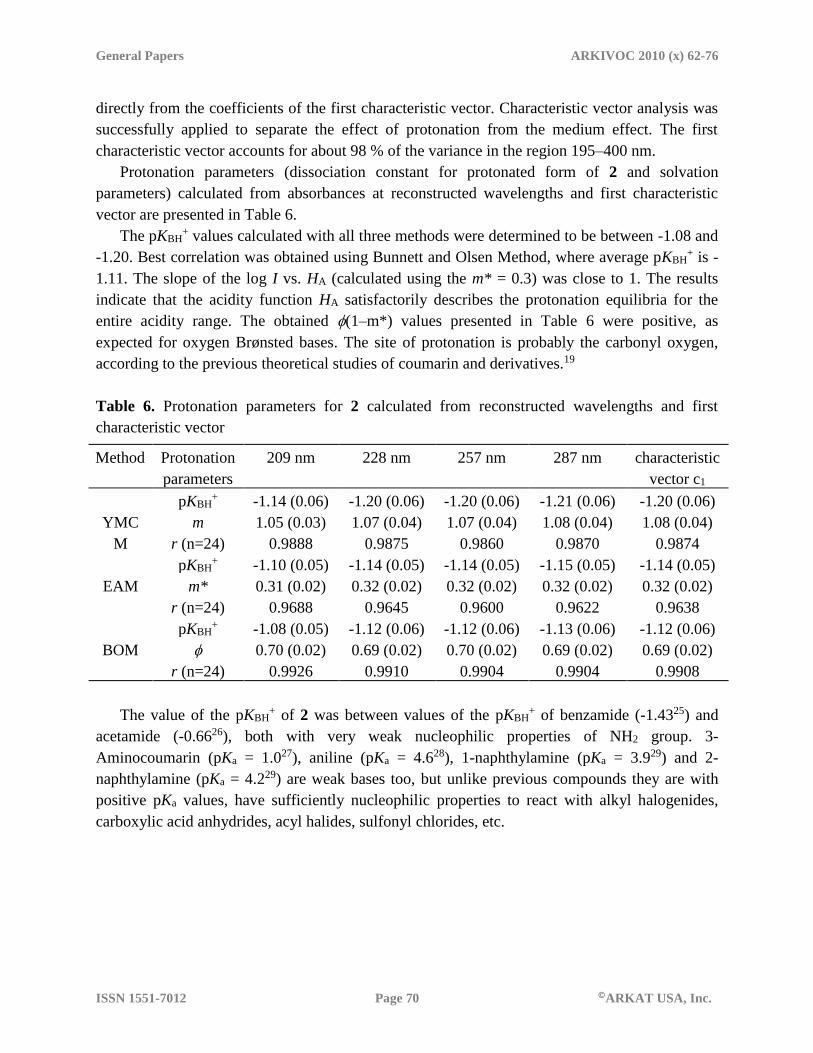
directly from the coefficients of the first characteristic vector. Characteristic vector analysis was
successfully applied to separate the effect of protonation from the medium effect. The first
characteristic vector accounts for about 98 % of the variance in the region 195–400 nm.
Protonation parameters (dissociation constant for protonated form of 2 and solvation
parameters) calculated from absorbances at reconstructed wavelengths and first characteristic
vector are presented in Table 6.
The pKBH+ values calculated with all three methods were determined to be between -1.08 and
-1.20. Best correlation was obtained using Bunnett and Olsen Method, where average pKBH+ is -
1.11. The slope of the log I vs. HA (calculated using the m* = 0.3) was close to 1. The results
indicate that the acidity function HA satisfactorily describes the protonation equilibria for the
entire acidity range. The obtained (1–m*) values presented in Table 6 were positive, as
expected for oxygen Brønsted bases. The site of protonation is probably the carbonyl oxygen,
according to the previous theoretical studies of coumarin and derivatives.19
Table 6. Protonation parameters for 2 calculated from reconstructed wavelengths and first
characteristic vector
Method Protonation
parameters
209 nm 228 nm 257 nm 287 nm characteristic
vector c1
YMC
M
pKBH+ -1.14 (0.06) -1.20 (0.06) -1.20 (0.06) -1.21 (0.06) -1.20 (0.06)
m 1.05 (0.03) 1.07 (0.04) 1.07 (0.04) 1.08 (0.04) 1.08 (0.04)
r (n=24) 0.9888 0.9875 0.9860 0.9870 0.9874
EAM
pKBH+ -1.10 (0.05) -1.14 (0.05) -1.14 (0.05) -1.15 (0.05) -1.14 (0.05)
m* 0.31 (0.02) 0.32 (0.02) 0.32 (0.02) 0.32 (0.02) 0.32 (0.02)
r (n=24) 0.9688 0.9645 0.9600 0.9622 0.9638
BOM
pKBH+ -1.08 (0.05) -1.12 (0.06) -1.12 (0.06) -1.13 (0.06) -1.12 (0.06)
0.70 (0.02) 0.69 (0.02) 0.70 (0.02) 0.69 (0.02) 0.69 (0.02)
r (n=24) 0.9926 0.9910 0.9904 0.9904 0.9908
The value of the pKBH+ of 2 was between values of the pKBH
+ of benzamide (-1.4325) and
acetamide (-0.6626), both with very weak nucleophilic properties of NH2 group. 3-
Aminocoumarin (pKa = 1.027), aniline (pKa = 4.628), 1-naphthylamine (pKa = 3.929) and 2-
naphthylamine (pKa = 4.229) are weak bases too, but unlike previous compounds they are with
positive pKa values, have sufficiently nucleophilic properties to react with alkyl halogenides,
carboxylic acid anhydrides, acyl halides, sulfonyl chlorides, etc.

General Papers ARKIVOC 2010 (x) 62-76
ISSN 1551-7012 Page 71 ARKAT USA, Inc.
Conclusions
The structure of 2 was determined with single crystal X-ray analysis. The full data from NMR
and IR spectra were reported for the first time. The calculated values with quantum chemical
methods were in accordance with the experimental. The C7-N1 bond distance indicates a
considerable degree of double bond character. The conjugation of NH2 is additionally confirmed
by the infrared spectra. This is most probably the reason for very low nucleophility of nitrogen
atom in molecule of 2. Moreover, the values of pKBH+ correlate with the week nucleophilic
properties of 2.
Considering the above mentioned and our experience in performing reactions with 2, an
attempt to synthesize N-substituted 4-aminocoumarins from 2 would be bad strategy.
Experimental Section
General. Ammonium acetate and 4-hydroxycoumarin were purchased commercially and used
without further purification. Melting points were determined on a Reichert hot-stage apparatus.
The crystallographic analysis was carried out on an Enraf–Nonius CAD4 diffractometer, using
graphite monochromated Mo-Kα radiation (λ = 0.71073 Å) at room temperature. An orange
irregular block was cut off from a large crystal and selected for measurement of the intensities.
Unit cell parameters were determined from centering 22 reflections in the θ range (17.94–19.58)º
and refined by the least-squares method. Maximum 2θ was 27.98º and scan mode: ω/2θ. Three
standard reflections were monitored every 500 reflections during data collection and no
significant intensity decay was observed. All diffracted intensities were corrected for Lorentz
and polarization effects.30 No absorption correction was employed. After merging of equivalent
reflections, 1836 independent reflections were obtained (Rint = 0.0896), which were used for the
solution and refinement of the structure. The structure was solved by direct methods and was
refined by the full-matrix least-squares method using SHELXS97 and SHELXL97 computer
programs,31 respectively, in the space group C 2/c (no. 15). All non-hydrogen atoms were refined
with anisotropic displacement parameters. H atoms were placed at idealized positions using
standard geometric criteria. The final refinement of the structure converged to the final indices
R1 = 0.0578 and wR2 = 0.1538 for 1836 reflections with [I > 2σ(I)]. The ORTEP program32 was
used to generate the ellipsoid plot and the figures involving H-bonds and packing were drawn
using Mercury.33 Further relevant crystallographic data are summarized in Table 7.
The FTIR spectra (4000-400 cm-1) were recorded at ambient temperature as KBr pellets on
Perkin-Elmer System 2000.
The NMR spectra were run on a Bruker DRX 250 spectrometer using standard Bruker software
in solvent DMSO-d6. The residual solvent signal was used as an internal standard for the 1H ( =
2.5 ppm) and 13C ( = 39.5 ppm) NMR spectra.

General Papers ARKIVOC 2010 (x) 62-76
ISSN 1551-7012 Page 72 ARKAT USA, Inc.
The UV spectra of the 2 (altogether 26 samples) and appropriate blanks were recorded in a range
of sulfuric acid solutions from 0.0 to 15.0 mol dm−3. The UV spectra were recorded, immediately
after preparing the solutions, on a Varian Cary 50 Spectrophotometer in 1 cm quartz cell, with
resolution of 1 nm, in the range from 190 to 400 nm, at room temperature. Since the absorption
spectra are affected by medium effects (well-defined isosbestic points have not been observed),
corrections were made by means of the characteristic vector analysis using the developed
procedure in MathCad environment, on preprocessed experimental data (all normalized to unit
area under the curves).
Table 7. Crystal data and structure refinement for 4-aminocoumarin
Crystal formula C9H7NO2
Formula weight 161.16
Crystal dimensions (mm) 0.20x0.18x0.14
Temp (K) 290(2)
Crystal system Monoclinic
Space group C 2/c
a (Ǻ) 10.830(4)
b (Ǻ) 10.758(4)
c (Ǻ) 13.213(5)
α (º) 90.0
β (º) 93.12(2)
γ (º) 90.0
V (Ǻ3) 1537.1(1)
Z; Dcalc, (g m-3) 8; 1.393
F(000) 672
Range of θ (º) 2.7-27.98
μ (MoKα) (mm-1) 0.10
Reflections collected 3596
Independent reflections 1836 (Rint = 0.0896)
Absorption correction none
R[F2 > 2σ(F2)]/Rw (F2 ) 0.0578/0.1538
GOF 0.94
Final shift 0.000
(Δρ)min, (Δρ)max, (e Ȧ-3) -0.21/0.16
The ab initio restricted Hartree–Fock (RHF) method and DFT are used to obtain equilibrium
geometry of 4-aminocoumarin molecule. All calculations have been performed with the standard
GAUSSIAN software (AIX, version 1998).34 DFT method, employed in the present study is
B3LYP—Becke’s three parameter hybrid method35 using the correlation functional of Lee, Yang
and Parr.36 The standard 6-31+G** basis set was applied in all calculations. The local minimum

General Papers ARKIVOC 2010 (x) 62-76
ISSN 1551-7012 Page 73 ARKAT USA, Inc.
was verified by establishing that the matrix of the energy second derivatives (Hessian) has no
negative eigenvalues. The theoretical vibrational spectra were interpreted by means of potential
energy distributions (PEDs) using VEDA 4 program.18 For a better correspondence between
experimental and calculated values, we modified the results using the empirical scaling factors.17
1H and 13C NMR chemical shifts were calculated by using the GIAO method37 at the HF/6-
311+G** level of theory (reference compound TMS was calculated at the same level); a solvent
was not considered.
4-Aminocoumarin (2). A mixture of well powdered 4-hydroxycoumarin (1,07 g, 0,0066 mol)
and ammonium acetate (7,87 g, 0,1 mol) was melted in an oil bath (max. 130 ºС). Liquid mixture
was stirred 3 hours and was left to cool to ambient temperature. At the cooled mixture, water was
added. Crude product (max. 74 %) was isolated as yellow crystals by simple filtration. First
purification was made by dissolving the crystals in ethanol and precipitation with water. Melting
point 226-228 oC (from ethanol). Various melting points were found in the literature, 161.5-162
ºС,38 199 ºС,13a 232-234 ºС,12a,39 241-243 ºС.40 Anal.Calcd. for C9H7NO2: C, 67.07%; H, 4.38%;
N, 8.69%. Found: C, 66.94%; H, 4.61%; N, 8.61%.
Reactions of 2 with 3 (Scheme 1) were performed in different solvents (H2O, dioxane,
tetrahydrofuran, pyridine, DMSO and DMF), without and with presence of triethylamine,
Na2CO3 or pyridine. Also, same reactions were performed at room temperature and by heating
the reaction mixture. In addition, two reactions were performed by refluxing the solution of 2 in
acetic anhydride or ethyl acetate.
Supplementary material
CCDC 754032 contains the supplementary crystallographic data for this paper. These data can be
obtained free of charge via www.ccdc.cam.ac.uk/conts/retrieving.html (or from the Cambridge
Crystallographic Data Centre, 12, Union Road, Cambridge CB2 1EZ, UK; fax: +44 1223
336033).
Acknowledgements
This work was supported by the Bulgarian National Science Fund (Contract BM-02/07) and
Macedonian Ministry of Education and Science (Contract 03-1586).

General Papers ARKIVOC 2010 (x) 62-76
ISSN 1551-7012 Page 74 ARKAT USA, Inc.
References
1. (a) Mandhane, P. G.; Joshi, R. S.; Ghawalkar, A. R.; Jadhav, G. R.; Gill, C. H. Bull. Korean
Chem. Soc. 2009, 30(12), 2969. (b) Matloubi Moghaddam, F.; Mirjafary, Z.; Saeidian, H.
Scientica Iranica, Transactions C: Chemistry and Chemical Engineering 2009, 16, 12. (c)
Shaabani, A.; Ghadari, R.; Rahmati, A.; Rezayan, A. H. J. Iran. Chem. Soc. 2009, 6, 710. (d)
Chavana, F.; Madjea, B.; Bharada, J.; Ubalea, M.; Wareb, M.; Shingareb, M.; Shindec, N.
Bulletin of the Ctalysis Society of India 2008, 7, 41. (e) Kumar, S.; Saini, A.; Sandhu, J. S.
Arkivoc 2007, (xv), 18. (f) Rajitha, B.; Naveen Kumar, V.; Someshwar, P.; Venu Madhav, J.;
Narsimha Reddy, P.; Thirupathi Reddy, Y. Arkivoc 2006, (xii), 23.
2. (a) Naveen Kumar, V.; Thirupathi Reddy, Y.; Narasimhareddy, P.; Rajithaa, B.; De Clercq,
E. Arkivoc 2006, (xv), 181. (b) Satyanarayana, V. S. V.; Sreevani, P.; Sivakumar, A.;
Vijayakumar, V. Arkivoc 2008, (xvii), 22. (c) Rajendra Prasad, Y.; Ravi Kumar, P.; Jesse
Smiles, D.; Ajay Babu, P. Arkivoc 2008, (xi), 266. (d) Kaswala, P. B.; Chikhalia, K. H.;
Shah, N. K.; Patel, D. P.; Patel, D. H.; Mudaliar G. V. Arkivoc 2009, (xi), 326. (e)
Manojkumar, P.; Kochupappy Ravi, T.; Subbuchettiar, G. Acta Pharm. 2009, 59, 159.
3. (a) Heide, L. Methods Enzymol. 2009, 459, 437. (b) Anderle, C.; Stieger, M.; Burrell, M.;
Reinelt, S.; Maxwell, A.; Page, M.; Heide L. Antimicrob. Agents Chemother. 2008, 52,1982.
(c) Anderle, C.; Li, S.; Kammerer, B.; Gust, B.; Heide, L. J. Antibiot. 2007, 60, 504. (d)
Fridman, M.; Balibar, C. J.; Lupoli, T.; Kahne, D.; Walsh, C. T.; Garneau-Tsodikova S.
Biochemistry 2007, 46(28), 8462. (e) Kudale, A. A.; Kendall, J; Warford, C. C.; Wilkins, N.
D.; Bodwell, G. J. Tetrahedron Lett. 2007, 48, 5077. (f) Li, S.; Heide, L. Curr. Med. Chem.
2005, 12, 419. (g) Freitag, A.; Galm, U.; Li, S.; Heide, L. J. Antibiot. 2004, 57, 205. (h) Xu,
H.; Heide, L.; Li, S. Chem. Biol. 2004, 11, 655.
4. Maddi, V.; Mamledesai, S. N.; Satyanarayana, D.; Swamy, S. Indian J. Pharm. Sci. 2007, 69,
847.
5. Al-Haiza, M. A.; Mostafa, M. S.; El-Kady, M. Y. Scientific Journal of King Faisal
University (Basic and Applied Sciences) 2005, 6, 1426.
6. (a) Bhojya Naik, R.; Bhojya Naik, H. S.; Harish Kumar, H. N.; Hosamani, K. M.;
Mahadevan, K. M. Arkivoc 2009, (ii), 11. (b) Wagner, B. D. Molecules 2009, 14, 210. (c)
Choi, J. Y.; Park, E. J.; Chang, S. H.; Kang, T. J. Bull. Korean Chem. Soc. 2009, 30, 1452.
7. (a) Li, J.; Yao, S. Q. Org. Lett. 2009, 11, 405. (b) Lewis, E. K.; Haaland, W. C.; Nguyen, F.;
Heller, D. A.; Allen, M. J.; MacGregor, R. R.; Berger, C. S.; Willingham, B.; Burns, L. A.;
Scott, G. B. I.; Kittrell, C.; Johnson, B. R.; Curl, R. F.; Metzker, M. L. PNAS 2005, 102,
5346.
8. Kokotos, G.; Theodorou, V.; TzougrakiDieter, C.; Deforce, L. D.; Van den Eeckhout, E. G.
Bioorg. Med. Chem. Lett. 1998, 7, 2165.
9. (a) Di Braccio, M.; Grossi, G.; Roma, G.; Marzano, C.; Bacccichetti, F.; Simonato, M.;
Bordin, F. Farmaco 2003, 58, 1083. (b) Roma, G.; Di Braccio, M.; Carrieri, A.; Grossi, G.;
Leoncini, G.; Signorello, M. G.; Carotti, A. Biorg. Med. Chem. 2003, 11, 123. (c) El-Saghier,

General Papers ARKIVOC 2010 (x) 62-76
ISSN 1551-7012 Page 75 ARKAT USA, Inc.
A. M. M.; Naili, M. B.; Rammash, B. Kh.; Saleh, N. A.; Kreddanc, K. M. Arkivoc 2007,
(xvi), 83.
10. (a) Majumdar, K. C.; Bhattacharyya, T. Tetrahedron Lett. 2001, 42, 4231. (b) Jacquot, Y.;
Laios, I.; Cleeren, A.; Nonclercq, D.; Bermont, L.; Refouvelet, B.; Boubekeur, K.; Xicluna,
A.; Leclercq, G.; Laurent, G. Biorg. Med. Chem. 2007, 15, 2269. (c) Lai., J.; Kuo, P.; Gau,
Y.; Yang, D. Tetrahedron Lett. 2007, 48, 7796. (d) Giraud, A.; Vanelle, P.; Giraud, L.
Tetrahedron Lett. 1999, 40, 4321. (e) Papoutsis, I.; Spyroudis, S.; Varvoglis, A.; Callies, J.
A.; Zhdankin, V. V. Tetrahedron Lett. 1997, 38, 8401. (f) Ivanov, I. C.; Karagiosov, S. K.
Synthesis 1995, 633.
11. (a) Kantlehner, W.; Vettel, M.; Lehmann, H.; Edelmann, K.; Stieglitz, R.; Ivanov, I. C. J.
Prakt. Chem. 1998, 340, 408. (b) Shepard, M. S.; Carreira, E. M. Tetrahedron 1997, 53,
16253. (c) Connor, D. T.; Young, P. A.; von Strandimann, M. J. Heterocycl. Chem. 1981, 18,
697.
12. (a) Ivanov, I. C.; Karagiosov, S. K.; Manolov, I. Arch. Pharm. (Weinheim) 1991, 324, 61. (b)
Zagorevskii, V. A.; Savellev, V. L.; Meshcheryakova, L. M. Chem. Heterocycl. Comp. 1970,
6, 944.
13. (a) Chavan, A. P. J. Chem. Res. 2006, 179. (b) Stoyanov, E. V.; Ivanov, I. C. Molecules
2004, 9, 627. (c) Liao, Y.; Kuo, P.; Yang, D. Tetrahedron Lett. 2003, 44, 1599.
14. Chopra, D.; Venugopala, K. N.; Rao G. K.; Guru, T. N. Row, Acta Cryst. E. 2007, 63, 2826.
15. Allen, F. H.; Kennard, O.; Watson, D. G.; Brammer, L.; Orpen, A. G. Tables, J. Chem. Soc.
Perkin Trans. II 1987, S1.
16. Spectral Database for Organic Compounds, www.aist.go.jp
17. Merrick, J. P.; Moran, D.; Radom, L. J. Phys. Chem. A 2007, 111, 11683.
18. Michal, H. J. Vibrational Energy Distribution Analysis, VEDA 4, Warsaw, 2004.
19. Sokolova, I. V.; Loboda, L. I. Struct. Chem. 1983, 23, 848.
20. Kirpichënok, M. A.; Baukulev, V. M.; Karandashova, L. A.; Grandberg, I. I. J. Chem.
Heterocycl. Comp. 1991, 27, 1193.
21. Sizova, Z. A.; Karasev, A. A.; Lukatskaya, L. L.; Rubtsov, M. I.; Doroshenko, A. O. Theor.
Exp. Chem. 2002, 38, 168.
22. Yates, K.; McClelland, R. A. J. Am. Chem. Soc. 1967, 89, 2686.
23. Bunnet, J. F.; Olsen, F. P. Can. J. Chem. 1966, 44, 1899.
24. Cox, R. A.; Yates, K. J. Am. Chem. Soc. 1978, 100, 3861.
25. Stojković, G.; Popovski, E.; J. Serb. Chem. Soc. 2006, 71, 1061.
26. Bagno, A.; Lovato, G.; Scorrano, J. Chem. Soc. Perkin Trans. 2 1993, 1091.
27. Subba Rao, R. V.; Krishnamurthy, M.; Dogra, S. K. Journal of Photochemistry, 1986, 34, 55.
28. Bordwell, F. G. Acc. Chem. Res. 1988, 21, 456.
29. Montgomery, J. H. Groundwater Chemicals Desk Reference, 4th Edn.; CRC Press, Taylor &
Francis Group: Boca Raton, 2007; pp 783-785.
30. CAD-4 EXPRESS. Version 5.1/1.2. Enraf Nonius, Delft, the Netherlands.
31. Sheldrick, G. M. Acta Cryst. A. 2008, 64, 112

General Papers ARKIVOC 2010 (x) 62-76
ISSN 1551-7012 Page 76 ARKAT USA, Inc.
32. Farrugia, J. L. J. Appl. Cryst. 1997, 30, 565.
33. Macrae, C. F.; Edgington, P. R.; McCabe, P.; Pidcock, E.; Shields, G. P.; Taylor, R.; Towler,
M.; van de Streek, J. J. Appl. Cryst. 2006, 39, 453.
34. Frisch, M. J.; Trucks, G. W.; Schlegel, H. B.; Scuseria, G. E.; Robb, M. A.; Cheeseman, J.
R.; Zakrzewski, V. G.; Montgomery, Jr., J. A.; Stratmann, R. E.; Burant, J. C.; Dapprich, S.;
Millam, J. M.; Daniels, A. D.; Kudin, K. N.; Strain, M. C.; Farkas, O.; Tomasi, J.; Barone,
V.; Cossi, M.; Cammi, R.; Mennucci, B.; Pomelli, C.; Adamo, C.; Clifford, S.; Ochterski, J.;
Petersson, G. A.; Ayala, P. Y.; Cui, Q.; Morokuma, K.; Malick, D. K.; Rabuck, A. D.;
Raghavachari, K.; Foresman, J. B.; Cioslowski, J.; Ortiz, J. V.; Baboul, A. G.; Stefanov, B.
B.; Liu, G.; Liashenko, A.; Piskorz, P.; Komaromi, I.; Gomperts, R.; Martin, R. L.; Fox, D.
J.; Keith, T.; Al-Laham, M. A.; Peng, C. Y.; Nanayakkara, A.; Gonzalez, C.; Challacombe,
M.; Gill, P. M. W.; Johnson, B.; Chen, W.; Wong, M. W.; Andres, J. L.; Head-Gordon, M.;
Replogle, E. S.; Pople, J. A. Gaussian 98, Revision A.7, Gaussian, Inc., Pittsburgh PA, 1998.
35. Becke, A. D. Phys. Rev. A 1988, 38, 3098.
36. Lee, C.; Yang, W.; Parr, R. G. Phys. Rev. B 1988, 37, 785.
37. Ditchfield, R. Mol. Phys. 1974, 27, 789.
38. Zagorevskii, V. A.; Dudykina, N. V. Zh. Obsch. Khim. 1962, 32, 2384.
39. Manolov, I.; Danchev, N. D. Eur. J. Med. Chen. Chim. Ther. 1995, 30, 531.
40. Uno, H.; Kurokawa, M.; Nishimura, H. Chem. Pharm. Bull. 1976, 24, 644.