experimental infection with brazilian newcastle disease ... · infection with brazilian newcastle...
TRANSCRIPT

V
Ed
APa
b
(
a
A
R
A
A
J
K
G
C
E
R
S
I
Necvs
h1B
b r a z i l i a n j o u r n a l o f m i c r o b i o l o g y 4 7 (2 0 1 6) 231–242
h t tp : / /www.bjmicrobio l .com.br /
eterinary Microbiology
xperimental infection with Brazilian Newcastleisease virus strain in pigeons and chickens
driano de Oliveira Torres Carrascoa,∗, Meire Christina Sekia, Jyan Lucas Benevenutea,riscila Ikedaa, Aramis Augusto Pintob
Departamento de Medicina Veterinária, Universidade Estadual do Centro-Oeste (UNICENTRO), Guarapuava, PR, BrazilDepartamento de Patologia Veterinária, Faculdade de Ciências Agrárias e Veterinárias da Universidade Estadual Paulista
FCAV/UNESP), Jaboticabal, SP, Brazil
r t i c l e i n f o
rticle history:
eceived 19 May 2015
ccepted 29 May 2015
ssociate Editor: João Pessoa Araújo
unior
eywords:
allus gallus
olumba livia
xperimental infection
T-PCR
erology
a b s t r a c t
This study was designed with the goal of adding as much information as possible about
the role of pigeons (Columba livia) and chickens (Gallus gallus) in Newcastle disease virus
epidemiology. These species were submitted to direct experimental infection with New-
castle disease virus to evaluate interspecies transmission and virus-host relationships. The
results obtained in four experimental models were analyzed by hemagglutination inhibi-
tion and reverse transcriptase polymerase chain reaction for detection of virus shedding.
These techniques revealed that both avian species, when previously immunized with a
low pathogenic Newcastle disease virus strain (LaSota), developed high antibody titers that
significantly reduced virus shedding after infection with a highly pathogenic Newcastle dis-
ease virus strain (São Joao do Meriti) and that, in chickens, prevent clinical signs. Infected
pigeons shed the pathogenic strain, which was not detected in sentinel chickens or control
birds. When the presence of Newcastle disease virus was analyzed in tissue samples by
RT-PCR, in both species, the virus was most frequently found in the spleen. The vaccination
regimen can prevent clinical disease in chickens and reduce viral shedding by chickens or
pigeons. Biosecurity measures associated with vaccination programs are crucial to maintain
a virulent Newcastle disease virus-free status in industrial poultry in Brazil.
© 2015 Sociedade Brasileira de Microbiologia. Published by Elsevier Editora Ltda. This is
ntroduction
ewcastle disease (ND) is an acute, highly contagious viral dis-ase of poultry that can cause high mortality (up to 100%) in
hicken, the most important natural host of the disease. Theirus can also affect a wide variety of avian species causingevere disease. The disease is regarded as endemic in many∗ Corresponding author.E-mail: [email protected] (A.d.O.T. Carrasco).
ttp://dx.doi.org/10.1016/j.bjm.2015.07.001517-8382/© 2015 Sociedade Brasileira de Microbiologia. Published by EY-NC-ND license (http://creativecommons.org/licenses/by-nc-nd/4.0/)
an open access article under the CC BY-NC-ND license
(http://creativecommons.org/licenses/by-nc-nd/4.0/).
countries, and is caused by an avian Paramyxovirus type 1(APMV 1), a member of the genus Avulavirus, from the Paramyx-oviridae family.1 As demonstrated in intensive surveys, nearly236 free-living species from 27 of the 50 orders of birds havebeen reported to be susceptible to either natural or experi-
mental infection with ND.2 On several occasions, Newcastledisease virus (NDV) was isolated from wildlife birds,3 and mostoutbreaks of NDV arise in unvaccinated susceptible animals.4lsevier Editora Ltda. This is an open access article under the CC.

i c r o
232 b r a z i l i a n j o u r n a l o f mTo keep ND under control, large-scale prophylactic vaccina-tion is used in most member states of the European Unionand elsewhere in the world.4,5 Although vaccination in generalprovides good protection against clinical disease and mor-tality, it may not provide sufficient protection against virustransmission to prevent ND outbreaks.6,7 The most commonstrains used worldwide in vaccination are LaSota and B1.5
Conventional vaccine strains may protect against clinical dis-ease caused by virulent NDV, but viral infection can still occurin vaccinated birds,8,9 which may be the route of virus spreadin poultry facilities farms.10 However, vaccinated birds mayshow significantly reduction in virus shedding compared withunvaccinated ones.9,10
The susceptibility of pigeons (Columba livia) and othermembers of the Columbidae family to NDV has been reportedby several authors.11–15 It is now clear that the disease occursin pigeons as a result of virus dissemination from affectedchicken flocks, and it occurs in poultry flocks when the virusis disseminated from domesticated or feral pigeons.16 Thesource of ND infection to chicken flocks may be food contam-inated with feces of feral pigeons infected with NDV.16,17
Many aspects of ND infection in pigeons are unclear, andexperimental infection could answer many questions aboutNDV epidemiology, such as virus pathogenicity, infectivity,and shedding. As previously described by experimental stud-ies, adult pigeons infected via eye drops with a chickenpathogenic APMV-1 strain shed the virus both through thecloaca and the mouth for up to 21 days post-infection (dpi).11
Pigeon Paramyxovirus type 1 (PPMV-1) isolates may be trans-mitted from infected pigeons to chickens that were in contactwith them, with replication in the chickens (as demonstratedby the excretion of the virus by cloacal route), and antibodyresponse against the virus.16 In experimental infections con-ducted with a PPMV-1 strain, mortality rates greater than70% were observed in pigeons, but no infected chicken died.In spite of these findings, there are few comparative stud-ies on pigeons and chickens infected with the same PPMV-1strain, making it difficult to determine the significance of theseresults.15
Thus, little is known about the course of the infec-tion, the significance of humoral antibody response, viralshedding, clinical signs, and contact transmission of a Brazil-ian pathogenic NDV strain between pigeons and chickens(experimental infection). To some extent, viral replicationcomplex may play a role in the pigeon-to-chicken transmis-sion, although further studies are needed to investigate thefactors that are determinant for interspecies transmission.15
Some studies were carried out to evaluate the prevalence ofNewcastle disease in commercial birds in poultry-producingareas in Brazil,18,19 and the results showed that industrialpoultry produced in the nine Brazilian states analyzed wasfree of Newcastle disease.18
The nationwide efficiency of poultry production makesBrazil a competitive nation in international markets, evenin the absence of economic subsidies. In order to guaranteebetter sanitary conditions for Brazilian avian products, the
National Program for Poultry Health (PNSA) was implementedfor the control of Newcastle disease in the country.19Considering the potential risk of contamination of poul-try species by pigeons carrying NDV, it is important to
b i o l o g y 4 7 (2 0 1 6) 231–242
study the pathogenesis of the disease both in pigeons andin chickens. This study was designed to evaluate humoralimmune response, viral shedding, and contact transmissionafter experimental infection of pigeons and chickens with apathogenic NDV isolate of chicken origin under experimentaland controlled conditions.
Materials and methods
Birds
In an attempt to reproduce natural conditions of NDV trans-mission, a total of fifty-two free-living adult domestic pigeons(C. livia) were used in this study. Animals were clinicallyhealthy, had non-specific levels of hemagglutination inhi-bition (HI) antibodies (HI Titers ≤ 2), and were negative inreverse transcriptase polymerase chain reaction (RT-PCR) forthe presence of NDV in cloacal swab samples. After capture,pigeons were housed in adequate facilities (away from chickenfacilities) for 90 days to be adapted to captivity. Similarly,twenty-nine 90-day-old, SPF (specific pathogen free) chickenswere used in the study. The two species were kept in separatefacilities until the beginning of the experimental study.
On the day of experimental infection, pigeons and chickensdivided in four experimental groups were moved to NegativePressure Isolators (Alesco®, Brazil), under controlled labora-tory and biosafety conditions.
All animal procedures were performed according to theEthical Principles in Animal Research adopted by the BrazilianCollege of Animal Experimentation, and to the 2000 Report ofthe AVMA Panel on Euthanasia.20
Viruses
The experimental infection was performed using the Sao Joaodo Meriti (SJM) strain (Gene Bank Number: EF534701), a highlypathogenic NDV strain (APMV-1) for chickens (mean deathtime in chicken embryos = 48 h; ICPI in day-old chicken = 1.78).The virus was propagated twice in the allantoic cavity of 9to 10-day-old embryonated SPF eggs by inoculation of 0.1 mLof infectious allantoic fluid. A virus stock was produced byharvesting allantoic fluid from chicken embryos, freezing itat −70 ◦C, and storing it. This virus stock titer was 109.0
median embryo lethal dose/mL (ELD50), determined on daythree before experimental infection. All virus dilutions werecarried out with sterile phosphate buffered saline (PBS), pH7.2.
Live vaccines containing LaSota (LS) NDV strain (New Vac-LS – Fort Dodge Saúde Animal Ltda®, Brazil) were used in thevaccination procedures. This strain is commercially used inthe immunization of chickens in Brazil.
Experimental infection and sampling
Pigeons and chickens were randomly divided into four groups,
as described below. After vaccination/experimental infection,birds were monitored daily for presence of any clinical signs,such as diarrhea, torticollis, incoordination, apathy, tremors,ocular and nasal discharge, abnormal posture, and flying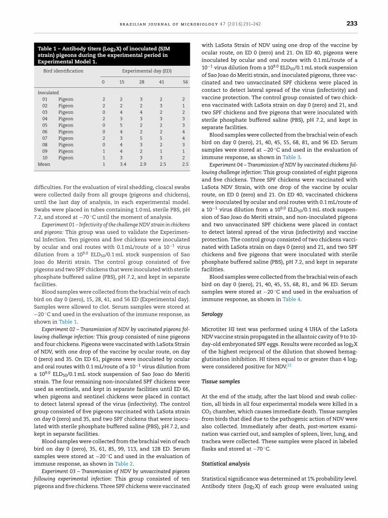
b r a z i l i a n j o u r n a l o f m i c r o b i
Table 1 – Antibody titers (Log2X) of inoculated (SJMstrain) pigeons during the experimental period inExperimental Model 1.
Bird identification Experimental day (ED)
0 15 28 41 56
Inoculated01 Pigeon 2 2 3 2 202 Pigeon 2 2 2 3 103 Pigeon 0 4 4 2 204 Pigeon 2 3 3 3 305 Pigeon 0 5 2 2 306 Pigeon 0 4 2 2 407 Pigeon 2 3 5 5 408 Pigeon 0 4 3 2 309 Pigeon 1 4 2 1 110 Pigeon 1 3 3 3 2
dwuS7
atbdJppf
bS−s
lao0aasuwtgolk
bsi
fp
Statistical analysis
Mean 1 3.4 2.9 2.5 2.5
ifficulties. For the evaluation of viral shedding, cloacal swabsere collected daily from all groups (pigeons and chickens),ntil the last day of analysis, in each experimental model.wabs were placed in tubes containing 1.0 mL sterile PBS, pH.2, and stored at −70 ◦C until the moment of analysis.
Experiment 01 – Infectivity of the challenge NDV strain in chickensnd pigeons: This group was used to validate the Experimen-al Infection. Ten pigeons and five chickens were inoculatedy ocular and oral routes with 0.1 mL/route of a 10−1 virusilution from a 109.0 ELD50/0.1 mL stock suspension of Sao
oao do Meriti strain. The control group consisted of fiveigeons and two SPF chickens that were inoculated with sterilehosphate buffered saline (PBS), pH 7.2, and kept in separateacilities.
Blood samples were collected from the brachial vein of eachird on day 0 (zero), 15, 28, 41, and 56 ED (Experimental day).amples were allowed to clot. Serum samples were stored at20 ◦C and used in the evaluation of the immune response, as
hown in Table 1.Experiment 02 – Transmission of NDV by vaccinated pigeons fol-
owing challenge infection: This group consisted of nine pigeonsnd four chickens. Pigeons were vaccinated with LaSota Strainf NDV, with one drop of the vaccine by ocular route, on day
(zero) and 35. On ED 61, pigeons were inoculated by ocularnd oral routes with 0.1 mL/route of a 10−1 virus dilution from
109.0 ELD50/0.1 mL stock suspension of Sao Joao do Merititrain. The four remaining non-inoculated SPF chickens weresed as sentinels, and kept in separate facilities until ED 66,hen pigeons and sentinel chickens were placed in contact
o detect lateral spread of the virus (infectivity). The controlroup consisted of five pigeons vaccinated with LaSota strainn day 0 (zero) and 35, and two SPF chickens that were inocu-
ated with sterile phosphate buffered saline (PBS), pH 7.2, andept in separate facilities.
Blood samples were collected from the brachial vein of eachird on day 0 (zero), 35, 61, 85, 99, 113, and 128 ED. Serumamples were stored at −20 ◦C and used in the evaluation ofmmune response, as shown in Table 2.
Experiment 03 – Transmission of NDV by unvaccinated pigeonsollowing experimental infection: This group consisted of tenigeons and five chickens. Three SPF chickens were vaccinated
o l o g y 4 7 (2 0 1 6) 231–242 233
with LaSota Strain of NDV using one drop of the vaccine byocular route, on ED 0 (zero) and 21. On ED 40, pigeons wereinoculated by ocular and oral routes with 0.1 mL/route of a10−1 virus dilution from a 109.0 ELD50/0.1 mL stock suspensionof Sao Joao do Meriti strain, and inoculated pigeons, three vac-cinated and two unvaccinated SPF chickens were placed incontact to detect lateral spread of the virus (infectivity) andvaccine protection. The control group consisted of two chick-ens vaccinated with LaSota strain on day 0 (zero) and 21, andtwo SPF chickens and five pigeons that were inoculated withsterile phosphate buffered saline (PBS), pH 7.2, and kept inseparate facilities.
Blood samples were collected from the brachial vein of eachbird on day 0 (zero), 21, 40, 45, 55, 68, 81, and 96 ED. Serumsamples were stored at −20 ◦C and used in the evaluation ofimmune response, as shown in Table 3.
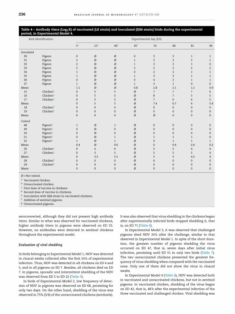
Experiment 04 – Transmission of NDV by vaccinated chickens fol-lowing challenge infection: This group consisted of eight pigeonsand five chickens. Three SPF chickens were vaccinated withLaSota NDV Strain, with one drop of the vaccine by ocularroute, on ED 0 (zero) and 21. On ED 40, vaccinated chickenswere inoculated by ocular and oral routes with 0.1 mL/route ofa 10−1 virus dilution from a 109.0 ELD50/0.1 mL stock suspen-sion of Sao Joao do Meriti strain, and non-inoculated pigeonsand two unvaccinated SPF chickens were placed in contactto detect lateral spread of the virus (infectivity) and vaccineprotection. The control group consisted of two chickens vacci-nated with LaSota strain on days 0 (zero) and 21, and two SPFchickens and five pigeons that were inoculated with sterilephosphate buffered saline (PBS), pH 7.2, and kept in separatefacilities.
Blood samples were collected from the brachial vein of eachbird on day 0 (zero), 21, 40, 45, 55, 68, 81, and 96 ED. Serumsamples were stored at −20 ◦C and used in the evaluation ofimmune response, as shown in Table 4.
Serology
Microtiter HI test was performed using 4 UHA of the LaSotaNDV vaccine strain propagated in the allantoic cavity of 9 to 10-day-old embryonated SPF eggs. Results were recorded as log2Xof the highest reciprocal of the dilution that showed hemag-glutination inhibition. HI titers equal to or greater than 4 log2
were considered positive for NDV.21
Tissue samples
At the end of the study, after the last blood and swab collec-tion, all birds in all four experimental models were killed in aCO2 chamber, which causes immediate death. Tissue samplesfrom birds that died due to the pathogenic action of NDV werealso collected. Immediately after death, post-mortem exami-nation was carried out, and samples of spleen, liver, lung, andtrachea were collected. These samples were placed in labeledflasks and stored at −70 ◦C.
Statistical significance was determined at 1% probability level.Antibody titers (log2X) of each group were evaluated using

234 b r a z i l i a n j o u r n a l o f m i c r o b i o l o g y 4 7 (2 0 1 6) 231–242
Table 2 – Antibody titers (Log2X) of vaccinated (LS strain) and inoculated (SJM strain) birds during the experimentalperiod, in Experimental Model 2.
Bird identification Experimental Day (ED)
0a 35b 61c 66d 85 99 113 128
Inoculated11 Pigeon 1 2 4 Ø 5 4 4 412 Pigeon 2 2 2 Ø 3 4 4 213 Pigeon 0 0 4 Ø 3 2 2 214 Pigeon 2 2 4 Ø 6 5 5 415 Pigeon 2 2 4 Ø 5 5 5 416 Pigeon 2 3 5 Ø 6 4 3 317 Pigeon 2 2 4 Ø 4 3 3 318 Pigeon 2 2 4 Ø 7 7 5 719 Pigeon 1 5 3 Ø 5 5 4 3
Mean 1.6 2.3 3.8 Ø 4.9 4.4 3.9 3.66 Chickenf Ø Ø Ø 0 0 0 0 07 Chickenf Ø Ø Ø 1 1 0 1 08 Chickenf Ø Ø Ø 0 1 0 0 09 Chickenf Ø Ø Ø 0 0 0 0 0
Mean Ø Ø Ø 0.25 0.5 0 0.25 0
ControlP 38 Pigeone 1 2 3 Ø 3 2 2 2P 39 Pigeone 0 2 2 Ø 3 2 2 2P 40 Pigeone 0 2 3 Ø 3 3 2 2P 41 Pigeone 1 1 2 Ø 2 1 2 1P 42 Pigeone 0 3 3 Ø 2 3 1 1
Mean 0.4 2 2.6 Ø 2.6 2.2 1.8 1.6C 20 Chickenf Ø Ø Ø 0 0 0 0 0C 21 Chickenf Ø Ø Ø 0 0 0 0 0Mean Ø Ø Ø 0 0 0 0 0
Ø = Not tested.a First dose of vaccine in pigeons.b Second dose of vaccine in pigeons.c Inoculation with SJM strain in vaccinated pigeons.d Addition of sentinel chickens.e Vaccinated pigeon.f Unvaccinated chicken.
the Statistical Analysis Systems − SAS® (Statistical Analy-sis Systems Institute Inc., Cary, NC, USA). To evaluate theinteraction between time and treatment, Tukey–Kramer testwas used.
Viral shedding
This assay was based on the fusion (F) gene of NDV. RNAextraction from cloacal swab samples was performed usingthe QIamp Viral RNA Mini Kit (Qiagen, USA), according to themanufacturer’s protocol. For tissue samples, the Total RNAExtraction Kit (Mini), Real Genomics®, was used according tothe manufacturer’s protocol. The LaSota NDV vaccine strain(New Vac-LS® – Fort Dodge Saúde Animal Ltda) and ultra-purewater were used as positive and negative controls, respec-tively. Controls were submitted to the same procedures carriedout with experimental chickens and pigeons. RT-PCR was per-
formed using primers targeting a conserved region of the NDVgenome (Fusion – F gene), as previously described.22 Theseprimers are targeted to a conserved genome region that isable to amplify any NDV strain, no matter the pathogenicity,yielding a 362-bp fragment,22 and were validated in otherstudies.14,23–25 The primer sequence was as follows: P1F(sense) 5′-TTG ATG GCA GGC CTC TTG C-3′ and P2R (anti-sense) 5′-GGA GGA TGT TGG CAG CAT T-3′. cDNA synthesisand PCR were performed according to Jestin and Jestin.23
Samples were analyzed by electrophoresis on 1.5% agarose(w/v) gels (Invitrogen®, USA) stained with ethidium bromide0.5 �g/mL (Invitrogen®). The electrophoresis run was carriedout at 100 volts/50 min. Positive samples showed a 362-bp DNAfragment under UV light.
For the validation of our results, the genes that encodeprotein F were sequenced from the material shed in thefeces of the animals. These sequences were compared withstandard samples: the pathogenic NDV strain São Joao doMeriti (EF534701), and the LaSota vaccine strain (EF534702).Positive samples in RT-PCR were sequenced using the BigDyeTerminator Kit (Applied Biosystems, Foster City, CA, USA).
®
Results obtained were analyzed with the aid of the BioEditsoftware. The sequences obtained were analyzed in an onlinedatabase,26 confirming nucleotide homology with São Joao doMeriti and LaSota strains.
b r a z i l i a n j o u r n a l o f m i c r o b i o l o g y 4 7 (2 0 1 6) 231–242 235
Table 3 – Antibody titers (Log2X) of vaccinated (LS strain) and inoculated (SJM strain) birds during the experimentalperiod, in Experimental Model 3.
Bird Identification Experimental day (ED)
0c 21d 40e 45f 55 68 81 96
Inoculated20 Pigeon 0 0 Ø Ø 2 3 3 221 Pigeon 2 1 Ø Ø 2 4 3 222 Pigeon 2 1 Ø Ø 3 4 2 223 Pigeon 2 2 Ø Ø 2 2 1 124 Pigeon 1 1 Ø Ø 5 5 3 125 Pigeon 1 0 Ø Ø 4 4 3 226 Pigeon 1 0 Ø Ø 4 4 2 327 Pigeon 1 1 Ø Ø 4 3 3 428 Pigeon 0 0 Ø Ø 3 3 3 329 Pigeon 0 0 Ø Ø 3 3 3 3
Mean 1 0.6 Ø Ø 3.2 3.5 3 2.310 Chickena 0 5 Ø 6 5 5 5 511 Chickena 0 5 Ø 6 5 5 5 512 Chickena 0 4 Ø 4 4 4 3 3
Mean 0 4.7 Ø 5.4 4.7 4.7 4.4 4.413 Chickenb 0 0 Ø 0 0 0 4 414 Chickenb 0 0 Ø 0 0 0 5 4
Mean 0 0 Ø 0 0 0 4.5 4
Control43 Pigeong 1 1 Ø Ø 0 0 0 044 Pigeong 0 0 Ø Ø 0 0 0 045 Pigeong 0 0 Ø Ø 0 0 0 046 Pigeong 0 1 Ø Ø 1 1 0 047 Pigeong 0 1 Ø Ø 1 1 1 1
Mean 0.2 0.6 Ø Ø 0 0.4 0.2 0.222 Chickena 0 4 Ø 6 5 5 4 423 Chickena 0 3 Ø 5 5 5 5 4
Mean 0 3.5 Ø 5.5 5 5 4.5 424 Chickenb 0 0 Ø 0 0 0 0 025 Chickenb 0 0 Ø 0 0 0 0 0
Mean 0 0 0 0 0 0 0 0
Ø = Not tested.a Vaccinated chickens.b Unvaccinated chickens.c First dose of vaccine in chickens.d Second dose of vaccine in chickens.e Inoculation with SJM strain in vaccinated pigeons.f Addition of sentinel chickens.
R
S
Iecwsdn
hew
g Unvaccinated pigeon.
esults
erology
n Experimental Model 1, non-immunized pigeons and chick-ns were experimentally infected. Although pigeons remainedlinically healthy during the whole trial, antibody productionas fast, reaching high HI titers from ED 15 to ED 56, as pre-
ented in Table 1. However, all five chickens died on the 7thay after virus inoculation (ED 7), and blood samples couldot be collected.
In pigeons of Experimental Model 2, vaccination led to
igh antibody titers even before experimental infection. Afterxperimental infection (ED 61), higher titers of HI antibodiesere detected on ED 85, which remained practically stableuntil the end of the study, on ED 128. In sentinel chickensof Experimental Model 2, which were placed in contact withinfected pigeons on ED 66, no seroconversion (Table 2) and noclinical signs compatible with ND were observed.
In Experimental Model 3, at the moment the chickens wereplaced in contact with infected birds (ED 45), vaccinated chick-ens already presented high levels of antibodies, as shownin Table 3. In unvaccinated chickens (sentinels), seroconver-sion was only observed on ED 81, whereas in experimentallyinfected pigeons, high titers of antibodies were detected fromED 55 on, that is, since the 15th day after the inoculation withstrain SJM.
In Experimental Model 4, it was observed that vaccinatedchickens presented high titers of antibodies, and the highestlevel was observed on ED 55 (Table 4) due to the experimen-tal infection carried out after vaccination. Sentinel pigeons
236 b r a z i l i a n j o u r n a l o f m i c r o b i o l o g y 4 7 (2 0 1 6) 231–242
Table 4 – Antibody titers (Log2X) of vaccinated (LS strain) and inoculated (SJM strain) birds during the experimentalperiod, in Experimental Model 4.
Bird identification Experimental day (ED)
0c 21d 40e 45f 55 68 81 96
Inoculated30 Pigeon 0 Ø Ø 0 3 3 1 131 Pigeon 2 Ø Ø 1 2 2 2 132 Pigeon 2 Ø Ø 1 3 2 1 033 Pigeon 2 Ø Ø 2 2 2 2 234 Pigeon 1 Ø Ø 0 2 2 1 135 Pigeon 1 Ø Ø 1 3 3 1 136 Pigeon 0 Ø Ø 0 4 2 1 137 Pigeon 1 Ø Ø 1 3 1 0 0
Mean 1.1 Ø Ø 0.8 2.8 2.1 1.1 0.915 Chickena 0 5 5 Ø 7 7 7 616 Chickena 0 5 5 Ø 8 7 5 517 Chickena 0 5 5 Ø 7 6 6 5
Mean 0 5 5 Ø 7.4 6.7 6 5.418 Chickenb 0 0 0 Ø 0 0 0 019 Chickenb 0 0 0 Ø 0 0 0 0
Mean 0 0 0 Ø Ø 0 0 0
Control48 Pigeong 1 Ø 1 Ø 0 0 0 049 Pigeong 0 Ø 0 Ø 0 0 0 050 Pigeong 0 Ø 0 Ø 0 0 0 051 Pigeong 0 Ø 1 Ø 1 1 1 052 Pigeong 1 Ø 1 Ø 0 1 1 1
Mean 0.4 Ø 0.6 Ø 0 0.4 0.4 0.226 Chickena 0 4 6 Ø 5 5 4 427 Chickena 0 3 5 Ø 5 5 5 4
Mean 0 3.5 5.5 Ø 5 5 4.5 428 Chickenb 0 0 0 Ø 0 0 0 029 Chickenb 0 0 0 Ø 0 0 0 0
Mean 0 0 0 Ø 0 0 0 0
Ø = Not tested.a Vaccinated chicken.b Unvaccinated chicken.c First dose of vaccine in chickens.d Second dose of vaccine in chickens.e Inoculation with SJM strain in vaccinated chickens.f
Addition of sentinel pigeons.g Unvaccinated pigeons.seroconverted, although they did not present high antibodytiters. Similar to what was observed for vaccinated chickens,higher antibody titers in pigeons were observed on ED 55.However, no antibodies were detected in sentinel chickensthroughout the experimental model.
Evaluation of viral shedding
In birds belonging to Experimental Model 1, NDV was detectedin cloacal swabs collected after the first 24 h of experimentalinfection. Thus, NDV was detected in all chickens on ED 4 and5, and in all pigeons on ED 7. Besides, all chickens died on ED7. In pigeons, sporadic and intermittent shedding of the NDVwas observed from ED 5 to ED 22 (Table 5).
In birds of Experimental Model 2, low frequency of detec-tion of NDV in pigeons was observed on ED 68, persisting foronly two days. On the other hand, shedding of the virus wasobserved in 75% (3/4) of the unvaccinated chickens (sentinels).
It was also observed that virus shedding in the chickens beganafter experimentally infected birds stopped shedding it, thatis, on ED 71 (Table 6).
In Experimental Model 3, it was observed that challengedpigeons shed NDV 24 h after the challenge, similar to thatobserved in Experimental Model 1. In spite of the short dura-tion, the greatest number of pigeons shedding the virusoccurred on ED 47, that is, seven days after initial virusinfection, persisting until ED 51 in only two birds (Table 7).The two unvaccinated chickens presented the greatest fre-quency of virus shedding when compared with the vaccinatedones. Only one of them did not show the virus in cloacalswabs.
In Experimental Model 4 (Table 8), NDV was detected bothin vaccinated and unvaccinated chickens, but not in sentinel
pigeons. In vaccinated chicken, shedding of the virus beganon ED 42, that is, 48 h after the experimental infection of thethree vaccinated and challenged chicken. Viral shedding was
b r a z i l i a n j o u r n a l o f m i c r o b i o l o g y 4 7 (2 0 1 6) 231–242 237
Table 5 – Cloacal shedding of NDV as determined by RT-PCR in samples collected from pigeons and chickensexperimentally infected with SJM strain, in Experimental Model 1.
ED P1 P2 P3 P4 P5 P6 P7 P8 P9 P10 C1 C2 C3 C4 C5
0 − − − − − − − − − − − − − − −1 − − + − − − − − − − + − + − −2 − − + − − − − − − − − + + + +3 − − + − + − − − + − − − − − −4 − − + − + − − − − − + + + + +5 − − − + + − − − − − + + + + +6 + + + + + + + + − + + + + ¥ ¥
7 + + + + + + + + + + ¥ ¥ ¥ ¥ ¥
8 − − − − − − − − − −9 − − − − − − − − − −
10 − − − + − − − + − +11 − + − − − + + − − +12 − − − − − − − − − −13 − − − − − − − + − −14 − − + + − − − − − −15 − − + + − − + − − −16 − − − − − − − + − +17 − + − − − − − − − −18 − − − − − − − − − −19 + − − − + − − + − −20 − − − − − − − − − −21 − − − − − − − − − +22 − − − − − − − + − −23 ... ... ... ... ... ... ... ... ... ...56 − − − − − − − − − −
icken
ftd
iiccs
ED, experimental day; ¥, dead; −, negative; +, positive; P, pigeon; C, ch
ast and did not last longer than three alternate days. However,he two sentinel chickens that shed the virus during four daysid not die.
In relation to the evaluation of the presence of the virusn tissue samples from pigeons, detection was not uniformn the four experimental models. From the samples of organs
ollected from 37 pigeons (not considering the animals in theontrol group), NDV was detected in 29.73% (11/37) of spleenamples, in 16.22% (6/37) of liver samples, in 10.81% (4/37) ofTable 6 – Cloacal shedding of NDV as determined by RT-PCR in
and chickens vaccinated (LS strain) and experimentally infected
ED P 11 P 12 P 13 P 14 P 15 P 16
0 − − − − − −
66 − − − − − −
67 − − − − − −
68 − + − + − −
69 − + − − + +
70 − − − − − −
71 − − − − − −
72 − − − − − −
73 − − − − − −
74 − − − − − −
75 − − − − − −
76 − − − − − −
77 − − − − − −
78 − − − − − −
79 ... ... ... ... ... ...
123 − − − − − −
ED, experimental day; −, negative; +, positive; P, pigeon; C, chicken.
.
lung samples, and in 2.7% (1/37) of trachea samples. Viruseswere observed in all tissue samples of only one individual inExperimental Model 2.
In chickens, the distribution of positive tissue samples ineach experimental model was also uneven. The frequency of
positive results in the spleen, liver, lungs, and trachea was,respectively, 42.1% (8/19), 36.85% (7/19), 31.5% (6/19), and 5.26%(1/19). None of the tissue samples that came from control birdswas positive for NDV.samples collected from sentinel birds and from pigeons with SJM strain, in Experimental Model 2.
P 17 P 18 P 19 C 6 C 7 C 8 C 9
− − − − − − −− − − − − − −− − − − − − −− − − − − − −− − − − − − −− − − − − − −− − − − + − −− − − − + − −− − − − − − −− − − − − − −− − − − + − +− − − − + + −− − − − + + +− − − − − + −... ... ... ... ... ... ...− − − − − − −

238 b r a z i l i a n j o u r n a l o f m i c r o b i o l o g y 4 7 (2 0 1 6) 231–242
Table 7 – NDV as determined by RT-PCR in cloacal samples collected from sentinel birds and from chickens vaccinated (LSstrain) and pigeons and chickens experimentally infected with SJM strain, in Experimental Model.
ED P20 P21 P22 P23 P24 P25 P26 P27 P28 P29 C10a C11a C12a C13b C14b
0 − − − − − − − − − − − − − − −40 − − − − − − − − − − − − − − −41 − + + + − + − − + − − − − − −42 − − + − + − − + + − − − − − −43 + − − − + + − + + + − − − − −44 + + − − − − − − − − − − − − −45 − − + + + − + − − + − − − − −46 − − − − − − + + − + − − − + −47 + + + + + + + + + + − − − + −48 + − + − + + − − + − − − − − +49 − + + − − − − − − − + + − + +50 + − − − + − + + − + + − − + +51 − − − + − + − − − − + − − + −52 − − − − − − − − − − + + − + +53 − − − − − − − − − − − − − − −54 − − − − − − − − − − − − − + −55 − − − − − − − − − − − − − + +56 − − − − − − − − − − + + − + +57 − − − − − − − − − − − − − − +58 − − − − − − − − − − − − − + +59 − − − − − − − − − − − − − + −60 − − − − − − − − − − − − − − +61 − − − − − − − − − − − − − + −62 ... ... ... ... ... ... ... ... ... ... ... ... ... ... ...96 − − − − − − − − − − − − − − −
ED, experimental day; −, negative; +, positive; P, pigeon; C, chicken.
a Vaccinated chicken.b Sentinel chicken.All genetic material shed in the samples (swabs andtissues) that were positive in RT-PCR was sequenced andshowed to be genetically homologous with São Joao de Meriti(EF534701) strain.
Discussion
This study was designed to evaluate, under experimentaland controlled conditions, humoral antibody response, virusexcretion, and contact transmission of pathogenic Newcas-tle disease Virus of chicken origin used in the experimentalinfection of pigeons (C. livia) and chickens (Gallus gallus). Apathogenic strain that was isolated from a Brazilian outbreakin the 1970s was used in the experimental infection.
In general, it is accepted that the dissemination of NDVamong pigeons and chickens takes place in natural condi-tions by indirect contact, or when these species get in contactwith each other.27 Experimental models of infection, undercontrolled conditions, enable better evaluation of the infec-tivity and transmissibility of the virus, besides the evaluationof vaccine effects (particularly in relation to mortality), onsetof clinical signs of the disease, and prevention and reduc-tion of virus shedding. The oculonasal route was used toreproduce the natural route of infection. This type of trans-
mission by direct contact has already been described amongducks and chickens, as well as environmental contaminationby viruses shed in the feces.28 It is important to deter-mine whether pigeons would be able to assemble adequateantibody response against LaSota vaccine strain and get pro-tected against the challenge with APMV-1, considering theabsence of commercial vaccines for PPMV and APMV inpigeons.
Therefore, given the results observed in ExperimentalModel 1 and the already expected death of all chickens onED 6, it was confirmed that São Joao do Meriti (SJM) strainis highly pathogenic to this species, as it had already beendemonstrated.14 Besides, these results validate those obtainedin the other Experimental Models (models 2, 3, and 4). Chick-ens that died showed respiratory signs, and secondarily,nervous signs, both of which are compatible with ND.
In relation to the analysis of the genome of the virus, itwas demonstrated that pigeons shed the virus intermittentlythroughout the experimental period since ED 1, persistinguntil ED 22. Peak virus shedding was observed on ED 7, whenNDV was detected in cloacal swabs collected from all pigeons.In chickens, virus detection took place from ED 1 to ED 6. OnED 5 and 6, virus was detected in all birds until they died. Theresults obtained for the virus shedding period after experi-mental infection in pigeons in this study were similar to thosereported by other authors.12,29,30 However, the use of an exoticNewcastle disease virus strain that was responsible for themost recent outbreak in California (USA) in an experimentalinfection of adult pigeons yielded a morbidity rate of 20% andtwo pigeons were euthanized because they displayed severe
clinical signs of ND.13 These results are important to comparethe different strains of NDV responsible for outbreaks in com-mercial poultry from different countries, and to evaluate their
b r a z i l i a n j o u r n a l o f m i c r o b i o l o g y 4 7 (2 0 1 6) 231–242 239
Table 8 – NDV as determined by RT-PCR in cloacal samples collected from chickens vaccinated (LS strain) andexperimentally infected with SJM strain in contact with sentinel pigeons and chickens, in Experimental Model 4.
ED P30 P31 P32 P33 P34 P35 P36 P37 C15a C16a C17a C18b C19b
0 − − − − − − − − − − − − −40 − − − − − − − − − − − − −41 − − − − − − − − − − − − −42 − − − − − − − − + − − + +43 − − − − − − − − − − − − +44 − − − − − − − − − + − + +45 − − − − − − − − − − + − +46 − − − − − − − − − + − − −47 − − − − − − − − − − + + −48 − − − − − − − − − − − − −49 − − − − − − − − − − − + −50 − − − − − − − − − − + − −51 ... ... ... ... ... ... ... ... ... ... ... ... ...96 − − − − − − − − − − − − −
ED, experimental day; −, negative; +, positive; P, pigeon; C, chicken.
veooTrs
d1pei
Airoalaprsb
sbrsmllss
dp
a Vaccinated chicken.b Sentinel chicken.
irulence to different avian species. The fact that NDV wasxcreted for up to 20 days (24 dpi) without any clinical signsf the disease is particularly significant for the organizationf control programs and knowledge on NDV epidemiology.14
hese data reinforce the importance of studying virus-hostelationships focusing on each individual virus strain, as eachtrain shows specific pathobiological patterns.
The different behavior of NDV strain when inoculated inifferent hosts was also observed in the present study, once00% of the chickens died in a short period of time, whereasigeons that were in contact with these chickens (or werexperimentally infected) remained healthy, without any clin-cal signs compatible with ND.
Chickens infected by ocular and oral route with strainPMV-1, which is pathogenic to pigeons, did not present clin-
cal signs of ND, either.30 Still, these findings confirm theesults that showed that chicken infected with a PPMV-1 strainf high pathogenicity to pigeons remained clinically healthyfter the challenge.15,31 It was reported that virus strains iso-ated from wild birds need some passages in chicken to bedapted to this species and, consequently, to show increasedathogenicity.32 Therefore, it may be inferred that, in natu-al field conditions, ND outbreaks may occur if a pathogenictrain is introduced in a new area that, until then, had noteen challenged.33
The reason for such different viral behavior in different birdpecies has been investigated. The analysis of the molecularasis of Paramyxovirus pathogenesis shows that virulence is aesult of a complex relationship between the virus and its host,o that the ability of a virus strain to induce the disease is inti-ately related with the presence of proteases that are able to
yse some polypeptide structures of the virus.34,35 Neverthe-ess, host specificity is not a typical characteristic of APMV-1,ince a great number of hosts are susceptible to different NDVtrains.34
From a serological viewpoint, the levels of antibodiesetected by HI in Experimental Model 1 presented a commonattern observed in this disease, with the peak HI around ED 15
(Table 1), followed by a gradual decrease. On the other hand,the other models showed that when SJM strain was used invaccinated animals (chickens and pigeons), the presence ofantibodies led to a significant reduction in virus shedding.Besides, it was observed that LaSota strain was able to pro-tect the chickens during ND clinical phase, even when theywere in contact with infected birds. These results had alreadybeen reported by Jeon et al.9
Persistent virus shedding, even in immunized animals,may suggest that vaccinated birds may remain as reservoirsor sources of infection, especially their feces and/or contami-nated material.9,36 On the other hand, these authors reportedthat, in some ND outbreaks, reduction in virus shedding isof crucial importance in the control of virus dissemination.That is, although vaccination protects the bird against thedisease, it does not prevent infection and consequent virusshedding.8,10,36
In fact, there is a correspondence between high titers ofhemagglutination inhibition antibodies and resistance of thebird to the challenge of a virulent NDV strain, once high HItiters may lead to protection lasting for up to six months.37
In this context, HI antibody titers over 64 were able to protectthe birds from the challenges, as demonstrated in Models 2,3, and 4 (Tables 2–4). Kapczynski and King8 agree with thisfinding and show that high titers of HI antibodies (≥28) rule outthe use of larger doses of the vaccines, because they providesimilar protection against the challenge, including in terms ofvirus shedding. In broilers, a large part of the flock (≥85%) hasto have high antibody titers (log2 titer ≥ 3) after vaccination toprevent any epidemic spread in vaccinated populations.6
In relation to prophylactic measures used against ND, vac-cination is indispensable and routinely applied in the poultryproduction sector. Therefore, in the design of the presentstudy, LaSota virus strain was chosen because it is the mostcommon strain used in the immunization of poultry flocks,
given its ability to induce adequate protection level, no matterthe pathogenic strain involved.4,9 However, there are reportson failures in immunization programs in outbreaks involving
i c r o
out with financial support from the Fundacão de Amparo
240 b r a z i l i a n j o u r n a l o f m
highly virulent strains.30,36 This fact, though, was not observedin the present study, in which LaSota strain was able to pre-vent the occurrence of clinical signs in chickens infected withhighly pathogenic strains (SJM). Mild infections caused by NDVvaccine can stimulate local immunity and prevent the occur-rence of clinical signs. It should be emphasized that theseresults are related to a vaccination program carried out in con-trolled conditions, in which the correct vaccine dose was usedin all animals. According to Dortmans et al.,4 a live vaccineadapted to an outbreak strains does not increase protectionnor reduce viral shedding compared with a classic live vac-cine. Therefore, in a challenge in field conditions, the immuneresponse of a flock against a vaccine strain may not be uni-form. Vaccination of a large number of chickens against NDis usually carried out using non-virulent live virus admin-istrated by spray or in drinking water. These administrationtechniques usually produce considerable variation in individ-ual antibody responses.6
Although vaccination protected the animals, NDV wasdetected in the feces of 4 from 10 vaccinated pigeons (Model2) only on ED 3 and 4, that is, when shedding was significantlylower (p < 0.01), compared with unvaccinated pigeons of Exper-imental Model 1. Besides, it should be emphasized that 3 fromthe 4 chickens in contact with these pigeons have shed NDVwithout seroconversion or clinical signs of the disease. Con-ventional vaccine strains may protect against clinical diseasecaused by virulent NDV, but virus infection can still occur invaccinated birds.6,8,9 However, virus shedding may be signifi-cantly reduced when compared with unvaccinated chickens.9
The fact of some birds shed NDV without seroconversionor clinical signs of the disease may be due to a change inthe infectivity of the pathogenic strain, with the consequentshedding of defective virus particles or particles modified byvaccination. In these conditions, a modified virus strain wouldbe unable to stimulate the immune system of the bird, asproven by the lack of antibodies or clinical signs of the dis-ease (Tables 2 and 4). Another hypothesis is related to the lowfidelity of RNA polymerase, as well as to the possibility of pro-duction of new virus particles that are intimately related, butdifferent from each other, called quasispecies.38 This findingwas not observed in the present study, once positive samplesin RT-PCR were genetically homologous with the São Joao doMeriti strain.
In this context, it is important to emphasize that RT-PCRenables the detection of virus genome fragments, but not thedetection of the virion. Therefore, sentinel chickens wouldsurely get in contact with fragments of the NDV genome thatare unable to cause the disease and, consequently, to producean immune response in these birds. In these conditions, frag-ments of the NDV genome have to be detected by a techniqueof high analytical sensitivity, such as RT-PCR. This characteris-tic of molecular techniques is one of the differentials of thesetechniques compared with virus isolation, as samples that losttheir infectivity may still be detected by RT-PCR, as reportedby Kho et al.39 Care should be taken when drawing conclu-sions based on RT-PCR or Real Time PCR, especially whenconsidering testing of virus infection and elucidation of virus
epidemiology. Although these methods are sensitive, resultsobtained with them sometimes contradict clinical studies onvirus isolation in NDV/PPMV-1 outbreaks.15,40b i o l o g y 4 7 (2 0 1 6) 231–242
In the evaluation of the presence of the virus in tissuesamples, the different organs showed different rates of detec-tion. In general, frequency was greater in samples of spleen,followed by liver, lung, and trachea, both in pigeons and chick-ens. In general, non-uniform distribution of positive tissuesamples may be attributed to the lack of infection of the tis-sues analyzed at the moment of collection, or the action ofthe immune system in the different organs. In an experi-mental infection with a PPMV-1 strain, not all tissue samplesfrom dead pigeons had detectable virus RNA, which was onlydetected in six tissue samples, including the kidneys, lungs,brain, and trachea.15 The greatest frequency of positive spleensamples found in the present study corroborated the resultsobtained by Wakamatsu et al.,41 indicating the spleen as themain target organ affected by the virus. Accordingly, pigeonsexperimentally infected with PPMV-1 strains and analyzed byRT-PCR presented greater detection rates of NDV in the spleen,lungs, cecum, tonsils, and kidneys.31 Therefore, these authorsrecommend that these organs are preferentially consideredin NDV detection. Kho et al.39 and Barbezange and Jestin42
observed that RT-PCR was able to detect NDV in brain, lung,trachea, and spleen samples, at the same rates in these differ-ent organs. Differently, Creelan et al.43 observed that detectionof NDV in tissue samples should not be the main focus in anattempted diagnosis, due to the fact that there is no tissuetropism pattern in different virus strains.
Several differences have been observed in virus strainsisolated from different species of birds, mainly free-livingbirds in different locations throughout the planet. It is veryimportant to devise more accurate and precise methods toevaluate the virulence of NDV isolates, especially in hostsother than chicken, and further studies are needed to investi-gate the determinant factors of interspecies transmission.15
These strains circulate in bird populations, generally with-out causing the disease, in a parasite vs. host balance. Whenthese free-living birds get in contact with commercial birds,outbreaks may occur, with considerable losses to countriesthat raise and export poultry and poultry products. Biosecuritymeasures associated with vaccination programs as postulatedby the International Animal Health Code are crucial for thepreservation of the virulent NDV-free status for industrialpoultry in Brazil.19
Conflicts of interest
The authors declare no conflicts of interest.
Acknowledgements
The authors wish to thank Bosque Municipal Dr. Fábio Bar-reto, Ribeirão Preto, São Paulo, Brazil. This research was carried
à Pesquisa do Estado de São Paulo and from the ConselhoNacional de Desenvolvimento Científico e Tecnológico-CNPq,Brazil.
r o b i
r
b r a z i l i a n j o u r n a l o f m i c
e f e r e n c e s
1. Mayo MA. Virus taxonomy – Houston 2002. Arch Virol.2002;147:1071–1076.
2. Wan H, Chen L, Wu L, Liu X. Newcastle disease in geese:natural occurrence and experimental infection. Avian Pathol.2004;33:216–221.
3. Hoque MA, Burgess GW, Karo-Karo D, Cheam AL, Skerratt LF.Monitoring of wild birds for Newcastle disease virus in northQueensland, Australia. Prev Vet Med. 2012;103:49–62.
4. Dortmans JCFM, Peeters BPH, Koch G. Newcastle diseasevirus outbreaks: vaccine mismatch or inadequateapplication? Vet Microbiol. 2012;160:17–22.
5. Costa-Hurtado M, Afonso CL, Miller PJ, et al. Virusinterference between H7N2 low pathogenic avian influenzavirus and lentogenic Newcastle disease virus inexperimental co-infections in chickens and turkeys. Vet Res.2014;45:1.
6. van Boven M, Bouma A, Fabri THF, Katsma E, Hartog L, KochG. Herd immunity to Newcastle disease virus in poultry byvaccination. Avian Pathol. 2008;37:1–5.
7. Esaki M, Godoy A, Rosenberger JK, et al. Protection andantibody response caused by turkey herpesvirus vectorNewcastle disease vaccine. Avian Dis. 2013;57:750–755.
8. Kapczynski DR, King DJ. Protection of chickens against overtclinical disease and determination of viral sheddingfollowing vaccination with commercially availableNewcastle disease virus vaccines upon challenge with highlyvirulent virus from the California 2002 exotic Newcastledisease outbreak. Vaccine. 2005;23:3424–3433.
9. Jeon WJ, Lee EK, Lee YJ, et al. Protective efficacy ofcommercial inactivated Newcastle disease virus vaccines inchickens against a recent Korean epizootic strain. J Vet Sci.2008;9:295–300.
10. Choi KS, Kye SJ, Kim JY, Lee HS. Genetic and antigenicvariation of shedding viruses from vaccinated chickens afterchallenge with virulent Newcastle disease virus. Avian Dis.2013;57:303–306.
11. Erickson GA, Brugh M, Beard CW. Viscerotropic velogenicNewcastle disease in pigeons: clinical disease andimmunization. Avian Dis. 1980;24:257–267.
12. Biancifiori F, Fioroni A. An occurrence of Newcastle diseasein pigeons: virological and serological studies on theisolates. Comp Immunol Microbiol Infect Dis. 1983;6:247–252.
13. Kapczynski DR, Wise MG, King DJ. Susceptibility andprotection of naive and vaccinated racing pigeons (Columbialivia) against exotic Newcastle disease virus from theCalifornia 2002–2003 outbreak. Avian Dis. 2006;50:336–341.
14. de Oliveira Torres Carrasco A, Seki MC, de Freitas Raso T,Paulillo AC, Pinto AA. Experimental infection of Newcastledisease virus in pigeons (Columba livia): humoral antibodyresponse, contact transmission and viral genome shedding.Vet Microbiol. 2008;129:89–96.
15. Guo H, Liu X, Xu Y, et al. A comparative study of pigeons andchickens experimentally infected with PPMV-1 to determineantigenic relationships between PPMV-1 and NDV strains.Vet Microbiol. 2014;168:88–97.
16. Alexander DJ, Parsons G. Avian paramyxovirus type 1infections of racing pigeons: 2 pathogenicity experiments inpigeons and chickens. Vet Rec. 1984;114:466–469.
17. Toro H, Hoerr FJ, Farmer K, Dykstra CC, Roberts SR, Perdue M.Pigeon paramyxovirus: association with common avianpathogens in chickens and serologic survey in wild birds.
Avian Dis. 2005;49:92–98.18. Orsi MA, Doretto L Jr, Camillo SCA, et al. A survey formaintenance of virulent newcastle disease virus-free area inpoultry production in Brazil. Braz J Microbiol. 2010;41:368–375.
o l o g y 4 7 (2 0 1 6) 231–242 241
19. Orsi MA, Doretto L Jr, Camillo SCA, et al. Prevalence ofnewcastle disease virus in broiler chickens (Gallus gallus) inBrazil. Braz J Microbiol. 2010;41:349–357.
20. AVMA Panel on Euthanasia, American Veterinary MedicalAssociation. Report of the AVMA panel on Euthanasia. J AmVet Med Assoc. 2001;218:669–696.
21. Office International des Epizooties: Newcastle Disease.Manual of Standards for Diagnostic Tests and Vaccines, Paris. 5thed; 2004.
22. Oberdörfer A, Werner O. Newcastle disease virus: detectionand characterization differing in pathogenicity. Avian Pathol.1998;27:237–243.
23. Jestin V, Jestin A. Detection of Newcastle disease virus RNAin infected allantoic fluids by in vitro enzymaticamplification (PCR). Arch Virol. 1991;118:151–161.
24. Soares PBM, Demétrio C, Sanfilippo L, Kawanoto AH,Brentano L, Durigon EL. Standardization of a duplex RT-PCRfor the detection of Influenza A and Newcastle diseaseviruses in migratory birds. J Virol Methods. 2005;123:125–130.
25. Carrasco AOT, Seki MC, de Sousa RL, Raso TF, Pinto AA.Protection levels of vaccinated pigeons (Columba livia)against a highly pathogenic Newcastle disease virus strain.Trop Anim Health Prod. 2009;41:1325–1330.
26. Microbial Nucleotide BLAST. Available at:http://www.ncbi.nlm.nih.gov/sutils/genom table.cgi.
27. Ujvári D, Wehmann E, Kaleta EF, et al. Phylogenetic analysisreveals extensive evolution of avian paramyxovirus type 1strains of pigeons (Columba livia) and suggests multiplespecies transmission. Virus Res. 2003;96:63–73.
28. Otim Onapa M, Christensen H, Mukiibi GM, Bisgaard M. Apreliminary study of the role of ducks in the transmission ofNewcastle disease virus to in-contact rural free-rangechickens. Trop Anim Health Prod. 2006;38:285–289.
29. Alexander DJ, Parsons G. Pathogenicity for chickens of avianparamyxovirus type 1 isolates obtained from pigeons inGreat Britain during 1983–85. Avian Pathol. 1986;15:487–493.
30. Panshin A, Shihmanter E, Weisman Y, Örvell C, Lipkind M.Antigenic heterogeneity among the field isolates ofNewcastle disease virus (NDV) in relation to the vaccinestrain: 1 Studies on viruses isolated from wild birds in Israel.Comp Immunol Microbiol Infect Dis. 2002;25:95–108.
31. Barbezange C, Jestin V. Monitoring of pigeon paramyxovirustype-1 in organs of pigeons naturally infected withSalmonella Typhimurium. Avian Pathol. 2003;32:277–283.
32. Kommers GD, King DJ, Seal BS, Brown CC. Virulence of sixheterogeneous-origin Newcastle disease virus isolatesbefore and after sequential passages in domestic chickens.Avian Pathol. 2003;32:81–93.
33. Alexander DJ. The epidemiology and control of avianinfluenza and Newcastle disease. J Comp Pathol.1995;112:105–126.
34. Nagai Y, Klenk HD, Rott R. Proteolytic cleavage of the viralglycoproteins and its significance for the virulence ofNewcastle disease virus. Virology. 1976;72:494–508.
35. Seal BS, Wise MG, Pedersen JC, et al. Genomic sequences oflow-virulence avian paramyxovirus-1 (Newcastle diseasevirus) isolates obtained from live-bird markets in NorthAmerica not related to commonly utilized commercialvaccine strains. Vet Microbiol. 2005;106:7–16.
36. Miller PJ, King DJ, Afonso CL, Suarez DL. Antigenic differencesamong Newcastle disease virus strains of differentgenotypes used in vaccine formulation affect viral sheddingafter a virulent challenge. Vaccine. 2007;25:7238–7246.
37. Richtzenhain LJ, Paulillo AC, Pinto AA, Kronka SN. Relationbetween the hemagglutination inhibition test and the
indirect Elisa in the serologic monitoring of laying henssubmitted to different systems of vaccination againstNewcastle disease. Rev Microbiol. 1993;24:187–191.
i c r o
differentiation of pathogenicity of avian paramyxovirus
242 b r a z i l i a n j o u r n a l o f m
38. Barbezange C, Jestin V. Molecular study of the quasispeciesevolution of a typical pigeon paramyxovirus type 1 afterserial passages in pigeons by contact. Avian Pathol.2005;34:111–122.
39. Kho CL, Mohd-Azmi ML, Arshad SS, Yusoff K. Performance ofan RT-nested PCR ELISA for detection of Newcastle diseasevirus. J Virol Methods. 2000;86:71–83.
40. Aldous EW, Alexander DJ. Detection and differentiation of
Newcastle disease virus (avian paramyxovirus type 1). AvianPathol. 2001;30:117–128.41. Wakamatsu N, King DJ, Kapczynski DR, Seal BS, Brown CC.Experimental pathogenesis for chickens, turkeys, and
b i o l o g y 4 7 (2 0 1 6) 231–242
pigeons of exotic Newcastle disease virus from an outbreakin California during 2002–2003. Vet Pathol. 2006;43:925–933.
42. Barbezange C, Jestin V. Development of a RT-nested PCR testdetecting pigeon Paramyxovirus-1 directly from organs ofinfected animals. J Virol Methods. 2002;106:197–207.
43. Creelan JL, Graham DA, McCullough SJ. Detection and
serotype 1 from field cases using one-step reversetranscriptase-polymerase chain reaction. Avian Pathol.2002;31:493–499.