expert consensus for multimodality imaging evaluation of ...expert consensus statement expert...
TRANSCRIPT
EXPERT CONSENSUS STATEMENT
From theClev
Federico II Un
Center, Wash
University of
Pennsylvania,
Boston, Mass
(J.G.); Jewish
(I.A.S.); the U
Oncology, M
Pennsylvania,
Chicago, Illino
Oslo, Norway
University of
Cancer Cente
Lisbon, Portu
(T.M.); CNR In
Rochester, Mi
The following
to this docum
Marielle Sche
Deborah A. A
Banchs, MD,
MD, PhD, FES
FASE, Andrei
PhD, FESC, H
FESC. The fol
interests: Ana
Genentech an
Expert Consensus for Multimodality ImagingEvaluation of Adult Patients during and after CancerTherapy: A Report from the American Society of
Echocardiography and the European Association ofCardiovascular Imaging
Juan Carlos Plana, MD, FASE, Chair, Maurizio Galderisi, MD, FESC, Co-Chair, Ana Barac, MD, PhD,Michael S. Ewer, MD, JD, Bonnie Ky, MD, FASE, Marielle Scherrer-Crosbie, MD, PhD, FASE,
Javier Ganame, MD, PhD, FASE, Igal A. Sebag, MD, FASE, Deborah A. Agler, RCT, RDCS, FASE,Luigi P. Badano, MD, PhD, FESC, Jose Banchs, MD, FASE, Daniela Cardinale, MD, PhD, FESC,
Joseph Carver, MD, Manuel Cerqueira, MD, Jeanne M. DeCara, MD, FASE, Thor Edvardsen, MD, PhD, FESC,Scott D. Flamm, MD, MBA, Thomas Force, MD, Brian P. Griffin, MD, Guy Jerusalem, MD, PhD,
Jennifer E. Liu, MD, FASE, Andreia Magalh~aes, MD, Thomas Marwick, MBBS, PhD, MPH,Liza Y. Sanchez, RCS, FASE, Rosa Sicari, MD, PhD, FESC, Hector R. Villarraga, MD, FASE,
and Patrizio Lancellotti, MD, PhD, FESC, Cleveland, Ohio; Naples, Padua, Milan, and Pisa, Italy; Washington,District of Columbia; Houston, Texas; Philadelphia, Pennsylvania; Boston, Massachusetts; Hamilton, Ontario andMontreal, Quebec, Canada; Chicago, Illinois; Oslo, Norway; Liege, Belgium; New York, New York; Lisbon, Portugal;
Hobart, Australia; Rochester, Minnesota
(J Am Soc Echocardiogr 2014;27:911-39.)
Keywords: Chemotherapy, Doxorubicin, Trastuzumab, Left ventricular dysfunction, Three-dimensionalechocardiography, Early detection, Strain, Biomarkers
eland Clinic, Cleveland, Ohio (J.C.P., D.A.A., M.C., S.D.F., and B.P.G.);
iversity Hospital, Naples, Italy (M.G.); Medstar Washington Hospital
ington, District of Columbia (A.B.); MD Anderson Cancer Center,
Texas, Houston, Texas (M.S.E., J.B., and L.Y.S.); the University of
Philadelphia, Pennsylvania (B.K.); Massachusetts General Hospital,
achusetts (M.S.-C.); McMaster University, Hamilton, Ontario, Canada
General Hospital and McGill University, Montreal, Quebec, Canada
niversity of Padua, Padua, Italy (L.P.B.); the European Institute of
ilan, Italy (D.C.); Abramson Cancer Center at the University of
Philadelphia, Pennsylvania (J.C.); University of Chicago Medicine,
is (J.M.D.C); Oslo University Hospital and the University of Oslo,
(T.E.); Temple University, Philadelphia, Pennsylvania (T.F.); the
Liege, Liege, Belgium (G.J. and P.L.); Memorial Sloan-Kettering
r, New York, New York (J.E.L.); Santa Marie University Hospital,
gal (A.M.); Menzies Research Institute Tasmania, Hobart, Australia
stitute of Clinical Physiology, Pisa, Italy (R.S.); and the Mayo Clinic,
nnesota (H.R.V.).
authors reported no actual or potential conflicts of interest in relation
ent: Juan Carlos Plana, MD, FASE, Maurizio Galderisi, MD, FESC,
rrer-Crosbie, MD, PhD, FASE, Javier Ganame, MD, PhD, FASE,
gler, RCT, RDCS, FASE, Luigi P. Badano, MD, PhD, FESC, Jose
FASE, Daniela Cardinale, MD, Joseph Carver, MD, Thor Edvardsen,
C, Brian Griffin, MD, Guy Jerusalem, MD, PhD, Jennifer E. Liu, MD,
a Magalh~aes, MD, Liza Y. Sanchez, RCS, FASE, Rosa Sicari, MD,
ector R. Villarraga, MD, FASE, and Patrizio Lancellotti, MD, PhD,
lowing authors reported relationships with one or more commercial
Barac, MD, received research funding and lectures honoraria from
d consultancy fees from Cell Therapeutics. Manuel Cerqueira, MD,
received a grant from Perceptive Informatics, Inc; consults for GE Healthcare,
FluoroPharma and Astellas; and serves on the speakers bureau for Astellas.
Jeanne M. DeCara, MD, served as a consultant for Epsilon Imaging and Methyl-
gene. Michael S. Ewer, MD, JD, consults or serves as advisory board member
for Roche Laboratories, Cell Therapeutics, GlaxoSmithKline, andBoehringer Ingel-
heim. Scott D. Flamm,MD,MBA, serves on advisory boards for Philips Healthcare,
Bayer Healthcare, and TeraRecon and received institutional research support from
Siemens Healthcare. Thomas Force, MD consults for GlaxoSmithKline. Bonnie Ky,
MD, FASE, received an investigator-initiated award from Pfizer, Inc. Thomas Mar-
wick, MBBS, PhD, MPH, received research funding from the National Health and
Medical Research Council in Australia and the Royal Hobart Hospital Foundation,
an equipment grant from Philips Medical Systems and Siemens, and a project
grant from GEMedical Systems. Igal A. Sebag, MD, FASE, serves on the speakers
bureau for Lantheus.
Attention ASE Members:
The ASE has gone green! Visit www.aseuniversity.org to earn free continuing
medical education credit through an online activity related to this article.
Certificates are available for immediate access upon successful completion
of the activity. Nonmembers will need to join the ASE to access this great
member benefit!
Reprint requests: American Society of Echocardiography, 2100 Gateway Centre
Boulevard, Suite 310, Morrisville, NC 27560 (E-mail: [email protected]).
0894-7317/$36.00
Copyright 2014 by the American Society of Echocardiography.
http://dx.doi.org/10.1016/j.echo.2014.07.012
911
Abbreviations
ASE = American Society ofEchocardiography
BNP = Brain-type natriureticpeptide
CAD = Coronary artery
disease
CMR = Cardiac magnetic
resonance
CTRCD = Cancer
therapeutics–related cardiac
dysfunction
DTI = Doppler tissue imaging
EACVI = European
Association of CardiovascularImaging
EAE = European Associationof Echocardiography
GLS = Global longitudinalstrain
HF = Heart failure
LGE = Late gadoliniumenhancement
LV = Left ventricular
LVEF = Left ventricularejection fraction
MUGA = Multigated bloodpool imaging
NT-proBNP = N-terminal
pro–B-type natriuretic peptide
RV = Right ventricular
STE = Speckle-trackingechocardiography
3D = Three-dimensional
3DE = Three-dimensionalechocardiography
TnI = Troponin I
2D = Two-dimensional
2DE = Two-dimensional
echocardiography
VEGF = Vascular endothelialgrowth factor
912 Plana et al Journal of the American Society of EchocardiographySeptember 2014
TABLE OF CONTENTS
B. CMR for Monitoring CTRCD 9281. CMR in theAssessment ofCardiac Structure and Function 928
I. Cancer Therapeutics–RelatedCardiac Dysfunction 912A. Definition, Classification,
and Mechanisms ofToxicity 9121. Definition of CancerTherapeutics–RelatedCardiac Dysfunction(CTRCD) 912
2. Classification byMechanism ofToxicity 913a. Type ICTRCD 913b. Type IICTRCD 913
II. Echocardiographic Evaluationof Cardiac Structure and Func-tion in Cancer Patients 914
A. LV Systolic Func-tion 914
B. LV Diastolic Func-tion 915
C. RV Function 915D. Valvular Heart Dis-
ease 916E. Pericardial Dis-
ease 917F. 3DE 918G. Contrast Echocardiog-
raphy 919H. Stress Echocardiogra-
phy 919I. Other 920
III. Detection of Subclinical LVDysfunction 920A. Detection of Subclinical LV
Dysfunction UsingImaging 9201. LVEF as a Tool toDetect Subclinical LVDysfunction 920
2. Diastolic Dysfunction:Early Signs andPrognostic Value 920
3. Detection of SubclinicalLV Dysfunction UsingDTI Velocities 922
4. Early Detection of LVDysfunction UsingStrain and StrainRate 922
B. Detection of Subclinical LVDysfunction Using Bio-markers 924
1. Troponins 9242. Other Biomarkers 926
C. An Integrated Approach of Imaging and Biomarkers 926D. Implications of Early Detection on Therapeutic Approaches 926
IV. Other Imaging Modalities 927A. Radionuclide Approaches for Monitoring Chemotherapy-Induced
Cardiotoxicity 9271. MUGA 927
2. MUGA Compared with Other Modalities 927
2. CMR and Echocardiography 9283. Beyond the LVEF: Advanced CMR Assessments 929
C. Specific Challenges 929V. Integrated Approach 930A. Baseline Assessment and Monitoring 930
1. Type I Agents 9302. Type II Agents 930
B. Detection of Subclinical LV Dysfunction 931Executive Summary 932Notice and Disclaimer 933References 933
I. CANCER THERAPEUTICS–RELATED CARDIAC
DYSFUNCTION
A. Definition, Classification, and Mechanisms of Toxicity
Cardiac dysfunction resulting from exposure to cancer therapeuticswas first recognized in the 1960s, with the widespread introductionof anthracyclines into the oncologic therapeutic armamentarium.1
Heart failure (HF) associated with anthracyclines was then recognizedas an important side effect. As a result, physicians learned to limit theirdoses to avoid cardiac dysfunction.2 Several strategies have been usedover the past decades to detect it. Two of them evolved over timeto be very useful: endomyocardial biopsies andmonitoring of left ven-tricular (LV) ejection fraction (LVEF) by cardiac imaging. Examinationof endomyocardial biopsies proved to be the most sensitive and spe-cific parameter for the identification of anthracycline-induced LVdysfunction and became the gold standard in the 1970s. However,the interest in endomyocardial biopsy has diminished over timebecause of the reduction in the cumulative dosages used to treat ma-lignancies, the invasive nature of the procedure, and the remarkableprogress made in noninvasive cardiac imaging. The noninvasiveevaluation of LVEF has gained importance, and notwithstanding thelimitations of the techniques used for its calculation, has emerged asthe most widely used strategy for monitoring the changes in cardiacfunction, both during and after the administration of potentially car-diotoxic cancer treatment.3-5
The timing of LV dysfunction can vary among agents. In the case ofanthracyclines, the damage occurs immediately after the exposure6;for others, the time frame between drug administration and detect-able cardiac dysfunction appears to be more variable. Nevertheless,the heart has significant cardiac reserve, and the expression of damagein the form of alterations in systolic or diastolic parameters may not beovert until a substantial amount of cardiac reserve has beenexhausted. Thus, cardiac damage may not become apparent untilyears or even decades after receiving the cardiotoxic treatment.This is particularly applicable to adult survivors of childhood cancers.
Not all cancer treatments affect the heart in the same way.Therefore these agents cannot be viewed as a single class of drugs.
1. Definition of Cancer Therapeutics–Related Cardiac
Dysfunction (CTRCD). Different definitions of CTRCD havebeen used historically.7 It is the consensus of this committee to defineCTRCD as a decrease in the LVEF of >10 percentage points, to avalue <53% (normal reference value for two-dimensional (2D) echo-cardiography (2DE) (see Section II). This decrease should beconfirmed by repeated cardiac imaging. The repeat study should be
Table 1 Characteristics of type I and II CTRCD
Type I Type II
Characteristic agent Doxorubicin Trastuzumab
Clinical course and typical response to
antiremodeling therapy (b-blockers, ACEinhibitors)
May stabilize, but underlying damage
appears to be permanent and irreversible;recurrence in months or years may be
related to sequential cardiac stress
High likelihood of recovery (to or near
baseline cardiac status) in 2–4 monthsafter interruption (reversible)
Dose effects Cumulative, dose related Not dose related
Effect of rechallenge High probability of recurrent dysfunction that
is progressive; may result in intractable
heart failure or death
Increasing evidence for the relative safety of
rechallenge (additional data needed)
Ultrastructure Vacuoles; myofibrillar disarray and dropout;
necrosis (changes resolve over time)
No apparent ultra structural abnormalities
(though not thoroughly studied)
ACE, Angiotensin-converting enzyme.
Journal of the American Society of EchocardiographyVolume 27 Number 9
Plana et al 913
performed 2 to 3 weeks after the baseline diagnostic study showingthe initial decrease in LVEF. LVEF decreasemay be further categorizedas symptomatic or asymptomatic, or with regard to reversibility:
� Reversible: to within 5 percentage points of baseline� Partially reversible: improved by $10 percentage points from the nadir butremaining >5 percentage points below baseline
� Irreversible: improved by <10 percentage points from the nadir and remain-ing >5 percentage points below baseline
� Indeterminate: patient not available for re-evaluation
In this expert consensus document, a classification of CTRCD onthe basis of the mechanisms of toxicity of the agents is used (Table 1).
2. Classification by Mechanism of Toxicity. a. Type I CTRCD.Doxorubicin is believed to cause dose-dependent cardiac dysfunctionthrough the generation of reactive oxygen species. Recently, investi-gators using an animal model proposed that doxorubicin-inducedCTRCD ismediated by topoisomerase-IIb in cardiomyocytes throughthe formation of ternary complexes (topoisomerase-IIb–anthracy-cline–deoxyribonucleic acid). These complexes induce deoxyribonu-cleic acid double-strand breaks and transcriptome changesresponsible for defective mitochondrial biogenesis, and reactive oxy-gen species formation.8 The damage caused by the anthracyclines oc-curs in a cumulative dose–dependent fashion. The expression ofdamage is related to preexisting disease, the state of cardiac reserveat the time of administration, coexisting damage, and individual vari-ability (including genetic variability). Electron microscopy of myocar-dial biopsies shows varying degrees of myocyte damage: vacuolarswelling progressing tomyofibrillar disarray and ultimately cell death.9
Once myocytes undergo cell death, they have minimal potential forreplacement via regeneration. In this regard, cardiac damage at thecellular level may be deemed irreversible, although cardiac functionmay be preserved and compensation optimized through antiremod-eling pharmacologic therapy, and/or less frequently, mechanicalintervention. Agents that are associated with type I CTRCD includeall of the anthracyclines (doxorubicin, epirubicin, and idarubicin) aswell as mitoxantrone. These agents are now considered to haveincreased potential for long-term cardiac dysfunction, increasedmorbidity, and mortality.10,11
b. Type II CTRCD. A number of agents do not directly cause celldamage in a cumulative dose–dependent fashion. There is consider-able evidence for this: first, the typical anthracycline-induced celldamage by electron microscopy is not seen with these agents, and
second, in many instances, these agents have been continued for de-cades, without the progressive cardiac dysfunction that would be ex-pected with type I agents. Finally, functional recovery of myocardialfunction is frequently (albeit not invariably) seen after their interrup-tion, assuming a type I agent was not given before or at the time oftherapy.10 This document uses trastuzumab as the classical exampleof type II CTRCD and presents evidence and consensusrecommendations for cardiac evaluation of patients receiving this tar-geted therapy, primarily indicated for HER2-positive breast cancer(summarized in Section V of this document). The role of cardiacassessment and imaging in patients receiving this regimen is furthercomplicated by the fact that type I (doxorubicin) and type II agents (tras-tuzumab), are often given sequentially or concurrently. Such sequentialor concurrent use may increase cell death indirectly by compromisingthe environment of marginally compensated cells, contributing to theconcern that type II agents can still result in cell death at the time ofadministration.We recognize that in the setting of a variety of predispos-ing factors, varying cumulative dosages of recognized cardiotoxicagents, and use of other agents that are known to increase oxidativestress and compromise myocyte stability, the algorithm proposed inthis document cannot be based on strong clinical data.
Since the approval of trastuzumab, numerous agents have enteredthe therapeutic armamentarium, including the small-molecule tyro-sine kinase inhibitors. It is difficult to make broad generalizationsabout these agents, because they often have different kinase targets.However, it appears that the most problematic are the agents thattarget vascular endothelial growth factor (VEGF) and VEGF recep-tors. These agents typically are associated with severe systemic arterialhypertension and ischemic events. The development of CTRCD inthese patients may be related to transient impairment of the contrac-tile elements within the cell or to the increased afterload on a compro-mised ventricle. The most concerning of this group are thenonselective agents, including sunitinib and sorafenib, because thesedrugs can target up to 50 different kinases, in addition to the intendedtarget.12 Because those ‘‘off-target’’ kinases play important roles in theheart and vasculature, the risk for toxicity is increased. As a result ofthe unspecific nature and predictability of myocardial damage, it isdifficult to provide general recommendations regarding how tomonitor patients receiving these agents. A number of attempts havebeen made to unify approaches to manage these patients, all stoppingshort of proposing guidelines; one attempt focused on arterial hyper-tension13 and the other on CTRCD.14 Careful management ofcomorbidities was urged in these documents.
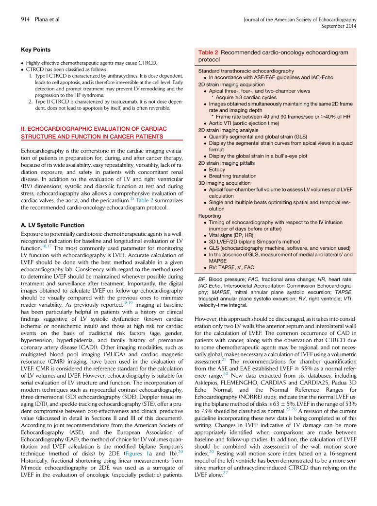
Table 2 Recommended cardio-oncology echocardiogramprotocol
Standard transthoracic echocardiography
� In accordance with ASE/EAE guidelines and IAC-Echo
2D strain imaging acquisition
� Apical three-, four-, and two-chamber views
* Acquire $3 cardiac cycles
� Images obtained simultaneously maintaining the same 2D framerate and imaging depth
914 Plana et al Journal of the American Society of EchocardiographySeptember 2014
Key Points
� Highly effective chemotherapeutic agents may cause CTRCD.� CTRCD has been classified as follows:
1. Type I CTRCD is characterized by anthracyclines. It is dose dependent,leads to cell apoptosis, and is therefore irreversible at the cell level. Earlydetection and prompt treatment may prevent LV remodeling and theprogression to the HF syndrome.
2. Type II CTRCD is characterized by trastuzumab. It is not dose depen-dent, does not lead to apoptosis by itself, and is often reversible.
* Frame rate between 40 and 90 frames/sec or $40% of HR
� Aortic VTI (aortic ejection time)
2D strain imaging analysis
� Quantify segmental and global strain (GLS)
� Display the segmental strain curves from apical views in a quadformat
� Display the global strain in a bull’s-eye plot
2D strain imaging pitfalls
� Ectopy
� Breathing translation
3D imaging acquisition
� Apical four-chamber full volume to assess LV volumes and LVEF
calculation� Single and multiple beats optimizing spatial and temporal res-
olution
II. ECHOCARDIOGRAPHIC EVALUATION OF CARDIAC
STRUCTURE AND FUNCTION IN CANCER PATIENTS
Echocardiography is the cornerstone in the cardiac imaging evalua-tion of patients in preparation for, during, and after cancer therapy,because of its wide availability, easy repeatability, versatility, lack of ra-diation exposure, and safety in patients with concomitant renaldisease. In addition to the evaluation of LV and right ventricular(RV) dimensions, systolic and diastolic function at rest and duringstress, echocardiography also allows a comprehensive evaluation ofcardiac valves, the aorta, and the pericardium.15 Table 2 summarizesthe recommended cardio-oncology-echocardiogram protocol.
Reporting
� Timing of echocardiography with respect to the IV infusion
(number of days before or after)
� Vital signs (BP, HR)� 3D LVEF/2D biplane Simpson’s method
� GLS (echocardiography machine, software, and version used)
� In the absence of GLS,measurement of medial and lateral s0 andMAPSE
� RV: TAPSE, s0, FAC
BP, Blood pressure; FAC, fractional area change; HR, heart rate;
IAC-Echo, Intersocietal Accreditation Commission Echocardiogra-
phy; MAPSE, mitral annular plane systolic excursion; TAPSE,
tricuspid annular plane systolic excursion; RV, right ventricle; VTI,velocity-time integral.
A. LV Systolic Function
Exposure to potentially cardiotoxic chemotherapeutic agents is a well-recognized indication for baseline and longitudinal evaluation of LVfunction.16,17 The most commonly used parameter for monitoringLV function with echocardiography is LVEF. Accurate calculation ofLVEF should be done with the best method available in a givenechocardiography lab. Consistency with regard to the method usedto determine LVEF should be maintained whenever possible duringtreatment and surveillance after treatment. Importantly, the digitalimages obtained to calculate LVEF on follow-up echocardiographyshould be visually compared with the previous ones to minimizereader variability. As previously reported,18,19 imaging at baselinehas been particularly helpful in patients with a history or clinicalfindings suggestive of LV systolic dysfunction (known cardiacischemic or nonischemic insult) and those at high risk for cardiacevents on the basis of traditional risk factors (age, gender,hypertension, hyperlipidemia, and family history of prematurecoronary artery disease [CAD]). Other imaging modalities, such asmultigated blood pool imaging (MUGA) and cardiac magneticresonance (CMR) imaging, have been used in the evaluation ofLVEF. CMR is considered the reference standard for the calculationof LV volumes and LVEF. However, echocardiography is suitable forserial evaluation of LV structure and function. The incorporation ofmodern techniques such as myocardial contrast echocardiography,three-dimensional (3D) echocardiography (3DE), Doppler tissue im-aging (DTI), and speckle-tracking echocardiography (STE), offer a pru-dent compromise between cost-effectiveness and clinical predictivevalue (discussed in detail in Sections II and III of this document).According to joint recommendations from the American Society ofEchocardiography (ASE), and the European Association ofEchocardiography (EAE), the method of choice for LV volumes quan-titation and LVEF calculation is the modified biplane Simpson’stechnique (method of disks) by 2DE (Figures 1a and 1b).20
Historically, fractional shortening using linear measurements fromM-mode echocardiography or 2DE was used as a surrogate ofLVEF in the evaluation of oncologic (especially pediatric) patients.
However, this approach should be discouraged, as it takes into consid-eration only two LV walls (the anterior septum and inferolateral wall)for the calculation of LVEF. The common occurrence of CAD inpatients with cancer, along with the observation that CTRCD dueto some chemotherapeutic agents may be regional, and not neces-sarily global, makes necessary a calculation of LVEF using a volumetricassessment.21 The recommendations for chamber quantificationfrom the ASE and EAE established LVEF $ 55% as a normal refer-ence range.20 New data extracted from six databases, includingAsklepios, FLEMENGHO, CARDIA5 and CARDIA25, Padua 3DEcho Normal, and the Normal Reference Ranges forEchocardiography (NORRE) study, indicate that the normal LVEF us-ing the biplane method of disks is 636 5%. LVEF in the range of 53%to 73% should be classified as normal.22-26 A revision of the currentguideline incorporating these new data is being completed as of thiswriting. Changes in LVEF indicative of LV damage can be moreappropriately identified when comparisons are made betweenbaseline and follow-up studies. In addition, the calculation of LVEFshould be combined with assessment of the wall motion scoreindex.20 Resting wall motion score index based on a 16-segmentmodel of the left ventricle has been demonstrated to be a more sen-sitive marker of anthracycline-induced CTRCD than relying on theLVEF alone.27
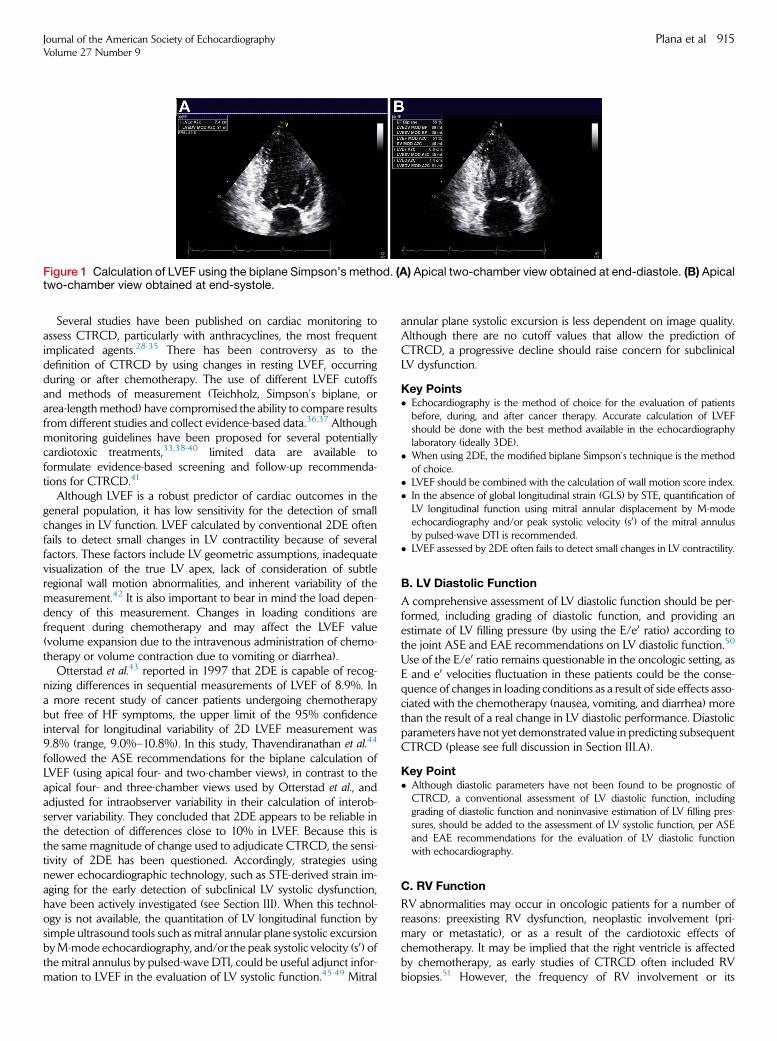
Figure 1 Calculation of LVEF using the biplane Simpson’s method. (A) Apical two-chamber view obtained at end-diastole. (B) Apicaltwo-chamber view obtained at end-systole.
Journal of the American Society of EchocardiographyVolume 27 Number 9
Plana et al 915
Several studies have been published on cardiac monitoring toassess CTRCD, particularly with anthracyclines, the most frequentimplicated agents.28-35 There has been controversy as to thedefinition of CTRCD by using changes in resting LVEF, occurringduring or after chemotherapy. The use of different LVEF cutoffsand methods of measurement (Teichholz, Simpson’s biplane, orarea-lengthmethod) have compromised the ability to compare resultsfrom different studies and collect evidence-based data.36,37 Althoughmonitoring guidelines have been proposed for several potentiallycardiotoxic treatments,33,38-40 limited data are available toformulate evidence-based screening and follow-up recommenda-tions for CTRCD.41
Although LVEF is a robust predictor of cardiac outcomes in thegeneral population, it has low sensitivity for the detection of smallchanges in LV function. LVEF calculated by conventional 2DE oftenfails to detect small changes in LV contractility because of severalfactors. These factors include LV geometric assumptions, inadequatevisualization of the true LV apex, lack of consideration of subtleregional wall motion abnormalities, and inherent variability of themeasurement.42 It is also important to bear in mind the load depen-dency of this measurement. Changes in loading conditions arefrequent during chemotherapy and may affect the LVEF value(volume expansion due to the intravenous administration of chemo-therapy or volume contraction due to vomiting or diarrhea).
Otterstad et al.43 reported in 1997 that 2DE is capable of recog-nizing differences in sequential measurements of LVEF of 8.9%. Ina more recent study of cancer patients undergoing chemotherapybut free of HF symptoms, the upper limit of the 95% confidenceinterval for longitudinal variability of 2D LVEF measurement was9.8% (range, 9.0%–10.8%). In this study, Thavendiranathan et al.44
followed the ASE recommendations for the biplane calculation ofLVEF (using apical four- and two-chamber views), in contrast to theapical four- and three-chamber views used by Otterstad et al., andadjusted for intraobserver variability in their calculation of interob-server variability. They concluded that 2DE appears to be reliable inthe detection of differences close to 10% in LVEF. Because this isthe same magnitude of change used to adjudicate CTRCD, the sensi-tivity of 2DE has been questioned. Accordingly, strategies usingnewer echocardiographic technology, such as STE-derived strain im-aging for the early detection of subclinical LV systolic dysfunction,have been actively investigated (see Section III). When this technol-ogy is not available, the quantitation of LV longitudinal function bysimple ultrasound tools such as mitral annular plane systolic excursionbyM-mode echocardiography, and/or the peak systolic velocity (s0) ofthe mitral annulus by pulsed-wave DTI, could be useful adjunct infor-mation to LVEF in the evaluation of LV systolic function.45-49 Mitral
annular plane systolic excursion is less dependent on image quality.Although there are no cutoff values that allow the prediction ofCTRCD, a progressive decline should raise concern for subclinicalLV dysfunction.
Key Points� Echocardiography is the method of choice for the evaluation of patientsbefore, during, and after cancer therapy. Accurate calculation of LVEFshould be done with the best method available in the echocardiographylaboratory (ideally 3DE).
� When using 2DE, the modified biplane Simpson’s technique is the methodof choice.
� LVEF should be combined with the calculation of wall motion score index.� In the absence of global longitudinal strain (GLS) by STE, quantification ofLV longitudinal function using mitral annular displacement by M-modeechocardiography and/or peak systolic velocity (s0) of the mitral annulusby pulsed-wave DTI is recommended.
� LVEF assessed by 2DE often fails to detect small changes in LV contractility.
B. LV Diastolic Function
A comprehensive assessment of LV diastolic function should be per-formed, including grading of diastolic function, and providing anestimate of LV filling pressure (by using the E/e0 ratio) according tothe joint ASE and EAE recommendations on LV diastolic function.50
Use of the E/e0 ratio remains questionable in the oncologic setting, asE and e0 velocities fluctuation in these patients could be the conse-quence of changes in loading conditions as a result of side effects asso-ciated with the chemotherapy (nausea, vomiting, and diarrhea) morethan the result of a real change in LV diastolic performance. Diastolicparameters have not yet demonstrated value in predicting subsequentCTRCD (please see full discussion in Section III.A).
Key Point� Although diastolic parameters have not been found to be prognostic ofCTRCD, a conventional assessment of LV diastolic function, includinggrading of diastolic function and noninvasive estimation of LV filling pres-sures, should be added to the assessment of LV systolic function, per ASEand EAE recommendations for the evaluation of LV diastolic functionwith echocardiography.
C. RV Function
RV abnormalities may occur in oncologic patients for a number ofreasons: preexisting RV dysfunction, neoplastic involvement (pri-mary or metastatic), or as a result of the cardiotoxic effects ofchemotherapy. It may be implied that the right ventricle is affectedby chemotherapy, as early studies of CTRCD often included RVbiopsies.51 However, the frequency of RV involvement or its
Figures 3 Transesophageal apical four-chamber and 3D reconstruction in an 86-year-old woman with marantic endocarditis, in thesetting of metastatic pancreatic cancer. Please note the diffuse involvement of the edge of the anterior and posterior leaflets.
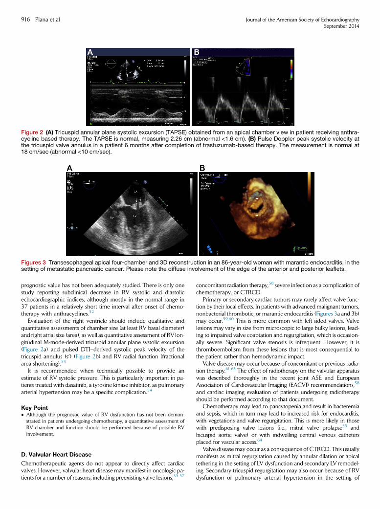
Figure 2 (A) Tricuspid annular plane systolic excursion (TAPSE) obtained from an apical chamber view in patient receiving anthra-cycline based therapy. The TAPSE is normal, measuring 2.26 cm (abnormal <1.6 cm). (B) Pulse Doppler peak systolic velocity atthe tricuspid valve annulus in a patient 6 months after completion of trastuzumab-based therapy. The measurement is normal at18 cm/sec (abnormal <10 cm/sec).
916 Plana et al Journal of the American Society of EchocardiographySeptember 2014
prognostic value has not been adequately studied. There is only onestudy reporting subclinical decrease in RV systolic and diastolicechocardiographic indices, although mostly in the normal range in37 patients in a relatively short time interval after onset of chemo-therapy with anthracyclines.52
Evaluation of the right ventricle should include qualitative andquantitative assessments of chamber size (at least RV basal diameter)and right atrial size (area), as well as quantitative assessment of RV lon-gitudinal M-mode-derived tricuspid annular plane systolic excursion(Figure 2a) and pulsed DTI–derived systolic peak velocity of thetricuspid annulus (s0) (Figure 2b) and RV radial function (fractionalarea shortening).53
It is recommended when technically possible to provide anestimate of RV systolic pressure. This is particularly important in pa-tients treated with dasatinib, a tyrosine kinase inhibitor, as pulmonaryarterial hypertension may be a specific complication.54
Key Point� Although the prognostic value of RV dysfunction has not been demon-strated in patients undergoing chemotherapy, a quantitative assessment ofRV chamber and function should be performed because of possible RVinvolvement.
D. Valvular Heart Disease
Chemotherapeutic agents do not appear to directly affect cardiacvalves. However, valvular heart disease may manifest in oncologic pa-tients for a number of reasons, including preexisting valve lesions,55-57
concomitant radiation therapy,58 severe infection as a complication ofchemotherapy, or CTRCD.
Primary or secondary cardiac tumors may rarely affect valve func-tion by their local effects. In patients with advancedmalignant tumors,nonbacterial thrombotic, or marantic endocarditis (Figures 3a and 3b)may occur.59,60 This is more common with left-sided valves. Valvelesions may vary in size from microscopic to large bulky lesions, lead-ing to impaired valve coaptation and regurgitation, which is occasion-ally severe. Significant valve stenosis is infrequent. However, it isthromboembolism from these lesions that is most consequential tothe patient rather than hemodynamic impact.
Valve disease may occur because of concomitant or previous radia-tion therapy.61-63 The effect of radiotherapy on the valvular apparatuswas described thoroughly in the recent joint ASE and EuropeanAssociation of Cardiovascular Imaging (EACVI) recommendations,58
and cardiac imaging evaluation of patients undergoing radiotherapyshould be performed according to that document.
Chemotherapy may lead to pancytopenia and result in bacteremiaand sepsis, which in turn may lead to increased risk for endocarditis,with vegetations and valve regurgitation. This is more likely in thosewith predisposing valve lesions (i.e., mitral valve prolapse55 andbicuspid aortic valve) or with indwelling central venous cathetersplaced for vascular access.64
Valve disease may occur as a consequence of CTRCD. This usuallymanifests as mitral regurgitation caused by annular dilation or apicaltethering in the setting of LV dysfunction and secondary LV remodel-ing. Secondary tricuspid regurgitation may also occur because of RVdysfunction or pulmonary arterial hypertension in the setting of
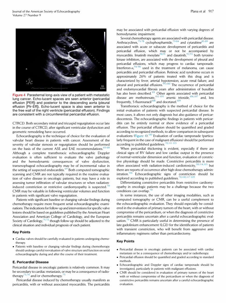
Figure 4 Parasternal long-axis view of a patient with metastaticlung cancer. Echo-lucent spaces are seen anterior (pericardialeffusion [PEff]) and posterior to the descending aorta (pleuraleffusion [Plr-Eff]). Echo-lucent space is also seen anterior tothe free wall of the right ventricle (pericardial effusion). Findingsare consistent with a circumferential pericardial effusion.
Journal of the American Society of EchocardiographyVolume 27 Number 9
Plana et al 917
CTRCD. Both secondary mitral and tricuspid regurgitation occur latein the course of CTRCD, after significant ventricular dysfunction andgeometric remodeling have occurred.
Echocardiography is the technique of choice for the evaluation ofvalvular heart disease in patients with cancer. Assessment of theseverity of valvular stenosis or regurgitation should be performedon the basis of the current ASE and EAE recommendations.65-68
Although a complete transthoracic echocardiographic Dopplerevaluation is often sufficient to evaluate the valve pathologyand the hemodynamic consequences of valve dysfunction,transesophageal echocardiography may be of incremental value inthe setting of suspected endocarditis.69 Both computed tomographicscanning and CMR are not typically required in the routine evalua-tion of valve disease in oncologic patients, but may have a role inassessing tumor infiltration of valvular structures or when radiation-induced constriction or restrictive cardiomyopathy is suspected.70
CMR may be valuable in following ventricular volumes and functionin patients with significant valve regurgitation.
Patients with significant baseline or changing valvular findings duringchemotherapy require more frequent serial echocardiographic exami-nations. The indications for follow-up and interventions for specificvalvelesions should be based on guidelines published by the American HeartAssociation and American College of Cardiology, and the EuropeanSociety of Cardiology,71,72 though follow-up should be adjusted to theclinical situation and individual prognosis of each patient.
Key Points
� Cardiac valves should be carefully evaluated in patients undergoing chemo-therapy.
� Patients with baseline or changing valvular findings during chemotherapyshould undergo careful reevaluation of valve structure and function on serialechocardiography during and after the course of their treatment.
E. Pericardial Disease
Pericardial disease in oncologic patients is relatively common. It maybe secondary to cardiac metastasis, or may be a consequence of radio-therapy73,74 and/or chemotherapy.75
Pericardial disease induced by chemotherapy usually manifests aspericarditis, with or without associated myocarditis. The pericarditis
may be associated with pericardial effusion with varying degrees ofhemodynamic impairment.
Several chemotherapy agents are associatedwith pericardial disease.Anthracyclines,75-78 cyclophosphamide,79-84 and cytarabine85-89 areassociated with acute or subacute development of pericarditis andpericardial effusion, which may or not be accompanied bymyocarditis. Imatinib mesylate90,91 and dasatinib,92,93 both tyrosine-kinase inhibitors, are associated with the development of pleural andpericardial effusions, which may progress to cardiac tamponade.Interferon-a,94-98 used in the treatment of melanoma, can causepericarditis and pericardial effusion. Retinoic acid syndrome occurs inapproximately 26% of patients treated with this drug and ischaracterized by fever, arterial hypotension, acute renal failure, andpleural and pericardial effusions.99,100 The occurrence of pericardialand endomyocardial fibrosis years after administration of busulfanhas also been described.101 Other agents associated with pericardialdisease are methotrexate,102-105 arsenic trioxide,106,107 and, lessfrequently, 5-fluorouracil108 and docetaxel.109
Transthoracic echocardiography is the method of choice for theinitial evaluation of patients with suspected pericardial disease. Inmost cases, it allows not only diagnosis but also guidance of pericar-diocentesis. The echocardiographic findings in patients with pericar-ditis can be entirely normal or show evidence of a pericardialeffusion. The pericardial effusion should be quantified and gradedaccording to recognized methods, to allow comparison in subsequentevaluations (Figure 4).110 Evaluation of cardiac tamponade (particu-larly frequent in the case of malignant effusions) should be performedaccording to published guidelines.70,111-113
When pericardial thickening is evident, especially if there areclinical signs of RV failure and low cardiac output in the presenceof normal ventricular dimension and function, evaluation of constric-tive physiology should be made. Constrictive pericarditis is moreoften associated with radiation-induced cardiotoxicity,58,114,115 butthere are reports of occurrence after high-dose chemotherapy admin-istration.116 Echocardiographic signs of constriction should beexplored according to published guidelines.70,110,117-119
Differentiating constrictive pericarditis from restrictive cardiomy-opathy in oncologic patients may be a challenge because the twoconditions can overlap.70
In some instances, the use of other imaging modalities, such ascomputed tomography or CMR, can be a useful complement tothe echocardiographic evaluation. They should especially be consid-ered in the evaluation of primary tumors of the heart, with or withoutcompromise of the pericardium, or when the diagnosis of constrictivepericarditis remains uncertain after a careful echocardiographic eval-uation.70 CMR is particularly useful in determining the presence oflate gadolinium enhancement (LGE) for the identification of patientswith transient constriction, who will benefit from aggressive anti-inflammatory regimens rather than pericardiectomy.
Key Points
� Pericardial disease in oncologic patients can be associated with cardiacmetastasis or be a consequence of chemotherapy and/or radiotherapy.
� Pericardial effusion should be quantified and graded according to standardmethods.
� Echocardiographic and Doppler signs of cardiac tamponade should beinvestigated, particularly in patients with malignant effusions.
� CMR should be considered in evaluation of primary tumors of the heartwith or without compromise of the pericardium or when the diagnosis ofconstrictive pericarditis remains uncertain after a careful echocardiographicevaluation.
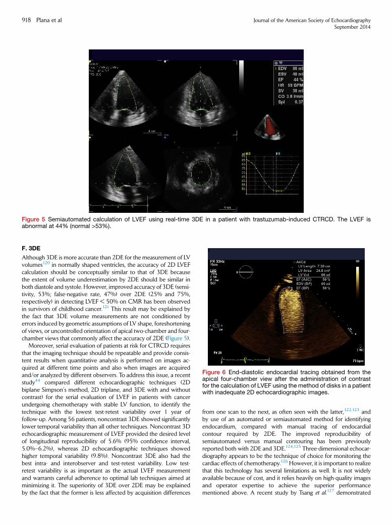
Figure 5 Semiautomated calculation of LVEF using real-time 3DE in a patient with trastuzumab-induced CTRCD. The LVEF isabnormal at 44% (normal >53%).
Figure 6 End-diastolic endocardial tracing obtained from theapical four-chamber view after the administration of contrastfor the calculation of LVEF using the method of disks in a patientwith inadequate 2D echocardiographic images.
918 Plana et al Journal of the American Society of EchocardiographySeptember 2014
F. 3DE
Although 3DE is more accurate than 2DE for the measurement of LVvolumes120 in normally shaped ventricles, the accuracy of 2D LVEFcalculation should be conceptually similar to that of 3DE becausethe extent of volume underestimation by 2DE should be similar inboth diastole and systole. However, improved accuracy of 3DE (sensi-tivity, 53%; false-negative rate, 47%) over 2DE (25% and 75%,respectively) in detecting LVEF < 50% on CMR has been observedin survivors of childhood cancer.121 This result may be explained bythe fact that 3DE volume measurements are not conditioned byerrors induced by geometric assumptions of LV shape, foreshorteningof views, or uncontrolled orientation of apical two-chamber and four-chamber views that commonly affect the accuracy of 2DE (Figure 5).
Moreover, serial evaluation of patients at risk for CTRCD requiresthat the imaging technique should be repeatable and provide consis-tent results when quantitative analysis is performed on images ac-quired at different time points and also when images are acquiredand/or analyzed by different observers. To address this issue, a recentstudy44 compared different echocardiographic techniques (2Dbiplane Simpson’s method, 2D triplane, and 3DE with and withoutcontrast) for the serial evaluation of LVEF in patients with cancerundergoing chemotherapy with stable LV function, to identify thetechnique with the lowest test-retest variability over 1 year offollow-up. Among 56 patients, noncontrast 3DE showed significantlylower temporal variability than all other techniques. Noncontrast 3Dechocardiographic measurement of LVEF provided the desired levelof longitudinal reproducibility of 5.6% (95% confidence interval,5.0%–6.2%), whereas 2D echocardiographic techniques showedhigher temporal variability (9.8%). Noncontrast 3DE also had thebest intra- and interobserver and test-retest variability. Low test-retest variability is as important as the actual LVEF measurementand warrants careful adherence to optimal lab techniques aimed atminimizing it. The superiority of 3DE over 2DE may be explainedby the fact that the former is less affected by acquisition differences
from one scan to the next, as often seen with the latter,122,123 andby use of an automated or semiautomated method for identifyingendocardium, compared with manual tracing of endocardialcontour required by 2DE. The improved reproducibility ofsemiautomated versus manual contouring has been previouslyreported both with 2DE and 3DE.124,125 Three-dimensional echocar-diography appears to be the technique of choice for monitoring thecardiac effects of chemotherapy.126 However, it is important to realizethat this technology has several limitations as well. It is not widelyavailable because of cost, and it relies heavily on high-quality imagesand operator expertise to achieve the superior performancementioned above. A recent study by Tsang et al.127 demonstrated
otoxicity
inLVEF
Predictivevalue/
correlations
)ProlongationofIVRT
>37%
predicteda
dropinLVEF>10%
(78%
sensitivity,
88%
specificity)
rted
13%
ofthosewith
reducedEwaveat
1.5
yhadreduced
fractional
shorteningatFU
)Allthosewith
decrease
dLVEFs
hadashortIVRTat
earlyFU
Journal of the American Society of EchocardiographyVolume 27 Number 9
Plana et al 919
that a quality improvement session dedicated to formally standardizethe analytic approach of the readers in the echocardiography labora-tory can eliminate the systematic bias and improve the agreementamong readers in the measurement of LV volumes. It is recommen-ded to include in the echocardiographic report the calculation ofLVEF by the biplane Simpson’s method, allowing comparison withprevious studies if this method was used. Where available, serial 3Dechocardiographic calculation of LVEF should be encouraged formonitoring CTRCD. It is to be expected that during the years tocome, less expensive, more automated, and user-friendly 3DEmachines that rely less on operator expertise could allow a widerapplication of this technique.
ivevalueofsubsequentcardi
Findings
Decrease
ongedIVRTand
celerationtimeat
wk
9/26(35%
ucedEwaveand
ducedE/A
ratio,
olongedIVRTand
decelerationtime
Notrepo
ucedmitralE
aveandDTIE
locityatearlyFU;
ore
pronounced
angesatlate
FU
4/16(25%
Key Points
� Three-dimensional echocardiography is the preferred technique for moni-toring LV function and detecting CTRCD in patients with cancer. Advan-tages include better accuracy in detecting LVEF below the lower limit ofnormal, better reproducibility, and lower temporal variability comparedwith 2DE in patients with cancer treated with chemotherapy.
� Costs, availability, high reliance on image quality, and need for training ofoperators currently limit the wide application of 3DE in the oncologicsetting.
Table
3Clinicalstudiesassessingdiastolic
functionindicesin
cancertreatm
entdemonstratingpredict
Study
Population
Treatm
ent
Tim
ingof
echocardiography
Stoddard
etal.
(1992)144
26adults;
mean
age,48y
Doxorubicin
>200
mg/m
2Base
line,3wk,and3
moaftertreatm
ent
Prol
de
3
Dorupetal.(2004)145
88ALL,66Wilm
s’tumor,agenot
reported
ALL:daunorubicin
90,
180,or270mg/m
2;
Wilm
s’tumor:
doxorubicin
303
mg/m
2
�1.5
and6.5
yafter
treatm
ent
Red re pr
E
Tassan-M
anginaetal.
(2006)48
16;meanage,38y
Doxorubicin
211
mg/m
2Before
chemotherapy,1–3
moand3.5
yafter
treatm
ent
Red w ve
m ch
ALL,Acute
lymphoblasticleukemia;FU,follo
w-up;IVRT,isovolumic
relaxationtime.
G. Contrast Echocardiography
Underestimation of volumes may occur when the endocardium is notadequately visualized.128 Endocardial border dropout can frequentlyoccur in patients undergoing chemotherapy (in particular patientswith breast cancer after mastectomy and chest irradiation).According to the ASE consensus statement on the clinical applicationsof ultrasonic contrast agents in echocardiography and EAE recom-mendations129,130 on myocardial contrast echocardiography, acontrast agent should be used when two contiguous LV segmentsfrom any apical view are not seen on noncontrast images (Figure 6).
There is limited literature to support the use of contrast for 3Dassessment of LV volumes in patients with cancer.122 A recent studyperformed in patients with cancer undergoing chemotherapy did notdemonstrate any advantage of using contrast-enhanced 3DE for themeasurement of LV volumes and LVEF (lower reproducibility andhigher temporal variability were noted compared with 3DE alone).44
There are two potential explanations for the findings. First, bloomingand attenuation artifacts may hinder the delineation of structures suchas the mitral valve, with the resultant variability in contouring of theleft ventricle. Second, most of the patients studied had adequateacoustic windows with harmonic imaging and therefore did notmeet traditional criteria for contrast administration.
Key Points
� The use of myocardial contrast agents could be potentially useful in chemo-therapy patients when endocardial dropout occurs.
� According to current recommendations, contrast should be used when twocontiguous LV segments are not well visualized on noncontrast apicalimages.
� Contrast agents are not recommended in conjunction with 3DE in thelongitudinal follow-up of patients with cancer.
H. Stress Echocardiography
Stress echocardiography, an established technique for the detectionand prognostication of stable CAD as recommended by guidelines,may be useful in the evaluation of patients with intermediate or
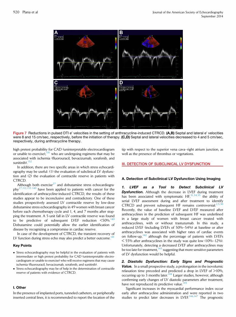
Figure 7 Reductions in pulsed DTI e0 velocities in the setting of anthracycline-induced CTRCD. (A,B) Septal and lateral e0 velocitieswere 8 and 15 cm/sec, respectively, before the initiation of therapy. (C,D) Septal and lateral velocities decreased to 4 and 5 cm/sec,respectively, during anthracycline therapy.
920 Plana et al Journal of the American Society of EchocardiographySeptember 2014
high pretest probability for CAD (uninterpretable electrocardiogramor unable to exercise),131 who are undergoing regimens that may beassociated with ischemia (fluorouracil, bevacizumab, sorafenib, andsunitinib).132
In addition, there are two specific areas in which stress echocardi-ography may be useful: (1) the evaluation of subclinical LV dysfunc-tion and (2) the evaluation of contractile reserve in patients withCTRCD.
Although both exercise27 and dobutamine stress echocardiogra-phy27,112,133-140 have been applied to patients with cancer for theidentification of anthracycline-induced CTRCD, the results of thesestudies appear to be inconclusive and contradictory. One of thesestudies prospectively assessed LV contractile reserve by low-dosedobutamine stress echocardiography in 49womenwith breast cancerbefore each chemotherapy cycle and 1, 4, and 7 months after stop-ping the treatment. A 5-unit fall in LV contractile reserve was foundto be predictive of subsequent LVEF reduction <50%.140
Dobutamine could potentially allow the earlier identification ofdisease by recognizing a compromise in cardiac reserve.
In case of the development of CTRCD, the transient recovery ofLV function during stress echo may also predict a better outcome.141
Key Points
� Stress echocardiography may be helpful in the evaluation of patients withintermediate or high pretest probability for CAD (uninterpretable electro-cardiogram or unable to exercise) who will receive regimens that may causeischemia (fluorouracil, bevacizumab, sorafenib, and sunitinib).
� Stress echocardiography may be of help in the determination of contractilereserve of patients with evidence of CTRCD.
I. Other
In the presence of implanted ports, tunneled catheters, or peripherallyinserted central lines, it is recommended to report the location of the
tip with respect to the superior vena cava–right atrium junction, aswell as the presence of thrombus or vegetations.
III. DETECTION OF SUBCLINICAL LV DYSFUNCTION
A. Detection of Subclinical LV Dysfunction Using Imaging
1. LVEF as a Tool to Detect Subclinical LV
Dysfunction. Although the decrease in LVEF during treatmenthas been associated with symptomatic HF,31,34,35 the ability ofserial LVEF assessment during and after treatment to identifyCTRCD and prevent subsequent HF remains controversial.2,5,30
Recently, the value of baseline LVEF and LVEF measured afteranthracyclines in the prediction of subsequent HF was underlinedin a large study of women with breast cancer treated withanthracyclines, with or without trastuzumab. In this study, areduced LVEF (including LVEFs of 50%–54%) at baseline or afteranthracyclines was associated with higher rates of cardiac eventson follow-up,142 although the percentage of patients with LVEFs< 55% after anthracyclines in the study was quite low (10%–12%).Unfortunately, detecting a decreased LVEF after anthracyclines maybe too late for treatment,143 suggesting that more sensitive parametersof LV dysfunction would be helpful.
2. Diastolic Dysfunction: Early Signs and Prognostic
Value. In a small prospective study, a prolongation in the isovolumicrelaxation time preceded and predicted a drop in LVEF of >10%,occurring up to 3 months later.144 Larger studies, however, althoughconfirming early changes of LV diastolic parameters after treatment,have not reproduced its predictive value.145
Significant increases in the myocardial performance index occurearly after anthracycline administration and were reported in twostudies to predict later decreases in LVEF.146,147 The prognostic
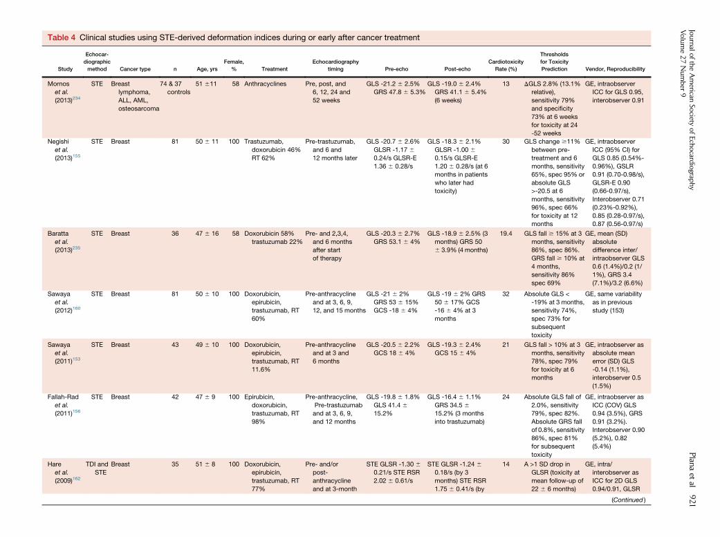
Table 4 Clinical studies using STE-derived deformation indices during or early after cancer treatment
Study
Echocar-
diographic
method Cancer type n Age, yrs
Female,
% Treatment
Echocardiography
timing Pre-echo Post-echo
Cardiotoxicity
Rate (%)
Thresholds
for Toxicity
Prediction Vendor, Reproducibility
Mornos
et al.
(2013)234
STE Breast
lymphoma,
ALL, AML,
osteosarcoma
74 & 37
controls
51 611 58 Anthracyclines Pre, post, and
6, 12, 24 and
52 weeks
GLS -21.2 6 2.5%
GRS 47.8 6 5.3%
GLS -19.0 6 2.4%
GRS 41.1 6 5.4%
(6 weeks)
13 DGLS 2.8% (13.1%
relative),
sensitivity 79%
and specificity
73% at 6 weeks
for toxicity at 24
-52 weeks
GE, intraobserver
ICC for GLS 0.95,
interobserver 0.91
Negishi
et al.
(2013)155
STE Breast 81 50 6 11 100 Trastuzumab,
doxorubicin 46%
RT 62%
Pre-trastuzumab,
and 6 and
12 months later
GLS -20.7 6 2.6%
GLSR -1.17 60.24/s GLSR-E
1.36 6 0.28/s
GLS -18.3 6 2.1%
GLSR -1.00 60.15/s GLSR-E
1.20 6 0.28/s (at 6
months in patients
who later had
toxicity)
30 GLS change $11%
between pre-
treatment and 6
months, sensitivity
65%, spec 95% or
absolute GLS
>-20.5 at 6
months, sensitivity
96%, spec 66%
for toxicity at 12
months
GE, intraobserver
ICC (95% CI) for
GLS 0.85 (0.54%-
0.96%), GSLR
0.91 (0.70-0.98/s),
GLSR-E 0.90
(0.66-0.97/s),
Interobserver 0.71
(0.23%-0.92%),
0.85 (0.28-0.97/s),
0.87 (0.56-0.97/s)
Baratta
et al.
(2013)235
STE Breast 36 47 6 16 58 Doxorubicin 58%
trastuzumab 22%
Pre- and 2,3,4,
and 6 months
after start
of therapy
GLS -20.3 6 2.7%
GRS 53.1 6 4%
GLS -18.9 6 2.5% (3
months) GRS 50
6 3.9% (4 months)
19.4 GLS fall $ 15% at 3
months, sensitivity
86%, spec 86%.
GRS fall $ 10% at
4 months,
sensitivity 86%
spec 69%
GE, mean (SD)
absolute
difference inter/
intraobserver GLS
0.6 (1.4%)/0.2 (1/
1%), GRS 3.4
(7.1%)/3.2 (6.6%)
Sawaya
et al.
(2012)160
STE Breast 81 50 6 10 100 Doxorubicin,
epirubicin,
trastuzumab, RT
60%
Pre-anthracycline
and at 3, 6, 9,
12, and 15 months
GLS -21 6 2%
GRS 53 6 15%
GCS -18 6 4%
GLS -19 6 2% GRS
50 6 17% GCS
-16 6 4% at 3
months
32 Absolute GLS <
-19% at 3 months,
sensitivity 74%,
spec 73% for
subsequent
toxicity
GE, same variability
as in previous
study (153)
Sawaya
et al.
(2011)153
STE Breast 43 49 6 10 100 Doxorubicin,
epirubicin,
trastuzumab, RT
11.6%
Pre-anthracycline
and at 3 and
6 months
GLS -20.5 6 2.2%
GCS 18 6 4%
GLS -19.3 6 2.4%
GCS 15 6 4%
21 GLS fall > 10% at 3
months, sensitivity
78%, spec 79%
for toxicity at 6
months
GE, intraobserver as
absolute mean
error (SD) GLS
-0.14 (1.1%),
interobserver 0.5
(1.5%)
Fallah-Rad
et al.
(2011)156
STE Breast 42 47 6 9 100 Epirubicin,
doxorubicin,
trastuzumab, RT
98%
Pre-anthracycline,
Pre-trastuzumab
and at 3, 6, 9,
and 12 months
GLS -19.8 6 1.8%
GLS 41.4 615.2%
GLS -16.4 6 1.1%
GRS 34.5 615.2% (3 months
into trastuzumab)
24 Absolute GLS fall of
2.0%, sensitivity
79%, spec 82%.
Absolute GRS fall
of 0.8%, sensitivity
86%, spec 81%
for subsequent
toxicity
GE, intraobserver as
ICC (COV) GLS
0.94 (3.5%), GRS
0.91 (3.2%).
Interobserver 0.90
(5.2%), 0.82
(5.4%)
Hare
et al.
(2009)162
TDI and
STE
Breast 35 51 6 8 100 Doxorubicin,
epirubicin,
trastuzumab, RT
77%
Pre- and/or
post-
anthracycline
and at 3-month
STE GLSR -1.30 60.21/s STE RSR
2.02 6 0.61/s
STE GLSR -1.24 60.18/s (by 3
months) STE RSR
1.75 6 0.41/s (by
14 A >1 SD drop in
GLSR (toxicity at
mean follow-up of
22 6 6 months)
GE, intra/
interobserver as
ICC for 2D GLS
0.94/0.91, GLSR
(Continued )
Journalo
ftheAmerican
Society
ofEchocard
iograp
hy
Volume27Number
9Plan
aet
al921
Table
4(Continued)
Study
Echocar-
diographic
method
Cancertype
nAge,yrs
Female,
%Treatm
ent
Echocardiography
timing
Pre-echo
Post-echo
Cardiotoxicity
Rate
(%)
Thresholds
forToxicity
Prediction
Vendor,Reproducibility
intervals
6-9
months)
0.94/0.91,GRS
0.86/0.50,GRSR
0.83/0.65
Mavinkurve-
Groothuis
etal.
(2013)236
STE
ALL
60,60
controls
6(2.2-15.4)
38
Anthracycline,
RT100%
Pre-anthracycline,
10weeks,
and12months
GLS-18.2
63.1%
GLSR-1.446
0.3/
sGRS66.8
61%
GCS-19.4
64.3
GLS-16.7
65.2%
GLSR-1.206
0.4/
sGRS55.2
616%
GCS-16.9
63.1%
(by12
months)
0Strain
valueswere
notpredictiveof
decreasein
LV
fractional
shortening
GE,nodata
AC,A
driamycin;A
LL,a
cute
lymphoblasticleukemia;A
ML,a
cute
myoblasticleukemia;C
HF,c
ongestiveheartfailure;C
REC,C
ardiacReviewandEvaluationCommittee;F
EC,5
-fluorouracil,
epirubicin,a
ndcyclophosphamide;4
CH,four-chamber;FS,fractionalshortening;G
CS,g
lobalcircumferentialstrain;G
LSR,g
loballongitudinalstrainrate;G
LSR-E,g
loballongitudinalearly
diastolic
strain
rate;GRS,globalradialstrain;GRSR,globalradialstrain
rate;LS,longitudinalstrain;LSR,longitudinalstrain
rate;NPV,negativepredictivevalue;PPV,positivepredictive
value;RSR,radialstrain
rate;SAX,short-axis;2CH,tw
o-chamber.
Reproducedwithperm
issionfrom
theJournalo
ftheAmericanCollegeofCardiology.
922 Plana et al Journal of the American Society of EchocardiographySeptember 2014
value of myocardial performance index could not be replicated insubsequent studies.148
Two studies have reported LV diastolic abnormalities late after an-thracycline administration; these abnormalities were associated withwall motion abnormalities despite a preserved LVEF.149 Anotherstudy reported that a reduced transmitral E/A ratio was associatedwith a reduction in longitudinal strain by STE in patients with normalLVEFs late after treatment.150 It is unclear, however, if these findingshave any clinical significance.
As a result, it can be concluded that the use of Doppler-derived dia-stolic indices is not useful in the early detection of CTRCD because oftheir inability to predict subsequent HF (Table 3).
3. Detection of Subclinical LV Dysfunction Using DTI
Velocities. Several investigators have demonstrated an early reduc-tion in e0 velocity of the mitral annulus in patients receiving anthra-cyclines (Figures 7c and 7d),48,150-152 which remained reducedduring treatment153 and several years thereafter.150 The reductionsin e0 velocity appear heterogeneous,150,151,154 suggesting differencesin regional wall stress, apoptosis, or fibrosis.
In a study by Negishi et al.,155 a 10% reduction in e0 velocity wasobserved in patients who developed CTRCD. Nevertheless, thereduction was not statistically significant (P = .09) or predictive ofsubsequent reduction in LVEF (P = 0.14).
A reduction in DTI-derived systolic velocity (s0) was reported inanimal models of doxorubicin-induced cardiac injury6 and in thechronic follow-up of patients treated with anthracyclines.150 Amarked early decrease in s0, and its value as a potential predictor ofchanges of LV systolic function after chemotherapy, was reported ina study of 42 patients with breast cancer treated with trastuzumabin the adjuvant setting.156 It is to be noted, however, that the rateof symptomatic HF in this study was of 24% at 6months of treatment,an unusually high rate in chemotherapy-treated populations.Whether these results can be generalized to patients with a lower inci-dence of HF is unknown.
Key Point
� A decreased LVEF at baseline or after anthracyclines is associated withhigher rates of cardiac events on follow-up.
� Although it has been suggested that alterations in LV diastolic function (asevaluated by Doppler indices of mitral inflow and e0 by pulsed DTI) precedealterations in systolic function, the evidence does not support the role ofthese indices for the prediction of later CTRCD.
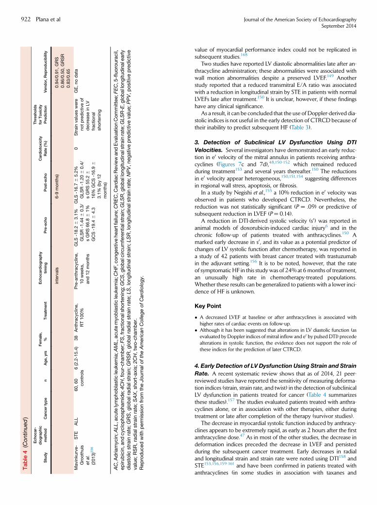
4. Early Detection of LV Dysfunction Using Strain and Strain
Rate. A recent systematic review shows that as of 2014, 21 peer-reviewed studies have reported the sensitivity of measuring deforma-tion indices (strain, strain rate, and twist) in the detection of subclinicalLV dysfunction in patients treated for cancer (Table 4 summarizesthese studies).157 The studies evaluated patients treated with anthra-cyclines alone, or in association with other therapies, either duringtreatment or late after completion of the therapy (survivor studies).
The decrease in myocardial systolic function induced by anthracy-clines appears to be extremely rapid, as early as 2 hours after the firstanthracycline dose.47 As in most of the other studies, the decrease indeformation indices preceded the decrease in LVEF and persistedduring the subsequent cancer treatment. Early decreases in radialand longitudinal strain and strain rate were noted using DTI158 andSTE153,156,159-161 and have been confirmed in patients treated withanthracyclines (in some studies in association with taxanes and
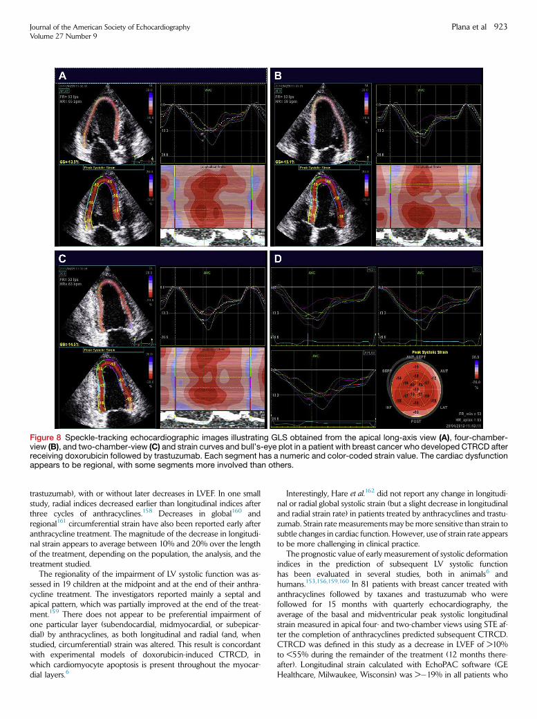
Figure 8 Speckle-tracking echocardiographic images illustrating GLS obtained from the apical long-axis view (A), four-chamber-view (B), and two-chamber-view (C) and strain curves and bull’s-eye plot in a patient with breast cancer who developed CTRCD afterreceiving doxorubicin followed by trastuzumab. Each segment has a numeric and color-coded strain value. The cardiac dysfunctionappears to be regional, with some segments more involved than others.
Journal of the American Society of EchocardiographyVolume 27 Number 9
Plana et al 923
trastuzumab), with or without later decreases in LVEF. In one smallstudy, radial indices decreased earlier than longitudinal indices afterthree cycles of anthracyclines.158 Decreases in global160 andregional161 circumferential strain have also been reported early afteranthracycline treatment. The magnitude of the decrease in longitudi-nal strain appears to average between 10% and 20% over the lengthof the treatment, depending on the population, the analysis, and thetreatment studied.
The regionality of the impairment of LV systolic function was as-sessed in 19 children at the midpoint and at the end of their anthra-cycline treatment. The investigators reported mainly a septal andapical pattern, which was partially improved at the end of the treat-ment.159 There does not appear to be preferential impairment ofone particular layer (subendocardial, midmyocardial, or subepicar-dial) by anthracyclines, as both longitudinal and radial (and, whenstudied, circumferential) strain was altered. This result is concordantwith experimental models of doxorubicin-induced CTRCD, inwhich cardiomyocyte apoptosis is present throughout the myocar-dial layers.6
Interestingly, Hare et al.162 did not report any change in longitudi-nal or radial global systolic strain (but a slight decrease in longitudinaland radial strain rate) in patients treated by anthracyclines and trastu-zumab. Strain rate measurements may bemore sensitive than strain tosubtle changes in cardiac function. However, use of strain rate appearsto be more challenging in clinical practice.
The prognostic value of early measurement of systolic deformationindices in the prediction of subsequent LV systolic functionhas been evaluated in several studies, both in animals6 andhumans.153,156,159,160 In 81 patients with breast cancer treated withanthracyclines followed by taxanes and trastuzumab who werefollowed for 15 months with quarterly echocardiography, theaverage of the basal and midventricular peak systolic longitudinalstrain measured in apical four- and two-chamber views using STE af-ter the completion of anthracyclines predicted subsequent CTRCD.CTRCD was defined in this study as a decrease in LVEF of >10%to <55% during the remainder of the treatment (12 months there-after). Longitudinal strain calculated with EchoPAC software (GEHealthcare, Milwaukee, Wisconsin) was >�19% in all patients who
924 Plana et al Journal of the American Society of EchocardiographySeptember 2014
later developed HF (Figures 8a–8d). Although reductions were seenin all three layers, neither radial nor circumferential strain was predic-tive of subsequent CTRCD.160 A predictive value of regional strainwas also reported in smaller studies with shorter follow-up pe-riods.153,156,159 Importantly, although the decrease in longitudinalstrain and LVEF appears to at least partially persist throughout theduration of the treatment,160 it is unknown what their evolutionwill be in subsequent years, and whether early deformation measure-ments will predict persistent decreases in LVEF or symptomatic HF.
Negishi et al.155 recently published a study looking for the optimalmyocardial deformation index to predict CTRCD at 12 months in81 women with breast cancer treated with trastuzumab, with orwithout anthracyclines. The strongest predictor of CTRCD wasDGLS measured at the 6-month visit. An 11% reduction (95% confi-dence interval, 8.3%–14.6%) was the optimal cutoff, with sensitivityof 65% and specificity of 94%. Of note, DGLS was superior tochanges in the count of abnormal segments, s0 and e0 velocities.They concluded that in patients with baseline strain measurements,the 95% confidence interval suggests that reductions of GLS of<8% compared with baseline appear not to be clinically meaningful,whereas those >15% are very likely to be of clinical significance (seeFigures 9a and 9b for example of calculation). They confirmed thefindings of Sawaya et al.,160 this time using the conventional calcula-tion of GLS averaging the 18 segments from the three apical views.They showed that in patients without baseline strain measurements,the proposed cutoff of �19% conforms to the confidence intervalaround �20.5% found in their study. Nevertheless the area underthe curve for absolute strain value is less, making the change in strainthe preferable approach.
Finally, four studies evaluated the deformation parameters inlong-term cancer survivors (range, 2–30 years after treat-ment).150,154,163,164 In two of the studies with longer follow-upand/or higher doses of anthracyclines, the LVEF (or fractional short-ening) was slightly decreased.163,164 In contrast, all four studiesdetected decreases in longitudinal and radial (and circumferentialwhen studied) parameters compared with age-matched controlpatients, underlining the sensitivity of these parameters in the detec-tion of subclinical LV dysfunction. STE appears therefore as the imag-ing technique of choice for detection of subclinical LV dysfunction.Normal values for GLS depend on the measurement position inthe myocardium, the vendor, and the version of the analysis software,resulting in considerable heterogeneity in the published literature.Two recently published large studies evaluating the normal rangesof LV 2D strain have shown an effect of gender in LV myocardialdeformation.165,166 The study of Kocabay et al.165 reported a meannormal GLS of �20.7 6 2 for men and �22.1 6 1.8 for women.These values are almost identical to the ones reported by theJapanese Ultrasound Speckle Tracking of the Left Ventricle(JUSTICE) study166 for the same vendor. There is also concern thatstrain values may decrease with age.166,167 As a result, it is notpossible to recommend universal normal values or lower limits ofnormal. We refer the reader to Table 5, which summarizes the findingsof the Japanese Ultrasound Speckle Tracking of the Left Ventriclestudy, providing mean values for GLS according to vendor, gender,and age. Cheng et al.168 recently evaluated the reproducibility of2D STE in the Offspring Cohort of the Framingham Heart Study.The interobserver intraclass correlation coefficient was $0.84 for allglobal strain measurements, with an average coefficient of variationfor GLS of #4%. The intraobserver intraclass correlation coefficientwas $0.91 among time points spanning a total 8-month period,with an average of #6% for GLS. The authors concluded that 2D
STE is reproducible when performed by trained operators.However, the technique has important limitations (Table 6). Thereare no data currently available as to the reproducibility of GLS atnonacademic centers or community hospitals. The presence of alearning curve for sonographers and interpreting physicians makesdedicated training and monitoring of quality (i.e., intra- and interob-server and test-retest variability) essential. When setting a strainprogram, it is recommended to initially designate one physicianand, where available, one technician to perform, interpret, andcompare studies over time. As experience is gained with the tech-nique, the effort may be expanded to include other physicians, tech-nicians, and trainees. Nevertheless, the most important limitation isintervendor variability.166,169 Different echocardigoraphy machinesor software packages can in fact produce different results, inparticular for circumferential and radial strain, making problematicintraindividual comparisons over time. Recognizing the critical needfor standardization in strain imaging, the EACVI and ASE invitedtechnical representatives from all interested vendors to participatein a concerted effort to reduce intervendor variability in strainmeasurement.170 Until that is achieved, it is recommended to usethe same vendor’s machine and software version to compare individ-ual patients with cancer when using 2D STE for the serial evaluationof systolic function.
Individual echocardiographic laboratories following patients withcancer should strive to incorporate strain assessment in their echocar-diography laboratory protocols.
Key Points
� Myocardial deformation (strain) can bemeasured using DTI or 2D STE. Thelatter is favored because of a lack of angle dependency.
� GLS is the optimal parameter of deformation for the early detection ofsubclinical LV dysfunction.
� Ideally, the measurements during chemotherapy should be compared withthe baseline value. In patients with available baseline strain measurements, arelative percentage reduction of GLS of <8% from baseline appears not tobe meaningful, and those >15% from baseline are very likely to beabnormal.
� When applying STE for the longitudinal follow-up of patients with cancer,the same vendor-specific ultrasound machine should be used.
B. Detection of Subclinical LV Dysfunction UsingBiomarkers
Biomarkers have the potential to fulfill a critical unmet need as arobust diagnostic tool for the early identification, assessment, andmonitoring of CTRCD. A biomarker approach is minimally invasiveand can be readily repeated without significant risk. Despite intrinsicassay variability, standardized assays typically have acceptable coeffi-cients of variation of <10%, potentially minimizing intra- and interob-server variability.171
1. Troponins. Cardiac troponins are the gold-standard biomarkersfor the diagnosis of myocardial injury.172,173 Troponin I (TnI) is asensitive and specific marker for myocardial injury in adults treatedwith anthracycline chemotherapy, and studies suggest that anelevation of troponin identifies patients at risk for the subsequentdevelopment of CTRCD.
The largest of these studies was performed in 703 patients with can-cer, in whomTnI was determined with each cycle of high-dose chemo-therapy and 1 month after chemotherapy.174 Patients were classifiedinto three subgroups on the basis of the combined presence of anydetectable TnI either within 72 hours (early) or 1 month after the lastadministration of chemotherapy (late). In 495 patients, both early
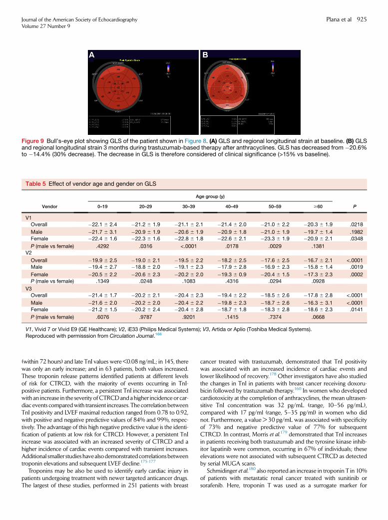
Figure 9 Bull’s-eye plot showing GLS of the patient shown in Figure 8. (A) GLS and regional longitudinal strain at baseline. (B) GLSand regional longitudinal strain 3 months during trastuzumab-based therapy after anthracyclines. GLS has decreased from �20.6%to �14.4% (30% decrease). The decrease in GLS is therefore considered of clinical significance (>15% vs baseline).
Table 5 Effect of vendor age and gender on GLS
Vendor
Age group (y)
P0–19 20–29 30–39 40–49 50–59 $60
V1Overall �22.1 6 2.4 �21.2 6 1.9 �21.1 6 2.1 �21.4 6 2.0 �21.0 6 2.2 �20.3 6 1.9 .0218
Male �21.7 6 3.1 �20.9 6 1.9 �20.6 6 1.9 �20.9 6 1.8 �21.0 6 1.9 �19.7 6 1.4 .1982
Female �22.4 6 1.6 �22.3 6 1.6 �22.8 6 1.8 �22.6 6 2.1 �23.3 6 1.9 �20.9 6 2.1 .0348
P (male vs female) .4292 .0316 <.0001 .0178 .0029 .1381V2
Overall �19.9 6 2.5 �19.0 6 2.1 �19.5 6 2.2 �18.2 6 2.5 �17.6 6 2.5 �16.7 6 2.1 <.0001
Male �19.4 6 2.7 �18.8 6 2.0 �19.1 6 2.3 �17.9 6 2.8 �16.9 6 2.3 �15.8 6 1.4 .0019
Female �20.5 6 2.2 �20.6 6 2.3 �20.2 6 2.0 �19.3 6 0.9 �20.4 6 1.5 �17.3 6 2.3 .0002
P (male vs female) .1349 .0248 .1083 .4316 .0294 .0928
V3Overall �21.4 6 1.7 �20.2 6 2.1 �20.4 6 2.3 �19.4 6 2.2 �18.5 6 2.6 �17.8 6 2.8 <.0001
Male �21.6 6 2.0 �20.2 6 2.0 �20.4 6 2.2 �19.8 6 2.3 �18.7 6 2.6 �16.3 6 3.1 <.0001Female �21.2 6 1.5 �20.2 6 2.4 �20.4 6 2.8 �18.7 6 1.8 �18.3 6 2.8 �18.6 6 2.3 .0141
P (male vs female) .6076 .9787 .9201 .1415 .7374 .0668
V1, Vivid 7 or Vivid E9 (GE Healthcare); V2, iE33 (Philips Medical Systems); V3, Artida or Aplio (Toshiba Medical Systems).
Reproduced with permisssion from Circulation Journal.166
Journal of the American Society of EchocardiographyVolume 27 Number 9
Plana et al 925
(within 72 hours) and late TnI values were <0.08 ng/mL; in 145, therewas only an early increase; and in 63 patients, both values increased.These troponin release patterns identified patients at different levelsof risk for CTRCD, with the majority of events occurring in TnI-positive patients. Furthermore, a persistent TnI increase was associatedwith an increase in the severity ofCTRCDandahigher incidence or car-diac events comparedwith transient increases. The correlation betweenTnI positivity and LVEF maximal reduction ranged from 0.78 to 0.92,with positive and negative predictive values of 84% and 99%, respec-tively. The advantage of this high negative predictive value is the identi-fication of patients at low risk for CTRCD. However, a persistent TnIincrease was associated with an increased severity of CTRCD and ahigher incidence of cardiac events compared with transient increases.Additional smaller studies havealsodemonstratedcorrelationsbetweentroponin elevations and subsequent LVEF decline.175-177
Troponins may be also be used to identify early cardiac injury inpatients undergoing treatment with newer targeted anticancer drugs.The largest of these studies, performed in 251 patients with breast
cancer treated with trastuzumab, demonstrated that TnI positivitywas associated with an increased incidence of cardiac events andlower likelihood of recovery.178 Other investigators have also studiedthe changes in TnI in patients with breast cancer receiving doxoru-bicin followed by trastuzumab therapy.160 In women who developedcardiotoxicity at the completion of anthracyclines, the mean ultrasen-sitive TnI concentration was 32 pg/mL (range, 10–56 pg/mL),compared with 17 pg/ml (range, 5–35 pg/ml) in women who didnot. Furthermore, a value > 30 pg/mL was associated with specificityof 73% and negative predictive value of 77% for subsequentCTRCD. In contrast, Morris et al.179 demonstrated that TnI increasesin patients receiving both trastuzumab and the tyrosine kinase inhib-itor lapatinib were common, occurring in 67% of individuals; theseelevations were not associated with subsequent CTRCD as detectedby serial MUGA scans.
Schmidinger et al.180 also reported an increase in troponin T in 10%of patients with metastatic renal cancer treated with sunitinib orsorafenib. Here, troponin T was used as a surrogate marker for
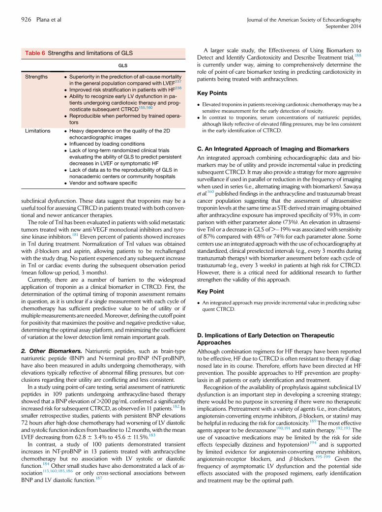
Table 6 Strengths and limitations of GLS
GLS
Strengths � Superiority in the prediction of all-cause mortality
in the general population compared with LVEF237
� Improved risk stratification in patients with HF238
� Ability to recognize early LV dysfunction in pa-
tients undergoing cardiotoxic therapy and prog-
nosticate subsequent CTRCD155,160
� Reproducible when performed by trained opera-tors
Limitations � Heavy dependence on the quality of the 2D
echocardiographic images� Influenced by loading conditions
� Lack of long-term randomized clinical trials
evaluating the ability of GLS to predict persistentdecreases in LVEF or symptomatic HF
� Lack of data as to the reproducibility of GLS in
nonacademic centers or community hospitals
� Vendor and software specific
926 Plana et al Journal of the American Society of EchocardiographySeptember 2014
subclinical dysfunction. These data suggest that troponins may be auseful tool for assessing CTRCD in patients treated with both conven-tional and newer anticancer therapies.
The role of TnI has been evaluated in patients with solid metastatictumors treated with new anti-VEGF monoclonal inhibitors and tyro-sine kinase inhibitors.181 Eleven percent of patients showed increasesin TnI during treatment. Normalization of TnI values was obtainedwith b-blockers and aspirin, allowing patients to be rechallengedwith the study drug. No patient experienced any subsequent increasein TnI or cardiac events during the subsequent observation period(mean follow-up period, 3 months).
Currently, there are a number of barriers to the widespreadapplication of troponin as a clinical biomarker in CTRCD. First, thedetermination of the optimal timing of troponin assessment remainsin question, as it is unclear if a single measurement with each cycle ofchemotherapy has sufficient predictive value to be of utility or ifmultiplemeasurements areneeded.Moreover, defining the cutoff pointfor positivity that maximizes the positive and negative predictive value,determining the optimal assay platform, and minimizing the coefficientof variation at the lower detection limit remain important goals.
2. Other Biomarkers. Natriuretic peptides, such as brain-typenatriuretic peptide (BNP) and N-terminal pro-BNP (NT-proBNP),have also been measured in adults undergoing chemotherapy, withelevations typically reflective of abnormal filling pressures, but con-clusions regarding their utility are conflicting and less consistent.
In a study using point-of-care testing, serial assessment of natriureticpeptides in 109 patients undergoing anthracycline-based therapyshowed that a BNP elevation of >200 pg/mL conferred a significantlyincreased risk for subsequent CTRCD, as observed in 11 patients.182 Insmaller retrospective studies, patients with persistent BNP elevations72 hours after high-dose chemotherapy had worsening of LV diastolicand systolic function indices frombaseline to 12months, with themeanLVEF decreasing from 62.8 6 3.4% to 45.6 6 11.5%.183
In contrast, a study of 100 patients demonstrated transientincreases in NT-proBNP in 13 patients treated with anthracyclinechemotherapy but no association with LV systolic or diastolicfunction.184 Other small studies have also demonstrated a lack of as-sociation113,160,185,186 or only cross-sectional associations betweenBNP and LV diastolic function.187
A larger scale study, the Effectiveness of Using Biomarkers toDetect and Identify Cardiotoxicity and Describe Treatment trial,188
is currently under way, aiming to comprehensively determine therole of point-of-care biomarker testing in predicting cardiotoxicity inpatients being treated with anthracyclines.
Key Points
� Elevated troponins in patients receiving cardiotoxic chemotherapymay be asensitive measurement for the early detection of toxicity.
� In contrast to troponins, serum concentrations of natriuretic peptides,although likely reflective of elevated filling pressures, may be less consistentin the early identification of CTRCD.
C. An Integrated Approach of Imaging and Biomarkers
An integrated approach combining echocardiographic data and bio-markers may be of utility and provide incremental value in predictingsubsequent CTRCD. It may also provide a strategy for more aggressivesurveillance if used in parallel or reduction in the frequency of imagingwhen used in series (i.e., alternating imaging with biomarkers). Sawayaet al.160 published findings in the anthracycline and trastuzumab breastcancer population suggesting that the assessment of ultrasensitivetroponin levels at the same time as STE-derived strain imaging obtainedafter anthracycline exposure has improved specificity of 93%, in com-parison with either parameter alone (73%). An elevation in ultrasensi-tive TnI or a decrease in GLS of >�19%was associated with sensitivityof 87% compared with 48% or 74% for each parameter alone. Somecenters use an integrated approachwith the use of echocardiography atstandardized, clinical preselected intervals (e.g., every 3 months duringtrastuzumab therapy) with biomarker assessment before each cycle oftrastuzumab (e.g., every 3 weeks) in patients at high risk for CTRCD.However, there is a critical need for additional research to furtherstrengthen the validity of this approach.
Key Point
� An integrated approach may provide incremental value in predicting subse-quent CTRCD.
D. Implications of Early Detection on TherapeuticApproaches
Although combination regimens for HF therapy have been reportedto be effective, HF due to CTRCD is often resistant to therapy if diag-nosed late in its course. Therefore, efforts have been directed at HFprevention. The possible approaches to HF prevention are prophy-laxis in all patients or early identification and treatment.
Recognition of the availability of prophylaxis against subclinical LVdysfunction is an important step in developing a screening strategy;there would be no purpose in screening if there were no therapeuticimplications. Pretreatment with a variety of agents (i.e., iron chelators,angiotensin-converting enzyme inhibitors, b-blockers, or statins) maybe helpful in reducing the risk for cardiotoxicity.189 Themost effectiveagents appear to be dexrazoxane190,191 and statin therapy.192,193 Theuse of vasoactive medications may be limited by the risk for sideeffects (especially dizziness and hypotension)194 and is supportedby limited evidence for angiotensin-converting enzyme inhibitors,angiotensin-receptor blockers, and b-blockers.195-199 Given thefrequency of asymptomatic LV dysfunction and the potential sideeffects associated with the proposed regimens, early identificationand treatment may be the optimal path.
Journal of the American Society of EchocardiographyVolume 27 Number 9
Plana et al 927
Treatment of subclinical LV dysfunction is based on a strategy ofearly detection of myocardial disease with either biomarkers or imag-ing. The attraction of this approach is that there is potential benefit forany patient, and those without dysfunction are not burdened by thetreatment. The disadvantages are that screening has to be sufficientlyaccurate to identify as closely as possible all at-risk patients and thatsome patients may have progressed to sufficient damage that treat-ment may provide only a partial response.
TnI release after high-dose chemotherapy in patients treated byanthracyclines was investigated by Cardinale and Sandri.171 Thisteam demonstrated the usefulness of enalapril in this population,178
when given before178 or early after143 the LVEF decrease. The initialstudy in 114 cancer survivors with TnI release demonstrated signifi-cant reductions in LVEF but increases in LV volumes only in untreatedpatients.178 The study, however, lacked placebo administration, wasunblinded, and lacked clinical end points. A further study demon-strated the role and limitations of an LVEF decrease initiating cardiactreatment in chemotherapy patients.143 In 201 consecutive patientswith LV dysfunction (LVEF < 45%) due to anthracycline cardiomyop-athy,143 enalapril (and, if possible, carvedilol) were initiated promptlyafter the detection of reduced LVEF. On the basis of sequential LVEFmeasurements over the subsequent 366 27months, 42% of patientswere considered responders, 13% were partial responders, and 45%were deemed nonresponders. Cardiac events were fewer in re-sponders than partial responders and nonresponders. The responserate progressively decreased with increasing time delay between theend of chemotherapy to the start of HF; no complete recovery ofLVEF was observed after 6 months.
Similar positive findings have been obtained with b-blockers inpatients with subclinical LV dysfunction after trastuzumab.200 Of 42patients with GLS decreases of $�11%, the 19 who were treatedshowed subsequent significant LVEF increases after 6 months (from52.6 6 5.6% to 57.4 6 6.0%) but patients not taking b-blockersshowed no change (from 56.7 6 5.9% to 56.0 6 5.2%, P = .001 be-tween groups). Additionally, data from a small number of recentstudies indicate that b-blockers have a role as novel therapeutic agentsin reducing tumor metastasis, tumor recurrence, and breast cancer–specific mortality.201
Key Point
� Small studies have suggested that a variety of agents (such as dexrazoxane,b-blockers, angiotensin-receptor blockers, and statins) may be helpful in theprevention or early treatment of CTRCD, but no definitive recommenda-tions can be set with the current available data.
IV. OTHER IMAGING MODALITIES
A. Radionuclide Approaches for MonitoringChemotherapy-Induced Cardiotoxicity
1. MUGA. Measurement of LV function using either first-pass orequilibrium radionuclide angiography (also known as MUGA) wasfirst used in the late 1970s to identify patients receiving anthracyclineswho had declines in LVEF before the development of clinical HFsymptoms.202 When anthracyclines were stopped with a clinicallyasymptomatic decline in LVEF, there was no further deteriorationin function, and in some patients, there was even recovery, especiallywhen aggressively treated with optimal HF medications. Thus, moni-toring byMUGA to detect an asymptomatic decline in LVEFwas pref-erable to waiting for the development of symptoms of congestive HF
symptoms, by which time CTRCD was irreversible. Serial imaging byMUGA had been reported to allow safe use of anthracyclines evenwhen baseline LVEF was abnormal.203 On the basis of these results,the following recommendations for the use of MUGA to monitoranthracycline-induced cardiotoxicity were proposed33:
1. LVEF > 50% at baselinea. Measurement at 250 to 300 mg/m2
b. Measurement at 450 mg/m2
c. Measurement before each dose above 450 mg/m2
d. Discontinue therapy if LVEF decreases by $10% from baseline andLVEF # 50%
2. LVEF < 50% at baselinea. Do not treat if LVEF is <30%b. Serial measurement before each dosec. Discontinue therapy if LVEF decreases by$10% from baseline or LVEF
# 30%
These early reports suggested that clinical symptoms of CTRCDdevelop after irreversible damage has occurred and that asymptom-atic declines in LVEF may represent an early sign of permanent dam-age. Efforts were made for earlier detection using stress testing anddetection of fibrosis that preceded decline in LV systolic function.The use of exercise stress to measure LV functional reserve, withnormal defined as an increase of $5 LVEF units, was shown tohave a higher sensitivity for early detection of CTRCD.202,204
Because of relatively low specificity, limited exercise capacity inmost patients with cancer, and the requirement for using supinebicycle exercise, this technique is seldom used today. As mentionedwith echocardiography, the significant variability in measurementsof LV diastolic function by MUGA limits its clinical application.205
2. MUGA Compared with Other Modalities. As a 3D imagingtechnique, MUGA has consistently outperformed standard 2DEwith respect to accuracy and reproducibility of LVEF measure-ments.206,207 In several studies, the values obtained by MUGAshowed much higher correlations with those obtained with other3D imaging tools, such as CMR and novel 3D echocardiographictechniques, but individual LV volumes and LVEF values still differedsignificantly across the techniques.206,208 Together, these findingspoint out that the LVEF results obtained by different techniques arenot interchangeable and suggest that choosing a single techniquemay provide the best option for serial monitoring of LVEF inpatients at risk.
The MUGA technique for monitoring anthracycline-inducedCTRCD has been standardized, shown to be highly reproducible,and widely available and effectively applied in academic centers,community hospitals, and physicians’ offices.33 On the basis ofthese findings, MUGA has been widely used in general clinicalpractice as well as in the efficacy trials for development of newchemotherapy agents for all tumor types.209 Advantages ofMUGA in evaluation of patients during or after cancer therapyinclude the following:
1. Its widespread use in clinical practice with extensive long-term follow up: Inthe 1980s, there were extensive publications establishing the efficacy ofMUGA for all types of adult and pediatric tumors treated with anthracy-clines. On the basis of this body of evidence, MUGA was used widely inclinical trials and carried over into clinical practice.
2. Few technical limitations: 99mTc red blood cell labeling and planar imagingcan be done in all patients without limitations due to obesity, poor acousticwindows, or the presence of cardiac devices such as pacemakers or defibril-lators. The technique is widely available, and cost is comparable with that ofalternative modalities.
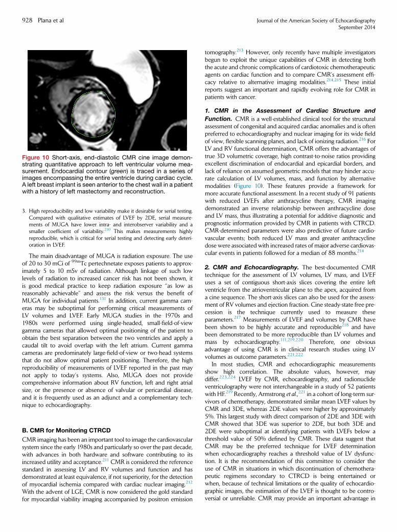
Figure 10 Short-axis, end-diastolic CMR cine image demon-strating quantitative approach to left ventricular volume mea-surement. Endocardial contour (green) is traced in a series ofimages encompassing the entire ventricle during cardiac cycle.A left breast implant is seen anterior to the chest wall in a patientwith a history of left mastectomy and reconstruction.
928 Plana et al Journal of the American Society of EchocardiographySeptember 2014
3. High reproducibility and low variability make it desirable for serial testing.Compared with qualitative estimates of LVEF by 2DE, serial measure-ments of MUGA have lower intra- and interobserver variability and asmaller coefficient of variability.210 This makes measurements highlyreproducible, which is critical for serial testing and detecting early deteri-oration in LVEF.
The main disadvantage of MUGA is radiation exposure. The useof 20 to 30 mCi of 99mTc pertechnetate exposes patients to approx-imately 5 to 10 mSv of radiation. Although linkage of such lowlevels of radiation to increased cancer risk has not been shown, itis good medical practice to keep radiation exposure ‘‘as low asreasonably achievable’’ and assess the risk versus the benefit ofMUGA for individual patients.131 In addition, current gamma cam-eras may be suboptimal for performing critical measurements ofLV volumes and LVEF. Early MUGA studies in the 1970s and1980s were performed using single-headed, small-field-of-viewgamma cameras that allowed optimal positioning of the patient toobtain the best separation between the two ventricles and apply acaudal tilt to avoid overlap with the left atrium. Current gammacameras are predominately large-field-of-view or two-head systemsthat do not allow optimal patient positioning. Therefore, the highreproducibility of measurements of LVEF reported in the past maynot apply to today’s systems. Also, MUGA does not providecomprehensive information about RV function, left and right atrialsize, or the presence or absence of valvular or pericardial disease,and it is frequently used as an adjunct and a complementary tech-nique to echocardiography.
B. CMR for Monitoring CTRCD
CMR imaging has been an important tool to image the cardiovascularsystem since the early 1980s and particularly so over the past decade,with advances in both hardware and software contributing to itsincreased utility and acceptance.211 CMR is considered the referencestandard in assessing LV and RV volumes and function and hasdemonstrated at least equivalence, if not superiority, for the detectionof myocardial ischemia compared with cardiac nuclear imaging.212
With the advent of LGE, CMR is now considered the gold standardfor myocardial viability imaging accompanied by positron emission
tomography.213 However, only recently have multiple investigatorsbegun to exploit the unique capabilities of CMR in detecting boththe acute and chronic complications of cardiotoxic chemotherapeuticagents on cardiac function and to compare CMR’s assessment effi-cacy relative to alternative imaging modalities.214,215 These initialreports suggest an important and rapidly evolving role for CMR inpatients with cancer.
1. CMR in the Assessment of Cardiac Structure and
Function. CMR is a well-established clinical tool for the structuralassessment of congenital and acquired cardiac anomalies and is oftenpreferred to echocardiography and nuclear imaging for its wide fieldof view, flexible scanning planes, and lack of ionizing radiation.216 ForLV and RV functional determination, CMR offers the advantages oftrue 3D volumetric coverage, high contrast-to-noise ratios providingexcellent discrimination of endocardial and epicardial borders, andlack of reliance on assumed geometric models that may hinder accu-rate calculation of LV volumes, mass, and function by alternativemodalities (Figure 10). These features provide a framework formore accurate functional assessment. In a recent study of 91 patientswith reduced LVEFs after anthracycline therapy, CMR imagingdemonstrated an inverse relationship between anthracycline doseand LV mass, thus illustrating a potential for additive diagnostic andprognostic information provided by CMR in patients with CTRCD.CMR-determined parameters were also predictive of future cardio-vascular events; both reduced LV mass and greater anthracyclinedose were associated with increased rates of major adverse cardiovas-cular events in patients followed for a median of 88 months.214
2. CMR and Echocardiography. The best-documented CMRtechnique for the assessment of LV volumes, LV mass, and LVEFuses a set of contiguous short-axis slices covering the entire leftventricle from the atrioventricular plane to the apex, acquired froma cine sequence. The short-axis slices can also be used for the assess-ment of RV volumes and ejection fraction. Cine steady-state free pre-cession is the technique currently used to measure theseparameters.217 Measurements of LVEF and volumes by CMR havebeen shown to be highly accurate and reproducible218 and havebeen demonstrated to be more reproducible than LV volumes andmass by echocardiography.111,219,220 Therefore, one obviousadvantage of using CMR is in clinical research studies using LVvolumes as outcome parameters.221,222
In most studies, CMR and echocardiographic measurementsshow high correlation. The absolute values, however, maydiffer.223,224 LVEF by CMR, echocardiography, and radionuclideventriculography were not interchangeable in a study of 52 patientswith HF.219 Recently, Armstrong et al.,221 in a cohort of long-term sur-vivors of chemotherapy, demonstrated similar mean LVEF values byCMR and 3DE, whereas 2DE values were higher by approximately5%. This largest study with direct comparison of 2DE and 3DE withCMR showed that 3DE was superior to 2DE, but both 3DE and2DE were suboptimal at identifying patients with LVEFs below athreshold value of 50% defined by CMR. These data suggest thatCMR may be the preferred technique for LVEF determinationwhen echocardiography reaches a threshold value of LV dysfunc-tion. It is the recommendation of this committee to consider theuse of CMR in situations in which discontinuation of chemothera-peutic regimens secondary to CTRCD is being entertained orwhen, because of technical limitations or the quality of echocardio-graphic images, the estimation of the LVEF is thought to be contro-versial or unreliable. CMR may provide an important advantage in
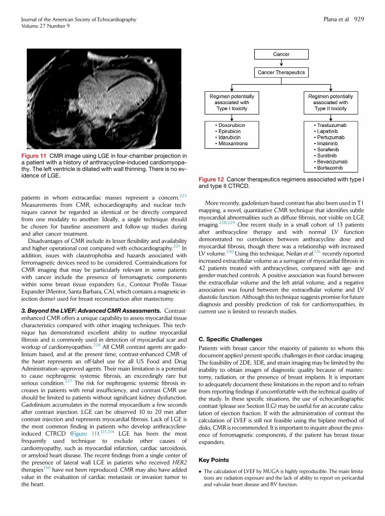
Figure 11 CMR image using LGE in four-chamber projection ina patient with a history of anthracycline-induced cardiomyopa-thy. The left ventricle is dilated with wall thinning. There is no ev-idence of LGE.
Figure 12 Cancer therapeutics regimens associated with type Iand type II CTRCD.
Journal of the American Society of EchocardiographyVolume 27 Number 9
Plana et al 929
patients in whom extracardiac masses represent a concern.225
Measurements from CMR, echocardiography and nuclear tech-niques cannot be regarded as identical or be directly comparedfrom one modality to another. Ideally, a single technique shouldbe chosen for baseline assessment and follow-up studies duringand after cancer treatment.
Disadvantages of CMR include its lesser flexibility and availabilityand higher operational cost compared with echocardiography.221 Inaddition, issues with claustrophobia and hazards associated withferromagnetic devices need to be considered. Contraindications forCMR imaging that may be particularly relevant in some patientswith cancer include the presence of ferromagnetic componentswithin some breast tissue expanders (i.e., Contour Profile TissueExpander [Mentor, Santa Barbara, CA], which contains a magnetic in-jection dome) used for breast reconstruction after mastectomy.
3. Beyond the LVEF: AdvancedCMRAssessments. Contrast-enhanced CMR offers a unique capability to assess myocardial tissuecharacteristics compared with other imaging techniques. This tech-nique has demonstrated excellent ability to outline myocardialfibrosis and is commonly used in detection of myocardial scar andworkup of cardiomyopathies.226 All CMR contrast agents are gado-linium based, and at the present time, contrast-enhanced CMR ofthe heart represents an off-label use for all US Food and DrugAdministration–approved agents. Their main limitation is a potentialto cause nephrogenic systemic fibrosis, an exceedingly rare butserious condition.227 The risk for nephrogenic systemic fibrosis in-creases in patients with renal insufficiency, and contrast CMR useshould be limited to patients without significant kidney dysfunction.Gadolinium accumulates in the normal myocardium a few secondsafter contrast injection. LGE can be observed 10 to 20 min aftercontrast injection and represents myocardial fibrosis. Lack of LGE isthe most common finding in patients who develop anthracycline-induced CTRCD (Figure 11).121,214 LGE has been the mostfrequently used technique to exclude other causes ofcardiomyopathy, such as myocardial infarction, cardiac sarcoidosis,or amyloid heart disease. The recent findings from a single center ofthe presence of lateral wall LGE in patients who received HER2therapies156 have not been reproduced. CMR may also have addedvalue in the evaluation of cardiac metastasis or invasion tumor tothe heart.
More recently, gadolinium-based contrast has also been used in T1mapping, a novel, quantitative CMR technique that identifies subtlemyocardial abnormalities such as diffuse fibrosis, not visible on LGEimaging.228,229 One recent study in a small cohort of 13 patientsafter anthracycline therapy and with normal LV functiondemonstrated no correlation between anthracycline dose andmyocardial fibrosis, though there was a relationship with increasedLV volume.230 Using this technique, Neilan et al.231 recently reportedincreased extracellular volume as a surrogate of myocardial fibrosis in42 patients treated with anthracyclines, compared with age- andgender-matched controls. A positive association was found betweenthe extracellular volume and the left atrial volume, and a negativeassociation was found between the extracellular volume and LVdiastolic function. Although this technique suggests promise for futurediagnosis and possibly prediction of risk for cardiomyopathies, itscurrent use is limited to research studies.
C. Specific Challenges
Patients with breast cancer (the majority of patients to whom thisdocument applies) present specific challenges in their cardiac imaging.The feasibility of 2DE, 3DE, and strain imaging may be limited by theinability to obtain images of diagnostic quality because of mastec-tomy, radiation, or the presence of breast implants. It is importantto adequately document these limitations in the report and to refrainfrom reporting findings if uncomfortable with the technical quality ofthe study. In these specific situations, the use of echocardiographiccontrast (please see Section II.G) may be useful for an accurate calcu-lation of ejection fraction. If with the administration of contrast thecalculation of LVEF is still not feasible using the biplane method ofdisks, CMR is recommended. It is important to inquire about the pres-ence of ferromagnetic components, if the patient has breast tissueexpanders.
Key Points
� The calculation of LVEF by MUGA is highly reproducible. The main limita-tions are radiation exposure and the lack of ability to report on pericardialand valvular heart disease and RV function.
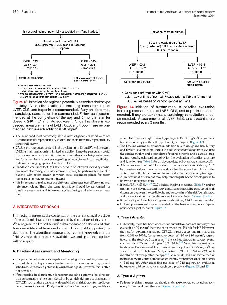
Figure 13 Initiation of a regimen potentially associatedwith typeI toxicity. A baseline evaluation including measurements ofLVEF, GLS, and troponin is recommended. If any are abnormal,a cardiology consultation is recommended. Follow-up is recom-mended at the completion of therapy and 6 months later fordoses < 240 mg/m2 or its equivalent. Once this dose is ex-ceeded, measurements of LVEF, GLS, and troponin are recom-mended before each additional 50 mg/m2.
Figure 14 Initiation of trastuzumab. A baseline evaluationincluding measurements of LVEF, GLS, and troponin is recom-mended. If any are abnormal, a cardiology consultation is rec-ommended. Measurements of LVEF, GLS, and troponins arerecommended every 3 months.
930 Plana et al Journal of the American Society of EchocardiographySeptember 2014
� The newer and most commonly used dual-head gamma cameras were notused in the initial reproducibility studies, and their interstudy reproducibilityis not well known.
� CMR is the reference standard in the evaluation of LVand RV volumes andLVEF. Its main limitation is its limited availability. It may be particularly usefulin situations in which discontinuation of chemotherapy is being entertainedand/or when there is concern regarding echocardiographic or equilibriumradionuclide angiographic calculation of LVEF.
� Standard precautions for CMR safety need to be followed, including consid-eration of electromagnetic interference. This may be particularly relevant inpatients with breast cancer, in whom tissue expanders placed for breastreconstruction may represent a hazard.
� It is important to realize that the different techniques use different normalreference values. Thus, the same technique should be performed forbaseline assessment and follow-up studies during and after cancer treat-ment.
V. INTEGRATED APPROACH
This section represents the consensus of the current clinical practicesof the academic institutions represented by the authors of this report.We recognize the limited scientific data available and the lack of classA evidence (derived from randomized clinical trials) supporting thealgorithms. The algorithms represent our current knowledge of thefield. As new data becomes available, we anticipate that updateswill be required.
A. Baseline Assessment and Monitoring
� Cooperation between cardiologists and oncologists is absolutely essential.� It would be ideal to perform a baseline cardiac assessment in every patientscheduled to receive a potentially cardiotoxic agent. However, this is oftennot possible.
� If not possible in all patients, it is recommended to perform a baseline car-diac assessment in those considered to be at high risk for development ofCTRCD, such as those patients with established or risk factors for cardiovas-cular disease, those with LV dysfunction, those >65 years of age, and those
scheduled to receive high doses of type I agents (>350 mg/m2) or combina-tion chemotherapy with both type I and type II agents (Figure 12).
� The baseline cardiac assessment, in addition to a thorough medical historyand physical examination, should include electrocardiography to evaluatethe cardiac rhythm and detect signs of resting ischemia and a cardiac imag-ing test (usually echocardiography) for the evaluation of cardiac structureand function (see Table 2 for cardio-oncology echocardiogram protocol).
� A baseline assessment of GLS and/or troponin is desirable. Although GLShas negative values in normal individuals, for the sake of simplicity in thissection, we will refer to it as an absolute value (without the negative sign).
� A pretreatment assessment may help cardiologists advise oncologists as toknown or anticipated risks.
� If the LVEF is <53%,22-26 GLS is below the limit of normal (Table 5), and/ortroponins are elevated, a cardiology consultation should be considered, withdiscussion between the cardiologist and oncologist of the risk/benefit ratio,and cancer treatment at the discretion of the oncologist (Figures 13–15).
� If the quality of the echocardiogram is suboptimal, CMR is recommended.� Follow-up assessment is recommended on the basis of the specific type ofanticancer agent received (Figure 13).
1. Type I Agents.
� Historically, there has been concern for cumulative doses of anthracyclinesexceeding 400 mg/m2, because of an associated 5% risk for HF. However,the risk for doxorubicin-related CTRCD is really a continuum that spansfrom 0.2% to 100%, for cumulative doses of 150 to 850 mg/m2, respec-tively. In the study by Swain et al.,30 the earliest step-up in cardiac eventsoccurred from 250 to 350 mg/m2 (9%–18%).232 New data evaluating pa-tients who have received low doses of anthracyclines (<375 mg/m2) re-vealed a rate of subclinical LV dysfunction (LVEF < 50%) of 26% at 6months of follow-up after therapy.233 As a result, this committee recom-mends follow-up at the completion of therapy for regimens including doses< 240 mg/m2. After exceeding the dose of 240 mg/m2, an evaluationbefore each additional cycle is considered prudent (Figures 13 and 15).
2. Type II Agents.
� Patients receiving trastuzumab should undergo follow-up echocardiographyevery 3 months during therapy (Figures 14 and 15).
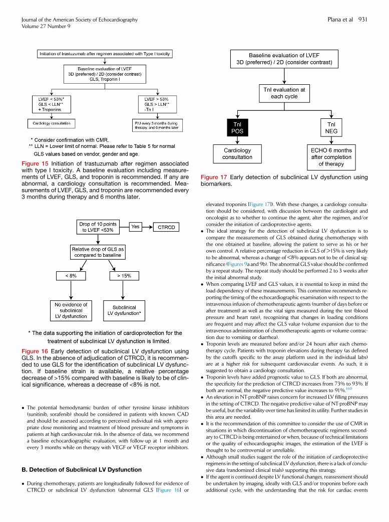
Figure 16 Early detection of subclinical LV dysfunction usingGLS. In the absence of adjudication of CTRCD, it is recommen-ded to use GLS for the identification of subclinical LV dysfunc-tion. If baseline strain is available, a relative percentagedecrease of >15% compared with baseline is likely to be of clin-ical significance, whereas a decrease of <8% is not.
Figure 15 Initiation of trastuzumab after regimen associatedwith type I toxicity. A baseline evaluation including measure-ments of LVEF, GLS, and troponin is recommended. If any areabnormal, a cardiology consultation is recommended. Mea-surements of LVEF, GLS, and troponin are recommended every3 months during therapy and 6 months later.
Figure 17 Early detection of subclinical LV dysfunction usingbiomarkers.
Journal of the American Society of EchocardiographyVolume 27 Number 9
Plana et al 931
� The potential hemodynamic burden of other tyrosine kinase inhibitors(sunitinib, sorafenib) should be considered in patients with known CADand should be assessed according to perceived individual risk with appro-priate close monitoring and treatment of blood pressure and symptoms inpatients at high cardiovascular risk. In the absence of data, we recommenda baseline echocardiographic evaluation, with follow-up at 1 month andevery 3 months while on therapy with VEGF or VEGF receptor inhibitors.
B. Detection of Subclinical LV Dysfunction
� During chemotherapy, patients are longitudinally followed for evidence ofCTRCD or subclinical LV dysfunction (abnormal GLS [Figure 16] or
elevated troponins [Figure 17]). With these changes, a cardiology consulta-tion should be considered, with discussion between the cardiologist andoncologist as to whether to continue the agent, alter the regimen, and/orconsider the initiation of cardioprotective agents.
� The ideal strategy for the detection of subclinical LV dysfunction is tocompare the measurements of GLS obtained during chemotherapy withthe one obtained at baseline, allowing the patient to serve as his or herown control. A relative percentage reduction in GLS of >15% is very likelyto be abnormal, whereas a change of <8% appears not to be of clinical sig-nificance (Figures 9a and 9b). The abnormal GLS value should be confirmedby a repeat study. The repeat study should be performed 2 to 3 weeks afterthe initial abnormal study.
� When comparing LVEF and GLS values, it is essential to keep in mind theload dependency of these measurements. This committee recommends re-porting the timing of the echocardiographic examination with respect to theintravenous infusion of chemotherapeutic agents (number of days before orafter treatment) as well as the vital signs measured during the test (bloodpressure and heart rate), recognizing that changes in loading conditionsare frequent and may affect the GLS value (volume expansion due to theintravenous administration of chemotherapeutic agents or volume contrac-tion due to vomiting or diarrhea).
� Troponin levels are measured before and/or 24 hours after each chemo-therapy cycle. Patients with troponin elevations during therapy (as definedby the cutoffs specific to the assay platform used in the individual labs)are at a higher risk for subsequent cardiovascular events. As such, it issuggested to obtain a cardiology consultation.
� Troponin levels have added prognostic value to GLS. If both are abnormal,the specificity for the prediction of CTRCD increases from 73% to 93%. Ifboth are normal, the negative predictive value increases to 91%.160
� An elevation in NT-proBNP raises concern for increased LV filling pressuresin the setting of CTRCD. The negative predictive value of NT-proBNP maybe useful, but the variability over time has limited its utility. Further studies inthis area are needed.
� It is the recommendation of this committee to consider the use of CMR insituations in which discontinuation of chemotherapeutic regimens second-ary to CTRCD is being entertained or when, because of technical limitationsor the quality of echocardiographic images, the estimation of the LVEF isthought to be controversial or unreliable.
� Although small studies suggest the role of the initiation of cardioprotectiveregimens in the setting of subclinical LV dysfunction, there is a lack of conclu-sive data (randomized clinical trials) supporting this strategy.
� If the agent is continued despite LV functional changes, reassessment shouldbe undertaken by imaging, ideally with GLS and/or troponins before eachadditional cycle, with the understanding that the risk for cardiac events
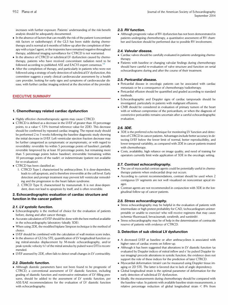
932 Plana et al Journal of the American Society of EchocardiographySeptember 2014
increases with further exposure. Patients’ understanding of the risk-benefitanalysis should be adequately documented.
� In the absence of factors that canmodify the risk of the patient (concomitantrisk factors or radiotherapy), if the GLS has been stable during chemo-therapy and is normal at 6 months of follow-up after the completion of ther-apy with a type I agent, or the troponins have remained negative throughouttherapy, additional imaging surveillance for CTRCD is not warranted.
� In the absence of CTRCD or subclinical LV dysfunction caused by chemo-therapy, patients who have received concomitant radiation need to befollowed according to published ASE and EACVI expert consensus.58
� After the completion of therapy, and particularly in patients who were notfollowed using a strategy of early detection of subclinical LV dysfunction, thiscommittee suggests a yearly clinical cardiovascular assessment by a healthcare provider, looking for early signs and symptoms of cardiovascular dis-ease, with further cardiac imaging ordered at the discretion of the provider.
EXECUTIVE SUMMARY
1. Chemotherapy related cardiac dysfunction
� Highly effective chemotherapeutic agents may cause CTRCD.� CTRCD is defined as a decrease in the LVEF of greater than 10 percentagepoints, to a value < 53% (normal reference value for 2DE). This decreaseshould be confirmed by repeated cardiac imaging. The repeat study shouldbe performed 2 to 3 weeks following the baseline diagnostic study showingthe initial decrease in LVEF. Left ventricular ejection fraction decrease maybe further categorized as symptomatic or asymptomatic, or with regard toreversibility: reversible (to within 5 percentage points of baseline); partiallyreversible (improved by at least 10 percentage points, but remaining morethan 5 percentage points below baseline); irreversible (remaining within10 percentage points of the nadir); or indeterminate (patient not availablefor re-evaluation).
� CTRCD has been classified as:1. CTRCD Type I, characterized by anthracyclines. It is dose-dependent,
leads to cell apoptosis, and is therefore irreversible at the cell level. Earlydetection and prompt treatment may prevent left ventricular remodel-ing and the progression to the heart failure syndrome.
2. CTRCD Type II, characterized by trastuzumab. It is not dose-depen-dent, does not lead to apoptosis by itself, and is often reversible.
2. Echocardiographic evaluation of cardiac structure andfunction in the cancer patient
2.1. LV systolic function.� Echocardiography is the method of choice for the evaluation of patientsbefore, during and after cancer therapy.
� Accurate calculation of LVEF should be donewith the best method availablein the echocardiography laboratory (ideally 3DE).
� When using 2DE, the modified biplane Simpson technique is the method ofchoice.
� LVEF should be combined with the calculation of wall motion score index.� In the absence of GLS by STE, quantification of LV longitudinal function us-ing mitral-annulus displacement by M-mode echocardiography, and/orpeak systolic velocity (s’) of the mitral annulus by pulsed-wave DTI is recom-mended.
� LVEF assessed by 2DE, often fails to detect small changes in LV contractility.
2.2. Diastolic function.� Although diastolic parameters have not been found to be prognostic ofCTRCD, a conventional assessment of LV diastolic function, includinggrading of diastolic function and noninvasive estimation of LV filling pres-sures, should be added to the assessment of LV systolic function, perASE/EAE recommendations for the evaluation of LV diastolic functionwith echocardiography.
2.3. RV function.� Although prognostic value of RV dysfunction has not been demonstrated inpatients undergoing chemotherapy, a quantitative assessment of RV cham-ber and function should be performed due to possible RV involvement.
2.4. Valvular disease.� Cardiac valves should be carefully evaluated in patients undergoing chemo-therapy.
� Patients with baseline or changing valvular findings during chemotherapyshould have careful re-evaluation of valve structure and function on serialechocardiograms during and after the course of their treatment.
2.5. Pericardial disease.� Pericardial disease in oncologic patients can be associated with cardiacmetastasis or be a consequence of chemotherapy/radiotherapy.
� Pericardial effusion should be quantified and graded according to standardmethods.
� Echocardiographic and Doppler signs of cardiac tamponade should beinvestigated, particularly in patients with malignant effusions.
� CMR should be considered in evaluation of primary tumors of the heartwith or without compromise of the pericardium, or when the diagnosis ofconstrictive pericarditis remains uncertain after a careful echocardiographicevaluation.
2.6. 3DE.� 3DE is the preferred echo technique for monitoring LV function and detec-tion of CTRCD in cancer patients. Advantages include better accuracy in de-tecting LVEF below the lower limit of normal, better reproducibility, andlower temporal variability, as compared with 2DE in cancer patients treatedwith chemotherapy.
� Costs, availability, high reliance on image quality, and need of training foroperators currently limit wide application of 3DE in the oncologic setting.
2.7. Contrast echocardiography.� The use of myocardial contrast agents could be potentially useful in chemo-therapy patients when endocardial drop out occurs.
� According to current recommendations, contrast should be used when 2contiguous LV segments are not well visualized on noncontrast apical im-ages.
� Contrast agents are not recommended in conjunction with 3DE in the lon-gitudinal follow-up of cancer patients.
2.8. Stress echocardiography.� Stress echocardiography may be helpful in the evaluation of patients withintermediate or high pretest probability for CAD, (echocardiogram uninter-pretable or unable to exercise) who will receive regimens that may causeischemia (fluorouacil, bevacizumab, sorafenib, and sunitinib).
� Stress echocardiography may be of help in the determination of contractilereserve of patients with evidence of CTRCD.
3. Detection of sub clinical LV dysfunction
� A decreased LVEF at baseline or after anthracyclines is associated withhigher rates of cardiac events on follow-up.
� Although it has been suggested that alterations in LV diastolic function (asevaluated by Doppler indices of mitral inflow and e’ by pulsed Doppler tis-sue imaging) precede alterations in systolic function, the evidence does notsupport the role of these indices for the prediction of later CTRCD.
� Myocardial deformation (strain) can be measured using Doppler tissue im-aging or 2D STE. The latter is favored due to lack of angle dependency.
� Global longitudinal strain is the optimal parameter of deformation for theearly detection of subclinical LV dysfunction.
� Ideally, the measurements during chemotherapy should be compared withthe baseline value. In patients with available baseline strain measurements, arelative percentage reduction of global longitudinal strain < 8% from
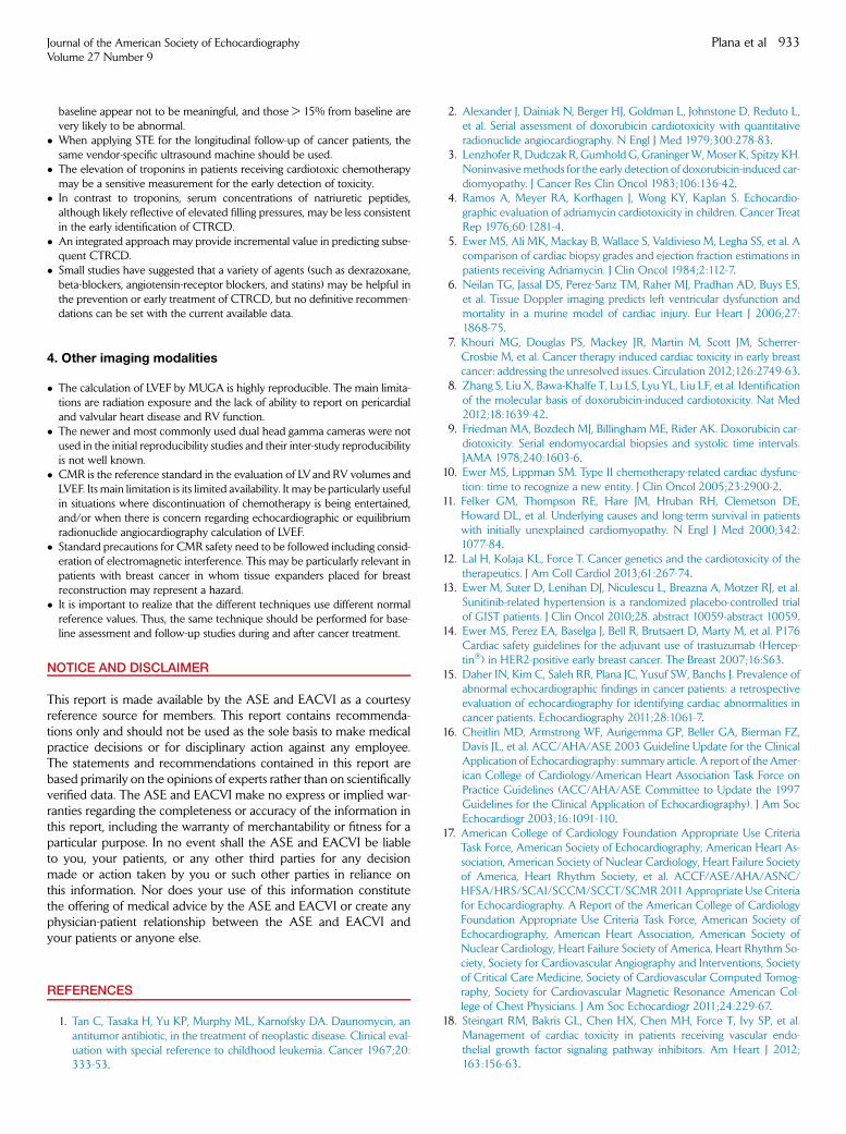
Journal of the American Society of EchocardiographyVolume 27 Number 9
Plana et al 933
baseline appear not to be meaningful, and those > 15% from baseline arevery likely to be abnormal.
� When applying STE for the longitudinal follow-up of cancer patients, thesame vendor-specific ultrasound machine should be used.
� The elevation of troponins in patients receiving cardiotoxic chemotherapymay be a sensitive measurement for the early detection of toxicity.
� In contrast to troponins, serum concentrations of natriuretic peptides,although likely reflective of elevated filling pressures, may be less consistentin the early identification of CTRCD.
� An integrated approach may provide incremental value in predicting subse-quent CTRCD.
� Small studies have suggested that a variety of agents (such as dexrazoxane,beta-blockers, angiotensin-receptor blockers, and statins) may be helpful inthe prevention or early treatment of CTRCD, but no definitive recommen-dations can be set with the current available data.
4. Other imaging modalities
� The calculation of LVEF by MUGA is highly reproducible. The main limita-tions are radiation exposure and the lack of ability to report on pericardialand valvular heart disease and RV function.
� The newer and most commonly used dual head gamma cameras were notused in the initial reproducibility studies and their inter-study reproducibilityis not well known.
� CMR is the reference standard in the evaluation of LVand RV volumes andLVEF. Its main limitation is its limited availability. It may be particularly usefulin situations where discontinuation of chemotherapy is being entertained,and/or when there is concern regarding echocardiographic or equilibriumradionuclide angiocardiography calculation of LVEF.
� Standard precautions for CMR safety need to be followed including consid-eration of electromagnetic interference. This may be particularly relevant inpatients with breast cancer in whom tissue expanders placed for breastreconstruction may represent a hazard.
� It is important to realize that the different techniques use different normalreference values. Thus, the same technique should be performed for base-line assessment and follow-up studies during and after cancer treatment.
NOTICE AND DISCLAIMER
This report is made available by the ASE and EACVI as a courtesyreference source for members. This report contains recommenda-tions only and should not be used as the sole basis to make medicalpractice decisions or for disciplinary action against any employee.The statements and recommendations contained in this report arebased primarily on the opinions of experts rather than on scientificallyverified data. The ASE and EACVI make no express or implied war-ranties regarding the completeness or accuracy of the information inthis report, including the warranty of merchantability or fitness for aparticular purpose. In no event shall the ASE and EACVI be liableto you, your patients, or any other third parties for any decisionmade or action taken by you or such other parties in reliance onthis information. Nor does your use of this information constitutethe offering of medical advice by the ASE and EACVI or create anyphysician-patient relationship between the ASE and EACVI andyour patients or anyone else.
REFERENCES
1. Tan C, Tasaka H, Yu KP, Murphy ML, Karnofsky DA. Daunomycin, anantitumor antibiotic, in the treatment of neoplastic disease. Clinical eval-uation with special reference to childhood leukemia. Cancer 1967;20:333-53.
2. Alexander J, Dainiak N, Berger HJ, Goldman L, Johnstone D, Reduto L,et al. Serial assessment of doxorubicin cardiotoxicity with quantitativeradionuclide angiocardiography. N Engl J Med 1979;300:278-83.
3. Lenzhofer R, Dudczak R, GumholdG, GraningerW,Moser K, Spitzy KH.Noninvasivemethods for the early detection of doxorubicin-induced car-diomyopathy. J Cancer Res Clin Oncol 1983;106:136-42.
4. Ramos A, Meyer RA, Korfhagen J, Wong KY, Kaplan S. Echocardio-graphic evaluation of adriamycin cardiotoxicity in children. Cancer TreatRep 1976;60:1281-4.
5. Ewer MS, Ali MK, Mackay B, Wallace S, Valdivieso M, Legha SS, et al. Acomparison of cardiac biopsy grades and ejection fraction estimations inpatients receiving Adriamycin. J Clin Oncol 1984;2:112-7.
6. Neilan TG, Jassal DS, Perez-Sanz TM, Raher MJ, Pradhan AD, Buys ES,et al. Tissue Doppler imaging predicts left ventricular dysfunction andmortality in a murine model of cardiac injury. Eur Heart J 2006;27:1868-75.
7. Khouri MG, Douglas PS, Mackey JR, Martin M, Scott JM, Scherrer-Crosbie M, et al. Cancer therapy-induced cardiac toxicity in early breastcancer: addressing the unresolved issues. Circulation 2012;126:2749-63.
8. Zhang S, Liu X, Bawa-Khalfe T, Lu LS, Lyu YL, Liu LF, et al. Identificationof the molecular basis of doxorubicin-induced cardiotoxicity. Nat Med2012;18:1639-42.
9. Friedman MA, Bozdech MJ, Billingham ME, Rider AK. Doxorubicin car-diotoxicity. Serial endomyocardial biopsies and systolic time intervals.JAMA 1978;240:1603-6.
10. Ewer MS, Lippman SM. Type II chemotherapy-related cardiac dysfunc-tion: time to recognize a new entity. J Clin Oncol 2005;23:2900-2.
11. Felker GM, Thompson RE, Hare JM, Hruban RH, Clemetson DE,Howard DL, et al. Underlying causes and long-term survival in patientswith initially unexplained cardiomyopathy. N Engl J Med 2000;342:1077-84.
12. Lal H, Kolaja KL, Force T. Cancer genetics and the cardiotoxicity of thetherapeutics. J Am Coll Cardiol 2013;61:267-74.
13. Ewer M, Suter D, Lenihan DJ, Niculescu L, Breazna A, Motzer RJ, et al.Sunitinib-related hypertension is a randomized placebo-controlled trialof GIST patients. J Clin Oncol 2010;28. abstract 10059-abstract 10059.
14. Ewer MS, Perez EA, Baselga J, Bell R, Brutsaert D, Marty M, et al. P176Cardiac safety guidelines for the adjuvant use of trastuzumab (Hercep-tin�) in HER2-positive early breast cancer. The Breast 2007;16:S63.
15. Daher IN, Kim C, Saleh RR, Plana JC, Yusuf SW, Banchs J. Prevalence ofabnormal echocardiographic findings in cancer patients: a retrospectiveevaluation of echocardiography for identifying cardiac abnormalities incancer patients. Echocardiography 2011;28:1061-7.
16. Cheitlin MD, Armstrong WF, Aurigemma GP, Beller GA, Bierman FZ,Davis JL, et al. ACC/AHA/ASE 2003 Guideline Update for the ClinicalApplication of Echocardiography: summary article. A report of the Amer-ican College of Cardiology/American Heart Association Task Force onPractice Guidelines (ACC/AHA/ASE Committee to Update the 1997Guidelines for the Clinical Application of Echocardiography). J Am SocEchocardiogr 2003;16:1091-110.
17. American College of Cardiology Foundation Appropriate Use CriteriaTask Force, American Society of Echocardiography, American Heart As-sociation, American Society of Nuclear Cardiology, Heart Failure Societyof America, Heart Rhythm Society, et al. ACCF/ASE/AHA/ASNC/HFSA/HRS/SCAI/SCCM/SCCT/SCMR2011Appropriate Use Criteriafor Echocardiography. A Report of the American College of CardiologyFoundation Appropriate Use Criteria Task Force, American Society ofEchocardiography, American Heart Association, American Society ofNuclear Cardiology, Heart Failure Society of America, Heart Rhythm So-ciety, Society for Cardiovascular Angiography and Interventions, Societyof Critical Care Medicine, Society of Cardiovascular Computed Tomog-raphy, Society for Cardiovascular Magnetic Resonance American Col-lege of Chest Physicians. J Am Soc Echocardiogr 2011;24:229-67.
18. Steingart RM, Bakris GL, Chen HX, Chen MH, Force T, Ivy SP, et al.Management of cardiac toxicity in patients receiving vascular endo-thelial growth factor signaling pathway inhibitors. Am Heart J 2012;163:156-63.
934 Plana et al Journal of the American Society of EchocardiographySeptember 2014
19. Eschenhagen T, Force T, Ewer MS, de Keulenaer GW, Suter TM,Anker SD, et al. Cardiovascular side effects of cancer therapies: a positionstatement from the Heart Failure Association of the European Society ofCardiology. Eur J Heart Fail 2011;13:1-10.
20. Lang RM, Bierig M, Devereux RB, Flachskampf FA, Foster E, Pellikka PA,et al. Recommendations for chamber quantification: a report from theAmerican Society of Echocardiography’s Guidelines and StandardsCommittee and the Chamber Quantification Writing Group, developedin conjunction with the European Association of Echocardiography, abranch of the European Society of Cardiology. J Am Soc Echocardiogr2005;18:1440-63.
21. Isner JM, Ferrans VJ, Cohen SR, Witkind BG, Virmani R, Gottdiener JS,et al. Clinical and morphologic cardiac findings after anthracyclinechemotherapy. Analysis of 64 patients studied at necropsy. Am J Cardiol1983;51:1167-74.
22. MuraruD, Badano LP, PelusoD, Dal Bianco L, Casablanca S, KocabayG,et al. Comprehensive analysis of left ventricular geometry and functionby three-dimensional echocardiography in healthy adults. J Am SocEchocardiogr 2013;26:618-28.
23. Rietzschel ER, De Buyzere ML, Bekaert S, Segers P, De Bacquer D,Cooman L, et al. Rationale, design, methods and baseline characteristicsof the Asklepios Study. Eur J Cardiovasc Prev Rehabil 2007;14:179-91.
24. Kuznetsova T, Herbots L, Lopez B, Jin Y, Richart T, Thijs L, et al. Preva-lence of left ventricular diastolic dysfunction in a general population. CircHeart Fail 2009;2:105-12.
25. Friedman GD, Cutter GR, Donahue RP, Hughes GH, Hulley SB,Jacobs DR Jr, et al. CARDIA: study design, recruitment, and some char-acteristics of the examined subjects. J Clin Epidemiol 1988;41:1105-16.
26. Lancellotti P, Badano LP, Lang RM, Akhaladze N, Athanassopoulos GD,Barone D, et al. Normal Reference Ranges for Echocardiography: ratio-nale, study design, and methodology (NORRE Study). Eur Heart J Car-diovasc Imaging 2013;14:303-8.
27. Bountioukos M, Doorduijn JK, Roelandt JR, Vourvouri EC, Bax JJ,Schinkel AF, et al. Repetitive dobutamine stress echocardiography forthe prediction of anthracycline cardiotoxicity. Eur J Echocardiogr 2003;4:300-5.
28. Ryberg M, Nielsen D, Skovsgaard T, Hansen J, Jensen BV,Dombernowsky P. Epirubicin cardiotoxicity: an analysis of 469 patientswith metastatic breast cancer. J Clin Oncol 1998;16:3502-8.
29. Nielsen D, Jensen JB, Dombernowsky P, Munck O, Fogh J, Brynjolf I,et al. Epirubicin cardiotoxicity: a study of 135 patients with advancedbreast cancer. J Clin Oncol 1990;8:1806-10.
30. Swain SM, Whaley FS, Ewer MS. Congestive heart failure in patientstreated with doxorubicin: a retrospective analysis of three trials. Cancer2003;97:2869-79.
31. Mitani I, Jain D, Joska TM, Burtness B, Zaret BL. Doxorubicin cardiotox-icity: prevention of congestive heart failure with serial cardiac functionmonitoring with equilibrium radionuclide angiocardiography in the cur-rent era. J Nucl Cardiol 2003;10:132-9.
32. Nousiainen T, Jantunen E, Vanninen E, Hartikainen J. Early decline in leftventricular ejection fraction predicts doxorubicin cardiotoxicity in lym-phoma patients. Br J Cancer 2002;86:1697-700.
33. Schwartz RG, McKenzie WB, Alexander J, Sager P, D’Souza A,Manatunga A, et al. Congestive heart failure and left ventricular dysfunc-tion complicating doxorubicin therapy. Seven-year experience using se-rial radionuclide angiocardiography. Am J Med 1987;82:1109-18.
34. Jensen BV, Skovsgaard T, Nielsen SL. Functional monitoring of anthracy-cline cardiotoxicity: a prospective, blinded, long-term observationalstudy of outcome in 120 patients. Ann Oncol 2002;13:699-709.
35. Steinherz LJ, Steinherz PG, Tan CT, Heller G, Murphy ML. Cardiactoxicity 4 to 20 years after completing anthracycline therapy. JAMA1991;266:1672-7.
36. Moja L, Tagliabue L, Balduzzi S, Parmelli E, Pistotti V, Guarneri V, et al.Trastuzumab containing regimens for early breast cancer. CochraneDatabase Syst Rev 2012;4:CD006243.
37. Seidman A, Hudis C, Pierri MK, Shak S, Paton V, Ashby M, et al. Cardiacdysfunction in the trastuzumab clinical trials experience. J Clin Oncol2002;20:1215-21.
38. Youssef G, Links M. The prevention and management of cardiovascularcomplications of chemotherapy in patients with cancer. Am J CardiovascDrugs 2005;5:233-43.
39. Keefe DL. Trastuzumab-associated cardiotoxicity. Cancer 2002;95:1592-600.
40. Schuchter LM, Hensley ML, Meropol NJ, Winer EP. American Society ofClinical Oncology Chemotherapy and Radiotherapy Expert Panel. 2002update of recommendations for the use of chemotherapy and radio-therapy protectants: clinical practice guidelines of the American Societyof Clinical Oncology. J Clin Oncol 2002;20:2895-903.
41. Carver JR, Shapiro CL, Ng A, Jacobs L, Schwartz C, Virgo KS, et al. Amer-ican Society of Clinical Oncology clinical evidence review on the ongoingcare of adult cancer survivors: cardiac and pulmonary late effects. J ClinOncol 2007;25:3991-4008.
42. Jacobs LD, Salgo IS, Goonewardena S, Weinert L, Coon P, Bardo D, et al.Rapid online quantification of left ventricular volume from real-timethree-dimensional echocardiographic data. Eur Heart J 2006;27:460-8.
43. Otterstad JE, Froeland G, St John Sutton M, Holme I. Accuracy andreproducibility of biplane two-dimensional echocardiographic measure-ments of left ventricular dimensions and function. Eur Heart J 1997;18:507-13.
44. Thavendiranathan P, Grant AD, Negishi T, Plana JC, Popovic ZB,Marwick TH. Reproducibility of echocardiographic techniques forsequential assessment of left ventricular ejection fraction and volumes:application to patients undergoing cancer chemotherapy. J Am Coll Car-diol 2013;61:77-84.
45. Ewer MS, Ewer SM. Long-term cardiac safety of dose-dense anthracy-cline therapy cannot be predicted from early ejection fraction data. JClin Oncol 2009;27:6073-5.
46. Karakurt C, Kocak G,Ozgen U. Evaluation of the left ventricular functionwith tissue tracking and tissue Doppler echocardiography in pediatricmalignancy survivors after anthracycline therapy. Echocardiography2008;25:880-7.
47. Ganame J, Claus P, Eyskens B, Uyttebroeck A, RenardM, D’hooge J, et al.Acute cardiac functional and morphological changes after Anthracyclineinfusions in children. Am J Cardiol 2007;99:974-7.
48. Tassan-Mangina S, Codorean D, Metivier M, Costa B, Himberlin C,Jouannaud C, et al. Tissue Doppler imaging and conventional echocardi-ography after anthracycline treatment in adults: early and late alterationsof left ventricular function during a prospective study. Eur J Echocardiogr2006;7:141-6.
49. Kapusta L, Thijssen JM, Groot-Loonen J, Antonius T, Mulder J,Daniels O. Tissue Doppler imaging in detection of myocardial dysfunc-tion in survivors of childhood cancer treated with anthracyclines. Ultra-sound Med Biol 2000;26:1099-108.
50. Nagueh SF, Appleton CP, Gillebert TC, Marino PN, Oh JK, Smiseth OA,et al. Recommendations for the evaluation of left ventricular diastolicfunction by echocardiography. Eur J Echocardiogr 2009;10:165-93.
51. Mason JW, Bristow MR, Billingham ME, Daniels JR. Invasive and noninvasive methods of assessing Adriamycin cardiotoxic effects in man: su-periority of histiopathologic assessment using endomyocardial biopsy.Cancer Treat Rep 1978;62:857-64.
52. Tanindi A, Demirci U, Tacoy G, Buyukberber S, Alsancak Y, Coskun U,et al. Assessment of right ventricular functions during cancer chemo-therapy. Eur J Echocardiogr 2011;12:834-40.
53. Rudski LG, Lai WW, Afilalo J, Hua L, Handschumacher MD,Chandrasekaran K, et al. Guidelines for the echocardiographic assess-ment of the right heart in adults: a report from the American Societyof Echocardiography endorsed by the European Association of Echocar-diography, a registered branch of the European Society of Cardiology,and the Canadian Society of Echocardiography. J Am Soc Echocardiogr2010;23:685-713: quiz 786–8.
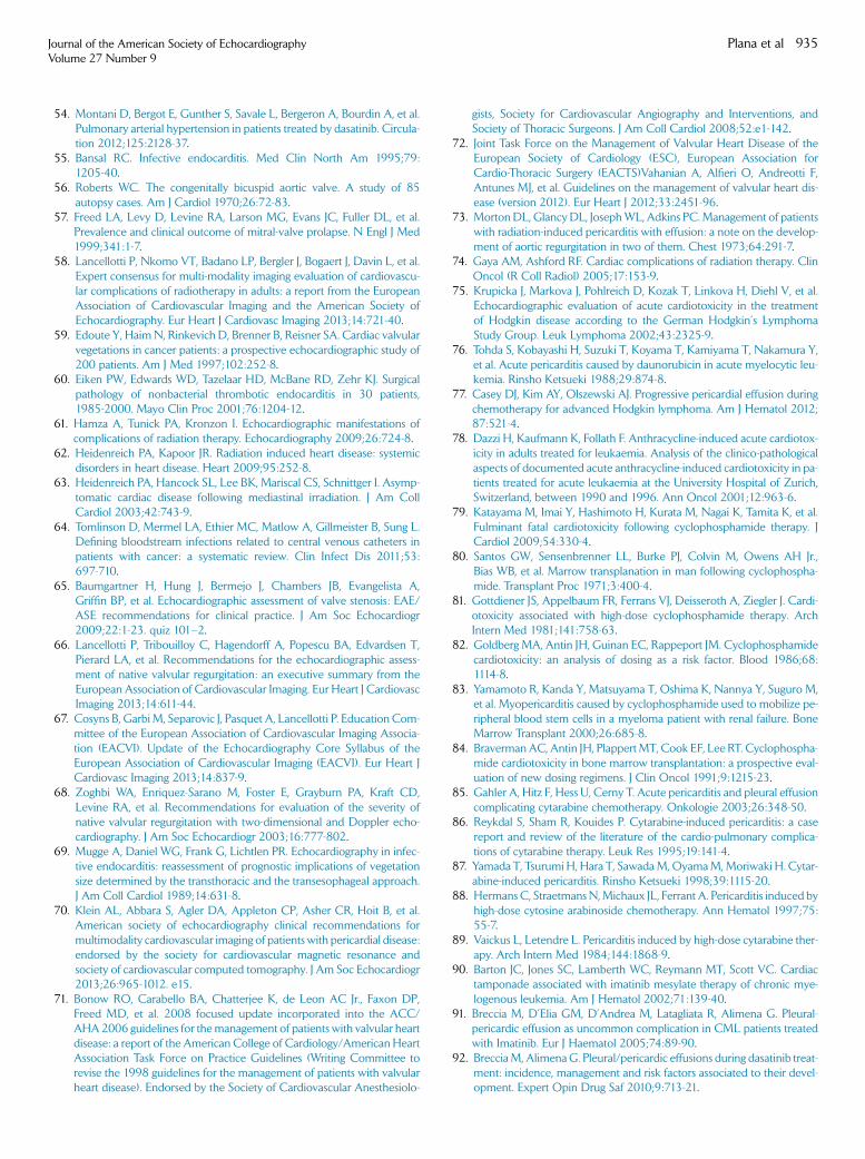
Journal of the American Society of EchocardiographyVolume 27 Number 9
Plana et al 935
54. Montani D, Bergot E, Gunther S, Savale L, Bergeron A, Bourdin A, et al.Pulmonary arterial hypertension in patients treated by dasatinib. Circula-tion 2012;125:2128-37.
55. Bansal RC. Infective endocarditis. Med Clin North Am 1995;79:1205-40.
56. Roberts WC. The congenitally bicuspid aortic valve. A study of 85autopsy cases. Am J Cardiol 1970;26:72-83.
57. Freed LA, Levy D, Levine RA, Larson MG, Evans JC, Fuller DL, et al.Prevalence and clinical outcome of mitral-valve prolapse. N Engl J Med1999;341:1-7.
58. Lancellotti P, Nkomo VT, Badano LP, Bergler J, Bogaert J, Davin L, et al.Expert consensus for multi-modality imaging evaluation of cardiovascu-lar complications of radiotherapy in adults: a report from the EuropeanAssociation of Cardiovascular Imaging and the American Society ofEchocardiography. Eur Heart J Cardiovasc Imaging 2013;14:721-40.
59. Edoute Y, HaimN, Rinkevich D, Brenner B, Reisner SA. Cardiac valvularvegetations in cancer patients: a prospective echocardiographic study of200 patients. Am J Med 1997;102:252-8.
60. Eiken PW, Edwards WD, Tazelaar HD, McBane RD, Zehr KJ. Surgicalpathology of nonbacterial thrombotic endocarditis in 30 patients,1985-2000. Mayo Clin Proc 2001;76:1204-12.
61. Hamza A, Tunick PA, Kronzon I. Echocardiographic manifestations ofcomplications of radiation therapy. Echocardiography 2009;26:724-8.
62. Heidenreich PA, Kapoor JR. Radiation induced heart disease: systemicdisorders in heart disease. Heart 2009;95:252-8.
63. Heidenreich PA, Hancock SL, Lee BK, Mariscal CS, Schnittger I. Asymp-tomatic cardiac disease following mediastinal irradiation. J Am CollCardiol 2003;42:743-9.
64. Tomlinson D, Mermel LA, Ethier MC, Matlow A, Gillmeister B, Sung L.Defining bloodstream infections related to central venous catheters inpatients with cancer: a systematic review. Clin Infect Dis 2011;53:697-710.
65. Baumgartner H, Hung J, Bermejo J, Chambers JB, Evangelista A,Griffin BP, et al. Echocardiographic assessment of valve stenosis: EAE/ASE recommendations for clinical practice. J Am Soc Echocardiogr2009;22:1-23. quiz 101–2.
66. Lancellotti P, Tribouilloy C, Hagendorff A, Popescu BA, Edvardsen T,Pierard LA, et al. Recommendations for the echocardiographic assess-ment of native valvular regurgitation: an executive summary from theEuropean Association of Cardiovascular Imaging. Eur Heart J CardiovascImaging 2013;14:611-44.
67. Cosyns B, Garbi M, Separovic J, Pasquet A, Lancellotti P. Education Com-mittee of the European Association of Cardiovascular Imaging Associa-tion (EACVI). Update of the Echocardiography Core Syllabus of theEuropean Association of Cardiovascular Imaging (EACVI). Eur Heart JCardiovasc Imaging 2013;14:837-9.
68. Zoghbi WA, Enriquez-Sarano M, Foster E, Grayburn PA, Kraft CD,Levine RA, et al. Recommendations for evaluation of the severity ofnative valvular regurgitation with two-dimensional and Doppler echo-cardiography. J Am Soc Echocardiogr 2003;16:777-802.
69. Mugge A, Daniel WG, Frank G, Lichtlen PR. Echocardiography in infec-tive endocarditis: reassessment of prognostic implications of vegetationsize determined by the transthoracic and the transesophageal approach.J Am Coll Cardiol 1989;14:631-8.
70. Klein AL, Abbara S, Agler DA, Appleton CP, Asher CR, Hoit B, et al.American society of echocardiography clinical recommendations formultimodality cardiovascular imaging of patients with pericardial disease:endorsed by the society for cardiovascular magnetic resonance andsociety of cardiovascular computed tomography. J Am Soc Echocardiogr2013;26:965-1012. e15.
71. Bonow RO, Carabello BA, Chatterjee K, de Leon AC Jr., Faxon DP,Freed MD, et al. 2008 focused update incorporated into the ACC/AHA2006 guidelines for themanagement of patients with valvular heartdisease: a report of the American College of Cardiology/American HeartAssociation Task Force on Practice Guidelines (Writing Committee torevise the 1998 guidelines for the management of patients with valvularheart disease). Endorsed by the Society of Cardiovascular Anesthesiolo-
gists, Society for Cardiovascular Angiography and Interventions, andSociety of Thoracic Surgeons. J Am Coll Cardiol 2008;52:e1-142.
72. Joint Task Force on the Management of Valvular Heart Disease of theEuropean Society of Cardiology (ESC), European Association forCardio-Thoracic Surgery (EACTS)Vahanian A, Alfieri O, Andreotti F,Antunes MJ, et al. Guidelines on the management of valvular heart dis-ease (version 2012). Eur Heart J 2012;33:2451-96.
73. MortonDL, GlancyDL, JosephWL, Adkins PC.Management of patientswith radiation-induced pericarditis with effusion: a note on the develop-ment of aortic regurgitation in two of them. Chest 1973;64:291-7.
74. Gaya AM, Ashford RF. Cardiac complications of radiation therapy. ClinOncol (R Coll Radiol) 2005;17:153-9.
75. Krupicka J, Markova J, Pohlreich D, Kozak T, Linkova H, Diehl V, et al.Echocardiographic evaluation of acute cardiotoxicity in the treatmentof Hodgkin disease according to the German Hodgkin’s LymphomaStudy Group. Leuk Lymphoma 2002;43:2325-9.
76. Tohda S, Kobayashi H, Suzuki T, Koyama T, Kamiyama T, Nakamura Y,et al. Acute pericarditis caused by daunorubicin in acute myelocytic leu-kemia. Rinsho Ketsueki 1988;29:874-8.
77. Casey DJ, Kim AY, Olszewski AJ. Progressive pericardial effusion duringchemotherapy for advanced Hodgkin lymphoma. Am J Hematol 2012;87:521-4.
78. Dazzi H, Kaufmann K, Follath F. Anthracycline-induced acute cardiotox-icity in adults treated for leukaemia. Analysis of the clinico-pathologicalaspects of documented acute anthracycline-induced cardiotoxicity in pa-tients treated for acute leukaemia at the University Hospital of Zurich,Switzerland, between 1990 and 1996. Ann Oncol 2001;12:963-6.
79. Katayama M, Imai Y, Hashimoto H, Kurata M, Nagai K, Tamita K, et al.Fulminant fatal cardiotoxicity following cyclophosphamide therapy. JCardiol 2009;54:330-4.
80. Santos GW, Sensenbrenner LL, Burke PJ, Colvin M, Owens AH Jr.,Bias WB, et al. Marrow transplanation in man following cyclophospha-mide. Transplant Proc 1971;3:400-4.
81. Gottdiener JS, Appelbaum FR, Ferrans VJ, Deisseroth A, Ziegler J. Cardi-otoxicity associated with high-dose cyclophosphamide therapy. ArchIntern Med 1981;141:758-63.
82. Goldberg MA, Antin JH, Guinan EC, Rappeport JM. Cyclophosphamidecardiotoxicity: an analysis of dosing as a risk factor. Blood 1986;68:1114-8.
83. Yamamoto R, Kanda Y, Matsuyama T, Oshima K, Nannya Y, Suguro M,et al. Myopericarditis caused by cyclophosphamide used to mobilize pe-ripheral blood stem cells in a myeloma patient with renal failure. BoneMarrow Transplant 2000;26:685-8.
84. BravermanAC, Antin JH, PlappertMT, Cook EF, Lee RT. Cyclophospha-mide cardiotoxicity in bone marrow transplantation: a prospective eval-uation of new dosing regimens. J Clin Oncol 1991;9:1215-23.
85. Gahler A, Hitz F, Hess U, Cerny T. Acute pericarditis and pleural effusioncomplicating cytarabine chemotherapy. Onkologie 2003;26:348-50.
86. Reykdal S, Sham R, Kouides P. Cytarabine-induced pericarditis: a casereport and review of the literature of the cardio-pulmonary complica-tions of cytarabine therapy. Leuk Res 1995;19:141-4.
87. Yamada T, Tsurumi H, Hara T, SawadaM,OyamaM,Moriwaki H. Cytar-abine-induced pericarditis. Rinsho Ketsueki 1998;39:1115-20.
88. Hermans C, StraetmansN,Michaux JL, Ferrant A. Pericarditis induced byhigh-dose cytosine arabinoside chemotherapy. Ann Hematol 1997;75:55-7.
89. Vaickus L, Letendre L. Pericarditis induced by high-dose cytarabine ther-apy. Arch Intern Med 1984;144:1868-9.
90. Barton JC, Jones SC, Lamberth WC, Reymann MT, Scott VC. Cardiactamponade associated with imatinib mesylate therapy of chronic mye-logenous leukemia. Am J Hematol 2002;71:139-40.
91. Breccia M, D’Elia GM, D’Andrea M, Latagliata R, Alimena G. Pleural-pericardic effusion as uncommon complication in CML patients treatedwith Imatinib. Eur J Haematol 2005;74:89-90.
92. BrecciaM, AlimenaG. Pleural/pericardic effusions during dasatinib treat-ment: incidence, management and risk factors associated to their devel-opment. Expert Opin Drug Saf 2010;9:713-21.
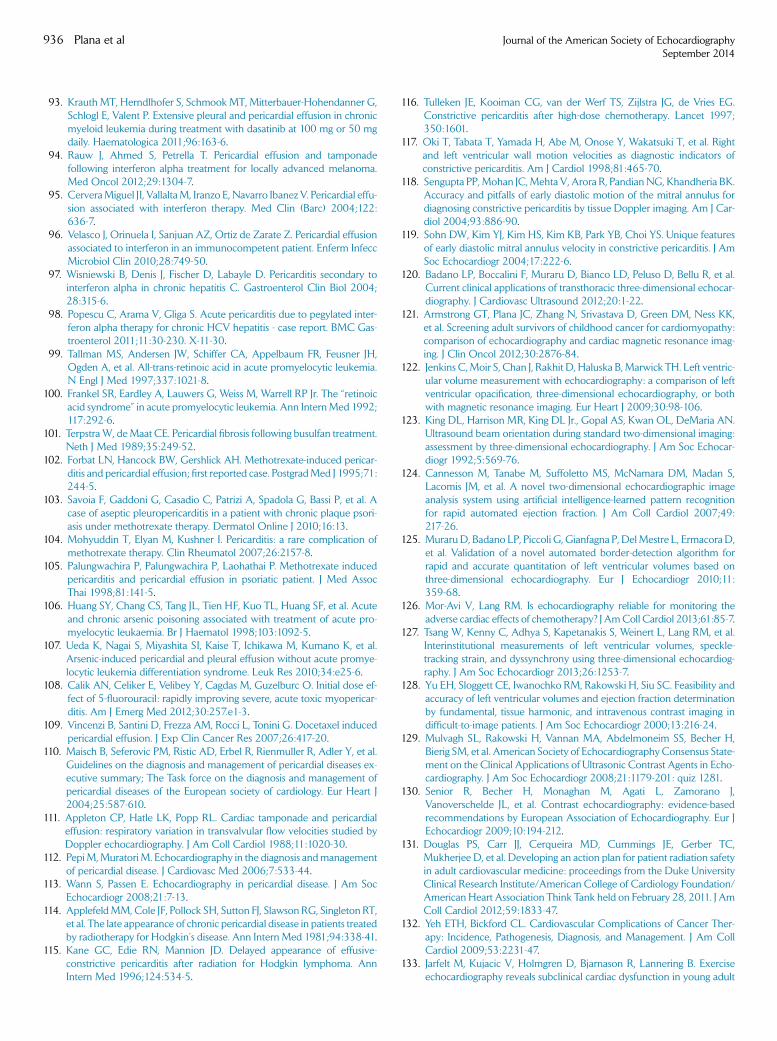
936 Plana et al Journal of the American Society of EchocardiographySeptember 2014
93. Krauth MT, Herndlhofer S, Schmook MT, Mitterbauer-Hohendanner G,Schlogl E, Valent P. Extensive pleural and pericardial effusion in chronicmyeloid leukemia during treatment with dasatinib at 100 mg or 50 mgdaily. Haematologica 2011;96:163-6.
94. Rauw J, Ahmed S, Petrella T. Pericardial effusion and tamponadefollowing interferon alpha treatment for locally advanced melanoma.Med Oncol 2012;29:1304-7.
95. CerveraMiguel JI, VallaltaM, Iranzo E, Navarro Ibanez V. Pericardial effu-sion associated with interferon therapy. Med Clin (Barc) 2004;122:636-7.
96. Velasco J, Orinuela I, Sanjuan AZ, Ortiz de Zarate Z. Pericardial effusionassociated to interferon in an immunocompetent patient. Enferm InfeccMicrobiol Clin 2010;28:749-50.
97. Wisniewski B, Denis J, Fischer D, Labayle D. Pericarditis secondary tointerferon alpha in chronic hepatitis C. Gastroenterol Clin Biol 2004;28:315-6.
98. Popescu C, Arama V, Gliga S. Acute pericarditis due to pegylated inter-feron alpha therapy for chronic HCV hepatitis - case report. BMC Gas-troenterol 2011;11:30-230. X-11-30.
99. Tallman MS, Andersen JW, Schiffer CA, Appelbaum FR, Feusner JH,Ogden A, et al. All-trans-retinoic acid in acute promyelocytic leukemia.N Engl J Med 1997;337:1021-8.
100. Frankel SR, Eardley A, Lauwers G, Weiss M, Warrell RP Jr. The ‘‘retinoicacid syndrome’’ in acute promyelocytic leukemia. Ann InternMed 1992;117:292-6.
101. TerpstraW, deMaat CE. Pericardial fibrosis following busulfan treatment.Neth J Med 1989;35:249-52.
102. Forbat LN, Hancock BW, Gershlick AH. Methotrexate-induced pericar-ditis and pericardial effusion; first reported case. PostgradMed J 1995;71:244-5.
103. Savoia F, Gaddoni G, Casadio C, Patrizi A, Spadola G, Bassi P, et al. Acase of aseptic pleuropericarditis in a patient with chronic plaque psori-asis under methotrexate therapy. Dermatol Online J 2010;16:13.
104. Mohyuddin T, Elyan M, Kushner I. Pericarditis: a rare complication ofmethotrexate therapy. Clin Rheumatol 2007;26:2157-8.
105. Palungwachira P, Palungwachira P, Laohathai P. Methotrexate inducedpericarditis and pericardial effusion in psoriatic patient. J Med AssocThai 1998;81:141-5.
106. Huang SY, Chang CS, Tang JL, Tien HF, Kuo TL, Huang SF, et al. Acuteand chronic arsenic poisoning associated with treatment of acute pro-myelocytic leukaemia. Br J Haematol 1998;103:1092-5.
107. Ueda K, Nagai S, Miyashita SI, Kaise T, Ichikawa M, Kumano K, et al.Arsenic-induced pericardial and pleural effusion without acute promye-locytic leukemia differentiation syndrome. Leuk Res 2010;34:e25-6.
108. Calik AN, Celiker E, Velibey Y, Cagdas M, Guzelburc O. Initial dose ef-fect of 5-fluorouracil: rapidly improving severe, acute toxic myopericar-ditis. Am J Emerg Med 2012;30:257.e1-3.
109. Vincenzi B, Santini D, Frezza AM, Rocci L, Tonini G. Docetaxel inducedpericardial effusion. J Exp Clin Cancer Res 2007;26:417-20.
110. Maisch B, Seferovic PM, Ristic AD, Erbel R, Rienmuller R, Adler Y, et al.Guidelines on the diagnosis and management of pericardial diseases ex-ecutive summary; The Task force on the diagnosis and management ofpericardial diseases of the European society of cardiology. Eur Heart J2004;25:587-610.
111. Appleton CP, Hatle LK, Popp RL. Cardiac tamponade and pericardialeffusion: respiratory variation in transvalvular flow velocities studied byDoppler echocardiography. J Am Coll Cardiol 1988;11:1020-30.
112. PepiM,Muratori M. Echocardiography in the diagnosis andmanagementof pericardial disease. J Cardiovasc Med 2006;7:533-44.
113. Wann S, Passen E. Echocardiography in pericardial disease. J Am SocEchocardiogr 2008;21:7-13.
114. ApplefeldMM, Cole JF, Pollock SH, Sutton FJ, Slawson RG, Singleton RT,et al. The late appearance of chronic pericardial disease in patients treatedby radiotherapy for Hodgkin’s disease. Ann Intern Med 1981;94:338-41.
115. Kane GC, Edie RN, Mannion JD. Delayed appearance of effusive-constrictive pericarditis after radiation for Hodgkin lymphoma. AnnIntern Med 1996;124:534-5.
116. Tulleken JE, Kooiman CG, van der Werf TS, Zijlstra JG, de Vries EG.Constrictive pericarditis after high-dose chemotherapy. Lancet 1997;350:1601.
117. Oki T, Tabata T, Yamada H, Abe M, Onose Y, Wakatsuki T, et al. Rightand left ventricular wall motion velocities as diagnostic indicators ofconstrictive pericarditis. Am J Cardiol 1998;81:465-70.
118. Sengupta PP, Mohan JC,Mehta V, Arora R, PandianNG, Khandheria BK.Accuracy and pitfalls of early diastolic motion of the mitral annulus fordiagnosing constrictive pericarditis by tissue Doppler imaging. Am J Car-diol 2004;93:886-90.
119. Sohn DW, Kim YJ, Kim HS, Kim KB, Park YB, Choi YS. Unique featuresof early diastolic mitral annulus velocity in constrictive pericarditis. J AmSoc Echocardiogr 2004;17:222-6.
120. Badano LP, Boccalini F, Muraru D, Bianco LD, Peluso D, Bellu R, et al.Current clinical applications of transthoracic three-dimensional echocar-diography. J Cardiovasc Ultrasound 2012;20:1-22.
121. Armstrong GT, Plana JC, Zhang N, Srivastava D, Green DM, Ness KK,et al. Screening adult survivors of childhood cancer for cardiomyopathy:comparison of echocardiography and cardiac magnetic resonance imag-ing. J Clin Oncol 2012;30:2876-84.
122. Jenkins C,Moir S, Chan J, Rakhit D, Haluska B, Marwick TH. Left ventric-ular volume measurement with echocardiography: a comparison of leftventricular opacification, three-dimensional echocardiography, or bothwith magnetic resonance imaging. Eur Heart J 2009;30:98-106.
123. King DL, Harrison MR, King DL Jr., Gopal AS, Kwan OL, DeMaria AN.Ultrasound beam orientation during standard two-dimensional imaging:assessment by three-dimensional echocardiography. J Am Soc Echocar-diogr 1992;5:569-76.
124. Cannesson M, Tanabe M, Suffoletto MS, McNamara DM, Madan S,Lacomis JM, et al. A novel two-dimensional echocardiographic imageanalysis system using artificial intelligence-learned pattern recognitionfor rapid automated ejection fraction. J Am Coll Cardiol 2007;49:217-26.
125. MuraruD, Badano LP, Piccoli G, Gianfagna P, DelMestre L, ErmacoraD,et al. Validation of a novel automated border-detection algorithm forrapid and accurate quantitation of left ventricular volumes based onthree-dimensional echocardiography. Eur J Echocardiogr 2010;11:359-68.
126. Mor-Avi V, Lang RM. Is echocardiography reliable for monitoring theadverse cardiac effects of chemotherapy? J AmColl Cardiol 2013;61:85-7.
127. Tsang W, Kenny C, Adhya S, Kapetanakis S, Weinert L, Lang RM, et al.Interinstitutional measurements of left ventricular volumes, speckle-tracking strain, and dyssynchrony using three-dimensional echocardiog-raphy. J Am Soc Echocardiogr 2013;26:1253-7.
128. Yu EH, Sloggett CE, Iwanochko RM, Rakowski H, Siu SC. Feasibility andaccuracy of left ventricular volumes and ejection fraction determinationby fundamental, tissue harmonic, and intravenous contrast imaging indifficult-to-image patients. J Am Soc Echocardiogr 2000;13:216-24.
129. Mulvagh SL, Rakowski H, Vannan MA, Abdelmoneim SS, Becher H,Bierig SM, et al. American Society of EchocardiographyConsensus State-ment on the Clinical Applications of Ultrasonic Contrast Agents in Echo-cardiography. J Am Soc Echocardiogr 2008;21:1179-201: quiz 1281.
130. Senior R, Becher H, Monaghan M, Agati L, Zamorano J,Vanoverschelde JL, et al. Contrast echocardiography: evidence-basedrecommendations by European Association of Echocardiography. Eur JEchocardiogr 2009;10:194-212.
131. Douglas PS, Carr JJ, Cerqueira MD, Cummings JE, Gerber TC,Mukherjee D, et al. Developing an action plan for patient radiation safetyin adult cardiovascular medicine: proceedings from the Duke UniversityClinical Research Institute/American College of Cardiology Foundation/American Heart Association Think Tank held on February 28, 2011. J AmColl Cardiol 2012;59:1833-47.
132. Yeh ETH, Bickford CL. Cardiovascular Complications of Cancer Ther-apy: Incidence, Pathogenesis, Diagnosis, and Management. J Am CollCardiol 2009;53:2231-47.
133. Jarfelt M, Kujacic V, Holmgren D, Bjarnason R, Lannering B. Exerciseechocardiography reveals subclinical cardiac dysfunction in young adult
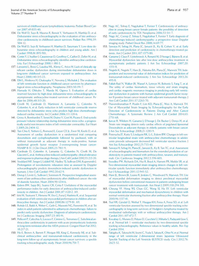
Journal of the American Society of EchocardiographyVolume 27 Number 9
Plana et al 937
survivors of childhood acute lymphoblastic leukemia. Pediatr Blood Can-cer 2007;49:835-40.
134. De Wolf D, Suys B, Maurus R, Benoit Y, Verhaaren H, Matthijs D, et al.Dobutamine stress echocardiography in the evaluation of late anthracy-cline cardiotoxicity in childhood cancer survivors. Pediatr Res 1996;39:504-12.
135. De Wolf D, Suys B, Verhaaren H, Matthys D, Taeymans Y. Low-dose do-butamine stress echocardiography in children and young adults. Am JCardiol 1998;81:895-901.
136. Cottin Y, L’huillier I, Casasnovas O, Geoffroy C, Caillot D, ZellerM, et al.Dobutamine stress echocardiography identifies anthracycline cardiotox-icity. Eur J Echocardiogr 2000;1:180-3.
137. Lanzarini L, Bossi G, LaudisaML, Klersy C, AricoM. Lack of clinically sig-nificant cardiac dysfunction during intermediate dobutamine doses inlong-term childhood cancer survivors exposed to anthracyclines. AmHeart J 2000;140:315-23.
138. Elbl L, Hrstkova H, Chaloupka V, Novotny J, Michalek J. The evaluationof left ventricular function in childhood cancer survivors by pharmaco-logical stress echocardiography. Neoplasma 2003;50:191-7.
139. Hamada H, Ohkubo T, Maeda M, Ogawa S. Evaluation of cardiacreserved function by high-dose dobutamine-stress echocardiography inasymptomatic anthracycline-treated survivors of childhood cancer. Pe-diatr Int 2006;48:313-20.
140. Civelli M, Cardinale D, Martinoni A, Lamantia G, Colombo N,Colombo A, et al. Early reduction in left ventricular contractile reservedetected by dobutamine stress echo predicts high-dose chemotherapy-induced cardiac toxicity. Int J Cardiol 2006;111:120-6.
141. GrosuA, Bombardini T, SenniM,Duino V, GoriM, Picano E. End-systolicpressure/volume relationship during dobutamine stress echo: a prognos-tically useful non-invasive index of left ventricular contractility. Eur HeartJ 2005;26:2404-12.
142. Tan-Chiu E, Yothers G, Romond E, Geyer CE Jr., Ewer M, Keefe D, et al.Assessment of cardiac dysfunction in a randomized trial comparingdoxorubicin and cyclophosphamide followed by paclitaxel, with orwithout trastuzumab as adjuvant therapy in node-positive, humanepidermal growth factor receptor 2-overexpressing breast cancer:NSABP B-31. J Clin Oncol 2005;23:7811-9.
143. Cardinale D, Colombo A, Lamantia G, Colombo N, Civelli M, DeGiacomiG, et al. Anthracycline-induced cardiomyopathy: clinical relevanceand response topharmacologic therapy. JAmCollCardiol 2010;55:213-20.
144. StoddardMF, Seeger J, Liddell NE, Hadley TJ, SullivanDM, Kupersmith J.Prolongation of isovolumetric relaxation time as assessed by Dopplerechocardiography predicts doxorubicin-induced systolic dysfunction inhumans. J Am Coll Cardiol 1992;20:62-9.
145. Dorup I, Levitt G, Sullivan I, Sorensen K. Prospective longitudinal assess-ment of late anthracycline cardiotoxicity after childhood cancer: the roleof diastolic function. Heart 2004;90:1214-6.
146. Eidem BW, Sapp BG, Suarez CR, Cetta F. Usefulness of the myocardialperformance index for early detection of anthracycline-induced cardio-toxicity in children. Am J Cardiol 2001;87:1120-2. A9.
147. Ishii M, Tsutsumi T, HimenoW, Eto G, Furui J, Hashino K, et al. Sequentialevaluation of left ventricular myocardial performance in children after an-thracycline therapy. Am J Cardiol 2000;86:1279-81. A9.
148. Rohde LE, Baldi A,Weber C, Geib G,Mazzotti NG, Fiorentini M, et al. Teiindex in adult patients submitted to adriamycin chemotherapy: failure topredict early systolic dysfunction. Diagnosis of adriamycin cardiotoxicity.Int J Cardiovasc Imaging 2007;23:185-91.
149. Pellicori P, Calicchia A, Lococo F, Cimino G, Torromeo C. Subclinical an-thracycline cardiotoxicity in patients with acute promyelocytic leukemiain long-term remission after the AIDA protocol. Congest Heart Fail 2012;18:217-21.
150. Ho E, Brown A, Barrett P, Morgan RB, King G, Kennedy MJ, et al. Sub-clinical anthracycline- and trastuzumab-induced cardiotoxicity in thelong-term follow-up of asymptomatic breast cancer survivors: a speckletracking echocardiographic study. Heart 2010;96:701-7.
151. Nagy AC, Tolnay E, Nagykalnai T, Forster T. Cardiotoxicity of anthracy-cline in young breast cancer female patients: the possibility of detectionof early cardiotoxicity by TDI. Neoplasma 2006;53:511-7.
152. Nagy AC, Cserep Z, Tolnay E, Nagykalnai T, Forster T. Early diagnosis ofchemotherapy-induced cardiomyopathy: a prospective tissue Dopplerimaging study. Pathol Oncol Res 2008;14:69-77.
153. Sawaya H, Sebag IA, Plana JC, Januzzi JL, Ky B, Cohen V, et al. Earlydetection and prediction of cardiotoxicity in chemotherapy-treated pa-tients. Am J Cardiol 2011;107:1375-80.
154. Ganame J, Claus P, Uyttebroeck A, Renard M, D’hooge J, Bijnens B, et al.Myocardial dysfunction late after low-dose anthracycline treatment inasymptomatic pediatric patients. J Am Soc Echocardiogr 2007;20:1351-8.
155. Negishi K, Negishi T, Hare JL, Haluska BA, Plana JC, Marwick TH. Inde-pendent and incremental value of deformation indices for prediction oftrastuzumab-induced cardiotoxicity. J Am Soc Echocardiogr 2013;26:493-8.
156. Fallah-Rad N, Walker JR, Wassef A, Lytwyn M, Bohonis S, Fang T, et al.The utility of cardiac biomarkers, tissue velocity and strain imaging,and cardiac magnetic resonance imaging in predicting early left ventric-ular dysfunction in patients with human epidermal growth factor recep-tor II-positive breast cancer treated with adjuvant trastuzumab therapy. JAm Coll Cardiol 2011;57:2263-70.
157. Thavendiranathan P, Poulin F, Lim KD, Plana JC, Woo A, Marwick TH.Use of Myocardial Strain Imaging by Echocardiography for the EarlyDetection of Cardiotoxicity in Patients During and After CancerChemotherapy: A Systematic Review. J Am Coll Cardiol 2014;63:2751-68.
158. Jurcut R, Wildiers H, Ganame J, D’hooge J, De Backer J, Denys H, et al.Strain rate imaging detects early cardiac effects of pegylated liposomalDoxorubicin as adjuvant therapy in elderly patients with breast cancer.J Am Soc Echocardiogr 2008;21:1283-9.
159. Poterucha JT, Kutty S, Lindquist RK, Li L, Eidem BW. Changes in left ven-tricular longitudinal strain with anthracycline chemotherapy in adoles-cents precede subsequent decreased left ventricular ejection fraction. JAm Soc Echocardiogr 2012;25:733-40.
160. Sawaya H, Sebag IA, Plana JC, Januzzi JL, Ky B, Tan TC, et al. Assessmentof echocardiography and biomarkers for the extended prediction of car-diotoxicity in patients treated with anthracyclines, taxanes, and trastuzu-mab. Circ Cardiovasc Imaging 2012;5:596-603.
161. Stoodley PW, Richards DA, Hui R, Boyd A, Harnett PR, Meikle SR, et al.Two-dimensional myocardial strain imaging detects changes in left ven-tricular systolic function immediately after anthracycline chemotherapy.Eur J Echocardiogr 2011;12:945-52.
162. Hare JL, Brown JK, Leano R, Jenkins C, Woodward N, Marwick TH. Useof myocardial deformation imaging to detect preclinical myocardialdysfunction before conventional measures in patients undergoing breastcancer treatment with trastuzumab. Am Heart J 2009;158:294-301.
163. Cheung YF, Hong WJ, Chan GC, Wong SJ, Ha SY. Left ventricularmyocardial deformation and mechanical dyssynchrony in children withnormal ventricular shortening fraction after anthracycline therapy. Heart2010;96:1137-41.
164. Tsai HR, Gjesdal O, Wethal T, Haugaa KH, Fossa A, Fossa SD, et al. Leftventricular function assessed by two-dimensional speckle tracking echo-cardiography in long-term survivors of Hodgkin’s lymphoma treated bymediastinal radiotherapy with or without anthracycline therapy. Am JCardiol 2011;107:472-7.
165. Kocabay G,Muraru D, Peluso D, Cucchini U, Mihaila S, Padayattil-Jose S,et al. Normal left 1 ventricular mechanics by two-dimensional speckletracking echocardiography. Reference values in healthy adults. Rev EspCardiol 2014.
166. Takigiku K, TakeuchiM, Izumi C, Yuda S, Sakata K, OhteN, et al. Normalrange of left ventricular 2-dimensional strain: Japanese UltrasoundSpeckle Tracking of the Left Ventricle (JUSTICE) study. Circ J 2012;76:2623-32.
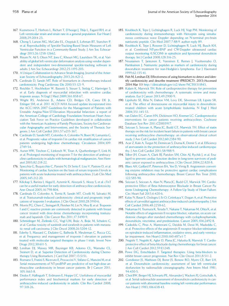
938 Plana et al Journal of the American Society of EchocardiographySeptember 2014
167. Kuznetsova T, Herbots L, Richart T, D’hooge J, Thijs L, Fagard RH, et al.Left ventricular strain and strain rate in a general population. Eur Heart J2008;29:2014-23.
168. Cheng S, Larson MG, McCabe EL, Osypiuk E, Lehman BT, Stanchev P,et al. Reproducibility of Speckle-Tracking-Based Strain Measures of LeftVentricular Function in a Community-Based Study. J Am Soc Echocar-diogr 2013;26:1258-12662.
169. Risum N, Ali S, Olsen NT, Jons C, Khouri MG, Lauridsen TK, et al. Vari-ability of global left ventricular deformation analysis using vendor depen-dent and independent two-dimensional speckle-tracking software inadults. J Am Soc Echocardiogr 2012;25:1195-203.
170. A Unique Collaboration to Advance Strain Imaging. Journal of the Amer-ican Society of Echocardiography 2013;26:A21-2.
171. Cardinale D, Sandri MT. Role of biomarkers in chemotherapy-inducedcardiotoxicity. Prog Cardiovasc Dis 2010;53:121-9.
172. Reichlin T, Hochholzer W, Bassetti S, Steuer S, Stelzig C, Hartwiger S,et al. Early diagnosis of myocardial infarction with sensitive cardiactroponin assays. N Engl J Med 2009;361:858-67.
173. Wright RS, Anderson JL, Adams CD, Bridges CR, Casey DE Jr.,Ettinger SM, et al. 2011 ACCF/AHA focused update incorporated intothe ACC/AHA 2007 Guidelines for the Management of Patients withUnstable Angina/Non-ST-Elevation Myocardial Infarction: a report ofthe American College of Cardiology Foundation/American Heart Asso-ciation Task Force on Practice Guidelines developed in collaborationwith the American Academy of Family Physicians, Society for Cardiovas-cular Angiography and Interventions, and the Society of Thoracic Sur-geons. J Am Coll Cardiol 2011;57:e215-367.
174. Cardinale D, Sandri MT, Colombo A, Colombo N, Boeri M, Lamantia G,et al. Prognostic value of troponin I in cardiac risk stratification of cancerpatients undergoing high-dose chemotherapy. Circulation 2004;109:2749-54.
175. Auner HW, Tinchon C, Linkesch W, Tiran A, Quehenberger F, Link H,et al. Prolonged monitoring of troponin T for the detection of anthracy-cline cardiotoxicity in adults with hematological malignancies. AnnHem-atol 2003;82:218-22.
176. Specchia G, Buquicchio C, Pansini N, Di Serio F, Liso V, Pastore D, et al.Monitoring of cardiac function on the basis of serum troponin I levels inpatients with acute leukemia treated with anthracyclines. J Lab Clin Med2005;145:212-20.
177. Kilickap S, Barista I, Akgul E, Aytemir K, Aksoyek S, Aksoy S, et al. cTnTcan be a useful marker for early detection of anthracycline cardiotoxicity.Ann Oncol 2005;16:798-804.
178. Cardinale D, Colombo A, Torrisi R, Sandri MT, Civelli M, Salvatici M,et al. Trastuzumab-induced cardiotoxicity: clinical and prognostic impli-cations of troponin I evaluation. J Clin Oncol 2010;28:3910-6.
179. Morris PG, Chen C, Steingart R, FleisherM, Lin N,Moy B, et al. TroponinI and C-reactive protein are commonly detected in patients with breastcancer treated with dose-dense chemotherapy incorporating trastuzu-mab and lapatinib. Clin Cancer Res 2011;17:3490-9.
180. Schmidinger M, Zielinski CC, Vogl UM, Bojic A, Bojic M, Schukro C,et al. Cardiac toxicity of sunitinib and sorafenib in patients with metasta-tic renal cell carcinoma. J Clin Oncol 2008;26:5204-12.
181. Ederhy S, Massard C, Dufaitre G, Balheda R, Meuleman C, Rocca CG,et al. Frequency and management of troponin I elevation in patientstreated with molecular targeted therapies in phase I trials. Invest NewDrugs 2012;30:611-5.
182. Lenihan DJ, Massey MR, Baysinger KB, Adorno CL, Warneke CL,Steinert D, et al. Superior Detection of Cardiotoxicity during Chemo-therapy Using Biomarkers. J Card Fail 2007;13:S151.
183. Romano S, Fratini S, Ricevuto E, Procaccini V, StifanoG,ManciniM, et al.Serial measurements of NT-proBNP are predictive of not-high-dose an-thracycline cardiotoxicity in breast cancer patients. Br J Cancer 2011;105:1663-8.
184. Dodos F, Halbsguth T, Erdmann E, Hoppe UC. Usefulness of myocardialperformance index and biochemical markers for early detection ofanthracycline-induced cardiotoxicity in adults. Clin Res Cardiol 2008;97:318-26.
185. Knobloch K, Tepe J, Lichtinghagen R, Luck HJ, Vogt PM. Monitoring ofcardiotoxicity during immunotherapy with Herceptin using simulta-neous continuous wave Doppler depending on N-terminal pro-brainnatriuretic peptide. Clin Med 2007;7:88-9. author reply 89.
186. Knobloch K, Tepe J, Rossner D, Lichtinghagen R, Luck HJ, Busch KH,et al. Combined NT-pro-BNP and CW-Doppler ultrasound cardiacoutput monitoring (USCOM) in epirubicin and liposomal doxorubicintherapy. Int J Cardiol 2008;128:316-25.
187. Nousiainen T, Jantunen E, Vanninen E, Remes J, Vuolteenaho O,Hartikainen J. Natriuretic peptides as markers of cardiotoxicity duringdoxorubicin treatment for non-Hodgkin’s lymphoma. Eur J Haematol1999;62:135-41.
188. Fish M, Lenihan DJ. Effectiveness of using biomarkers to detect and iden-tify cardiotoxicity and describe treatment (PREDICT). 2013;(Accessed2014 Mar 11) http://clinicaltrials.gov/ct2/show/NCT01311843.
189. Kalam K, Marwick TH. Role of cardioprotective therapy for preventionof cardiotoxicity with chemotherapy: A systematic review and meta-analysis. Eur J Cancer 2013;49:2900-9.
190. Lipshultz SE, Rifai N, Dalton VM, Levy DE, Silverman LB, Lipsitz SR,et al. The effect of dexrazoxane on myocardial injury in doxorubicin-treated children with acute lymphoblastic leukemia. N Engl J Med2004;351:145-53.
191. van Dalen EC, Caron HN, Dickinson HO, Kremer LC. Cardioprotectiveinterventions for cancer patients receiving anthracyclines. CochraneDatabase Syst Rev 2011;CD003917.
192. Seicean S, Seicean A, Plana JC, Budd GT, Marwick TH. Effect of statintherapy on the risk for incident heart failure in patients with breast cancerreceiving anthracycline chemotherapy: an observational clinical cohortstudy. J Am Coll Cardiol 2012;60:2384-90.
193. Acar Z, Kale A, TurgutM, Demircan S, Durna K, Demir S, et al. Efficiencyof atorvastatin in the protection of anthracycline-induced cardiomyopa-thy. J Am Coll Cardiol 2011;58:988-9.
194. Silber JH, Cnaan A, Clark BJ, Paridon SM, Chin AJ, Rychik J, et al. Ena-lapril to prevent cardiac function decline in long-term survivors of pedi-atric cancer exposed to anthracyclines. J Clin Oncol 2004;22:820-8.
195. Blaes AH, Gaillard P, Peterson BA, Yee D, Virnig B. Angiotensin convert-ing enzyme inhibitors may be protective against cardiac complicationsfollowing anthracycline chemotherapy. Breast Cancer Res Treat 2010;122:585-90.
196. Seicean S, Seicean A, Alan N, Plana JC, Budd GT, Marwick TH. Cardio-protective Effect of Beta-Adrenoceptor Blockade in Breast Cancer Pa-tients Undergoing Chemotherapy: A Follow-Up Study of Heart Failure.Circ Heart Fail 2013;6:420-6.
197. Kalay N, Basar E, Ozdogru I, Er O, Cetinkaya Y, Dogan A, et al. Protectiveeffects of carvedilol against anthracycline-induced cardiomyopathy. J AmColl Cardiol 2006;48:2258-62.
198. Nakamae H, Tsumura K, Terada Y, Nakane T, NakamaeM, Ohta K, et al.Notable effects of angiotensin II receptor blocker, valsartan, on acute car-diotoxic changes after standard chemotherapy with cyclophosphamide,doxorubicin, vincristine, and prednisolone. Cancer 2005;104:2492-8.
199. Cadeddu C, Piras A, Mantovani G, Deidda M, Dessi M, Madeddu C,et al. Protective effects of the angiotensin II receptor blocker telmisartanon epirubicin-induced inflammation, oxidative stress, and early ventricu-lar impairment. Am Heart J 2010;160:487.e1-7.
200. Negishi T, Negishi K, Agler D, Plana JC, Haluska B, Marwick T. Cardio-protective effect of beta-blockade during chemotherapy for breat cancer.J Am Coll Cardiol 2012;59:E1248.
201. Powe DG, Entschladen F. Targeted therapies: Using beta-blockers toinhibit breast cancer progression. Nat Rev Clin Oncol 2011;8:511-2.
202. Gottdiener JS, Mathisen DJ, Borer JS, Bonow RO, Myers CE, Barr LH,et al. Doxorubicin cardiotoxicity: assessment of late left ventriculardysfunction by radionuclide cineangiography. Ann Intern Med 1981;94:430-5.
203. Choi BW, Berger HJ, Schwartz PE, Alexander J, Wackers FJ, Gottschalk A,et al. Serial radionuclide assessment of doxorubicin cardiotoxicity in can-cer patients with abnormal baseline resting left ventricular performance.Am Heart J 1983;106:638-43.
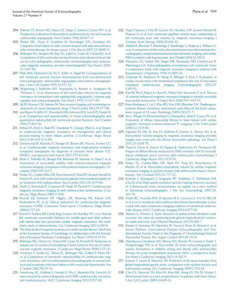
Journal of the American Society of EchocardiographyVolume 27 Number 9
Plana et al 939
204. Palmeri ST, Bonow RO, Myers CE, Seipp C, Jenkins J, Green MV, et al.Prospective evaluation of doxorubicin cardiotoxicity by rest and exerciseradionuclide angiography. Am J Cardiol 1986;58:607-13.
205. Pinder MC, Duan Z, Goodwin JS, Hortobagyi GN, Giordano SH.Congestive heart failure in older women treated with adjuvant anthracy-cline chemotherapy for breast cancer. J Clin Oncol 2007;25:3808-15.
206. Bellenger NG, Burgess MI, Ray SG, Lahiri A, Coats AJ, Cleland JG, et al.Comparison of left ventricular ejection fraction and volumes in heart fail-ure by echocardiography, radionuclide ventriculography and cardiovas-cular magnetic resonance; are they interchangeable? Eur Heart J 2000;21:1387-96.
207. Naik MM, Diamond GA, Pai T, Soffer A, Siegel RJ. Correspondence ofleft ventricular ejection fraction determinations from two-dimensionalechocardiography, radionuclide angiography and contrast cineangiogra-phy. J Am Coll Cardiol 1995;25:937-42.
208. Mogelvang J, Stokholm KH, Saunamaki K, Reimer A, Stubgaard M,Thomsen C, et al. Assessment of left ventricular volumes by magneticresonance in comparison with radionuclide angiography, contrast angi-ography and echocardiography. Eur Heart J 1992;13:1677-83.
209. Jiji RS, Kramer CM, SalernoM. Non-invasive imaging and monitoring car-diotoxicity of cancer therapeutic drugs. J Nucl Cardiol 2012;19:377-88.
210. van Royen N, Jaffe CC, Krumholz HM, Johnson KM, Lynch PJ, Natale D,et al. Comparison and reproducibility of visual echocardiographic andquantitative radionuclide left ventricular ejection fractions. Am J Cardiol1996;77:843-50.
211. Abbasi SA, Ertel A, Shah RV, Dandekar V, Chung J, Bhat G, et al. Impactof cardiovascular magnetic resonance on management and clinicaldecision-making in heart failure patients. J Cardiovasc Magn Reson2013;15:89-429 X-15-89.
212. Greenwood JP, Maredia N, Younger JF, Brown JM, Nixon J, Everett CC,et al. Cardiovascular magnetic resonance and single-photon emissioncomputed tomography for diagnosis of coronary heart disease (CE-MARC): a prospective trial. Lancet 2012;379:453-60.
213. Klein C, Nekolla SG, Bengel FM, Momose M, Sammer A, Haas F, et al.Assessment of myocardial viability with contrast-enhanced magneticresonance imaging: comparison with positron emission tomography. Cir-culation 2002;105:162-7.
214. Neilan TG,Coelho-FilhoOR, Pena-HerreraD, ShahRV, Jerosch-HeroldM,Francis SA, et al. Left ventricularmass in patientswith a cardiomyopathy af-ter treatment with anthracyclines. Am J Cardiol 2012;110:1679-86.
215. Smith G, Kotwinski P, Carpenter JP, Hugh M, Pennell D. Cardiovascularmagnetic resonance imaging in early anthracycline cardiotoxicity. J Car-diovasc Magn Reson 2009;11:P18.
216. Pennell DJ, Sechtem UP, Higgins CB, Manning WJ, Pohost GM,Rademakers FE, et al. Clinical indications for cardiovascular magneticresonance (CMR): Consensus Panel report. J Cardiovasc Magn Reson2004;6:727-65.
217. Kawel N, Turkbey EB,Carr JJ, Eng J, GomesAS, HundleyWG, et al. Normalleft ventricular myocardial thickness for middle-aged and older subjectswith steady-state free precession cardiac magnetic resonance: the multi-ethnic study of atherosclerosis. Circ Cardiovasc Imaging 2012;5:500-8.
218. The clinical role ofmagnetic resonance in cardiovascular disease. TaskForceof the European Society of Cardiology, in collaboration with the Associa-tion of European Paediatric Cardiologists. Eur Heart J 1998;19:19-39.
219. Bellenger NG, Davies LC, Francis JM, Coats AJ, Pennell DJ. Reduction insample size for studies of remodeling in heart failure by the use of cardio-vascular magnetic resonance. J Cardiovasc Magn Reson 2000;2:271-8.
220. Grothues F, Smith GC, Moon JC, Bellenger NG, Collins P, Klein HU,et al. Comparison of interstudy reproducibility of cardiovascular mag-netic resonance with two-dimensional echocardiography in normal sub-jects and in patients with heart failure or left ventricular hypertrophy. AmJ Cardiol 2002;90:29-34.
221. Armstrong AC, Gidding S, Gjesdal O, Wu C, Bluemke DA, Lima JA. LVmass assessed by echocardiography and CMR, cardiovascular outcomes,and medical practice. JACC Cardiovasc Imaging 2012;5:837-48.
222. Vogel-Claussen J, Finn JP, Gomes AS, Hundley GW, Jerosch-Herold M,Pearson G, et al. Left ventricular papillary muscle mass: relationship toleft ventricular mass and volumes by magnetic resonance imaging. JComput Assist Tomogr 2006;30:426-32.
223. Alfakih K, Bloomer T, Bainbridge S, Bainbridge G, Ridgway J, Williams G,et al.Acomparisonof left ventricularmassbetween two-dimensional echo-cardiography, using fundamental and tissue harmonic imaging, and cardiacMRI in patients with hypertension. Eur J Radiol 2004;52:103-9.
224. Missouris CG, Forbat SM, Singer DR, Markandu ND, Underwood R,MacGregor GA. Echocardiography overestimates left ventricular mass:a comparative study with magnetic resonance imaging in patients withhypertension. J Hypertens 1996;14:1005-10.
225. Goldman M, Matthews R, Meng H, Bilfinger T, Kort S. Evaluation ofcardiac involvement with mediastinal lymphoma: the role of innovativeintegrated cardiovascular imaging. Echocardiography 2012;29:E189-92.
226. Kim RJ, Wu E, Rafael A, Chen EL, Parker MA, Simonetti O, et al. The useof contrast-enhanced magnetic resonance imaging to identify reversiblemyocardial dysfunction. N Engl J Med 2000;343:1445-53.
227. Perez-Rodriguez J, Lai S, Ehst BD, Fine DM, Bluemke DA. Nephrogenicsystemic fibrosis: incidence, associations, and effect of risk factor assess-ment–report of 33 cases. Radiology 2009;250:371-7.
228. Iles L, Pfluger H, Phrommintikul A, Cherayath J, Aksit P, Gupta SN, et al.Evaluation of diffuse myocardial fibrosis in heart failure with cardiacmagnetic resonance contrast-enhanced T1 mapping. J Am Coll Cardiol2008;52:1574-80.
229. Ugander M, Oki AJ, Hsu LY, Kellman P, Greiser A, Aletras AH, et al.Extracellular volume imaging by magnetic resonance imaging providesinsights into overt and sub-clinical myocardial pathology. Eur Heart J2012;33:1268-78.
230. Tham E, Chow K, Spavor M, Pagano JJ, Haykowsky M, Thompson RJ.Degree of diffuse fibrosis measured by MRI correlates with LV remodel-ling in childhood cancer survivors after anthracycline chemotherapy. JCardiovasc Magn Reson 2011;13:P276.
231. Neilan TG, Coelho-Filho OR, Shah RV, Feng JH, Pena-Herrera D,Mandry D, et al. Myocardial extracellular volume by cardiac magneticresonance imaging in patients treated with anthracycline-based chemo-therapy. Am J Cardiol 2013;111:717-22.
232. Korinek J, Kjaergaard J, Sengupta PP, Yoshifuku S, McMahon EM,Cha SS, et al. High spatial resolution speckle tracking improves accuracyof 2-dimensional strain measurements: an update on a new methodin functional echocardiography. J Am Soc Echocardiogr 2007;20:165-70.
233. Drafts BC, Twomley KM, D’Agostino R Jr., Lawrence J, Avis N, Ellis LR,et al. Low to moderate dose anthracycline-based chemotherapy is asso-ciated with early noninvasive imaging evidence of subclinical cardiovas-cular disease. JACC Cardiovasc Imaging 2013;6:877-85.
234. Mornos C, Petrescu L. Early detection of anthracycline-mediated cardi-otoxicity: the value of considering both global longitudinal left ventricu-lar strain and twist. Can J Physiol Pharmacol 2013;91:601-7.
235. Baratta S, Damiano M, Marchese M, Trucco J, Rizzo M, Bernok F, et al.Serum Markers, Conventional Doppler Echocardiography and Two-dimensional Systolic Strain in the Diagnosis of Chemotherapy-InducedMyocardial Toxicity. Rev Argent Cardiol 2013;81:151-8.
236. Mavinkurve-Groothuis AM, Marcus KA, Pourier M, Loonen J, Feuth T,Hoogerbrugge PM, et al. Myocardial 2D strain echocardiography andcardiac biomarkers in children during and shortly after anthracyclinetherapy for acute lymphoblastic leukaemia (ALL): a prospective study.Eur Heart J Cardiovasc Imaging 2013;14:562-9.
237. Stanton T, Leano R, Marwick TH. Prediction of all-cause mortality fromglobal longitudinal speckle strain: comparison with ejection fraction andwall motion scoring. Circ Cardiovasc Imaging 2009;2:356-64.
238. Cho GY, Marwick TH, Kim HS, Kim MK, Hong KS, Oh DJ. Global 2-dimensional strain as a new prognosticator in patients with heart failure.J Am Coll Cardiol 2009;54:618-24.