exploiting the aerobic endospore-forming bacterial diversity in … · 2017-04-10 · members of...
TRANSCRIPT

RESEARCH ARTICLE Open Access
Exploiting the aerobic endospore-formingbacterial diversity in saline and hypersalineenvironments for biosurfactant productionCamila Rattes de Almeida Couto, Vanessa Marques Alvarez, Joana Montezano Marques,Diogo de Azevedo Jurelevicius and Lucy Seldin*
Abstract
Background: Biosurfactants are surface-active biomolecules with great applicability in the food, pharmaceutical andoil industries. Endospore-forming bacteria, which survive for long periods in harsh environments, are described asbiosurfactant producers. Although the ubiquity of endospore-forming bacteria in saline and hypersalineenvironments is well known, studies on the diversity of the endospore-forming and biosurfactant-producingbacterial genera/species in these habitats are underrepresented.
Methods: In this study, the structure of endospore-forming bacterial communities in sediment/mud samples fromVermelha Lagoon, Massambaba, Dois Rios and Abraão Beaches (saline environments), as well as the Praia Seca salterns(hypersaline environments) was determined via denaturing gradient gel electrophoresis. Bacterial strains were isolatedfrom these environmental samples and further identified using 16S rRNA gene sequencing. Strains presentingemulsification values higher than 30 % were grouped via BOX-PCR, and the culture supernatants of representativestrains were subjected to high temperatures and to the presence of up to 20 % NaCl to test their emulsifying activitiesin these extreme conditions. Mass spectrometry analysis was used to demonstrate the presence of surfactin.
Results: A diverse endospore-forming bacterial community was observed in all environments. The 110 bacterial strainsisolated from these environmental samples were molecularly identified as belonging to the genera Bacillus,Thalassobacillus, Halobacillus, Paenibacillus, Fictibacillus and Paenisporosarcina. Fifty-two strains showed emulsificationvalues of at least 30%, and they were grouped into18 BOX groups. The stability of the emulsification values variedwhen the culture supernatants of representative strains were subjected to high temperatures and to the presence ofup to 20% NaCl. The presence of surfactin was demonstrated in one of the most promising strains.
Conclusion: The environments studied can harbor endospore-forming bacteria capable of producing biosurfactantswith biotechnological applications. Various endospore-forming bacterial genera/species are presented for the first timeas biosurfactant producers.
Keywords: Biosurfactants, Endospore-forming bacteria, Saline and hypersaline environments, Microbial enhanced oilrecovery, Bioremediation
BackgroundAerobic endospore-forming bacteria are ubiquitousmembers of microbial communities in various environ-ments. Spores ensure the survival of bacteria throughlong periods of harsh conditions [1]. Endospore-formingbacteria are of particular interest because of their bio-technological potential such as the production of indus-
trially important enzymes and bioactive chemicals, thebiological degradation of pollutants and their use as bio-pesticides. Their roles in remediation, plant-growth pro-motion, biological control and other applications havebeen thoroughly presented by Mandic-Mulec and Pros-ser [2].Biosurfactants are one of the bioactive chemicals pro-
duced by aerobic endospore-forming bacteria. They areusually produced to the extracellular medium, and someof them have the unique property of reducing the
* Correspondence: [email protected]ório de Genética Microbiana, Instituto de Microbiologia Paulo deGóes, Universidade Federal do Rio de Janeiro, Centro de Ciências da Saúde,Bloco I, Ilha do Fundão, Rio de Janeiro, RJ CEP 21941-590, Brazil
© 2015 Couto et al. Open Access This article is distributed under the terms of the Creative Commons Attribution 4.0International License (http://creativecommons.org/licenses/by/4.0/), which permits unrestricted use, distribution, andreproduction in any medium, provided you give appropriate credit to the original author(s) and the source, provide a link tothe Creative Commons license, and indicate if changes were made. The Creative Commons Public Domain Dedication waiver(http://creativecommons.org/publicdomain/zero/1.0/) applies to the data made available in this article, unless otherwise stated.
Couto et al. BMC Microbiology (2015) 15:240 DOI 10.1186/s12866-015-0575-5

surface and interfacial tension of liquids. They are alsoimportant due to their environmentally friendly nature,selectivity and potential for large-scale production. Bio-surfactants have applications in the fields of agriculture,biomedical sciences, cosmetics, food processing, phar-maceuticals, and in the petrochemical industry. For ex-ample, their use in microbial-enhanced oil recovery(MEOR) is one of the most promising fields for biosur-factant application [3–5]. However, the biosurfactantsmust maintain their activity under the physical andchemical conditions (such as high temperatures and sa-linities) that they would be exposed to during the MEORprocess in oil reservoirs.Among the biosurfactants produced by the aerobic
endospore-forming bacteria, surfactin and lichenysin aretwo well-studied lipopeptide surfactants produced by B.subtilis and B. licheniformis, respectively [4]. They showeffectiveness under extreme temperature and pH condi-tions [6, 7] and exhibit a broad spectrum of activitiesthat confer them a great potential for development inbioprocesses [8].Potent biosurfactant-producing Bacillus species from
natural habitats have been reported [5], and variousstudies have focused on the isolation of more efficientsurfactin (and other biosurfactants) producers. As an ex-ample, some researchers have been investing in ap-proaches such as the search for surfactin producers inextreme habitats [9] and the development of novelmethods for the rapid screening of these producers invarious environments [2, 10]. However, very little isknown about the biosurfactants produced by othermembers of the group of endospore-forming bacteria.The ubiquity of endospore-forming bacteria in saline
and hypersaline environments is well known. Thesehalotolerant or halophilic endospore-forming bacteriamay offer a multitude of actual or potential applicationsin various fields of biotechnology such as oil recovery[11, 12]. However, studies on the diversity ofbiosurfactant-producing and endospore-forming bacter-ial genera/species in saline and hypersaline habitats areunderrepresented. The search for biosurfactant pro-ducers within halophilic/halotolerant bacteria appears tobe particularly promising because the biosurfactants ofthese organisms may have adaptations that can increasetheir stability in adverse environments. Therefore, in thisstudy, saline and hypersaline environments were chosenfor the isolation of potential biosurfactant producers be-longing to the endospore-forming bacterial group in anattempt to discover biosurfactants that are stable in highconcentrations of salt. Their stability at high tempera-tures was also considered. Firstly, the structure of aer-obic endospore-forming bacteria in these environmentalsamples was molecularly compared and correlated withthe differences in salinity to select environments not
only containing various concentrations of salt but alsovarious endospore-forming bacterial communities.
MethodsSample sitesSediment samples (50 g, 0 to 10 cm deep) from DoisRios Beach (DR), Abraão Beach (AB), MassambabaBeach (MA) (marine ecosystems), Vermelha Lagoon(VM), two different salterns (SA1 and SA2), as well as amud sample (LS) from one of the salterns (hypersalineecosystems) were collected in triplicate. The permissionfor sampling was given by “Instituto Estadual doAmbiente” – INEA 015/2015. DR and AB are located inIlha Grande State Park, Angra dos Reis, Rio de Janeiro,and MA, VM, SA1, SA2 and LS are located in the Mas-sambaba Environmental Protection Area, Saquarema,Rio de Janeiro, Brazil. The location and the physical,chemical and environmental proprieties of the samplesare described in Table 1. Other features of the VermelhaLagoon and Massambaba Beach were previously de-scribed in Jurelevicius et al. [13].
DNA extractionTotal microbial community DNA was extracted directlyfrom the sediment or mud samples (0.5 g of each samplein triplicate) using the FastDNA® Spin Kit for Soil (MPBiomedicals, Santa Ana, CA, USA). DNA preparationswere visualized via electrophoresis in 0.8 % agarose gelin 1x TBE buffer [14] and then stored at 4 °C prior toPCR amplification. The amount of DNA extracted fromeach sample was determined using a NanoDrop 1000spectrophotometer (Thermo Scientific, Suwanee, GA,USA).
PCR amplification of spore-forming bacterial 16S rRNAencoding geneA semi-nested PCR was used to amplify the 16SrRNA encoding gene (rrs) of spore-forming bacteria.The first reaction was performed using the pair ofprimers BacF [15] and L1401 [16] in a 25-μl mixturecontaining approximately 10 ng of DNA, 100 nM ofeach primer, 0.2 mM of each dNTP, 2.5 mM MgCl2,1.25 U Taq DNA polymerase (Promega, Madison, WI,USA) and 5 μl of 5X PCR buffer supplied by themanufacturer. The amplification conditions were asfollows: initial denaturation for 5 min at 94 °C; 35 cy-cles of 1 min at 94 °C, 1 min at 58 °C, and 2 min at72 °C; a final extension for 10 min at 72 °C; andcooling to 4 °C. Amplicons obtained in this first PCRreaction were then used as templates for a secondamplification with primers 968 F-GC and R1401 [16].The reaction mixture was the same as describedabove with the exception of MgCl2 (3.75 mM used).The amplification conditions were initial denaturation
Couto et al. BMC Microbiology (2015) 15:240 Page 2 of 17

for 5 min at 94 °C; 30 cycles of 1 min at 94 °C,1 min at 55 °C, and 2 min at 72 °C; a final extensionfor 10 min at 72 °C; and cooling to 4 °C.
Denaturing gradient gel electrophoresis (DGGE) andstatistical analysesDGGE analysis was carried out as previously described[17] using the Ingeny PhorU2 apparatus (Ingeny Inter-national BV, The Netherlands). PCR products wereloaded onto 8 % (w/v) polyacrylamide gels in 1X TAEbuffer (20 mM Tris-acetate, pH 7.4, 10 mM acetate,0.5 mM disodium EDTA). The polyacrylamide gels con-tained a denaturing gradient of urea and formamidevarying from 46.5 to 60 %. The gels were run for 17 h at140 V and 65 °C. After this period, they were soakedstained for 1 h in SYBR Green I nucleic acid staining so-lution (1.000X concentrated; Molecular Probes, TheNetherlands) and immediately photographed under UVlight. Dendrograms were constructed based on the pres-ence and absence of bands with the unweighted pairgroup method (UPGMA) with mathematical averagesand similarity coefficient of Dice using the BioNumericssoftware (Applied Maths, Ghent, Belgium). Additionally,the binary matrices generated from the DGGE laneswere exported to PAST software [18] for Non-metricalMultidimensional Scaling (NMDS) analysis.
Isolation of endospore-forming bacteriaFor the isolation of the endospore-forming bacteria, 1 gof each sediment/mud sample was resuspended in saline(9 ml), shaken for 20 min at room temperature and thenheated to 80 °C for 10 min. Serial decimal dilutions weresubsequently plated in triplicate onto trypticase soybroth (TSB), TSB +NaCl 3.5 %, TSB +NaCl 7 % andMarine broth (MB, Difco - containing high salt contentand numerous minerals that duplicate the major mineralcomposition of sea water), all supplemented with 1.2 %agar. Plates were incubated at 32 °C for up to 7 days.The bacterial population density was estimated throughthe determination of colony-forming units (CFU) in TSBagar plates (for non-halophiles and/or halotolerant bac-teria) and in MB agar plates (for halotolerant and/or
halophilic bacteria). Statistical analysis for CFU countswas performed using Tukey’s test (p < 0.05). Coloniespresenting different morphological characteristics ineach plate used (containing various amounts of salt)were selected for further purification. Bacterial pure cul-tures were stored at −80 °C in the same medium inwhich they were isolated with 10 % glycerol.The isolates were designated using the initials of the
place in which they were isolated (VM – VermelhaLagoon, DR – Dois Rios Beach, MA – MassambabaBeach, AB – Abraão Beach, SA1 – Saltern 1, SA2 –Saltern 2 and LS – Saltern 2 mud), followed by progres-sive numbers of isolation.
16S rRNA sequence analysis and identificationThe isolates were grown in the same culture mediumfrom which they were isolated at 32 °C for 48 h, and theZR Fungal and Bacterial DNA MiniPrep (Zymo Re-search, Irvine, CA, USA) was used according to themanufacturer’s instructions for their DNA extraction.16S rRNA gene amplification (1,562 bp) was performedusing the primers PA (5′ AGAGTTTGATCCTGGCT-CAG 3′) and PH (5′AAGGAGGTGATCCAGCCGCA3′), as described by Massol-Deya et al. [19]. Further, thePCR-amplified 16S rRNA gene of each isolated bacteriawere re-amplified and sequenced using the universalprimers 518 F (5′CCAGCAGCCGCGGTAATACG3′)and the facilities of Macrogen (Korea). The 16S rRNAsequences obtained were compared with the sequencespreviously deposited at the GenBank database using theBLAST-N facility (www.ncbi.nlm.nih.gov/blast). Forphylogenetic tree analyses, the sequences of closely re-lated bacterial strains were recovered from GenBankdatabase and aligned to the sequence obtained in thisstudy using the CLUSTAL-X software [20]. BioEdit ver-sion 7.0.5.3 (www.mbio.ncsu.edu/Bioedit/bioedit.html)was used for manual editing of the sequences. Phylogen-etic trees were constructed based on 16S rRNA se-quences via the unweighted pair group method with thearithmetic mean (UPGMA) method [21] using MEGA 5software [22]. The sequences obtained in this study are
Table 1 Physicochemical properties of the sediment and mud samples used in the study
Samples Coordinates Water temperature pH Salinity (%)
Dois Rios Beach 23°11‘S 44°11‘W 24 °C 7.00 3.5
Abraão Beach 23°08‘S 44°09‘W 24 °C 7.50 3.6
Massambaba Beach 22°57‘S 42°19‘W 24 °C 8.00 4.0
Vermelha Lagoon 22°55‘S 42°23‘W 28 °C 8.05 5.0
Saltern 1 22°54‘S 42°19‘W 35 °C 8.16 12
Saltern 2 22°54‘S 42°19‘W 39 °C 7.37 27
Saltern 2 (mud) 22°54‘S 42°19‘W 39 °C 7.00 27
Couto et al. BMC Microbiology (2015) 15:240 Page 3 of 17

available in GenBank under the accession numbersKR133283 - KR133392.
Screening for biosurfactant productionEmulsification assayThe emulsification assay used was previously de-scribed by Cooper and Goldenberg [23]. Cell-free cul-ture broth (200 μl) obtained as described above wasused to determine the emulsification of either kero-sene or hexadecane. In an Eppendorf tube containing600 μl of distilled water, 1.2 ml of kerosene/hexade-cane was mixed with each bacterial supernatant intriplicate. The mixture was vortex-shaken for 2 min,and the emulsion was allowed to stand for 24 h. Anegative control was performed only with water andkerosene or hexadecane. After the period of 24 h, theheight of the emulsion layer was measured. Theemulsification index was calculated based on the ratioof the height of emulsion layer and the total heightof the liquid [EI % = (emulsion/ total h) x 100]. Todetermine the stability of the emulsification ability ofthe biosurfactant, the emulsification index was alsodetermined after 15 and 30 days. The strains thatshowed emulsification indexes higher than 30 % wereselected for further studies.
Drop-collapse testA drop-collapse test was performed using all isolates fol-lowing the procedure described by Jain et al. [24]. Strainswere grown in TSB and/or MB medium for 7 days, and5 μl of the cell-free culture broth (supernatant centri-fuged 13.000 x g, 15 min) was dropped on a glass slidecovered with crude oil. The result was considered posi-tive for biosurfactant production when the drop diam-eter was at least 1 mm larger than that produced bydistilled water (negative control).
Oil spreading assayFor this assay, 10 μl of crude oil was added on thesurface of a Petri dish containing 40 ml of distilledwater to form a thin layer of oil, as described in Mor-ikawa et al. [25]. Culture supernatants (10 μl, ob-tained as above) were then placed in the center ofthe oil layer. If biosurfactant is present in the super-natant, the oil will be displaced with an oil-free clear-ing zone. A negative control was performed withdistilled water (without biosurfactant), and no oil dis-placement or clear zone was observed.
Hemolytic assayA hemolytic assay was performed using all isolates in5 % sheep blood agar plates as described by Mulliganet al. [26]. The cultures were spot-inoculated ontoplates containing blood agar and incubated at 32 °C
for 48 h. The plates were visually inspected for theformation of a clear zone (hemolysis) around the bac-terial spots.
Production of antimicrobial substance (AMS)The method described by Rosado and Seldin [27] wasused to detect the production of AMS. All isolatedstrains were grown in TSB at 32 °C for 24 h and thenspotted (5 μl) on the surface of TSB and/or MB agarplates that were incubated for 7 days at 32 °C. Afterincubation, the cells were killed by exposure tochloroform vapor for 30 min. Micrococcus sp. wasused as the indicator strain as described in von derWeid et al. [28, 29]. The production of AMS wassuggested by the presence of a clear zone of growthinhibition around the bacterial spots.
PCR screening for sfp geneScreening for the presence of the sfp gene from B. subtilisencoding 4′-phosphopantetheinyl transferase was carriedout using the primers sfpF- 5′ ATGAAGATTTACG-GAATTTA 3′ and sfpR- 5′ TTATAAAAGCTCTTCG-TACG 3′, which amplify a 675-bp fragment of thesurfactin gene [30]. The PCR reaction was performed inthe following mix: 1 μl (50–100 ng) of target DNA, 5 μl of5X PCR buffer (Promega, RJ, Brazil), 3.75 mM MgCl2,0.5 mM dNTP, 1 μM of each PCR primers and 1.25 U ofTaq polymerase in a 25-μl final volume. Thermal cyclingwas carried out with an initial denaturation of 94 °C for5 min followed by 35 cycles at 94 °C for 40 s, 48 °C for1 min, and 72 °C for 40 s, followed by a final extension of72 °C for 10 min. The PCR-amplified sfp gene of the dif-ferent isolates was re-amplified and sequenced using theprimers sfpF and Macrogen (Korea) facilities. The se-quences obtained were compared with the sequences pre-viously deposited in the GenBank database using theBLAST-N facility (www.ncbi.nlm.nih.gov/blast).
BOX-PCRAmplification reactions using the primer BOXA1R[31] were performed in the following mix: 1 μl (50–100 ng) of target DNA (from the strains that showedemulsification values higher than 30 %), 5 μl of 5XPCR buffer (Promega, RJ, Brazil), 3.75 mM MgCl2,0.5 mM dNTP, 1 μM of the primer BOXA1R, and1.25 U of Taq polymerase in a 25-μl final volume.The amplification conditions were as follows: initialdenaturation for 7 min at 94 °C; 35 cycles of 1 minat 94 °C, 1 min at 53 °C, and 8 min at 72 °C; a finalextension for 10 min at 72 °C; and cooling to 4 °C.Negative controls (without DNA) were run during allamplifications. Agarose gel electrophoresis of the PCRproducts was performed using 1.4 % agarose in 1XTBE buffer at 90 V for 4 h at room temperature. The
Couto et al. BMC Microbiology (2015) 15:240 Page 4 of 17

BOX results were collected into matrices indicatingthe presence or absence (scored as 1 or 0, respect-ively) of bands. Dendrograms were constructed usingDice similarity coefficients and the unweighted pairgroup method with arithmetic mean (UPGMA) withthe BioNumerics software package (Applied Maths,Ghent, Belgium).
Biosurfactant stability after salt and heat treatmentsStrains presenting emulsion values higher than 30 %were used for both tests. For salt stability, the emulsi-fication assay was repeated by replacing the waterwith different concentrations of saline solutions (NaCl5 %, 10 %, 15 % and 20 %). For heat resistance, theculture supernatants were heated to 100 °C and 121 °C and then submitted to the emulsification assay. Alltests were performed in triplicate using hexadecaneand kerosene, and the emulsion indexes were calcu-lated after 1, 15 and 30 days.
Crude biosurfactant extraction and mass spectrometryanalysisThe supernatant of different biosurfactant-producingstrains was separated from bacterial cells as describedabove and acidified with HCl to pH 2. The extractionwas performed with chloroform and methanol (2:1 –v/v) as described by Korenblum et al. [32]. The crudeextracts were analyzed via direct infusion using anESI–MS/MS triple quadrupole mass spectrometry
(API 2000 LC/MS/MS System - AB SCIEX, Massa-chusetts, USA) operating in the negative ion mode.Commercial surfactin (Sigma Aldrich) was used as apositive control.
ResultsDistribution of the endospore-forming bacteriaPCR-DGGE was used to analyze the structure anddistribution of endospore-forming bacterial communi-ties in various marine and hypersaline environments(Fig. 1). The results showed the presence of a com-plex endospore-forming bacterial community in allenvironments. In general, the triplicate samples ofeach environment showed great reproducibility ofband patterns; however, some outliers were observed.A dendrogram generated after DGGE analyses con-firmed the reproducibility of the triplicates andgrouped the samples in clusters. For example, theendospore-forming bacterial communities from hyper-saline samples containing more than 20 % salinity(Saltern 2 and Saltern 2 mud) were clustered withmore than 40 % similarity. However, endospore-forming bacterial communities present in two samplesof Dois Rios Beach shared less than 20 % similaritywith endospore-forming bacterial communities presentin all other environments studied.NMDS analyses confirmed the tendency of clustering
the samples in two groups based on the salinity of theenvironments: (i) the samples from environments with
Fig. 1 Denaturing gradient gel electrophoresis fingerprint and UPGMA cluster analysis based on16S rRNA coding gene fragments of theendospore-forming bacterial communities in the various marine and hypersaline environments sampled in Ilha Grande State Park, Angrados Reis and the Massambaba Environmental Protection Area, Saquarema, Rio de Janeiro
Couto et al. BMC Microbiology (2015) 15:240 Page 5 of 17

salinity ranging from 3.5 to 5.0 % and (ii) samples fromenvironments with salinity ranging from 12 to 27 %(Fig. 2). The structural diversity of endospore-formingbacterial communities in the different marine and hyper-saline environments studied here makes these environ-mental samples hotspots for the isolation of potentialbiosurfactant producers belonging to this bacterialgroup.
Enumeration and isolation of endospore-forming bacteriaNon-halophiles/halotolerant and halotolerant/halo-philic endospore-forming bacterial enumerations wereperformed using TSB and MB agar media, respect-ively (Fig. 3). The number of endospore-forming bac-teria varied from 1.3x10³ CFU g−1 (Saltern 2) to9.5x104 CFU g−1 (Vermelha Lagoon), both in MBagar medium. The CFU was usually lower in MB agarmedium compared with TSB agar, with the exceptionof that from Vermelha Lagoon. Significant differences(Tukey’s test, p <0.05) were observed between CFUquantification in Massambaba and Saltern 1, Saltern 2and Saltern 2 mud (TSB) and between the VermelhaLagoon and the remaining samples (MB) (Fig. 3).The isolation of halotolerant and halotolerant/halo-
philic endospore-forming bacteria was performedusing TSB agar, TSB agar with the addition of 3.5 or7 % NaCl and MB media. After 7 days of culture
incubation at 32 °C, 110 endospore-forming bacterialstrains were obtained. Among them, 26 endospore-forming strains were isolated from Vermelha Lagoon,4 strains from Dois Rios Beach, 28 strains fromMassambaba Beach, 5 strains from Abraão Beach, 18strains from Saltern 1, 9 strains from Saltern 2 and20 strains from Saltern 2 mud (Table 2). All of thesestrains were further identified by partial sequencing ofthe gene coding for 16S rRNA. Different endospore-forming bacterial genera were detected in saline andhypersaline samples, and 80.9 % of these bacterialstrains belonged to genera Bacillus (isolated from allsamples), 9.1 % to Fictibacillus (from Dois Rios Beachand Saltern 1), 3.6 % to Halobacillus (from VermelhaLagoon), 0.9 % Paenibacillus (from VermelhaLagoon), 3.6 % Paenisporosarcina (Saltern 2 mud) and1.8 % Thalassobacillus (from Vermelha Lagoon)(Table 2) when the first hit using BLASTn was con-sidered. As observed here, Vermelha Lagoon has themost diverse endospore-forming community amongthe environments studied. Based on the phylogenetictree constructed (Fig. 4), B. oceanisediminis - B. fir-mus as well as B. subtilis - B. tequilensis could not beseparated. These species predominated among thestrains belonging to the genus Bacillus (Table 2,Fig. 4). Bacillus horikoshii, B. vietnamensis, Paenispor-osarcina quisquiliarum and P. indica were isolated
Fig. 2 NMDS ordination diagram based on the genetic fingerprint data of the endospore-forming bacterial communities. The diagram corresponds tothe genetic fingerprint pattern presented in Fig. 1
Couto et al. BMC Microbiology (2015) 15:240 Page 6 of 17

only from the Saltern 2 mud in which the amount ofsalt reached 27 % (Tables 1 and 2).
Biosurfactant productionEmulsification assays were performed using eitherhexadecane or kerosene. More than half of the strainsisolated were able to emulsify at least one of the hy-drocarbons used. In contrast, 95 % and 66.7 % of theisolates did not show emulsification activity in par-ticular environments such as Saline 2 mud and Saline1 (hypersaline samples), respectively (Table 2). Intotal, 52 of the 110 isolated strains presented an EI %higher than 30 % and were chosen for further studies(11 from Vermelha Lagoon, 2 from Dois Rios Beach,22 from Massambaba Beach, 2 from Abraão Beach, 6from Saltern 1, 8 from Saltern 2 and only one fromsaltern 2 mud; Table 2). The great majority of thestrains presenting EI % higher than 30 % belonged tothe genus Bacillus. B. infantis was predominant inVermelha Lagoon, B. subtilis in Massambaba Beachand B. firmus in both salterns. Strains belonging toother genera were only found in Dois Rios Beach (F.barbaricus – one strain) and in Saltern 2 mud (P.indica – one strain).
Drop-collapse and oil spreading tests, hemolytic activityand production of antimicrobial substances (AMS)All strains that did not show emulsification activityalso gave negative results in the drop-collapse and oilspreading tests. Only 26.4 % and 22.7 % of the 110strains were positive in the drop-collapse and oilspreading tests, respectively (Table 2). Strains (39.1 %)belonging to different species presented hemolytic ac-tivity in 5 % sheep blood agar plates. Bacillus subtilis,B. licheniformis, B. infantis, B. aquimaris, B. tequilen-sis and F. barbaricus were species that predominantlypresented hemolytic activity (Table 2).Different biosurfactants produced by the aerobic
endospore-forming bacteria usually present two prop-erties (surfactant and antimicrobial); thus, all isolateswere screened for the production of antimicrobialcompounds against Micrococcus sp. Only 10 % of thestrains (mainly isolated from Massambaba Beach, Ver-melha Lagoon and Saltern 2 mud) inhibited thegrowth of the indicative strain (Table 2).
Detection of sfp gene coding for surfactin productionThe presence of the gene sfp coding for surfactin pro-duction was also screened among the isolates. The gene
Fig. 3 Enumeration of halotolerant and halotolerant/halophilic endospore-forming bacteria using TSB (a) and MB agar (b) media. Means withdifferent letters are significantly different (Tukey test, p < 0.05)
Couto et al. BMC Microbiology (2015) 15:240 Page 7 of 17

Table 2 Strains isolated from different environments, their molecular identification and biosurfactant production and othercharacteristics
Strains Isolationmedia
Identification (% identity) –accession number a
BOXgroup
E24-Hex E24-Ker DC OD(cm)
H sfp AMS
(EI %) (EI %)
1 day 15 days 30 days 1 day 15 days 30 days
VermelhaLagoon
VM 1.0 MB Bacillus aquimaris (99) -JF411268.1
27 ± 2.3 - - - - - - - - - -
VM 1.1 MB B. subterraneus (100) - KJ722431.1 - - - - - - - - - - -
VM 2.0 MB B. infantis (99) - KJ911913.1 - - - - - - - - - + +
VM 2.1 MB B. infantis (100) - KJ911913.1 10 ± 0 8 ± 0 - 24 ± 5.7 16 ± 8 12 ± 5.7 + - - + +
VM 3.0 MB Halobacillus trueperi (99) -KJ009504.1
- - - - - - - - - - -
VM 3.1 MB B. vietnamensis (100) - JF411279.1 14 38 ± 6.9 38 ± 6.9 38 ± 6.9 - - - + - - - +
VM 4.0 MB H. mangrovi (99) - JQ799099.1 - - - - - - - - - - -
VM 5.0 MB B. berkeleyi (99) - NR109459.1 - - - - - - - - - - -
VM 5.1 MB B. berkeleyi (99) - NR109459.1 - - - - - - - - - - -
VM 6.0 MB B. infantis (99) - KJ911913.1 15 19 ± 2.3 15 ± 1.2 15 ± 1.2 34 ± 2.8 27 ± 9.2 13 ± 8.3 - - - + +
VM 6.1 MB B. infantis (99) - KJ911913.1 16 47 ± 5.8 24 ± 6.9 21 ± 6.1 8 ± 83.5 6 ± 0 - - - - - -
VM 7.0 MB H. trueperi (99) - KJ009504.1 - - - - - - - - - + -
VM 7.1 MB Thalassobacillus hwangdonensis(100) - NR104552.1
- - - - - - - - - - -
VM 8.0 MB B. infantis (99) - KJ911913.1 9 19 ± 1.2 19 ± 1.2 15 ± 1.2 30 ± 2.8 17 ± 1.2 11 ± 1.2 - - - - -
VM 8.1 MB B. infantis (99) - KJ911913.1 21 ± 1.2 21 ± 1.2 21 ± 1.2 - - - + - + - -
VM 9.0 MB H. mangrove (99) - JQ799099.1 - - - - - - - - - + -
VM 10 TSB +3.5 % NaCl
B. subterraneus (99) - KJ722431.1 - - - - - - - - - + -
VM 20 TSB +3.5 % NaCl
B. infantis (99) - KJ911913.1 17 18 ± 3.5 12 ± 4 12 ± 4 32 ± 5.7 - - - - - - +
VM 30 TSB B. infantis (99) - KJ911913.1 4 33 ± 2.3 33 ± 2.3 30 ± 0 36 ± 2.8 28 ± 5.7 20 ± 5.7 + - - - -
VM 40 TSB B. infantis (99) - KJ911913.1 10 23 ± 2.3 23 ± 2.3 23 ± 2.3 36 ± 2.8 31 ± 9.5 31 ± 9.5 - - - - -
VM 41 TSB B. infantis (99) - KM077280.1 4 17 ± 6.1 16 ± 7.2 16 ± 7.2 32 ± 2.8 24 ± 3.5 22 ± 3.5 - - - - -
VM 50 TSB B. licheniformis (100) - LN774355.1 12 - - - 31 ± 2.3 - - - - + - +
VM 60 TSB Paenibacillus campinasensis (99) -JF830004.1
- - - - - - - - - - -
VM 70 TSB + 7 %NaCl
T. devorans (100) - JQ799100.1 - - - - - - - - - - -
VM 80 TSB +3.5 % NaCl
B. infantis (99) - KM077280.1 18 13 ± 1.2 13 ± 1.2 13 ± 1.2 38 ± 3.5 29 ± 8.3 19 ± 8.1 + - - - -
VM 81 TSB +3.5 % NaCl
B. infantis (99) - KJ911913.1 4 54 ± 7.2 47 ± 4.6 42 ± 3.5 27 ± 5.8 25 ± 4.6 19 ± 2.3 - - - - -
Dois RiosBeach
DR 1.0 TSB B. niacini (99) - KJ542777.1 - - - - - - - - - - -
DR 2.0 TSB B. asahii (99) - JN084129.1 7 - - - 32 ± 2.8 19 ± 9 14 ± 3.5 - - - - -
DR 3.0 MB Fictibacillus barbaricus (99) -KJ575018.1
- - - - - - - - - - -
DR 4.0 MB F. barbaricus (99) - KC778367.1 6 63 ± 2.3 63 ± 2.3 61 ± 1.2 - - - - - + - -
Couto et al. BMC Microbiology (2015) 15:240 Page 8 of 17

Table 2 Strains isolated from different environments, their molecular identification and biosurfactant production and othercharacteristics (Continued)
MassambabaBeach
MA 1.0 MB B. subtilis (99) - KJ668821.1 1 - - - 56 ± 6.4 46 ± 7.8 43 ± 4.2 + 2.3 + + -
MA 2.0 MB B. aquimaris (99) - EU624438.1 8 - - - 56 ± 2.1 31 ± 5.2 30 ± 3.5 - - - + -
MA 2.1 MB B. aquimaris (99) - KM677908.1 - - - - - - - - + + -
MA 3.0 MB B. subtilis (100) - KP100527.1 1 - - - 54 ± 3 46 ± 4.6 46 ± 4.6 + 2 + + +
MA 3.1 MB B. subtilis (100) - KP100527.1 3 28 ± 4.9 25 ± 1.7 24 ± 1.2 45 ± 0 44 ± 1.7 40 ± 5.2 - 1.5 + + +
MA 5.0 MB B. subtilis (100) - KP100527.1 2 34 ± 6 34 ± 6 32 ± 3.5 - - - + 2.5 + + -
MA 5.1 MB B. tequilensis (99) - KC851826.1 1 48 ± 5.5 26 ± 2.9 26 ± 2.9 27 ± 1.7 17 ± 6 17 ± 6 + 1.5 + + -
MA 6.0 MB B. aquimaris (100) - KM677908.1 - - - - - - - - - - -
MA 6.1 MB B. aquimaris (99) - KM677908.1 14 ± 0 13 ± 1.7 10 ± 3.5 - - - - - + + -
MA 7.0 MB B. subtilis (99) - KP100527.1 3 26 ± 2.9 - - 48 ± 4.2 41 ± 13.8 35 ± 9.2 + 1.7 + + -
MA 7.1 MB B. subtilis (99) - KJ668821.1 1 36 ± 3.5 30 ± 3.5 30 ± 3.5 13 ± 1.7 - - + 3 + + -
MA 8.0 TSB B. subtilis (99) - KP100527.1 3 30 ± 2.1 27 ± 1.7 25 ± 2.9 18 ± 4.6 14 ± 7.9 11 ± 5.2 + 1.7 + + -
MA 9.0 MB B. aquimaris (100) -KM677908.1 - - - - - - - - - + -
MA 9.1 MB B. aquimaris (100) -KM677908.1 - - - - - - - - + + -
MA 10.0 MB B. subtilis (100) -KP100527.1 1 9 ± 1.7 8 ± 0 - 45 ± 4.6 44 ± 3.5 35 ± 1.7 + 1.9 + + -
MA 10.1 MB B. subtilis (100) -KP100527.1 1 - - - 55 ± 1.7 49 ± 7.1 46 ± 4.6 + 1.8 + + -
MA 11.0 MB B. subtilis (99) -KP100527.1 3 - - - 46 ± 2.9 38 ± 8.7 34 ± 6 + 3.1 + + -
MA 11.1 MB B. subtilis (99) -KP100527.1 3 - - - 44 ± 1.2 44 ± 1.7 30 ± 1.7 + 2 + - -
MA 12.0 MB B. subtilis (99) -KF601955.1 - - - 14 ± 0 7 ± 4 7 ± 4 + 2.5 + + -
MA 12.1 MB B. subtilis (100) -KP100527.1 2 - - - 54 ± 3 50 ± 3.5 44 ± 1.7 + 1.6 + + -
MA 13.0 MB B. subtilis (100) KP100527.1 2 - - - 51 ± 0 25 ± 5.5 19 ± 6.9 + 2.0 + + -
MA 13.1 MB B. tequilensis (99) - KC851826.1 1 4 ± 1.2 - - 53 ± 4.6 48 ± 5.5 44 ± 1.7 + 2.7 + + -
MA 14.0 MB B. subtilis (100) KP100527.1 3 8 ± 0 8 ± 0 - 46 ± 2.9 32 ± 3.5 28 ± 0 + 2.5 + + -
MA 14.1 MB B. subtilis (100) KP100527.1 3 - - - 53 ± 1.7 45 ± 0 44 ± 1.7 + 1.1 + + -
MA 15.0 TSB +3.5 % NaCl
B. subtilis (100) KP100527.1 1 36 ± 7.5 31 ± 6 31 ± 6 18 ± 3.5 12 ± 4.6 12 ± 4.6 + 2 + + -
MA 15.1 TSB +3.5 % NaCl
B. licheniformis (100) -KP052704.1
11 32 ± 3.5 25 ± 2.9 23 ± 0 22 ± 4.6 22 ± 4.6 23 ± 5.2 + 1.6 + + +
MA 16.0 TSB +3.5 % NaCl
B. tequilensis (99) - KC851826.1 1 20 ± 0 20 ± 0 19 ± 1.7 51 ± 4.2 48 ± 4.2 43 ± 4.2 + 2.2 + + -
MA 16.1 TSB +3.5 % NaCl
B. subtilis subsp. subtilis (99) –KM972669.1
3 12 ± 3.5 12 ± 3.5 11 ± 4.6 46 ± 4.6 26 ± 7.1 18 ± 6.9 + 2.6 + + -
AbraãoBeach
AB 1.0 TSB +3.5 % NaCl
B. subtilis subsp. subtilis (99) –KM972669.1
- - - - - - + 1.8 + + -
AB 1.1 TSB +3.5 % NaCl
B. subtilis subsp. subtilis (99) –KM972669.1
3 14 ± 0 10 ± 3.5 10 ± 3.5 51 ± 0 37 ± 4.2 29 ± 3.5 + 2.5 + + -
AB 2.0 TSB +3.5 % NaCl
B. subtilis (100) KP100527.1 1 46 ± 2.9 32 ± 1.7 28 ± 0 59 ± 1.7 53 ± 3.5 53 ± 3.5 + 2 + + +
AB 3.0 TSB +3.5 % NaCl
B. aquimaris (100) - KM516787.1 - - - - - - - - - - -
AB 4.0 TSB +3.5 % NaCl
B. aquimaris (99) - KM516787.1 - - - 19 ± 1.7 - - - - - - -
Couto et al. BMC Microbiology (2015) 15:240 Page 9 of 17

Table 2 Strains isolated from different environments, their molecular identification and biosurfactant production and othercharacteristics (Continued)
Saltern 1
SA1 1 TSB F. barbaricus (100) - KJ831620.1 - - - - - - - - + + -
SA1 2 TSB + 7 %NaCl
F. barbaricus (100) - KJ831620.1 - - - - - - - - + - -
SA1 3 TSB + 7 %NaCl
F. barbaricus (100) - KJ831620.1 - - - - - - - - - - -
SA1 4 TSB + 7 %NaCl
F. barbaricus (98) -KJ575018.1 - - - - - - - - - - -
SA1 5 MB F. barbaricus (100) - KJ831620.1 - - - - - - - - - - -
SA1 6 MB B. oceanisediminis (100) -KM873030.1
- - - - - - - - + - -
SA1 7 MB B. firmus (99) - KP119100.1 - - - - - - - - - - -
SA1 8 MB B. firmus (99) - KF886286.1 - - - - - - - - - - -
SA1 10 MB B. firmus (99) - KP119100.1 2 32 ± 3.5 - - 49 ± 1.7 - - - - + - -
SA1 11 MB F. barbaricus (100) - KJ831620.1 - - - - - - - - + - -
SA1 12 MB F. barbaricus (99) - KJ831620.1 - - - - - - - - + - -
SA1 13 MB F. barbaricus (100) - KJ831620.1 - - - - - - - - + - -
SA1 14 MB B. firmus (100) -KP119100.1 2 - - - 51 ± 7.4 - - - - + - -
SA1 15 MB B. firmus (100) - KJ528874.1 2 - - - 52 ± 7.1 - - - - + - -
SA1 16 MB B. firmus (99) - KF886286.1 2 38 ± 3.5 38 ± 3.5 14 ± 0 52 ± 1.7 13 ± 1.7 - - - - - -
SA1 17 MB B. firmus (99) - KP119100.1 2 43 ± 2.9 - - 48 ± 0 17 ± 5.2 - - - - - -
SA1 18 TSB + 7 %NaCl
B. firmus (99) - KP119100.1 2 41 ± 1.7 14 ± 0 - 46 ± 2.9 15 ± 1.7 - - - - - -
SA1 19 TSB + 7 %NaCl
B. niabensis (99) -KF861607.1 - - - - - - - - - - -
Saltern 2
SA2 1 MB B. oceanisediminis (100) -KM873030.1
5 39 ± 1.7 14 ± 0 - 51 ± 4.2 - - - - - - -
SA2 2 MB B. firmus (100) - KP119100.1 5 36 ± 1.7 7 ± 4 7 ± 4 50 ± 6.4 - - - - - - -
SA2 3 MB B. firmus (100) - KP119100.1 2 - - - 65 ± 2.9 44 ± 10 38 ± 6.9 - - - - -
SA2 4 MB B. firmus (100) - KP119100.1 5 - - - 51 ± 7.4 43 ± 4.6 42 ± 3.5 - - - - -
SA2 5 MB B. firmus (100) - KP119100.1 2 - - - 51 ± 4.2 - - - - - - -
SA2 6 MB B. firmus (100) - KP119100.1 - - - - - - - - - - -
SA2 7 MB B. firmus (100) - KP119100.1 2 - - - 51 ± 8.5 38 ± 7 - - - - - -
SA2 8 MB B. oceanisediminis (100) -KM873030.1
2 45 - - - - - - - - - -
SA2 9 MB B. oceanisediminis (99) -KM873030.1
2 23 ± 8.1 - - 51 ± 7.4 48 ± 4.6 - - - - - -
Saltern 2(mud)
LS 1 TSB + 7 %NaCl
B. horikoshii (100) - KJ722424.1 - - - - - - - - - - -
LS 2 TSB + 7 %NaCl
B. horikoshii (100) - KJ722424.1 - - - - - - - - - - -
LS 3 TSB + 7 %NaCl
B. horikoshii (100) - KJ722424.1 - - - - - - - - - - -
LS 4 TSB + 7 %NaCl
B. horikoshii (100) - KJ722424.1 - - - - - - - - + - -
LS 5 TSB + 7 %NaCl
B. horikoshii (99) - KJ719363.1 - - - - - - - - - - -
Couto et al. BMC Microbiology (2015) 15:240 Page 10 of 17

sfp was found in 32.7 % of the isolates (Table 2), includ-ing endospore-forming bacteria related to different spe-cies from the genus Bacillus and to Halobacillustrueperi and H. mangrove (Table 2). DNA sequencinganalysis showed that the sequences of the gene sfp ob-tained from all Bacillus strains shared 99 % identity withthe sfp gene of B. subtilis, whereas the sfp sequences ofHalobacillus trueperi and H. mangrove shared only 86 %and 97 % sequence identity, respectively, with those ofthe B. subtilis sfp gene (data not shown).
Emulsion values (%) of the supernatant of onerepresentative strain of each BOX group at hightemperatures and in different amounts of NaClThe 52 strains, which presented an EI % higher than30 %, were divided into 18 groups (90 % similarity)according to the BOX dendrogram (Fig. 5, Table 2).Some groups were formed by more than 9 strains(groups 1, 2 and 3) and others by only one strain(groups 6 to 18; Table 2). One representative strainfrom each group was selected for testing the stabilityof their biosurfactants after the treatment with saltand heat.Emulsion values (%) of representative strains using
kerosene were usually higher than those obtained withhexadecane at 100 °C. The stability of the biosurfactantsat 121 °C was usually very low. Only two strains still
showed an EI % higher than zero - VM 40 and MA 15.1- using hexadecane (Table 3). However, the biosurfactantproduced by strain VM 40 (B. infantis) and tested withkerosene was stable for 30 days after being treated at121 °C (Table 3).When different concentrations of salt were added to
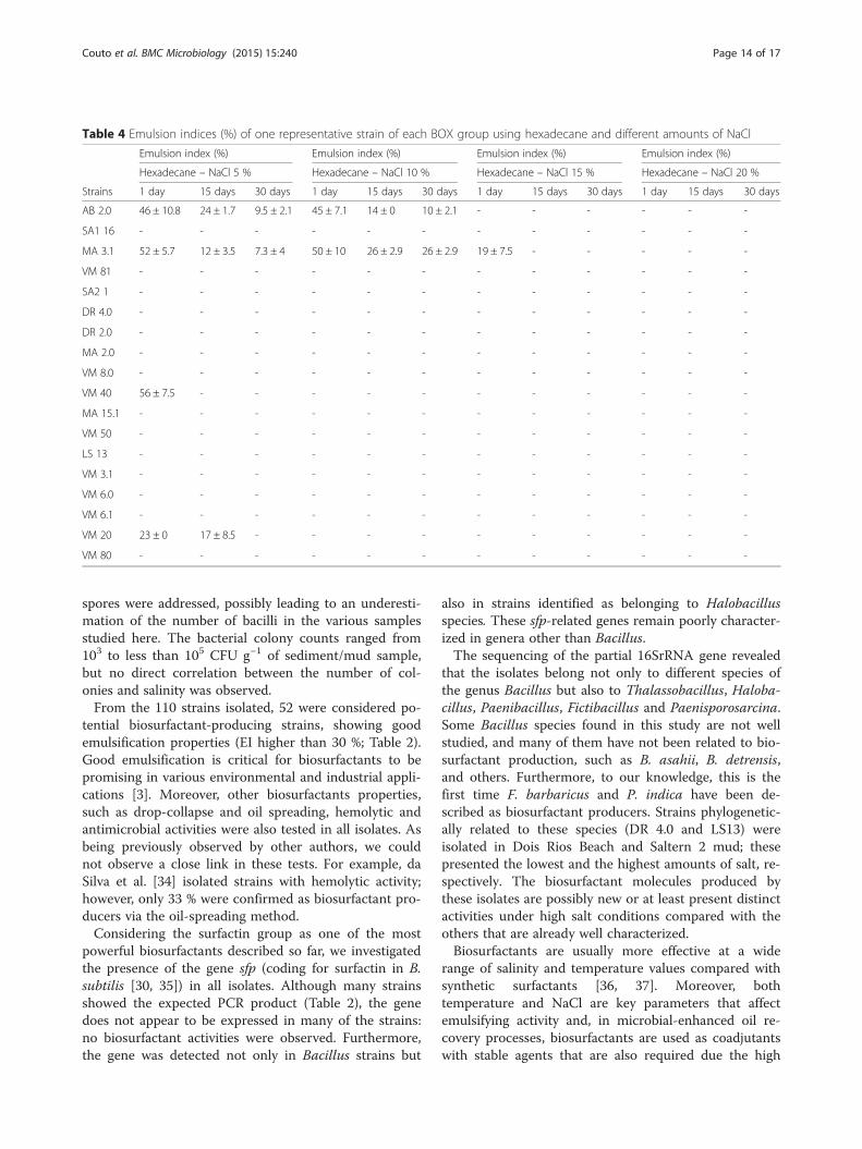
the supernatant of the biosurfactant producing strains,only two strains (AB 2.0 and MA 3.1) showed emulsifi-cation with up to 10 % NaCl using hexadecane (Table 4).Considering the same test but using kerosene as thehydrocarbon chosen, strains DR 2.0, VM 8.0, VM 40,VM 6.0 and VM 20 showed promising results with up to20 % NaCl added to the supernatants (Table 5). Particu-larly, strain DR 2.0 (B. asahii) was stable throughout theexperiment (30 days) after the addition of this highamount of salt (Table 5).
Detection of surfactin using ESI–MS/MS triple quadrupolemass spectrometryThe production of surfactin was analyzed in fourbiosurfactant-producing bacteria (DR 2.0, AB 2.0, VM6.0 and VM 8.0). A cluster of peaks with mass/charge(m/z) ratios between 1020 and 1034, which could be at-tributed to surfactin isoforms, was observed only in thecontrol (commercial surfactin; Fig. 6a) and in AB 2.0,identified as B. subtilis (Fig. 6c).
Table 2 Strains isolated from different environments, their molecular identification and biosurfactant production and othercharacteristics (Continued)
LS 6 MB B. vietnamensis (99) - JF411279.1 - - - - - - - - + - -
LS 7 MB B. vietnamensis (99) - JF411279.1 - - - - - - - - - - -
LS 8 MB B. vietnamensis (100) - JF411279.1 - - - - - - - - - - -
LS 10 MB B. vitnamensis (99) - JF411279.1 - - - - - - - - + - -
LS 11 MB B. foraminis (97) - KF724906.1 - - - - - - - - - - -
LS 12 TSB B. foraminis (99) - KF724906.1 - - - - - - - - - - -
LS 13 TSB Paenisporosarcina indica (99) -NR108473.1
13 - - - 59 ± 7.1 38 ± 7 - - - - - -
LS 14 TSB P. quisquiliarum (99) - JF309238.1 - - - - - - - - - - -
LS 15 TSB P. quisquiliarum (99) - JF309238.1 - - - - - - - - - - -
LS 16 TSB B. licheniformis (100) -LN774355.1
- - - - - - - - - - +
LS 17 TSB +3.5 % NaCl
B. aquimaris (99) - JF411268.1 - - - - - - - - - - -
LS 18 TSB +3.5 % NaCl
P. indica (99) - NR108473.1 - - - - - - - - - - -
LS 19 TSB +3.5 % NaCl
B. thioparans (99) - KJ911915.1 - - - - - - - - - - -
LS 20 TSB +3.5 % NaCl
B. foraminis (99) -KF724906.1 - - - - - - - - - - -
LS 21 TSB +3.5 % NaCl
B. foraminis (99) -KF724906.1 - - - - - - - - - - -
a First hit in GenBank with a known species; EI emulsification index, Hex hexadecane, Ker kerosene, DO drop collapse, OD oil displacement, H hemolysis, sfppresence of sfp gene, AMS production of an antimicrobial substance. (−) negative results. Strains in bold are those showing emulsification values higher than 30 %
Couto et al. BMC Microbiology (2015) 15:240 Page 11 of 17

DiscussionThe importance of endospore-forming bacteria as bio-surfactant producers and their presence in saline andhypersaline environments were combined in thisstudy. Both independent (utilizing primers that targetbacilli) and dependent cultivation-based approacheswere used to achieve our goals. The results obtainedhere (PCR-DGGE followed by NMDS analyses)showed a diverse endospore-forming bacterial com-munity in all environments and the grouping of theenvironmental samples based on their salinity. Like-wise, Wang et al. [33] demonstrated that salinity was
the environmental variable with the strongest influ-ence on the bacterial community composition of 32pristine Tibetan lakes that represent a broad salinityrange (freshwater to hypersaline). The diversity of theendospore-forming bacterial community observedamong the environmental samples analyzed supportedthe choice for a better characterization of thebiosurfactant-producing and endospore-forming bac-teria in these marine and hypersaline ecosystems.For cultivation-based methods, we heated all samples
to 80 °C for 10 min to facilitate the study of endospore-forming bacterial communities. Therefore, only the
Fig. 4 Phylogenetic tree of 16S rRNA gene sequences (approximately 800 bp) showing the relationship among the endospore-forming bacterialisolates. The tree was constructed based on the unweighted pair group method with arithmetic mean (UPGMA). Numbers on nodes representbootstrap values with 1,000 repetitions
Couto et al. BMC Microbiology (2015) 15:240 Page 12 of 17

Fig. 5 Dendrogram showing representative strains of each 18 groups formed via BOX-PCR
Table 3 Emulsion indices (%) of one representative strain of each BOX group using kerosene or hexadecane and high temperature
Emulsion index (%) Emulsion index (%) Emulsion index (%) Emulsion index (%)
Hexadecane - 100 °C Hexadecane - 121 °C Kerosene - 100 °C Kerosene - 121 °C
Strains BOX group 1 day 15 days 30 days 1 day 15 days 30 days 1 day 15 days 30 days 1 day 15 days 30 days
AB 2.0 1 46 ± 2.9 43 ± 0 40 ± 0 - - - 52 ± 8.6 51 ± 0 36 ± 7.5 - - -
SA1 16 2 - - - - - - - - - - - -
MA 3.1 3 52 ± 7.8 49 ± 7.8 49 ± 7.8 - - - 48 ± 10.3 49 ± 7.8 32 ± 8.6 6 ± 1.7 - -
VM 81 4 - - - - - - 39 ± 1.7 - - - - -
SA2 1 5 - - - - - - - - - - - -
DR 4.0 6 - - - - - - - - - - - -
DR 2.0 7 - - - - - - 56 ± 1.7 53 ± 4.6 40 ± 7.9 50 ± 1.7 - -
MA 2.0 8 - - - - - - 47 ± 9.9 30 ± 6.4 - 26 ± 2.9 - -
VM 8.0 9 63 ± 5.2 - - - - - 50 ± 2.1 - - - - -
VM 40 10 50 ± 1.7 36 ± 10.6 29 ± 7.8 13 ± 1.7 - - 55 ± 3.5 52 ± 8.1 48 ± 0 53 ± 2.1 48 ± 0 42 ± 2.1
MA 15.1 11 39 ± 9.6 39 ± 9.6 - 8 ± 0 8 ± 0 8 ± 0 44 ± 3.5 32 ± 3.5 10 ± 7 - - -
VM 50 12 - - - - - - 49 ± 5.7 30 ± 9.9 - - - -
LS 13 13 - - - - - - - - - - - -
VM 3.1 14 - - - - - - - - - - - -
VM 6.0 15 40 ± 5.2 - - - - - 37 ± 9.0 37 ± 12.7 - 36 ± 6.9 - -
VM 6.1 16 - - - - - - - - - - - -
VM 20 17 58 ± 4.6 42 ± 10.1 26 ± 8.6 - - - 59 ± 1.7 56 ± 2.1 47 ± 5.7 - - -
VM 80 18 - - - - - - - - - - -
Couto et al. BMC Microbiology (2015) 15:240 Page 13 of 17
spores were addressed, possibly leading to an underesti-mation of the number of bacilli in the various samplesstudied here. The bacterial colony counts ranged from103 to less than 105 CFU g−1 of sediment/mud sample,but no direct correlation between the number of col-onies and salinity was observed.From the 110 strains isolated, 52 were considered po-
tential biosurfactant-producing strains, showing goodemulsification properties (EI higher than 30 %; Table 2).Good emulsification is critical for biosurfactants to bepromising in various environmental and industrial appli-cations [3]. Moreover, other biosurfactants properties,such as drop-collapse and oil spreading, hemolytic andantimicrobial activities were also tested in all isolates. Asbeing previously observed by other authors, we couldnot observe a close link in these tests. For example, daSilva et al. [34] isolated strains with hemolytic activity;however, only 33 % were confirmed as biosurfactant pro-ducers via the oil-spreading method.Considering the surfactin group as one of the most
powerful biosurfactants described so far, we investigatedthe presence of the gene sfp (coding for surfactin in B.subtilis [30, 35]) in all isolates. Although many strainsshowed the expected PCR product (Table 2), the genedoes not appear to be expressed in many of the strains:no biosurfactant activities were observed. Furthermore,the gene was detected not only in Bacillus strains but
also in strains identified as belonging to Halobacillusspecies. These sfp-related genes remain poorly character-ized in genera other than Bacillus.The sequencing of the partial 16SrRNA gene revealed
that the isolates belong not only to different species ofthe genus Bacillus but also to Thalassobacillus, Haloba-cillus, Paenibacillus, Fictibacillus and Paenisporosarcina.Some Bacillus species found in this study are not wellstudied, and many of them have not been related to bio-surfactant production, such as B. asahii, B. detrensis,and others. Furthermore, to our knowledge, this is thefirst time F. barbaricus and P. indica have been de-scribed as biosurfactant producers. Strains phylogenetic-ally related to these species (DR 4.0 and LS13) wereisolated in Dois Rios Beach and Saltern 2 mud; thesepresented the lowest and the highest amounts of salt, re-spectively. The biosurfactant molecules produced bythese isolates are possibly new or at least present distinctactivities under high salt conditions compared with theothers that are already well characterized.Biosurfactants are usually more effective at a wide
range of salinity and temperature values compared withsynthetic surfactants [36, 37]. Moreover, bothtemperature and NaCl are key parameters that affectemulsifying activity and, in microbial-enhanced oil re-covery processes, biosurfactants are used as coadjutantswith stable agents that are also required due the high
Table 4 Emulsion indices (%) of one representative strain of each BOX group using hexadecane and different amounts of NaCl
Emulsion index (%) Emulsion index (%) Emulsion index (%) Emulsion index (%)
Hexadecane – NaCl 5 % Hexadecane – NaCl 10 % Hexadecane – NaCl 15 % Hexadecane – NaCl 20 %
Strains 1 day 15 days 30 days 1 day 15 days 30 days 1 day 15 days 30 days 1 day 15 days 30 days
AB 2.0 46 ± 10.8 24 ± 1.7 9.5 ± 2.1 45 ± 7.1 14 ± 0 10 ± 2.1 - - - - - -
SA1 16 - - - - - - - - - - - -
MA 3.1 52 ± 5.7 12 ± 3.5 7.3 ± 4 50 ± 10 26 ± 2.9 26 ± 2.9 19 ± 7.5 - - - - -
VM 81 - - - - - - - - - - - -
SA2 1 - - - - - - - - - - - -
DR 4.0 - - - - - - - - - - - -
DR 2.0 - - - - - - - - - - - -
MA 2.0 - - - - - - - - - - - -
VM 8.0 - - - - - - - - - - - -
VM 40 56 ± 7.5 - - - - - - - - - - -
MA 15.1 - - - - - - - - - - - -
VM 50 - - - - - - - - - - - -
LS 13 - - - - - - - - - - - -
VM 3.1 - - - - - - - - - - - -
VM 6.0 - - - - - - - - - - - -
VM 6.1 - - - - - - - - - - - -
VM 20 23 ± 0 17 ± 8.5 - - - - - - - - - -
VM 80 - - - - - - - - - - - -
Couto et al. BMC Microbiology (2015) 15:240 Page 14 of 17

temperature, pressure and salinity of the oil reservoirs[38]. Eighteen selected strains (belonging to the differentBOX groups) were tested for the loss of emulsifying ac-tivity treating the supernatant (cell-free broth) at 100 °Cand 121 °C and with different amounts of salt. Reduc-tions in emulsification activity at high temperatures and10 % NaCl were observed in nearly 80 % (100 °C) and90 % (121 °C and 10 % NaCl) of the strains using n-hexadecane (measurements made after 30 days). Nostrains showed emulsification when 15 % NaCl wasadded to the supernatants. The loss of emulsifier activitycould be explained by the denaturation of proteinaceouscompounds of the bioemulsifiers during heating. More-over, once the salinity decreases the viscosity, the in-crease in NaCl concentration may have influenced thequality of the emulsion, thereby reducing the emulsifica-tion capacity. The same loss of emulsifying activity wasreported for other microorganism surfactants [39, 40].Conversely, different strains showed emulsion-
stabilization in both temperatures and in up to 20 %NaCl using kerosene. Strains DR 2.0 (B. asahii), VM 8.0,VM 40 and VM 6.0 (all B. infantis) maintained theiremulsification activities for at least 15 days (with the ex-ception of VM 8.0, which lasted only 24 h) with theaddition of 20 % NaCl to their supernatants, therebyshowing a high tolerance to the salt concentrations. Nosurfactins were detected in these strains using ESI–MS/
MS triple quadrupole mass spectrometry; thus, thisstudy provides a new perspective to better characterizethese isolates and their biosurfactants.
ConclusionMolecular approaches showed the structural diversity ofendospore-forming bacterial communities in differentBrazilian marine and hypersaline environments from anEnvironmental Protection Area located in Rio de Janeirobased on the salinity of the environments. Therefore,these environmental samples were considered to be par-ticularly interesting for the isolation of potential biosur-factant producers belonging to this bacterial group.Different endospore-forming bacterial genera were iso-lated, but Bacillus spp. predominated in all the salineand hypersaline samples. For the first time, B. asahii, B.detrensis, F. barbaricus and P. indica were described asbiosurfactant producers. The use of biosurfactants pro-duced by endospore-forming bacterial isolates showingemulsification properties in high temperatures and/or inup to 20 % NaCl could be extremely valuable not onlyfor a possible bioremediation approach in saline and hy-persaline environments but also for microbial-enhancedoil recovery (MEOR).
Competing interestsThe authors declare that they have no competing interests.
Table 5 Emulsion indices (%) of one representative strain of each BOX group using kerosene and different amounts of NaCl
Emulsion index (%) Emulsion index (%) Emulsion index (%) Emulsion index (%)
kerosene – NaCl 5 % kerosene – NaCl 10 % kerosene – NaCl 15 % kerosene – NaCl 20 %
Strains 1 day 15 days 30 days 1 day 15 days 30 days 1 day 15 days 30 days 1 day 15 days 30 days
AB 2.0 53 ± 4.6 22 ± 1.7 10 ± 2.1 49 ± 1.7 12 ± 3.5 10 ± 6.4 - - - - - -
SA1 16 - - - - - - - - - - - -
MA 3.1 56 ± 6.4 26 ± 3.5 - - - - - - - - - -
VM 81 - - - - - - - - - - - -
SA 2 1 - - - - - - - - - - - -
DR 4.0 - - - - - - - - - - - -
DR 2.0 51 ± 7.6 44 ± 6.4 36 ± 6.2 53 ± 6.4 49 ± 8.1 47 ± 6.4 57 ± 0 54 ± 0 47 ± 1.4 52 ± 3.5 48 ± 7.4 44 ± 8.7
MA 2.0 - - - - - - - - - - - -
VM 8.0 51 ± 8.5 41 ± 3.5 33 ± 3.5 51 ± 3 46 ± 0 32 ± 8.1 57 ± 0 48 ± 0 34 ± 0 39 ± 9.6 - -
VM 40 55 ± 3.5 40 ± 5.2 - 51 ± 4.2 39 ± 2.1 - 59 ± 2.9 51 ± 5.5 - 63 ± 2.5 48 ± 8.1 -
MA 15.1 - - - - - - - - - - - -
VM 50 - - - - - - - - - - - -
LS 13 - - - - - - - - - - - -
VM 3.1 - - - - - - - - - - - -
VM 6.0 45 ± 7.4 41 ± 3.5 - 48 ± 0 43 ± 0 - 47 ± 4 45 ± 1.7 - 48 ± 2.5 43 ± 0 -
VM 6.1 - - - - - - - - - - - -
VM 20 52 ± 4.6 37 ± 7.9 - 44 ± 9.1 40 ± 10.4 - 43 ± 8.1 37 ± 12.5 - - - -
VM 80 - - - - - - - - - - - -
Couto et al. BMC Microbiology (2015) 15:240 Page 15 of 17

Fig. 6 ESI–MS/MS triple quadrupole mass spectrometry in negative ion mode of the crude extracts of four strains: (a) Commercial surfactin (SigmaAldrich) used as positive control, (b) B. asahii DR 2.0, (c) B. subtilis AB 2.0, (d) B. infantis VM 6.0 and (e) B. infantis VM 8.0
Couto et al. BMC Microbiology (2015) 15:240 Page 16 of 17

Authors’ contributionsCRAC, DAJ and LS designed the project. CRAC, VMA, JMM and DAJperformed the laboratory experiments. DAJ and LS prepared the manuscript.All of the authors examined and agreed with the final manuscript.
AcknowledgementsThis study was supported by grants from the Conselho Nacional deDesenvolvimento Científico e Tecnológico (CNPq), Coordenação deAperfeiçoamento de Pessoal de Nível Superior (CAPES) and Fundação deAmparo à Pesquisa do Estado do Rio de Janeiro (FAPERJ). The authors thankDr. Rodrigo de Souza and Thalita Barros for their help with the massspectrometry analyses.
Received: 24 August 2015 Accepted: 16 October 2015
References1. Singh A, Van Hamme JD, Ward OP. Surfactants in microbiology and
biotechnology: Part 2. Application aspects. Biotechnol Adv. 2007;25:99–121.2. Mandic-Mulec I, Prosser JI. Diversity of endospore-forming bacteria in soil:
Characterization and driving mechanisms. In: Logan NA, De Vos P, editors.Endospore-forming Soil Bacteria, Soil Biology 27, Chapter 2. Heidelberg:Springer-Verlag Berlin; 2011.
3. Banat IM, Makkar RS, Cameotra SS. Potential commercial applications ofmicrobial surfactants. Appl Microbiol Biotechnol. 2000;53:495–508.
4. Sekhon KK, Khanna S, Cameotra SS. Biosurfactant production and potentialcorrelation with esterase activity. J Pet Environ Biotechnol. 2012;3:7. http://dx.doi.org/10.4172/2157-7463.1000133.
5. Joshi SJ, Suthar H, Yadav AK, Hingurao K, Nerurkar A. Occurrence ofbiosurfactant producing Bacillus spp. in diverse habitats. ISRN Biotechnol.2013;2013:6. article ID 652340.
6. Rodrigues LR, Teixeira JA, van der Mei HC, Oliveira R. Isolation and partialcharacterization of a biosurfactant produced by Streptococcus thermophilusA. Colloids Surf B: Biointerfaces. 2006;53:105–12.
7. Kaloorazi NA, Choobari MFS. Biosurfactants: Properties and applications. JBiol Today’s World. 2013;2:235–41.
8. Jacques P. Surfactin and other lipopeptides from Bacillus spp. In: Soberon-Chavez G, editor. Biosurfactants. From genes to applications. Heidelberg:Springer-Verlag Berlin; 2011. p. 32–43.
9. Das P, Mukherjee S, Sen R. Antimicrobial potential of a lipopeptidebiosurfactant derived from a marine Bacillus circulans. J Appl Microbiol.2008;104:1675–84.
10. das Neves LCM, Kobayashi MJ, Rodrigues TM, Converti A, Penna TCV.Biomonitoring of biosurfactant production by green fluorescent proteinmarked Bacillus subtilis W1012. J Chem Technol Biotechnol. 2009;84:112–8.
11. Yakimov MM, Timmis KN, Wray V, Fredrickson HL. Characterization of a newlipopeptide surfactant produced by thermotolerant and halotolerantsubsurface Bacillus licheniformis BAS50. Appl Environ Microbiol.1995;61(5):1706–13.
12. Margesin R, Schinner F. Biodegradation and bioremediation ofhydrocarbons in extreme environments. Appl Microbiol Biotechnol.2001;56(5–6):650–63.
13. Jurelevicius D, Alvarez VM, Marques JM, Lima LRF, Dias FA, Seldin L. Bacterialcommunity response to petroleum hydrocarbon amendments infreshwater, marine and hypersaline water-containing microcosms. ApplEnviron Microbiol. 2013;79(19):5927–35.
14. Sambrook J, Fritsch EF, Manniatis T. Molecular cloning: a laboratory manual.New York, N.Y., USA: Cold Spring Harbor Laboratory Press; 1989.
15. Garbeva P, van Veen JA, van Elsas JD. Predominant Bacillus spp. inagricultural soil under different management regimes detected via PCR-DGGE. Microbial Ecol. 2003;45:302–16.
16. Nübel U, Engelen B, Felske A, Snaidr J, Wieshuber A, Amann RI, et al.Sequence heterogeneities of genes encoding 16S rRNAs in Paenibacilluspolymyxa detected by temperature gradient gel electrophoresis. J Bacteriol.1996;19:5636–43.
17. Muyzer G, de Wall EC, Uitterlinden AG. Profiling of complex microbialpopulation by denaturing gradient gel electrophoresis analysis ofpolymerase chain reaction – amplified genes coding for 16S rRNA. ApplEnviron Microbiol. 1993;59:695–700.
18. Hammer Ø, Harper DAT, Ryan PD. PAST: Paleontological statistics softwarepackage for education and data analysis. Palaeontol Electron. 2001;4(1):9.http://palaeo-electronica.org/2001_1/past/issue1_01.htm.
19. Massol-Deya AA, Odelson DA, Hichey RP, Tiedje JM. Bacterial communityfingerprinting of amplified 16S and 16-23S ribosomal DNA gene sequencesand restriction endonuclease analysis (ARDRA). In: Akkermans ADL, van ElsasJD, de Bruijn FJ, editors. Molecular Microbial Ecology Manual. Section 3.3.2.The Netherlands: Kluwer Academic Publishers; 1995. p. 1–8.
20. Thompson JD, Gibson TJ, Plewniak F, Jeanmougin F, Higgins DG. TheCLUSTAL_X windows interface: Flexible strategies for multiple sequencealignment aided by quality analysis tools. Nucleic Acids Res. 1997;25:4876–82.
21. Sokal RR, Michener CD. A statistical method for evaluating systematicrelationships. Univ Kans Sci Bull. 1958;38:1409–38.
22. Tamura K, Peterson D, Peterson N, Stecher G, Nei M, Kumar S. MEGA 5:Molecular Evoutionary Genetics Analysis using Maximum Likelihood,Evolutionary Distance, and Maximum Parsimony Methods. Mol Biol Evol.2011;28:2731–9.
23. Cooper D, Goldenberg B. Surface-active agents from 2 Bacillus species. ApplEnviron Microbiol. 1987;53(2):224–9.
24. Jain D, Collins-Thompson D, Lee H. A drop-collapsing test for screeningsurfactant-producing microorganisms. J Microbiol Methods. 1991;13(4):271–9.
25. Morikawa M, Hirata Y, Imanaka T. A study on the structure–functionrelationship of the lipopeptide biosurfactants. Biochim Biophys Acta.2000;1488:211–8.
26. Mulligan C, Cooper D, Neufeld R. Selection of microbes producingbiosurfactants in media without hydrocarbons. J Fermentation Technol.1984;62:311–4.
27. Rosado AS, Seldin L. Production of a potentially novel anti-microbialsubstance by Bacillus polymyxa. World J Microbiol Biotechnol. 1993;90:521–8.
28. von der Weid I, Alviano DS, Santos ALS, Soares RMA, Alviano CS, Seldin L.Antimicrobial activity of Paenibacillus peoriae against a broad spectrum ofphytopathogenic bacteria and fungi. J Appl Microbiol. 2003;95:1152–60.
29. von der Weid I, Maraha N, Seldin L, Jansson JK. Antifungal and root surfacecolonization properties of GFP-tagged Paenibacillus brasilensis PB177. WorldJ Microbiol Biotechnol. 2005;21:1591–7.
30. Hsieh FC, Li MC, Lin TC, Kao SS. Rapid detection and characterization ofsurfactin-producing Bacillus subtilis and closely related species based onPCR. Curr Microbiol. 2004;49:186–91.
31. Versalovic J, Schneider M, De Bruijn FJ, Lupski JR. Genomic fingerprinting ofbacteria using repetitive sequence-based polymerase chain reaction.Methods Mol Cell Biol. 1994;5:25–40.
32. Korenblum E, Araujo LV, Guimarães CR, Souza LM, Sassaki G, Abreu F, et al.Purification and characterization of a surfactin-like molecule produced byBacillus sp. H2O-1 and its antagonistic effect against sulfate reducingbacteria. BMC Microbiol. 2012;12:252.
33. Wang J, Yang D, Zhang Y, Shen J, van der Gast C, Hahn MW, et al. Dopatterns of bacterial diversity along salinity gradients differ from thoseobserved for macroorganisms? PLoS ONE. 2011;6(11):e27597. doi:10.1371/journal.pone.0027597.
34. da Silva FSP, Pylro VS, Fernandes PL, Barcelos GS, Kalks KH, Schaefer CE, etal. Unexplored Brazilian oceanic island host high salt tolerant biosurfactant-producing bacterial strains. Extremophiles. 2015;19(3):561–72.
35. Nakano MM, Corbell N, Besson J, Zuber P. Isolation and characterization ofsfp: a gene that functions in the production of the lipopeptidebiosurfactant, surfactin, in Bacillus subtilis. Mol Gen Genet. 1992;232:313–21.
36. Banat IM, Franzetti A, Gandolfi I, Bestetti G, Martinotti MG, Fracchia L, et al.Microbial biosurfactants production, applications and future potential. ApplMicrobiol Biotechnol. 2010;87:427–44.
37. Cameotra SS, Makkar RS, Kaur J, Mehta SK. Synthesis of biosurfactants andtheir advantages to microorganisms and mankind. Adv Exp Med Biol.2010;672:261–80.
38. Uad I, Silva-Castro GA, Pozo C, González-López J, Calvo C. Biodegradativepotential and characterization of bioemulsifiers of marine bacteria isolatedfrom samples of seawater, sediment and fuel extracted at 4000 m of depth(Prestige wreck). Int Biodeterior Biodegradation. 2010;64:511–8.
39. Sarubbo LA, Luna JM, Campos-Takaki GM. Production and stability studiesof the bioemulsifier obtained from a new strain of Candida glabrata UCP1002. Electron J Biotechnol. 2006;9:400–6.
40. Luna JM, Rufino RD, Campos-Takaki GM, Sarubbo LA. Properties of thebiosurfactant produced by Candida sphaerica cultivated in low-costsubstrates. Chem Eng Trans. 2012;27:67–72.
Couto et al. BMC Microbiology (2015) 15:240 Page 17 of 17