exploring pericyte and cardiac stem cell secretome unveils ... · cular ecs; mmps,...
TRANSCRIPT
Ellison-Hughes, G. M., & Madeddu, P. (2017). Exploring pericyte andcardiac stem cell secretome unveils new tactics for drug discovery.Pharmacology and Therapeutics, 171(2017), 1-12. DOI:10.1016/j.pharmthera.2016.11.007
Publisher's PDF, also known as Version of record
License (if available):CC BY
Link to published version (if available):10.1016/j.pharmthera.2016.11.007
Link to publication record in Explore Bristol ResearchPDF-document
This is the final published version of the article (version of record). It first appeared online via Pharmacology &Therapeutics at http://www.sciencedirect.com/science/article/pii/S0163725816302339 . Please refer to anyapplicable terms of use of the publisher.
University of Bristol - Explore Bristol ResearchGeneral rights
This document is made available in accordance with publisher policies. Please cite only the publishedversion using the reference above. Full terms of use are available:http://www.bristol.ac.uk/pure/about/ebr-terms
Pharmacology & Therapeutics 171 (2017) 1–12
Contents lists available at ScienceDirect
Pharmacology & Therapeutics
j ourna l homepage: www.e lsev ie r .com/ locate /pharmthera
Exploring pericyte and cardiac stem cell secretome unveils new tacticsfor drug discovery☆
Georgina M. Ellison-Hughes a, Paolo Madeddu b,⁎a Centre of Human&Aerospace Physiological Sciences, Centre for Stem Cells and RegenerativeMedicine, Faculty ofMedicine & Life Sciences, Guy's Campus, King's College London, London SE1 1UL,United Kingdomb Chair Experimental CardiovascularMedicine, Bristol Heart Institute, School of Clinical Sciences University of Bristol Level 7, Bristol Royal Infirmary, UpperMaudlin Street, Bristol BS2 8HW, UnitedKingdom
Abbreviations:Abi3bp, ABI FamilyMember 3 Binding Pheart disease; DPP-4, Dipeptidyl peptidase-4; ESCs, EmbryGLP1, Glucagon-like peptide-1; EPCs, Endothelial progenitcolony stimulating factor; HF, Heart failure; HGF, Hepatocycular ECs; MMPs, Metalloproteinases; MI, Myocardial infNeuregulin 1; PDGFβ, Platelet-derived growth factor betagrowth factor beta1; TNF-α, Tumor necrosis factor; LC-M☆ Supplementary material available at www.arthroplas⁎ Corresponding author.
E-mail address: [email protected] (P. Madeddu).
http://dx.doi.org/10.1016/j.pharmthera.2016.11.0070163-7258/© 2016 The Authors. Published by Elsevier Inc
a b s t r a c t
a r t i c l e i n f oAvailable online 2 December 2016
Ischaemic diseases remain a major cause of morbidity and mortality despite continuous advancements in med-ical and interventional treatments.Moreover, available drugs reduce symptoms associatedwith tissue ischaemia,without providing a definitive repair. Cardiovascular regenerativemedicine is an expanding field of research thataims to improve the treatment of ischaemic disorders through restorative methods, such as gene therapy, stemcell therapy, and tissue engineering. Stem cell transplantation has salutary effects through direct and indirect ac-tions, the latter being attributable to growth factors and cytokines released by stem cells and influencing the en-dogenousmechanisms of repair. Autologous stemcell therapies offer less scope for intellectual property coverageand have limited scalability. On the other hand, off-the-shelf cell products and derivatives from the stem cellsecretome have a greater potential for large-scale distribution, thus enticing commercial investors and recipro-cally producing more significant medical and social benefits. This review focuses on the paracrine properties ofcardiac stem cells and pericytes, two stem cell populations that are increasingly attracting the attention of regen-erative medicine operators. It is likely that new cardiovascular drugs are introduced in the next future by apply-ing different approaches based on the refinement of the stem cell secretome.© 2016 The Authors. Published by Elsevier Inc. This is an open access article under the CC BY license(http://creativecommons.org/licenses/by/4.0/).
Keywords:Cardiac stem cellsPericytesSecretomeRegenerative medicineDrug discovery
Contents
1. Introduction. . . . . . . . . . . . . . . . . . . . . . . . . . . . . . . . . . . . . . . . . . . . . . . . 22. Generation of a therapeutic product from stem cell secretome . . . . . . . . . . . . . . . . . . . . . . . . . 23. Exploiting the secretome of CSCs and pericytes for cardiovascular repair . . . . . . . . . . . . . . . . . . . . 34. Evidence for paracrine healing properties of cardiac stem/progenitor cells . . . . . . . . . . . . . . . . . . . . 35. Conclusions . . . . . . . . . . . . . . . . . . . . . . . . . . . . . . . . . . . . . . . . . . . . . . . . 4Conflict of interest . . . . . . . . . . . . . . . . . . . . . . . . . . . . . . . . . . . . . . . . . . . . . . . 5Acknowledgment . . . . . . . . . . . . . . . . . . . . . . . . . . . . . . . . . . . . . . . . . . . . . . . . 6References . . . . . . . . . . . . . . . . . . . . . . . . . . . . . . . . . . . . . . . . . . . . . . . . . . . 6
3669999
rotein; Ang, Angiopoietin; CSCs, Cardiac stem cells; CDCs, Cardiosphere-derived cells; CM, Conditionedmedium; CHD, Coronaryonic stem cells; ECs, ECs; EPCs, Endothelial progenitor cells; bFGF, Fibroblast growth factor; FDA, Food andDrug Administration;or cells; eNOS, Endothelial nitric oxide synthase; FAECs, Fetal aorta ECs; FOXO1, Forkhead box protein O1; G-CSF, Granulocyte-te growth factor; IGF-1, Insulin growth factor-1; IL, Interleukin; HGF, Hepatocyte growth factor; HUVECs, Human umbilical vas-arction; MCP-1, Monocyte chemoattractant protein-1; MSCs, Mesenchymal stem cells; NHS, National Health System; NRG-1,; sFRP1, Secreted frizzled-related protein 1; SCF, Stem cell factor; SDF-1, Stromal cell-derived factor-1; TGF-β1, TransformingS/MS, Tandem Mass Spectrometry Detection; VEGF-A, Vascular growth factor A; VPCs, Vascular progenitor cells.tyjournal.org.
. This is an open access article under the CC BY license (http://creativecommons.org/licenses/by/4.0/).
2 G.M. Ellison-Hughes, P. Madeddu / Pharmacology & Therapeutics 171 (2017) 1–12
1. Introduction
Coronary heart disease (CHD) caused by the narrowing of arteriesthat feed the heart is the UK's single biggest killer, being responsiblefor ~73,000 deaths each year, an average of 200 people each day.Acute myocardial infarctionl (MI) represents the most harmful form ofCHD. Over the last decade, mortality due to CHD has declined in theUK, but more people live with secondary consequences. In fact, mostof the current treatments are palliative, i.e. they reduce symptoms asso-ciated with heart dysfunction, without providing a definitive repair.Consequently, CHD patients undergo a progressive decline in thepumping function of the heart that ultimately leads to heart failure(HF). Today, post-infarct HF is the leading cause of invalidity, hospitali-zation and mortality in patients over 65. In 2012–13, the UK NationalHealth System (NHS) expenditure for cardiovascular disease was£7.02billion, 63% of which devoted to secondary care (Bhatnagar,Wickramasinghe, Williams, Rayner, & Townsend, 2015) The NHS ana-lysts have predicted a mismatch between total budget and patientneeds of nearly £30 billion by 2020/21. Therefore, efficiency actions toincrease quality and reduce expenditure growth are essential for all ser-vices, including those for treatment and care of CHD patients. However,efficiency alone may not suffice without the introduction of new tech-nologies having a transformative impact on this unmet clinical field.
1.1. The urgent need for new therapies
Current care of CHD comprises pharmacotherapy andrevascularisation. However, medical treatment can be ineffective as inthe case of refractory angina (which has an estimated prevalence of1.8 million in the USA and an incidence of 30–50,000/year in Europe).Additionally, a steadily increasing number of patients fall into thecategory in which revascularization cannot be applied or fails becauseof restenosis. This is especially true of patients with occlusive pathologyextending to the microcirculation and diabetic or elderly patients whohave had multiple bypasses and stenting operations. Also, the mostimportant limitation of current treatments is that they do not replacecells irreversibly damaged by ischaemia.
Cardiovascular regenerative medicine is a fast-growing field of re-search that aims to improve the treatment of CHD through innovativerestorative methods, such as gene therapy, stem cell therapy and tissueengineering (Assmus et al., 2002; Wollert et al., 2004). Clinical studieswith skeletal myoblasts, bone marrow-derived cells, mesenchymalstem cells (MSCs) and cardiac stem cells (CSCs) have shown feasibilityand initial evidence of efficacy (Assmus et al., 2002; de Jong,Houtgraaf, Samiei, Boersma, & Duckers, 2014; Hare et al., 2009;Menasche et al., 2008; Sant'anna et al., 2010). After multiple systematicreviews and meta-analyses, the consensus is that transplantation ofadult bonemarrow cellsmodestly improves ventricular function, infarctsize, and remodeling in patients with CHD compared with standardtherapy, and these benefits persist during long-term follow-up(Martin-Rendon, 2016). Bone marrow cell transplantation also reducesthe incidence of death, recurrent MI, and stent thrombosis in patientswith CHD (Jeevanantham et al., 2012). Moreover, Steven Chamuleau,Andreas Zieher and collegaues have recently utilized interactionmodelsin a multivariable fashion to identify subgroups of patients that are de-fined as potential treatment responders, while simultaneouslycorrecting for relevant factors that affect general disease outcome. Thiskind of approach could be the next step towards optimal cell therapyin clinical care (Zwetsloot et al., 2016).
The SCIPIO clinical trial, the first in man to investigate c-kit+ CSCs,reported that 16 patients with ischemic cardiomyopathy receivedintracoronary infusions of 0.5-1 × 10⁶ c-kit+, autologous CSCs andcompared to controls these patients benefited from an 8 and 12 unitincrease in left ventricular ejection fraction, 4 and 12months after infu-sion, respectively (Bolli et al., 2011). A subset of 7 patients was subjectto cMRI analysis, which showed that the infarct region had significantly
decreased in size by ~10 g up to 12months following c-kit+ CSC trans-plantation (Bolli et al., 2011).
However, there is a persisting dispute regarding the mechanismsunderpinning the benefit of cell therapy. The direct contribution oftransplanted cells in vascular and cardiac reconstitution has beenquestioned (Balsam et al., 2004; Murry et al., 2004), and presently theconcept of paracrine promotion of spontaneous healing processesprevails (Gnecchi et al., 2005; Tang et al., 2016a,b). Indeed, the generalconsensus is that cell therapy and resultant improvements in cardiacfunction and decreased infarct size in human trials is due to a ‘paracrine’effect (Tang et al., 2016a,b). However, the lack of cardiomyocyte differ-entiation capability of bonemarrow cells or CSCs could be due to lack ofcharacterisation of the transplanted cell type, poor cell survival and re-tention, hostile host environment and subsequent restriction of cell pro-liferation, integration and differentiation in this damage-regenerationinfarct model.
Despite the adult mammalian heart being composed of terminallydifferentiated cardiomyocytes that are permanently withdrawn fromthe cell cycle (Chien & Olson, 2002; Nadal-Ginard, 1978), it is now ap-parent that the adult heart has the capacity, albeit low, to self-renewcardiomyocytes over the human lifespan (Bergmann et al., 2012,2015). This is supported by the detection of small, newly-formed,immature cardiomyocytes, which incorporate BrdU/EdU and/or stainpositive for Ki67, Aurora B, and embryonic/neonatal myosin heavychain, aswell as cardiomyocytes undergoingmitosis, under normal con-ditions and in response to diverse pathological and physiological stimuli(Bergmann et al., 2015; Bostrom et al., 2010; Ellison et al., 2013;Urbanek et al., 2003, 2005; Waring et al., 2014). The source of thesenewly formed cardiomyocytes is a matter of debate (Laflamme &Murry, 2011). Three main sources of origin of the new cardiomyocyteshave been claimed: (a) circulating progenitors, which throughthe bloodstream home to the myocardium and differentiate intocardiomyocytes (Quaini et al., 2002); (b) mitotic division of the pre-existing cardiomyocytes (Bersell, Arab, Haring, & Kuhn, 2009; Bostromet al., 2010; Engel, Hsieh, Lee, & Keating, 2006; Senyo et al., 2013);and (c) a small population of residentmultipotent stem cells able to dif-ferentiate into the main cell types of the heart (i.e., cardiomyocytes,smooth and endothelial vascular and connective tissue cells)(Rasmussen et al., 2014; Torella, Ellison, Karakikes, & Nadal-Ginard,2007).
Blood-borne precursors, althoughwell documented for having a rolein inflammation and healing, and when adult mouse bonemarrow cellswere injected into the chick embryo they converted to a myocardialphenotype (Eisenberg, Burch, & Eisenberg, 2006), their cardiomyogenicpotential in the damaged adult heart is very limited, if any (Ellison et al.,2013; Loffredo, Steinhauser, Gannon, & Lee, 2011). The evidence so farpresented in support of re-entry of terminally differentiatedcardiomyocytes into the cell cycle has been limited to show division ofcells that express proteins of the contractile apparatus in their cyto-plasm (Bersell et al., 2009; Bostrom et al., 2010; Kuhn et al., 2007;Senyo et al., 2013). This evidence is equally compatible with newmyocyte formation from the pool of multipotent cardiac stem/progeni-tor cells, which as precursor cells express contractile proteins and be-cause newly born myocytes are not yet terminally differentiated theyare capable of a few rounds of division before irreversibly withdrawingfrom the cell cycle (Nadal-Ginard, 1978; Nadal-Ginard, Kajstura,Anversa, & Leri, 2003). However, the mechanisms underlying a strictpostmitotic state in the heart during pathological remodeling have yetto be fully elucidated (Zebrowski, Becker, & Engel, 2016).
The best documented source of the small, immature, newly formedcardiomyocytes in the adult mammalian heart, including the human(Torella, Ellison, Mendez-Ferrer, Ibanez, & Nadal-Ginard, 2006), is asmall population of endogenous cardiac stem and progenitor cells(eCSCs) distributed throughout the atria and ventricles, which areclonogenic, self-renewing and can give rise to functional cardiomyocytesand vasculature in vitro and in vivo. Importantly, owing to genetic

3G.M. Ellison-Hughes, P. Madeddu / Pharmacology & Therapeutics 171 (2017) 1–12
labelling and transitional tracking it is now documented that newlyformed cardiomyocytes observed in the adult mammalian heart arethe product of eCSC differentiation (Ellison et al., 2013; Hsieh et al.,2007; van Berlo & Molkentin, 2014).
Here, we provide an overview of current knowledge regardingthe therapeutic potential of using the stem cell-derived secretome in-stead of source stem cell therapy to repair and regenerate the damagedheart. Furthermore, we illustrate methodological aspects of secretome-based cardiovascular regenerative medicine, with particular referenceto functional transcriptomics and proteomics as a combinatory strategyto cherry pick the most beneficial components of the stem cellsecretome. Finally, we report current evidence regarding the salutaryaspects of the secretome from two stem cell populations, namely CSCsand pericytes.
2. Generation of a therapeutic product from stem cell secretome
Stem cells secrete potent combinations of cytokines, growth factors,enzymes,microvesicles/exosomes and geneticmaterial, whichhelp car-diac repair and regeneration at multiple points. The stem cell secretomesupports cardiomyocyte survival and proliferation, differentiation ofresident stem cells, and neovascularization, while limiting inflammato-ry and pro-fibrotic processes (Baraniak &McDevitt, 2010; Gnecchi et al.,2005; Rao et al., 2015; Zhou et al., 2011). Some paracrine factors havepleiotropic actions. For instance, one of the key pathways in stem cell-based cardiac repair is the stromal cell-derived factor-1 (SDF-1)/CXCR4 axis. It was proposed that substituting SDF-1 gene therapy forsource stem cells might represent a sensible therapeutic approach(Penn, 2009). The blinded placebo-controlled STOP-HF trial demon-strated a single endocardial administration of plasmid SDF-1 is safe, at-tenuates left ventricle remodeling and improves ejection fraction inischaemic cardiomyopathy (Chung et al., 2015). However, there arealso paracrine factors that boost specific responses, thereby providinga more defined approach for selective treatments inspired by the stemcell secretome. For instance, mesenchymal stem cell-derived InsulinGrowth Factor-1 (IGF-1) and ABI Family Member 3 Binding Protein(Abi3bp) reportedly induce resident CSC mobilization (Mourkioti &Rosenthal, 2005) and differentiation to the cardiac lineage, (Engelset al., 2014; Hodgkinson et al., 2014) while CSC-derived basic FibroblastGrowth Factor (bFGF), Vascular Growth Factor-A (VEGF-A) andHepato-cyte Growth Factor (HGF) exert potent pro-angiogenic effects (Raoet al., 2015; Zhou et al., 2011).
Intracoronary administration of IGF-1 and HGF, in doses rangingfrom 0.5 to 2 μg HGF and 2 to 8 μg IGF-1, just below the site of left ante-rior descendent occlusion, 30 min after MI during coronary reperfusionin the pig, triggered a regenerative response from the CSCs, which is po-tent and able to produce physiologically significant regeneration of thedamaged myocardium (Ellison et al., 2011). IGF-1 and HGF inducedCSC migration, proliferation and functional cardiomyogenic andmicrovasculature differentiation. Furthermore, IGF-1/HGF, in a dose-dependent manner, improved cardiomyocyte survival, and reducedfibrosis and cardiomyocyte reactive hypertrophy. Interestingly, theeffects of a single administration of IGF-1/HGF is still measurable2 months after its application, suggesting the existence of a feedbackloop triggered by the external stimuli that activate the production ofgrowth and survival factors by the targeted cells, which explains thepersistence and long duration of the regenerative myocardial response.These histological changes were correlated with a reduced infarct sizeand an improved ventricular segmental contractility and ejection frac-tion at the end of the follow-up assessed by cMRI (Ellison et al., 2011).
Neuregulin-1 (NRG-1) is another key factor implicated in stimu-lating cardiac repair and regeneration (Wadugu & Kuhn, 2012;Waring et al., 2014). An Ig-domain containing form of NRG-1β,also known as glial growth factor 2 (GG2) has been shown to im-prove left ventricular ejection fraction and remodeling in pigspost-MI, compared to controls (Galindo et al., 2014). It is thought
that NRG-1 imparts functional benefits by activating and increasingeCSC proliferation (Waring et al., 2014), inducing cardiomyocyte re-placement (Bersell et al., 2009; Cohen et al., 2014; Polizzotti et al.,2015). A bioengineered hydrogel system enables targeted andsustained intramyocardial delivery of NRG-1, activating the cardio-myocyte cell cycle and enhancing ventricular function in a murinemodel of ischemic cardiomyopathy (Cohen et al., 2014), protectingcardiomyocytes from apoptosis and improving mitochondrial func-tion (Galindo et al., 2014). However, the role of NRG-1 in inducingcardiomyocyte proliferation in the adult heart has been challenged,with NRG1β1 treatment not increasing cardiomyocyte DNA synthe-sis and consequent cardiomyocyte renewal in normal or infarctedadult mouse hearts (Reuter, Soonpaa, Firulli, Chang, & Field, 2014).Therefore, the role of NRG-1 administration in inducing cardiomyo-cyte proliferation and replacement in the adult failing heart remainscontroversial.
The stem cell secretome is collected in a form of cell culture condi-tioned medium (CM) or supernatant. The use of stem cell-derivedsecretome has several advantages compared to the use of stem cells,as CM can be manufactured, sterilized, freeze-dried, packaged, storedand transportedmore easily. Therefore, the stemcell-derived secretomehas a promising prospect to become a successful pharmaceutical/me-dicinal product for regenerativemedicine. On the other hand, variabilityrepresents a major limitation. The first level of inconsistency is repre-sented by inter-individual variability, due to the patient's characteris-tics. Culture conditions, growth media and passages also affect thecomposition of CM, as recently reviewed (Pawitan, 2014). In someocca-sions, changes in culture conditions are intentionally introduced to en-hance the production of certain biological components. For instance,stem cells could be exposed to hypoxic conditions, with the aim of stim-ulating the production of growth factors (Di Santo et al., 2009;Ranganath, Levy, Inamdar, & Karp, 2012). The three-dimensional spher-oid culture of human adipose-derived stem cells with clinically relevantmedium composed of amino acids, vitamins, glucose, and human serumleads to 23- to 27-fold higher production of angiogenic factors than thatby conventional monolayer culture (Bhang et al., 2014). On the otherhand, the purity of preparations may become an issue when completegrowth medium is used, or collection of CM is performed shortly afterremoval of growth medium supplements. In this case, contaminationmay derive from the presence of left-over serum carrier proteins, dueto dynamic recycling of extracellular proteins by cellular vesicles (Isoet al., 2014). The presence of serum or culture media in the CM- orsupernatant-derived product might elicit immune responses and/orside effects. In this respect, alternative methods to culture cells (suchas the adoption of serum-free culture, etc) and the refinement of theCM-derived secretome products should be considered. However, a re-cent study indicates that the formation of complexes of serum-derivedimmunoglobulins with paracrine factors could enhance the stabilityand biological activity of the CM therapeutic component (Rao et al.,2015). A challenging objective of future research is to define thewhole composition of crude CM preparations, measure activity of themain components and determine therapeutic doses. In this respect,manufacturing and quality control protocols need to be further refinedand standardized to accomplish the classical regulatory path for drugs.The goal is to generate synthetic versions, inspired by the stem cellsecretome, containing consistent dosages of therapeutic factors for thetreatment of thousand patients.
2.1. Methodologies to determine the therapeutic component
Different methods have been used for studying the secretomecomposition and for distinguishing the essential components thatare advantageous for therapeutic applications. Multiplex antibody-based techniques, such as antibody arrays, have high sensitivity(in the range of 1–10 pg/ml), specificity, and reproducibility acrossa broad range of concentrations. High-throughput analysis of the
4 G.M. Ellison-Hughes, P. Madeddu / Pharmacology & Therapeutics 171 (2017) 1–12
human MSC secretome using a human cytokine antibody arrayhas identified about 40 proteins with high expression levels(Parekkadan et al., 2007). Studies using multiplex antibody-basedarrays showed the contribution of MSC-derived paracrine factorsin determining cardiac improvement in a swine MI models(Nguyen et al., 2010) and the participation of angiogenin, a secretedribonuclease that inhibits protein translation under stress condi-tions, in promotion of cardiomyocyte survival by intramyocardiallyinjected bone marrow-derived mononuclear cells (Ascione et al.,2015). However, a common drawback of antibody-based techniques(e.g., ELISA and antibody arrays) consists of the limited availability ofantibodies to detect secreted proteins.
Liquid Chromatography with TandemMass Spectrometry Detection(LC-MS/MS) has beenwidely used for characterizing the secretomepro-file. For example, a study using this technique demonstrated that pre-conditioning of human adipose-derived MSCs with Tumor NecrosisFactor-alpha (TNF-α) leads to enhanced expression of cytokines andchemokines such as Interleukin-6 (IL-6), Interleukin-8 (IL-8),Monocytechemoattractant protein-1 (MCP-1), metalloproteinases (MMPs),Pentraxin-related protein, and Cathepsin L. in CM (Lee et al., 2010).On the other hand, gel-based and LC-MS/MS techniques have limitedsensitivity to detect molecules that are present in fmol concentrations(Ranganath et al., 2012).
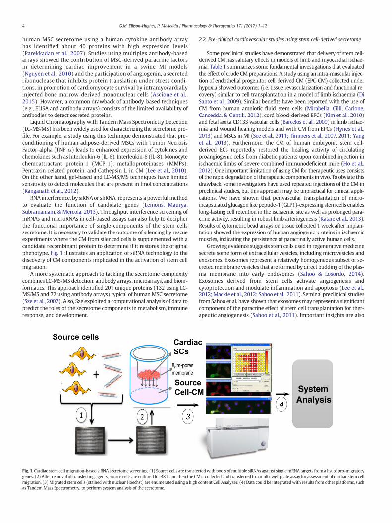
RNA interference, by siRNA or shRNA, represents a powerful methodto evaluate the function of candidate genes (Lemons, Maurya,Subramaniam, & Mercola, 2013). Throughput interference screening ofmRNAs and microRNAs in cell-based assays can also help to decipherthe functional importance of single components of the stem cellssecretome. It is necessary to validate the outcome of silencing by rescueexperiments where the CM from silenced cells is supplemented with acandidate recombinant protein to determine if it restores the originalphenotype. Fig. 1 illustrates an application of siRNA technology to thediscovery of CM components implicated in the activation of stem cellmigration.
A more systematic approach to tackling the secretome complexitycombines LC-MS/MS detection, antibody arrays,microarrays, and bioin-formatics. This approach identified 201 unique proteins (132 using LC-MS/MS and 72 using antibody arrays) typical of humanMSC secretome(Sze et al., 2007). Also, Sze exploited a computational analysis of data topredict the roles of the secretome components in metabolism, immuneresponse, and development.
Fig. 1. Cardiac stem cellmigration-based siRNA secretome screening. (1) Source cells are transfegenes. (2) After removal of transfecting agents, source cells are cultured for 48 h and then the CMmigration. (3) Migrated stem cells (stainedwith nuclear Hoechst) are enumerated using a highas TandemMass Spectrometry, to perform system analysis of the secretome.
2.2. Pre-clinical cardiovascular studies using stem cell-derived secretome
Some preclinical studies have demonstrated that delivery of stem cell-derived CM has salutary effects in models of limb and myocardial ischae-mia. Table 1 summarizes some fundamental investigations that evaluatedthe effect of crude CMpreparations. A study using an intra-muscular injec-tion of endothelial progenitor cell-derived CM (EPC-CM) collected underhypoxia showed outcomes (i.e. tissue revascularization and functional re-covery) similar to cell transplantation in a model of limb ischaemia (DiSanto et al., 2009). Similar benefits have been reported with the use ofCM from human amniotic fluid stem cells (Mirabella, Cilli, Carlone,Cancedda, & Gentili, 2012), cord blood-derived EPCs (Kim et al., 2010)and fetal aorta CD133 vascular cells (Barcelos et al., 2009) in limb ischae-mia and wound healing models and with CM from EPCs (Hynes et al.,2013) and MSCs in MI (See et al., 2011; Timmers et al., 2007, 2011; Yanget al., 2013). Furthermore, the CM of human embryonic stem cell-derived ECs reportedly restored the healing activity of circulatingproangiogenic cells from diabetic patients upon combined injection inischaemic limbs of severe combined immunodeficient mice (Ho et al.,2012). One important limitation of using CM for therapeutic uses consistsof the rapid degradation of therapeutic components in vivo. To obviate thisdrawback, some investigators have used repeated injections of the CM inpreclinical studies, but this approach may be unpractical for clinical appli-cations. We have shown that perivascular transplantation of micro-incapsulated glucagon like peptide-1 (GLP1)-expressing stemcells enableslong-lasting cell retention in the ischaemic site as well as prolonged para-crine activity, resulting in robust limb arteriogenesis (Katare et al., 2013).Results of cytometric bead arrays on tissue collected 1 week after implan-tation showed the expression of human angiogenic proteins in ischaemicmuscles, indicating the persistence of paracrinally active human cells.
Growing evidence suggests stem cells used in regenerativemedicinesecrete some form of extracellular vesicles, including microvesicles andexosomes. Exosomes represent a relatively homogeneous subset of se-cretedmembrane vesicles that are formedby direct budding of the plas-ma membrane into early endosomes (Sahoo & Losordo, 2014).Exosomes derived from stem cells activate angiogenesis andcytoprotection and modulate inflammation and apoptosis (Lee et al.,2012;Mackie et al., 2012; Sahoo et al., 2011). Seminal preclinical studiesfrom Sahoo et al. have shown that exosomesmay represent a significantcomponent of the paracrine effect of stem cell transplantation for ther-apeutic angiogenesis (Sahoo et al., 2011). Important insights are also
SystemAnalysis
ctedwith pools ofmultiple siRNAs against singlemRNA targets from a list of pro-migratoryis collected and transferred to amulti-well plate assay for assessment of cardiac stem cell
content Cell Analyzer. (4) Data could be integratedwith results from other platforms, such
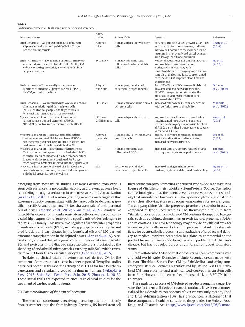
Table 1Cardiovascular preclinical trials using stem cell-derived secretome.
Disease/deliveryAnimalmodel Source of CM Outcome Reference
Limb ischaemia— Daily injection of 40 μl of humanadipose-derived stem cell (ADSC) CM for 7 daysinto the gracilis muscle
Athymicmice
Human adipose-derived stemcells
Enhanced endothelial cell growth, CD34+ cellmobilization from bone marrow, and bonemarrow cell homing to the ischemic region,resulting in improved blood vessel density,limb salvage, and blood perfusion.
Bhang et al.(2014)
Limb ischaemia— Single injection of human embryonicstem cell-derived endothelial-like cell (ESC-EC) CMand/or circulating proangiogenic cells (PACs) intothe gracilis muscle
SCID mice Human embryonic stemcell-derived endothelial-likecells
Neither diabetic PACs nor CM from ESC-ECsimprove blood flow recovery andangiogenesis. In contrast, bothtransplantations of proangiogenic cells fromcontrols or diabetic patients supplementedwith ESC-ECs CM improve blood flow andangiogenesis.
Ho et al.(2012)
Limb ischaemia— Three weekly intramuscularinjections of endothelial progenitor cells (EPCs),EPC-CM, or control medium
Athymicnude rats
Human peripheral bloodendothelial progenitor cells
Both EPC-CM and EPCs increase limb bloodflow assessed and neovascularization.EPC-CM transplantation stimulates themobilization and recruitment of bonemarrow-derived EPCs.
Di Santoet al. (2009)
Limb ischaemia— Two intramuscular weekly injectionsof human amniotic liquid derived stem cells(AFSC) CM (topically applied to thigh muscles)for a total treatment-duration of two weeks
SCID mice Human amniotic liquid derivedcKit stem cells
Increased arteriogenesis, capillary density,total perfusion area, and mobility.
Mirabellaet al. (2012)
Myocardial infarction— Peri-infarct injection ofhuman adipose-derived stem cells (ADSC),ADSC-CM or control medium immediately after MI
SCID andC57BL/6 mice
Human adipose-derived stemcells
Improved cardiac function, reduced infarctsize, increased reparative angiogenesis,reduced cardiomyocyte apoptosis The effectof ADSCs on the first 3 outcomes was superiorto that of ADSC-CM.
Yang et al.(2013)
Myocardial infarction— Intramyocardial injectionsof either concentrated CM derived from STRO-3-mesenchymal precursor cells cultured in serum-freemedium or control medium at 48 h after MI
Athymicnude rats
Human STRO-3- mesenchymalprecursor cells
Improved ventricular function, reducedventricular dilatation, and infarct size,increased neovascularization.
See et al.(2011)
Myocardial infarction— Intravenous treatment withCM from human embryonic stem cells-derived MSCsor control medium initiated 4 h after coronary arteryligation with the treatment continued for 7 daystwice daily via a catheter inserted into the jugular vein
Pigs Human embryonic stemcells-derived MSCs
Increased capillary density, reduced infarctsize improved myocardial performance.
Timmerset al. (2011)
Myocardial infarction— At the end of 2 h reperfusion,three cycles of intracoronary infusion CM from porcineendothelial progenitor cells or vehicle
Pigs Porcine peripheral bloodendothelial progenitor cells
Increased angiogenesis, improvedcardiomyocyte remodeling and contractility.
Hynes et al.(2013)
5G.M. Ellison-Hughes, P. Madeddu / Pharmacology & Therapeutics 171 (2017) 1–12
emerging from mechanistic studies. Exosomes derived from variousstem cells enhance the myocardial viability and prevent adverse heartremodeling through a reduction in oxidative stress and Akt activation(Arslan et al., 2013). Furthermore, exciting new research suggests thatexosomes directly communicate with the target cells by delivering spe-cific microRNAs and other small RNAs characteristic of their parentalcell of origin (Mackie et al., 2012; Yuan et al., 2009). Analysis ofmicroRNAs expression in embryonic stem cell-derived exosomes re-vealed high expression of embryonic-specific microRNAs belonging tothe miR-294 family. This microRNA regulates fundamental propertiesof embryonic stem cells (ESCs), including pluripotency, cell cycle, andproliferation and participates in the beneficial effect of ESC-derivedexosomes transplantation in the injured heart (Khan et al., 2015). A re-cent study showed the pathogenic communication between vascularECs and pericytes in the diabetic microvasculature is mediated by theshedding of endothelial microparticles carrying miR-503, which trans-fer miR-503 from ECs to vascular pericytes (Caporali et al., 2015).
To date, no clinical trial employing stem cell-derived CM for thetreatment of cardiovascular disease has been reported. Twopilot studiesdescribed potential therapeutic activity of MSC CM for hair follicle re-generation and resurfacing wound healing in humans (Fukuoka &Suga, 2015; Shin, Ryu, Kwon, Park, & Jo, 2015; Zhou et al., 2013).These initial trials are expected to encourage clinical studies for thetreatment of cardiovascular patients.
2.3. Commercialization of the stem cell secretome
The stem cell secretome is receiving increasing attention not onlyfrom researchers but also from industry. Recently, US-based stem cell
therapeutic company Stemedica announced worldwide manufacturinglicense of VitriLife to their subsidiary StemProtein (Source: StemedicaCell Technologies, Inc.). The patent covers a novel vaporization technol-ogy that immobilizes biologicals in glassy carbohydrates (a VitriLife™state) thus allowing storage at room temperature for several years.The company claims VitriLife-preserved proteins are superior in activitycompared to lyophilized proteins. The thermostable mixture fromVitriLife processed stem cell-derived CM contains therapeutic biologi-cals, such as cytokines, chemokines, growth factors, proteins, mRNAs,and microRNAs. The new technology may provide an efficient way forconverting stem cell-derived factors into powders that retain natural ef-ficacy for eventual bulk processing and packaging of product and deliv-ery to medical markets. Stemedica has plans to commercialize theproduct formany disease conditions, from skin problems to Alzheimer'sdisease, but has not released yet any information about regulatorystatus.
Stemcell-derived CM in cosmetic products has been commercializedand sold world-wide. Examples include Regenica cream made withHuman Fibroblast Serum Free CM by SkinMedica, anti-aging non-embryonic stem cell extracts manufactured by Lifeline Skin Care, stabi-lized CM from placenta- and umbilical cord-derived human stem cellsfrom Blue Horizon, and serum-free adipose-derived MSC CM fromCelprogen.
The regulatory process of CM-derived products remains vague. De-spite the fact stem cell-derived cosmetic products have been commer-cialized for a decade as components of skin creams, only recently Foodand Drug Administration (FDA) has pronounced a statement thatthese compounds should be considered drugs under the Federal Food,Drug, and Cosmetic Act (http://www.ipscell.com/2016/08/3-more-
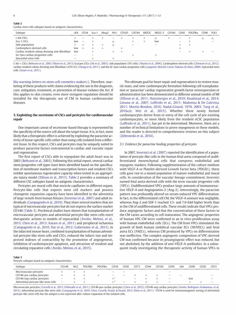
Table 2Cardiac stem cells subtypes based on antigenic characteristics.
Subtype cKit CD34 Sca-1 Abcg2 Wt1 CD105 GATA4 MEF2C NKX2–5 CD166 CD45 PDGFRα CD90 FLK1
c-Kit CSCs + − + + − + + + ± + − + ± ±Sca-1 CSCs low − + ± + + low − + −Side population + + + + ± − − + +Cardiosphere-derived cells low + + + + +Cardiac resident colony-forming unit-fibroblast low + + + − + + −Isl-1pos cardiac progenitor cells − − + +Epicardial stem cells − + +
C-kit+ CSCs (Beltrami et al., 2003; Ellison et al., 2013) Sca1pos CSCs (Oh et al., 2003); side population (SP) cells) (Martin et al., 2004); Cardiosphere-derived cells (Chimenti et al., 2012);cardiac resident colony-forming unit-fibroblast (cCFU-Fs) (Chong et al., 2011); and the Isl-1pos cardiac progenitor cells (Laugwitz, Moretti, Caron, Nakano, & Chien, 2008); Epicardial stemcells (Smart et al., 2011).
6 G.M. Ellison-Hughes, P. Madeddu / Pharmacology & Therapeutics 171 (2017) 1–12
fda-warning-letters-to-stem-cell-cosmetics-makers/). Therefore, mar-keting of these productswith claims evidencing the use in the diagnosis,cure, mitigation, treatment, or prevention of disease violates the Act. Ifthis applies to skin creams, even more stringent regulation should beinstalled for the therapeutic use of CM in human cardiovasculardiseases.
3. Exploiting the secretome of CSCs and pericytes for cardiovascularrepair
One important caveat of secretome-based therapy is represented bythe specificity of the source cell about the target tissue. It is, in fact, morelikely that a therapeutic effect is achieved by exploiting the paracrine ac-tivity of tissue-specific cells rather than using cells isolated froma differ-ent tissue. In this respect, CSCs and pericytes may be uniquely suited toproduce paracrine factors instrumental to cardiac and vascular repairand regeneration.
The first report of CSCs able to repopulate the adult heart was in2003 (Beltrami et al., 2003). Following this initial report, several cardiacstem/progenitor cell types have been identified based on the specifica-tion of membrane markers and transcription factors and resident CSCsexhibit spontaneous regenerative capacity when tested in an appropri-ate injury model (Ellison et al., 2013). Table 2 provides a summary ofdifferent CSC subtypes based on antigenic characteristics.
Pericytes are mural cells that encircle capillaries in different organs.Pericyte-like cells that express stem cell markers and possessclonogenic expansion capacity have been identified in the adventitiaof large vessels fromhuman fetuses (Invernici et al., 2007) and adult in-dividuals (Campagnolo et al., 2010). They share severalmarkers that aretypical ofmicrovascular pericytes but do not express the surfacemarkerCD146 (Table 3). Preclinical studies have shown that transplantation ofmicrovascular pericytes and adventitial pericyte-like stem cells exerttherapeutic actions in models of myocardial (Avolio, Meloni, et al.,2015; Chen et al., 2013; Katare et al., 2011) and peripheral ischaemia(Campagnolo et al., 2010; Dar et al., 2012; Gubernator et al., 2015). Inthe infarctedmouse heart, combined transplantation of human adventi-tial pericyte-like stem cells and CSCs, reduced the infarct size and im-proved indexes of contractility by the promotion of angiogenesis,inhibition of cardiomyocyte apoptosis, and attraction of resident andcirculating reparative cells (Avolio, Meloni, et al., 2015).
Table 3Pericyte subtypes based on antigenic characteristics.
Subtype CD146 NG2 PDGFRβ PDGFRα CD34
Microvascular pericytes + + + −CD146-pos cardiac pericytes + + + + −CD146-neg cardiac pericytes − + + +*Adventitial pericyte-like stem cells − + + +*
Microvascular pericytes (Corselli et al., 2013; Dellavalle et al., 2011); CD146-pos cardiac pericy2015); Adventitial pericyte-like stem cells (Campagnolo et al., 2010; Chen, Corselli, Peault, & Hpericyte-like stem cells but the antigen is not expressed after culture expansion of the isolated
Theultimate goal for heart repair and regeneration is to restoremus-cle mass, and new cardiomyocyte formation following cell transplanta-tion or ‘paracrine’ cardiac regenerative growth factor overexpression oradministration has been demonstrated in different animal models of MI(Ellison et al., 2011; Hatzistergos et al., 2010; Koudstaal et al., 2014;Limana et al., 2007; Loffredo et al., 2011; Madonna & De Caterina,2011; Martin-Rendon, 2016; Nadal-Ginard, 1978, 2003; Tang et al.,2016a,b; Wei et al., 2015). Whether these newly formedcardiomyocytes derive from re-entry of the cell cycle of pre-existingcardiomyocytes, or more likely from the resident eCSC population(Loffredo et al., 2011), has yet to be determined. Moreover, there are anumber of technical limitations to prove myogenesis in these models,and the reader is directed to comprehensive reviews on this subject(Zebrowski et al., 2016).
3.1. Evidence for paracrine healing properties of pericytes
In 2007, Invernici et al. (2007) reported the identification of a popu-lation of pericyte-like cells in the human fetal aorta composed of undif-ferentiated mesenchymal cells that coexpress endothelial andmyogenic markers. Following supplementation of the culture mediumwith VEGF-A or Platelet-derived Growth Factor beta (PDGFβ), thesecells gave rise to a mixed population of mature endothelial and muralcells. In consideration of the vascular lineage commitment, Invernicinamed fetal aorta-derived cells with the term vascular progenitor cells(VPCs). Undifferentiated VPCs produce large amounts of immunoreac-tive VEGF-A and Angiopoietin-2 (Ang-2). Interestingly, the paracrinepattern was profoundly altered on serum-induced VPC differentiation.In fact, in the differentiated cell CM, the VEGF-A amount was negligible,whereas Ang-2 and SDF-1 reached 3.5- and 7.0-fold higher levels thanin the CM of undifferentiated cells. These results indicate that VPCs pro-duce angiogenic factors and that the concentration of these factors inthe CM varies according to cell maturation. The angiogenic propertiesof human VPC CM were confirmed in an in vitro proliferation assaywith human endothelial cells (ECs). The CM from VPCs stimulated thegrowth of both human umbilical vascular ECs (HUVECs) and fetalaorta ECs (FAECs), whereas CM produced by VPCs on differentiationwas ineffective. The complex angiogenic composition of VPC-derivedCM was confirmed because its proangiogenic effect was reduced, butnot abolished, by the addition of anti-VEGF-A antibodies. In a subse-quent study investigating the therapeutic activity of human VPCs in
CD45 CD73 CD44 CD105 CD45 cKit CD90 CD26 CD31
− −− + + + − − −
− + + − low −− + + + − + + −
tes (Chen et al., 2015); CD146-neg cardiac pericytes (Avolio, Rodriguez-Arabaolaza, et al.,uard, 2012; Klein et al., 2011). *CD34 is used for immunomagnetic sorting of adventitialcells.
7G.M. Ellison-Hughes, P. Madeddu / Pharmacology & Therapeutics 171 (2017) 1–12
ischemic wound healing, Barcelos et al. (2009) used a cytokine beadarray to identify additional proangiogenic factors. They found that un-differentiated VPCs secrete high levels of IL-6 and IL-8, VEGF-A,granulocyte-colony stimulating factor (G-CSF) and MCP-1. Administra-tion of the CM from undifferentiated VPCs instead of cells supportedwound closure and neovascularization in mice, whereas the CM of dif-ferentiated VPCs was ineffective. Furthermore, neutralizing antibodiesagainst VEGF-A or IL-8 inhibited the healing effect of VPC-derived CM,thus confirming the key role of both factors in wound closure andcapillarization, whereas capturing IL-6 was ineffective. Importantly,the study delineated the signaling pathway activated by the VPCsecretome in target cells. In fact, VPC-derived CM induced the phos-phorylation of Akt at Ser473 as well as of endothelial nitric oxide syn-thase (eNOS) and Forkhead box protein O1 (FOXO1) in HUVECs. Also,transfection of HUVECs with dominant-negative Akt remarkably re-duced the pro-survival effect of CM. In consideration of the link betweenthe VEGF-A and Wnt pathways, Barcelos further explored the involve-ment of Wnt in the promotion of angiogenesis by VPCs. They foundthat the Wnt antagonist Secreted frizzled-related protein 1 (sFRP1),but not Dkk-1 an inhibitor of the canonical Wnt signaling pathway,abolishes the facilitation of wound closure and reparative angiogenesisby VPC-derived CM. The editorial comment to Barcelos' study (Chenet al., 2014) highlights this was the first to demonstrate the efficacyand associated healingmechanisms of topical therapywith human pro-genitor cell-derived CM in a preclinical model of diabetic ischemic footulcer. Adding a level of complexity to this system, miR-15a and miR-16-1 can antagonize Wnt signaling (Bonci et al., 2008). Importantly,the two microRNAs impair the biological functions of proangiogeniccells, and their expression is increased in the proangiogenic cells andserum of patients with critical limb ischaemia (Spinetti et al., 2013).
Pericyte-like cells isolated and expanded from the adult saphenousvein produce large amounts of angiogenic factors, in particular, VEGF-A, VEGF-B, Ang-1, and miR-132, which are delivered to neighboringECs through the establishment of integrin-mediated interactions(Campagnolo et al., 2010; Katare et al., 2011). Secretion of VEGF-A,Ang-1 and miR-132 is further augmented by hypoxia/starvation,which mimics in vitro the environment encountered by cells upontransplantation into ischaemic tissues (Katare et al., 2011). Importantly,anti-miR-132-transfected pericytes were inferior to naïve pericytes or
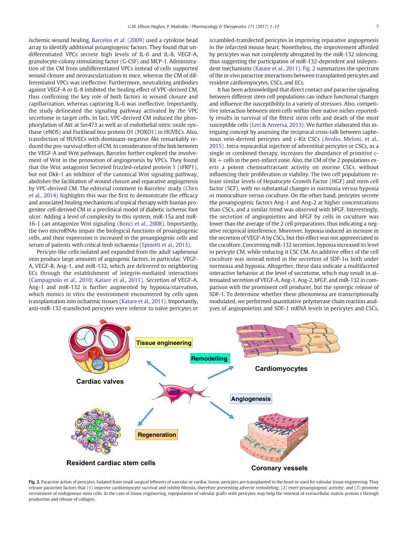
Regeneration
Remo
Tissue engineering
Fig. 2. Paracrine action of pericytes. Isolated from small surgical leftovers of vascular or cardiacrelease paracrine factors that (1) improve cardiomyocyte survival and inhibit fibrosis, thererecruitment of endogenous stem cells. In the case of tissue engineering, repopulation of valvuproduction and release of collagen.
scrambled-transfected pericytes in improving reparative angiogenesisin the infarcted mouse heart. Nonetheless, the improvement affordedby pericytes was not completely abrogated by the miR-132 silencing,thus suggesting the participation of miR-132-dependent and indepen-dent mechanisms (Katare et al., 2011). Fig. 2 summarizes the spectrumof the in vivo paracrine interactions between transplanted pericytes andresident cardiomyocytes, CSCs, and ECs.
It has been acknowledged that direct contact and paracrine signalingbetween different stem cell populations can induce functional changesand influence the susceptibility to a variety of stressors. Also, competi-tive interaction between stem cells within their native niches reported-ly results in survival of the fittest stem cells and death of the mostsusceptible cells (Leri & Anversa, 2013). We further elaborated this in-triguing concept by assessing the reciprocal cross-talk between saphe-nous vein-derived pericytes and c-Kit CSCs (Avolio, Meloni, et al.,2015). Intra-myocardial injection of adventitial pericytes or CSCs, as asingle or combined therapy, increases the abundance of primitive c-Kit + cells in the peri-infarct zone. Also, the CM of the 2 populations ex-erts a potent chemoattractant activity on murine CSCs, withoutinfluencing their proliferation or viability. The two cell populations re-lease similar levels of Hepatocyte Growth Factor (HGF) and stem cellfactor (SCF), with no substantial changes in normoxia versus hypoxiaor monoculture versus coculture. On the other hand, pericytes secretethe proangiogenic factors Ang-1 and Ang-2 at higher concentrationsthan CSCs, and a similar trend was observed with bFGF. Interestingly,the secretion of angiopoietins and bFGF by cells in coculture waslower than the average of the 2 cell preparations, thus indicating a neg-ative reciprocal interference. Moreover, hypoxia induced an increase inthe secretion of VEGF-A by CSCs, but this effectwas not appreeeciated inthe coculture. ConcerningmiR-132 secretion, hypoxia increased its levelin pericyte CM, while reducing it CSC CM. An additive effect of the cellcoculture was instead noted in the secretion of SDF-1α both undernormoxia and hypoxia. Altogether, these data indicate a multifacetedinteractive behavior at the level of secretome, which may result in at-tenuated secretion of VEGF-A, Ang-1, Ang-2, bFGF, andmiR-132 in com-parison with the prominent cell producer, but the synergic release ofSDF-1. To determine whether these phenomena are transcriptionallymodulated, we performed quantitative polymerase chain reaction anal-yses of angiopoietins and SDF-1 mRNA levels in pericytes and CSCs,
Angiogenesis
delling
tissue, pericytes are transplanted in the heart or used for valvular tissue engineering. Theyfore preventing adverse remodeling; (2) exert proangiogenic activity; and (3) promotelar grafts with pericytes may help the renewal of extracellular matrix protein s through
8 G.M. Ellison-Hughes, P. Madeddu / Pharmacology & Therapeutics 171 (2017) 1–12
before and after exposure to each other's CM. Results indicate CM re-duces the expression of Ang-2 in both the cell types. Also, CSCs exposedto pericyte CM showed reduced SDF-1 mRNA levels as compared withCSCs exposed to the unconditioned medium. These data suggest a tran-scriptional interference on Ang-2 and SDF-1. However, while Ang-2 ex-pression was reduced at mRNA and protein level, the increase in SDF-1content in coculture media was not attributed to the induction of genetranscription, but rather to an increase in secretion rate. Exploring a pos-sible involvement of Dipeptidyl peptidase-4 (DPP-4) we found higherexpression levels by CSCs. Furthermore, DPP-4 is downregulated atmRNAand protein level in CSCs exposed to pericytes CM. These findingsmay have important implication for association cell therapy, where theoutcomemay depend on the balance between cooperative and compet-itive interactions at the level of the secretome. For instance, in our study,the combined transplantation of pericytes and CSCs additively reducedthe infarct size and promoted vascular proliferation and arteriogenesis,but did not surpass single therapies concerning contractility indexes(Avolio, Meloni, et al., 2015).
Skeletal muscle-derived stem cells have been among the first celltherapy models for regenerative treatment of the infarcted heart(Menasche, 2008). A recent study investigated the therapeutic potentialof human skeletal muscle pericytes for treating ischaemic heart diseaseand mediating associated repair mechanisms in mice (Chen et al.,2013). The authors found that pericyte transplantation attenuates leftventricular dilatation andmyocardialfibrosis and improves cardiac con-tractility in infarcted mouse hearts. In line with findings in saphenousvein-derived pericytes, hypoxia-induced the expression of VEGF-A aswell as PDGF-β, TransformingGrowth Factor beta1 (TGF-β1), and corre-sponding receptors while expression of bFGF, HGF, and Ang-1 wasrepressed.
3.2. Resident cardiac pericytes: are they better suited for the heart?
New knowledge on microvessel-associated regenerative precursorcells in cardiacmuscle opens prospectives for organ-specific treatmentsof patients with congenital and acquired heart defects. Peault's teamshowed that microvascular pericytes within the human myocardiumexhibit phenotypes and multipotency similar to their anatomicallyand developmentally distinct counterparts (Chen et al., 2015). Howev-er, they have no ability for skeletalmyogenesis, diverging in this respectfrom pericytes of all other origins. In contrast, a cardiomyogenic poten-tial was evidenced both in vitro and after intra-myocardial transplanta-tion in vivo.
Using the same standard operating protocol employed for the deri-vation of pericytes from adult saphenous veins, we have isolated andexpanded pericyte-like progenitors from small biopsies of congenitallydefective hearts (antigenic features illustrated in Table 3) (Avolio,Rodriguez-Arabaolaza, et al., 2015). The long-term objective of researchon neonatal cardiac pericytes is their perusal for definitive correction ofcongenital heart defects (Avolio, Caputo, &Madeddu, 2015). A spectrumof prostheses in the form of conduits, patches, and valves is employed incongenital cardiac surgery, but none of them is perfect. In particular,currently, available grafts do not possess growing capacity and becomeincompetent with time, thus requiring surgical replacement. The basicconcept of tissue engineering is to create living material made bycellularized grafts that, once implanted into the heart, grows and re-models in parallel with the recipient organ. Neonatal pericytes producea powerful secretome uniquely fit for the purpose. This includes growthfactors that promote vasculogenesis and cardiomyogenesis andchemokines able to attract ECs and endothelial progenitor cellsinstrumental to graft endothelialisation. Comparison with saphenousvein–derived pericytes indicates similarity of the 2 cell populations,though cardiac pericytes secrete more HGF (6-fold), Ang-2 (8-fold),bFGF (4-fold), and VEGF-A (6-fold) than saphenous vein–derivedpericytes. Furthermore, neonatal cardiac pericytes release procollagen
type 1, a major constituent of the cardiac extracellular matrix, whichis fundamental to the maintenance of graft integrity.
The umbilical cord represents a valuable source of stem cells imme-diately available at birth for corrective strategies. In a recent study,human cord-derived pericytes or cord blood-derived MSCs were deliv-ered before or after alveolar injury into the airways of newborn rats ex-posed to hyperoxia, a model of bronchopulmonary dysplasia whichcomplicates extreme prematurity and currently lacks efficient treat-ment (Pierro et al., 2013). Rat pups exposed to hyperoxia showed typi-cal alteration in alveolar growth with enlargement and loss of lungcapillaries. Both human cord-derived pericytes and cord blood-derivedMSCs partially prevented and rescued lung function and structure.However, the low cell engraftment suggested a paracrine effect. Inline, cell free-derived CM from both cell types manifested significantbenefit when used either prophylactically or therapeutically. Altogeth-er, these studies open new avenues to exploit pericytes in neonatalregenerative medicine.
4. Evidence for paracrine healing properties of cardiac stem/progen-itor cells
There is a growing consensus that the beneficial effects of any myo-cardial autologous or allogeneic cell therapy so far tested is, at least inpart, mediated by a paracrine effect on the patient's cells at risk and ac-tivation of the host's CSCs by growth factors secreted by thetransplanted cells (Braunwald, 2015; Lorkeers et al., 2014). Major con-tributors to this cardioprotective and CSC stimulatory effect are HGFand IGF-1 acting through their relative receptors present both on themyocytes, vascular and CSCs of the recipient's heart (Ellison et al.,2011).
MSCs have a broad repertoire of secreted trophic and immunomod-ulatory cytokines; however, they also secrete factors that negativelymodulate cardiomyocyte apoptosis, inflammation, scar formation, andpathological remodeling (Ranganath et al., 2012). Moreover, it is ques-tionablewhether they are themost optimal cell to use regarding surviv-al and homing to and engraftment in the myocardium. Furthermore,cells can become entrapped in the microvasculature and impede cellentry into the myocardium.
The safety and efficacy of transplanting 2 million allogeneic, mis-matched Cardiosphere-derived cells (CDCs) was tested in infarctedrats. Three weeks post-MI, animals that received allogeneic CDCs exhib-ited smaller scar size, increased infarcted wall thickness, and attenua-tion of ventricular remodeling. Allogeneic CDC transplantationresulted in an improved fractional area change (∼12%), ejection fraction(∼20%), and fractional shortening (∼10%), and this was sustained for atleast 6 months. Furthermore, allogeneic CDCs stimulated endogenousregenerative mechanisms (cardiomyocyte cycling, recruitment of en-dogenous CSCs, angiogenesis) and increased levels of myocardialVEGF, IGF-1, and HGF (Malliaras et al., 2014).
We have previously shown that CSCs that express high levels of thetranscription factor GATA-4 exert a paracrine survival effect oncardiomyocytes through increased IGF-1.
secretion and induction of the IGF-1R signaling pathway(Kawaguchi et al., 2010). Furthermore, unlike bone marrow derivedcells (Hofmann et al., 2005), CSCs have a very high tropism for themyo-cardium (Ellison et al., 2013). This cardiac tropism is governed by theSDF1-CXCR4 signaling axis, and when CSCs are injected eitherintracoronary or systemically, they home to and nest into the damagedheart with a high efficiency and significantly restore the myocardium,anatomically and functionally (Ellison et al., 2013).
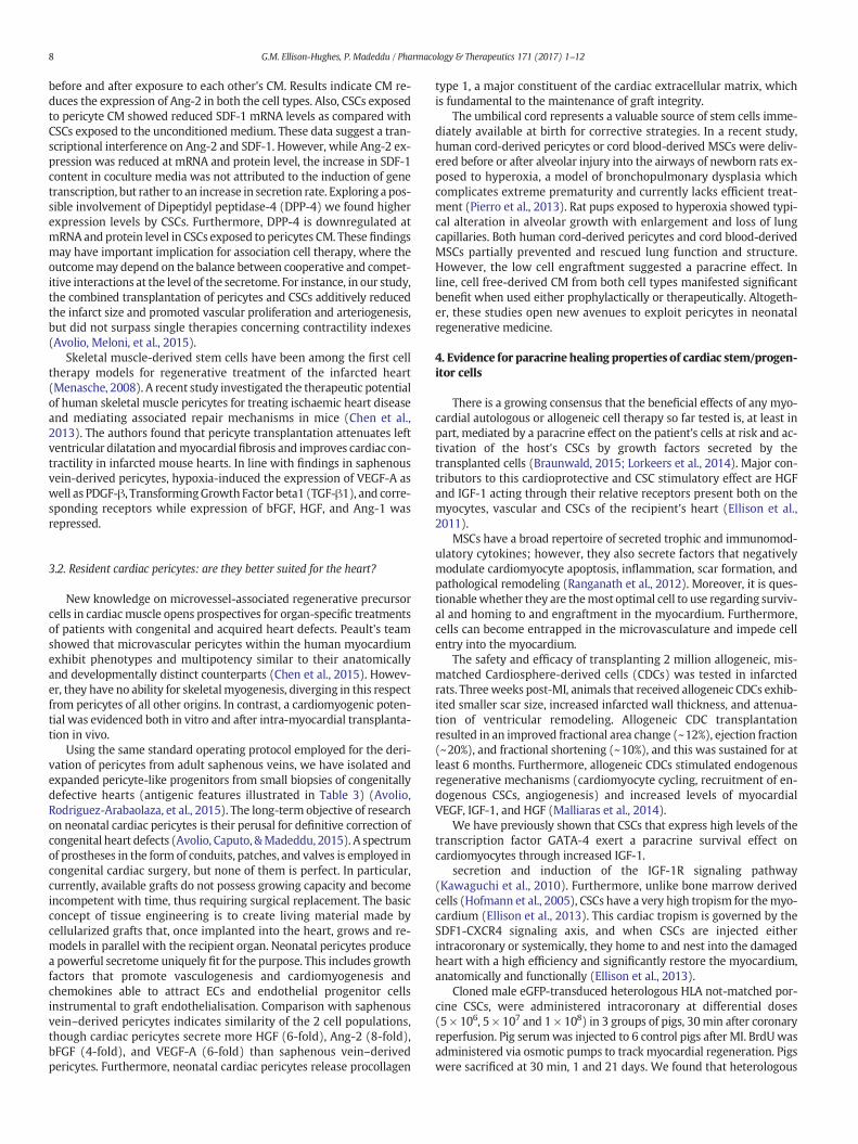
Cloned male eGFP-transduced heterologous HLA not-matched por-cine CSCs, were administered intracoronary at differential doses(5 × 10⁶, 5 × 10⁷ and 1 × 10⁸) in 3 groups of pigs, 30min after coronaryreperfusion. Pig serumwas injected to 6 control pigs after MI. BrdUwasadministered via osmotic pumps to track myocardial regeneration. Pigswere sacrificed at 30 min, 1 and 21 days. We found that heterologous

Fig. 3. Through paracrine mechanisms, c-kitpos heterologous HLA non-matched CSC treatment preserves myocardial wall structure and attenuates remodeling in a porcine MI model.(A) CSC treatment led to significantly decreased myocyte hypertrophy in the border region. *P b 0.05 vs. CTRL. (B) Representative H&E staining showing a band of hypertrophicmyocytes in the border region of CTRL pig myocardium. (C) CSC treatment significantly decreased percent number of apoptotic (caspase 3 positive) myocytes in the border region.*P b 0.05 vs. CTRL. (D & E) Representative images of sirius red staining to identify fibrotic tissue (red) and muscle (yellow) in the infarct region of CTRL (D) and CSC-treated (E) pighearts. (F) CSC-treated pig hearts had a decreased percentage area fraction of fibrosis in the infarct zone.*P b 0.05 vs. CTRL.
9G.M. Ellison-Hughes, P. Madeddu / Pharmacology & Therapeutics 171 (2017) 1–12
CSC administrationwaswell tolerated andwithout adverse effects. CSCsnested into the damaged myocardium with an efficiency of N95%, at30min through to 1 day after MI. Minimal spill over of CSCswas detect-ed in the coronary sinus, spleen, lung or live and all injected CSCs haddisappeared from the myocardium at 21 days. CSC-treated infarctedpig hearts showed a significant increase in the number of endogenousc-kitpos (GFPneg) CSCs in the border and infarct regions, compared toCTRL. CSC-treated hearts exhibited an increase in the number of small,newly formed BrdUpos myocytes and capillaries. CSC injection signifi-cantly preserved myocardial wall structure and attenuated remodelingby reducing hypertrophy, apoptosis and fibrosis (Fig. 3). Moreover, car-diac function was significantly preserved/improved by heterologousCSC-treatment. To summarize, intracoronary injection of heterologousCSCs after MI in pigs, which is a clinically relevant MI model, activatesthe endogenous CSCs through a paracrine mechanism resulting in im-proved myocardial cell survival and physiologically meaningful regen-eration (Ellison et al., 2009).
Allogeneic CSC therapy is conceptually and practically different fromany presently in clinical use. The proposed cell therapy is only a differentform of growth factor therapy, where the cells naturally home to thedamaged myocardium, deliver a more complex mixture of growth fac-tors, elicit a ‘paracrine’ effect and activate the endogenous target cells.Then the allogeneic CSCs are eliminated, and the regeneration triggeredby activated endogenous CSCs is completely autologous. Therefore, theallogeneic CSCs survived long enough in the allogeneic host to producetheir paracrine effect before being eliminated by the host immune sys-tem. Once more information is available, the allogeneic cells could beused either alone or in combination with the available factor therapyto improve the activation of the CSCs and the maturation of theirprogeny.
5. Conclusions
The market analysis predicts protein and peptide-based drugs willcompose N50%of novel drugswithin the next 10 years. Pharmacologicalcompanies have been using unbiased discovery methods to generatenew druggable compounds since many years. The approach is now
increasingly used in academic research. It is likely that new cardiovascu-lar drugs will be introduced in the next future by applying these ap-proaches to the study of stem cell paracrine function. Due to thevariability of the product and high costs associated with clinical gradeproduction, stem cell-based therapy is not amenable to the majority ofischaemic patients. The use of drugs inspired by the stem cell secretomewill instead offer unprecedented therapeutic opportunities, resulting ina fundamental shift in the initial concept of regenerative medicine.
Conflict of interest
The authors do not have any conflict of interest to declare.
Acknowledgment
Unravelling mechanisms of stem cell depletion for preservation ofregenerative fitness in patients with diabetes 30545.
References
Arslan, F., Lai, R. C., Smeets, M. B., Akeroyd, L., Choo, A., Aguor, E. N., ... de Kleijn, D. P.(2013). Mesenchymal stem cell-derived exosomes increase ATP levels, decrease oxi-dative stress and activate PI3K/Akt pathway to enhance myocardial viability and pre-vent adverse remodeling after myocardial ischemia/reperfusion injury. Stem CellResearch 10(3), 301–312.
Ascione, R., Rowlinson, J., Avolio, E., Katare, R., Meloni, M., Spencer, H. L., ... Madeddu, P.(2015). Migration towards SDF-1 selects angiogenin-expressing bonemarrowmono-cytes endowed with cardiac reparative activity in patients with previous myocardialinfarction. Stem Cell Research & Therapy 6, 53.
Assmus, B., Schachinger, V., Teupe, C., Britten, M., Lehmann, R., Dobert, N., ... Zeiher, A. M.(2002). Transplantation of progenitor cells and regeneration enhancement in acutemyocardial infarction (TOPCARE-AMI). Circulation 106(24), 3009–3017.
Avolio, E., Caputo, M., & Madeddu, P. (2015a). Stem cell therapy and tissue engineering forcorrection of congenital heart disease. Frontiers in Cell and Development Biology 3, 39.
Avolio, E., Meloni, M., Spencer, H. L., Riu, F., Katare, R., Mangialardi, G., ... Madeddu, P.(2015b). Combined intramyocardial delivery of human pericytes and cardiac stemcells additively improves the healing of mouse infarcted hearts through stimulationof vascular and muscular repair. Circulation Research 116(10), e81–e94.
Avolio, E., Rodriguez-Arabaolaza, I., Spencer, H. L., Riu, F., Mangialardi, G., Slater, S. C., ...Madeddu, P. (2015c). Expansion and characterization of neonatal cardiac pericytesprovides a novel cellular option for tissue engineering in congenital heart disease.Journal of the American Heart Association 4(6), e002043.
10 G.M. Ellison-Hughes, P. Madeddu / Pharmacology & Therapeutics 171 (2017) 1–12
Balsam, L. B., Wagers, A. J., Christensen, J. L., Kofidis, T., Weissman, I. L., & Robbins, R. C.(2004). Haematopoietic stem cells adopt mature haematopoietic fates in ischaemicmyocardium. Nature 428(6983), 668–673.
Baraniak, P. R., & McDevitt, T. C. (2010). Stem cell paracrine actions and tissue regenera-tion. Regenerative Medicine 5(1), 121–143.
Barcelos, L. S., Duplaa, C., Krankel, N., Graiani, G., Invernici, G., Katare, R., ... Madeddu, P.(2009). Human CD133+ progenitor cells promote the healing of diabetic ischemiculcers by paracrine stimulation of angiogenesis and activation of Wnt signaling.Circulation Research 104(9), 1095–1102.
Beltrami, A. P., Barlucchi, L., Torella, D., Baker, M., Limana, F., Chimenti, S., ... Anversa, P.(2003). Adult cardiac stem cells are multipotent and support myocardial regenera-tion. Cell 114(6), 763–776.
Bergmann, O., Zdunek, S., Felker, A., Salehpour, M., Alkass, K., Bernard, S., ... Frisen, J.(2015). Dynamics of cell generation and turnover in the human heart. Cell 161(7),1566–1575.
Bergmann, O., Zdunek, S., Frisen, J., Bernard, S., Druid, H., & Jovinge, S. (2012). Cardiomyo-cyte renewal in humans. Circulation Research 110(1), e17–e18 (author reply e19-21).
Bersell, K., Arab, S., Haring, B., & Kuhn, B. (2009). Neuregulin1/ErbB4 signaling inducescardiomyocyte proliferation and repair of heart injury. Cell 138(2), 257–270.
Bhang, S. H., Lee, S., Shin, J. Y., Lee, T. J., Jang, H. K., & Kim, B. S. (2014). Efficacious and clin-ically relevant conditioned medium of human adipose-derived stem cells for thera-peutic angiogenesis. Molecular Therapy 22(4), 862–872.
Bhatnagar, P., Wickramasinghe, K., Williams, J., Rayner, M., & Townsend, N. (2015).The epidemiology of cardiovascular disease in the UK 2014. Heart 101(15),1182–1189.
Bolli, R., Chugh, A. R., D'Amario, D., Loughran, J. H., Stoddard, M. F., Ikram, S., ... Anversa, P.(2011). Cardiac stem cells in patients with ischaemic cardiomyopathy (SCIPIO): initialresults of a randomised phase 1 trial. Lancet 378(9806), 1847–1857. http://dx.doi.org/10.1016/S0140-6736(11)61590-0. Epub 2011 Nov 14.
Bonci, D., Coppola, V., Musumeci, M., Addario, A., Giuffrida, R., Memeo, L., ... De Maria, R.(2008). The miR-15a-miR-16-1 cluster controls prostate cancer by targeting multipleoncogenic activities. Nature Medicine 14(11), 1271–1277.
Bostrom, P., Mann, N., Wu, J., Quintero, P. A., Plovie, E. R., Panakova, D., ... Spiegelman, B.M.(2010). C/EBPbeta controls exercise-induced cardiac growth and protects againstpathological cardiac remodeling. Cell 143(7), 1072–1083.
Braunwald, E. (2015). The war against heart failure: The lancet lecture. Lancet 385(9970),812–824.
Campagnolo, P., Cesselli, D., Al Haj Zen, A., Beltrami, A. P., Krankel, N., Katare, R., ... Madeddu, P.(2010). Human adult vena saphena contains perivascular progenitor cells endowedwithclonogenic and proangiogenic potential. Circulation 121(15), 1735–1745.
Caporali, A., Meloni, M., Nailor, A., Mitic, T., Shantikumar, S., Riu, F., ... Emanueli, C. (2015).p75(NTR)-dependent activation of NF-kappaB regulates microRNA-503 transcriptionand pericyte-endothelial crosstalk in diabetes after limb ischaemia. NatureCommunications 6, 8024.
Chen, W. C., Baily, J. E., Corselli, M., Diaz, M. E., Sun, B., Xiang, G., ... Peault, B. (2015).Human myocardial pericytes: Multipotent mesodermal precursors exhibiting cardiacspecificity. Stem Cells 33(2), 557–573.
Chen, C. W., Corselli, M., Peault, B., & Huard, J. (2012). Human blood-vessel-derived stemcells for tissue repair and regeneration. Journal of Biomedicine & Biotechnology 2012,597439.
Chen, C. W., Okada, M., Proto, J. D., Gao, X., Sekiya, N., Beckman, S. A., ... Huard, J. (2013).Human pericytes for ischemic heart repair. Stem Cells 31(2), 305–316.
Chen, W. C., Saparov, A., Corselli, M., Crisan, M., Zheng, B., Peault, B., & Huard, J. (2014).Isolation of blood-vessel-derived multipotent precursors from human skeletal mus-cle. Journal of Visualized Experiments 90, e51195.
Chien, K. R., & Olson, E. N. (2002). Converging pathways and principles in heart develop-ment and disease: CV@CSH. Cell 110(2), 153–162.
Chimenti, I., Gaetani, R., Barile, L., Forte, E., Ionta, V., Angelini, F., ... Giacomello, A. (2012).Isolation and expansion of adult cardiac stem/progenitor cells in the form ofcardiospheres from human cardiac biopsies and murine hearts. Methods inMolecular Biology 879, 327–338.
Chong, J. J., Chandrakanthan, V., Xaymardan, M., Asli, N. S., Li, J., Ahmed, I., ... Harvey, R. P.(2011). Adult cardiac-resident MSC-like stem cells with a proepicardial origin. CellStem Cell 9(6), 527–540.
Chung, E. S., Miller, L., Patel, A. N., Anderson, R. D., Mendelsohn, F. O., Traverse, J., ... Penn,M. S. (2015). Changes in ventricular remodelling and clinical status during the yearfollowing a single administration of stromal cell-derived factor-1 non-viral gene ther-apy in chronic ischaemic heart failure patients: The STOP-HF randomized phase IItrial. European Heart Journal 36(33), 2228–2238.
Cohen, J. E., Purcell, B. P., MacArthur, J. W., Jr., Mu, A., Shudo, Y., Patel, J. B., ... Woo, Y. J.(2014). A bioengineered hydrogel system enables targeted and sustainedintramyocardial delivery of neuregulin, activating the cardiomyocyte cell cycle andenhancing ventricular function in a murine model of ischemic cardiomyopathy.Circulation Heart Failure 7(4), 619–626.
Corselli, M., Crisan, M., Murray, I. R., West, C. C., Scholes, J., Codrea, F., ... Peault, B. (2013).Identification of perivascular mesenchymal stromal/stem cells by flow cytometry.Cytometry Part A 83(8), 714–720.
Dar, A., Domev, H., Ben-Yosef, O., Tzukerman, M., Zeevi-Levin, N., Novak, A., ... Itskovitz-Eldor, J. (2012). Multipotent vasculogenic pericytes from human pluripotent stemcells promote recovery of murine ischemic limb. Circulation 125(1), 87–99.
de Jong, R., Houtgraaf, J. H., Samiei, S., Boersma, E., & Duckers, H. J. (2014). Intracoronarystem cell infusion after acute myocardial infarction: A meta-analysis and update onclinical trials. Circulation Cardiovascular Interventions 7(2), 156–167.
Dellavalle, A., Maroli, G., Covarello, D., Azzoni, E., Innocenzi, A., Perani, L., ... Cossu, G.(2011). Pericytes resident in postnatal skeletal muscle differentiate intomuscle fibresand generate satellite cells. Nature Communications 2, 499.
Di Santo, S., Yang, Z., Wyler von Ballmoos, M., Voelzmann, J., Diehm, N., Baumgartner, I., &Kalka, C. (2009). Novel cell-free strategy for therapeutic angiogenesis: In vitro gener-ated conditioned medium can replace progenitor cell transplantation. PloS One 4(5),e5643.
Eisenberg, C. A., Burch, J. B., & Eisenberg, L. M. (2006). Bone marrow cellstransdifferentiate to cardiomyocytes when introduced into the embryonic heart.Stem Cells 24(5), 1236–1245.
Ellison, G. M., Torella, D., Dellegrottaglie, S., Perez-Martinez, C., Perez de Prado, A.,Vicinanza, C., ... Nadal-Ginard, B. (2011). Endogenous cardiac stem cell activation byinsulin-like growth factor-1/hepatocyte growth factor intracoronary injection fosterssurvival and regeneration of the infarcted pig heart. Journal of the American College ofCardiology 58(9), 977–986.
Ellison, G. M., Torella, D., Trigueros, C., Gonzalez, A., Waring, C., Perez-Martinez, C., ...Nadal-Ginard, B. (2009). Use of heterologous non-matched Cardiac Stem Cells(CSCs) without immunosuppression as an effective regenerating agent in a porcinemodel of acute myocardial infarction. European Heart Journal 30, 495.
Ellison, G. M., Vicinanza, C., Smith, A. J., Aquila, I., Leone, A., Waring, C. D., ... Nadal-Ginard,B. (2013). Adult c-kit(pos) cardiac stem cells are necessary and sufficient for func-tional cardiac regeneration and repair. Cell 154(4), 827–842.
Engel, F. B., Hsieh, P. C., Lee, R. T., & Keating, M. T. (2006). FGF1/p38 MAP kinase inhibitortherapy induces cardiomyocyte mitosis, reduces scarring, and rescues function aftermyocardial infarction. Proceedings of the National Academy of Sciences of the UnitedStates of America 103(42), 15546–15551.
Engels, M. C., Rajarajan, K., Feistritzer, R., Sharma, A., Nielsen, U. B., Schalij, M. J., ... Wu, S.M. (2014). Insulin-like growth factor promotes cardiac lineage induction in vitro byselective expansion of early mesoderm. Stem Cells 32(6), 1493–1502.
Fukuoka, H., & Suga, H. (2015). Hair regeneration treatment using adipose-derived stemcell conditioned medium: Follow-up with Trichograms. Eplasty 15, e10.
Galindo, C. L., Kasasbeh, E., Murphy, A., Ryzhov, S., Lenihan, S., Ahmad, F. A., ... Cleator, J. H.(2014). Anti-remodeling and anti-fibrotic effects of the neuregulin-1beta glial growthfactor 2 in a large animal model of heart failure. Journal of the American Heart Associ-ation 3(5), e000773.
Gnecchi, M., He, H., Liang, O. D., Melo, L. G., Morello, F., Mu, H., ... Dzau, V. J. (2005). Para-crine action accounts for marked protection of ischemic heart by Akt-modified mes-enchymal stem cells. Nature Medicine 11(4), 367–368.
Gubernator, M., Slater, S. C., Spencer, H. L., Spiteri, I., Sottoriva, A., Riu, F., ... Madeddu, P.(2015). Epigenetic profile of human adventitial progenitor cells correlates with ther-apeutic outcomes in amousemodel of limb ischemia. Arteriosclerosis, Thrombosis, andVascular Biology 35(3), 675–688.
Hare, J. M., Traverse, J. H., Henry, T. D., Dib, N., Strumpf, R. K., Schulman, S. P., ... Sherman,W. (2009). A randomized, double-blind, placebo-controlled, dose-escalation study ofintravenous adult human mesenchymal stem cells (prochymal) after acute myocar-dial infarction. Journal of the American College of Cardiology 54(24), 2277–2286.
Hatzistergos, K. E., Quevedo, H., Oskouei, B. N., Hu, Q., Feigenbaum, G. S., Margitich, I. S., ...Hare, J. M. (2010). Bone marrowmesenchymal stem cells stimulate cardiac stem cellproliferation and differentiation. Circulation Research 107(7), 913–922.
Ho, J. C. Y., Lai, W. H., Li, M. F., Au, K. W., Yip, M. C., Wong, N. L. Y., ... Tse, H. F. (2012). Re-versal of endothelial progenitor cell dysfunction in patients with type 2 diabetesusing a conditioned medium of human embryonic stem cell-derived endothelialcells. Diabetes-Metabolism Research and Reviews 28(5), 462–473.
Hodgkinson, C. P., Gomez, J. A., Payne, A. J., Zhang, L., Wang, X., Dal-Pra, S., ... Dzau, V. J.(2014). Abi3bp regulates cardiac progenitor cell proliferation and differentiation.Circulation Research 115(12), 1007–1016.
Hofmann, M.,Wollert, K. C., Meyer, G. P., Menke, A., Arseniev, L., Hertenstein, B., ... Drexler,H. (2005). Monitoring of bonemarrow cell homing into the infarcted humanmyocar-dium. Circulation 111(17), 2198–2202.
Hsieh, P. C., Segers, V. F., Davis, M. E., MacGillivray, C., Gannon, J., Molkentin, J. D., ... Lee, R.T. (2007). Evidence from a genetic fate-mapping study that stem cells refresh adultmammalian cardiomyocytes after injury. Nature Medicine 13(8), 970–974.
Hynes, B., Kumar, A. H. S., O'Sullivan, J., Buneker, C. K., Leblond, A. L., Weiss, S., ... Caplice, N.M. (2013). Potent endothelial progenitor cell-conditioned media-related anti-apoptotic, cardiotrophic, and pro-angiogenic effects post-myocardial infarction aremediated by insulin-like growth factor-1. European Heart Journal 34(10), 782–789.
Invernici, G., Emanueli, C., Madeddu, P., Cristini, S., Gadau, S., Benetti, A., ... Alessandri, G.(2007). Human fetal aorta contains vascular progenitor cells capable of inducingvasculogenesis, angiogenesis, and myogenesis in vitro and in a murine model of pe-ripheral ischemia. The American Journal of Pathology 170(6), 1879–1892.
Iso, Y., Rao, K. S., Poole, C. N., Zaman, A. K., Curril, I., Sobel, B. E., ... Spees, J. L. (2014).Priming with ligands secreted by human stromal progenitor cells promotesgrafts of cardiac stem/progenitor cells after myocardial infarction. Stem Cells32(3), 674–683.
Jeevanantham, V., Butler, M., Saad, A., Abdel-Latif, A., Zuba-Surma, E. K., & Dawn, B.(2012). Adult bone marrow cell therapy improves survival and induces long-termimprovement in cardiac parameters: A systematic review and meta-analysis.Circulation 126(5), 551–568.
Katare, R., Riu, F., Mitchell, K., Gubernator, M., Campagnolo, P., Cui, Y., ... Madeddu, P.(2011). Transplantation of human pericyte progenitor cells improves the repair of in-farcted heart through activation of an angiogenic program involving micro-RNA-132.Circulation Research 109(8), 894–906.
Katare, R., Riu, F., Rowlinson, J., Lewis, A., Holden, R., Meloni, M., ... Madeddu, P. (2013).Perivascular delivery of encapsulated mesenchymal stem cells improvespostischemic angiogenesis via paracrine activation of VEGF-A. Arteriosclerosis,Thrombosis, and Vascular Biology 33(8), 1872–1880.
Kawaguchi, N., Smith, A. J., Waring, C. D., Hasan, M. K., Miyamoto, S., Matsuoka, R., &Ellison, G. M. (2010). C-kit(pos) GATA-4 high rat cardiac stem cells Foster adult car-diomyocyte survival through IGF-1 paracrine signalling. PloS One 5(12).
11G.M. Ellison-Hughes, P. Madeddu / Pharmacology & Therapeutics 171 (2017) 1–12
Khan, M., Nickoloff, E., Abramova, T., Johnson, J., Verma, S. K., Krishnamurthy, P., ...Kishore, R. (2015). Embryonic stem cell-derived exosomes promote endogenous re-pair mechanisms and enhance cardiac function following myocardial infarction.Circulation Research 117(1), 52–64.
Kim, J. Y., Song, S. H., Kim, K. L., Ko, J. J., Im, J. E., Yie, S. W., ... Suh, W. (2010). Human cordblood-derived endothelial progenitor cells and their conditioned media exhibit ther-apeutic equivalence for diabetic wound healing. Cell Transplantation 19(12),1635–1644.
Klein, D., Weisshardt, P., Kleff, V., Jastrow, H., Jakob, H. G., & Ergun, S. (2011). Vascularwall-resident CD44+ multipotent stem cells give rise to pericytes and smooth mus-cle cells and contribute to new vessel maturation. PloS One 6(5), e20540.
Koudstaal, S., Bastings, M. M., Feyen, D. A., Waring, C. D., van Slochteren, F. J., Dankers, P. Y., ... Chamuleau, S. A. (2014). Sustained delivery of insulin-like growth factor-1/hepatocyte growth factor stimulates endogenous cardiac repair in the chronic infarct-ed pig heart. Journal of Cardiovascular Translational Research 7(2), 232–241.
Kuhn, B., del Monte, F., Hajjar, R. J., Chang, Y. S., Lebeche, D., Arab, S., & Keating, M. T.(2007). Periostin induces proliferation of differentiated cardiomyocytes and pro-motes cardiac repair. Nature Medicine 13(8), 962–969.
Laflamme, M. A., & Murry, C. E. (2011). Heart regeneration. Nature 473(7347), 326–335.Laugwitz, K. L., Moretti, A., Caron, L., Nakano, A., & Chien, K. R. (2008). Islet1 cardiovascu-
lar progenitors: A single source for heart lineages? Development 135(2), 193–205.Lee, M. J., Kim, J., Kim, M. Y., Bae, Y. S., Ryu, S. H., Lee, T. G., & Kim, J. H. (2010). Proteomic
analysis of tumor necrosis factor-alpha-induced secretome of human adipose tissue-derived mesenchymal stem cells. Journal of Proteome Research 9(4), 1754–1762.
Lee, C., Mitsialis, S. A., Aslam, M., Vitali, S. H., Vergadi, E., Konstantinou, G., ... Kourembanas,S. (2012). Exosomes mediate the cytoprotective action of mesenchymal stromal cellson hypoxia-induced pulmonary hypertension. Circulation 126(22), 2601–2611.
Lemons, D., Maurya, M. R., Subramaniam, S., & Mercola, M. (2013). Developing microRNAscreening as a functional genomics tool for disease research. Frontiers in Physiology 4.
Leri, A., & Anversa, P. (2013). Stem cells and myocardial regeneration cooperation winsover competition. Circulation 127(2), 165–168.
Limana, F., Zacheo, A., Mocini, D., Mangoni, A., Borsellino, G., Diamantini, A., ... Capogrossi,M. C. (2007). Identification of myocardial and vascular precursor cells in human andmouse epicardium. Circulation Research 101(12), 1255–1265.
Loffredo, F. S., Steinhauser, M. L., Gannon, J., & Lee, R. T. (2011). Bone marrow-derived celltherapy stimulates endogenous cardiomyocyte progenitors and promotes cardiac re-pair. Cell Stem Cell 8(4), 389–398.
Lorkeers, S. J. J. O., Eding, J. E. C., van der Spoel, T. I. G., Vesterinen, H. M., Sena, E. S.,Duckers, H. J., ... Chamuleau, S. A. J. (2014). Similar effect of autologous and allogeneiccell therapy for ischaemic heart disease: Systematic review and meta-analysis oflarge animal studies. European Journal of Clinical Investigation 44, 5–6.
Mackie, A. R., Klyachko, E., Thorne, T., Schultz, K. M., Millay, M., Ito, A., ... Losordo, D. W.(2012). Sonic hedgehog-modified human CD34+ cells preserve cardiac functionafter acute myocardial infarction. Circulation Research 111(3), 312–321.
Madonna, R., & De Caterina, R. (2011). Stem cells and growth factor delivery systems forcardiovascular disease. Journal of Biotechnology 154(4), 291–297.
Malliaras, K., Makkar, R. R., Smith, R. R., Cheng, K., Wu, E., Bonow, R. O., ... Marban, E.(2014). Intracoronary cardiosphere-derived cells after myocardial infarction: Evi-dence of therapeutic regeneration in the final 1-year results of the CADUCEUS trial(CArdiosphere-derived aUtologous stem CElls to reverse ventricUlar dySfunction).Journal of the American College of Cardiology 63(2), 110–122.
Martin, C. M., Meeson, A. P., Robertson, S. M., Hawke, T. J., Richardson, J. A., Bates, S., ...Garry, D. J. (2004). Persistent expression of the ATP-binding cassette transporter,Abcg2, identifies cardiac SP cells in the developing and adult heart. DevelopmentalBiology 265(1), 262–275.
Martin-Rendon, E. (2016). Meta-analyses of human cell-based cardiac regeneration ther-apies: What can systematic reviews tell us about cell therapies for ischemic heart dis-ease? Circulation Research 118(8), 1264–1272.
Menasche, P. (2008). Skeletal myoblasts and cardiac repair. Journal of Molecular andCellular Cardiology 45(4), 545–553.
Menasche, P., Alfieri, O., Janssens, S., McKenna, W., Reichenspurner, H., Trinquart, L., ...Hagege, A. A. (2008). The myoblast autologous grafting in ischemic cardiomyopathy(MAGIC) trial: First randomized placebo-controlled study of myoblast transplanta-tion. Circulation 117(9), 1189–1200.
Mirabella, T., Cilli, M., Carlone, S., Cancedda, R., & Gentili, C. (2012). Amniotic liquid de-rived stem cells as reservoir of secreted angiogenic factors capable of stimulatingneoarteriogenesis in an ischemic model (vol 32, pg 3689, 2011). Biomaterials33(20), 5175.
Mourkioti, F., & Rosenthal, N. (2005). IGF-1, inflammation and stem cells: Interactionsduring muscle regeneration. Trends in Immunology 26(10), 535–542.
Murry, C. E., Soonpaa, M. H., Reinecke, H., Nakajima, H., Nakajima, H. O., Rubart, M., ...Field, L. J. (2004). Haematopoietic stem cells do not transdifferentiate into cardiacmyocytes in myocardial infarcts. Nature 428(6983), 664–668.
Nadal-Ginard, B. (1978). Commitment, fusion and biochemical differentiation of a myo-genic cell line in the absence of DNA synthesis. Cell 15(3), 855–864.
Nadal-Ginard, B., Kajstura, J., Anversa, P., & Leri, A. (2003). A matter of life and death: Car-diac myocyte apoptosis and regeneration. The Journal of Clinical Investigation 111(10),1457–1459.
Nguyen, B. K., Maltais, S., Perrault, L. P., Tanguay, J. F., Tardif, J. C., Stevens, L. M., ... Noiseux,N. (2010). Improved function and myocardial repair of infarcted heart byintracoronary injection of mesenchymal stem cell-derived growth factors. Journal ofCardiovascular Translational Research 3(5), 547–558.
Oh, H., Bradfute, S. B., Gallardo, T. D., Nakamura, T., Gaussin, V., Mishina, Y., ... Schneider,M. D. (2003). Cardiac progenitor cells from adult myocardium: Homing, differentia-tion, and fusion after infarction. Proceedings of the National Academy of Sciences ofthe United States of America 100(21), 12313–12318.
Parekkadan, B., van Poll, D., Suganuma, K., Carter, E. A., Berthiaume, F., Tilles, A. W., &Yarmush, M. L. (2007). Mesenchymal stem cell-derived molecules reverse fulminanthepatic failure. PloS One 2(9), e941.
Pawitan, J. A. (2014). Prospect of stem cell conditionedmedium in regenerative medicine.BioMed Research International.
Penn, M. S. (2009). Importance of the SDF-1:CXCR4 axis in myocardial repair. CirculationResearch 104(10), 1133–1135.
Pierro, M., Ionescu, L., Montemurro, T., Vadivel, A., Weissmann, G., Oudit, G., ... Thebaud, B.(2013). Short-term, long-term and paracrine effect of human umbilical cord-derivedstem cells in lung injury prevention and repair in experimental bronchopulmonarydysplasia. Thorax 68(5), 475–484.
Polizzotti, B. D., Ganapathy, B., Walsh, S., Choudhury, S., Ammanamanchi, N., Bennett, D. G., ...Kuhn, B. (2015). Neuregulin stimulation of cardiomyocyte regeneration in mice andhuman myocardium reveals a therapeutic window. Science Translational Medicine7(281), 281ra245.
Quaini, F., Urbanek, K., Beltrami, A. P., Finato, N., Beltrami, C. A., Nadal-Ginard, B., ...Anversa, P. (2002). Chimerism of the transplanted heart. The New England Journal ofMedicine 346(1), 5–15.
Ranganath, S. H., Levy, O., Inamdar, M. S., & Karp, J. M. (2012). Harnessing the mesenchy-mal stem cell secretome for the treatment of cardiovascular disease. Cell Stem Cell10(3), 244–258.
Rao, K. S., Aronshtam, A., McElory-Yaggy, K. L., Bakondi, B., VanBuren, P., Sobel, B. E., & Spees,J. L. (2015). Human epicardial cell-conditioned medium contains HGF/IgG complexesthat phosphorylate RYK and protect against vascular injury. Cardiovascular Research107(2), 277–286.
Rasmussen, J. G., Frobert, O., Holst-Hansen, C., Kastrup, J., Baandrup, U., Zachar, V., ...Simonsen, U. (2014). Comparison of human adipose-derived stem cells and bonemarrow-derived stem cells in a myocardial infarction model. Cell Transplantation23(2), 195–206.
Reuter, S., Soonpaa, M. H., Firulli, A. B., Chang, A. N., & Field, L. J. (2014). Recombinantneuregulin 1 does not activate cardiomyocyte DNA synthesis in normal or infarctedadult mice. PloS One 9(12), e115871.
Sahoo, S., & Losordo, D. W. (2014). Exosomes and cardiac repair after myocardial infarc-tion. Circulation Research 114(2), 333–344.
Sahoo, S., Klychko, E., Thorne, T., Misener, S., Schultz, K. M., Millay, M., ... Losordo, D. W.(2011). Exosomes from human CD34(+) stem cells mediate their proangiogenicparacrine activity. Circulation Research 109(7), 724–U735.
Sant'anna, R. T., Kalil, R. A., Pretto Neto, A. S., Pivatto Junior, F., Fracasso, J., Sant'anna, J. R., ...Nesralla, I. A. (2010). Global contractility increment in nonischemic dilated cardiomy-opathy after free wall-only intramyocardial injection of autologous bone marrowmononuclear cells: An insight over stem cells clinical mechanism of action. CellTransplantation 19(8), 959–964.
See, F., Seki, T., Psaltis, P. J., Sondermeijer, H. P., Gronthos, S., Zannettino, A. C. W., ... Itescu,S. (2011). Therapeutic effects of human STRO-3-selected mesenchymal precursorcells and their soluble factors in experimental myocardial ischemia. Journal ofCellular and Molecular Medicine 15(10), 2117–2129.
Senyo, S. E., Steinhauser, M. L., Pizzimenti, C. L., Yang, V. K., Cai, L., Wang, M., ... Lee, R. T.(2013). Mammalian heart renewal by pre-existing cardiomyocytes. Nature 493(7432),433–436.
Shin, H., Ryu, H. H., Kwon, O., Park, B. S., & Jo, S. J. (2015). Clinical use of conditionedmediaof adipose tissue-derived stem cells in female pattern hair loss: A retrospective caseseries study. International Journal of Dermatology 54(6), 730–735.
Smart, N., Bollini, S., Dube, K. N., Vieira, J. M., Zhou, B., Davidson, S., ... Riley, P. R. (2011). Denovo cardiomyocytes from within the activated adult heart after injury. Nature474(7353), 640–644.
Spinetti, G., Fortunato, O., Caporali, A., Shantikumar, S., Marchetti, M., Meloni, M., ...Emanueli, C. (2013). MicroRNA-15a and microRNA-16 impair human circulatingproangiogenic cell functions and are increased in the proangiogenic cells andserum of patients with critical limb ischemia. Circulation Research 112(2),335–346.
Sze, S. K., de Kleijn, D. P., Lai, R. C., KhiaWay Tan, E., Zhao, H., Yeo, K. S., ... Lim, S. K. (2007).Elucidating the secretion proteome of human embryonic stem cell-derived mesen-chymal stem cells. Molecular & Cellular Proteomics 6(10), 1680–1689.
Tang, X. L., Li, Q., Rokosh, G., Sanganalmath, S., Chen, N., Ou, Q., ... Bolli, R. (2016a). Long-term outcome of administration of c-kitPOS cardiac progenitor cells after acute myo-cardial infarction: Transplanted cells do not become cardiomyocytes, but structuraland functional improvement and proliferation of endogenous cells persist for atleast one year. Circulation Research.
Tang, X. L., Li, Q., Rokosh, G., Sanganalmath, S. K., Chen, N., Ou, Q., ... Bolli, R. (2016b). Long-term outcome of administration of c-kit(POS) cardiac progenitor cells after acutemyocardial infarction: Transplanted cells do not become cardiomyocytes, but struc-tural and functional improvement and proliferation of endogenous cells persist forat least one year. Circulation Research 118(7), 1091–1105.
Timmers, L., Lim, S. K., Arslan, F., Armstrong, J. S., Hoefer, I. E., Doevendans, P. A., ... deKleijn, D. P. (2007). Reduction of myocardial infarct size by human mesenchymalstem cell conditioned medium. Stem Cell Research 1(2), 129–137.
Timmers, L., Lim, S. K., Hoefer, I. E., Arslan, F., Lai, R. C., van Oorschot, A. A. M., ... deKleijn, D. P. V. (2011). Human mesenchymal stem cell-conditioned mediumimproves cardiac function following myocardial infarction. Stem Cell Research6(3), 206–214.
Torella, D., Ellison, G. M., Karakikes, I., & Nadal-Ginard, B. (2007). Resident cardiac stemcells. Cellular and Molecular Life Sciences 64(6), 661–673.
Torella, D., Ellison, G.M., Mendez-Ferrer, S., Ibanez, B., & Nadal-Ginard, B. (2006). Residenthuman cardiac stem cells: Role in cardiac cellular homeostasis and potential for myo-cardial regeneration. Nature Clinical Practice Cardiovascular Medicine 3(Suppl. 1),S8–13.
12 G.M. Ellison-Hughes, P. Madeddu / Pharmacology & Therapeutics 171 (2017) 1–12
Urbanek, K., Quaini, F., Tasca, G., Torella, D., Castaldo, C., Nadal-Ginard, B., ... Anversa, P.(2003). Intense myocyte formation from cardiac stem cells in human cardiac hyper-trophy. Proceedings of the National Academy of Sciences of the United States of America100(18), 10440–10445.
Urbanek, K., Torella, D., Sheikh, F., De Angelis, A., Nurzynska, D., Silvestri, F., ... Anversa, P.(2005). Myocardial regeneration by activation of multipotent cardiac stem cells in is-chemic heart failure. Proceedings of the National Academy of Sciences of the UnitedStates of America 102(24), 8692–8697.
van Berlo, J. H., & Molkentin, J. D. (2014). An emerging consensus on cardiac regeneration.Nature Medicine 20(12), 1386–1393.
Wadugu, B., & Kuhn, B. (2012). The role of neuregulin/ErbB2/ErbB4 signaling in the heartwith special focus on effects on cardiomyocyte proliferation. American Journal of Phys-iology. Heart and Circulatory Physiology 302(11), H2139–H2147.
Waring, C. D., Vicinanza, C., Papalamprou, A., Smith, A. J., Purushothaman, S., Goldspink, D.F., ... Ellison, G. M. (2014). The adult heart responds to increased workload with phys-iologic hypertrophy, cardiac stem cell activation, and new myocyte formation.European Heart Journal 35(39), 2722–2731.
Wei, K., Serpooshan, V., Hurtado, C., Diez-Cunado, M., Zhao, M., Maruyama, S., ... Ruiz-Lozano, P. (2015). Epicardial FSTL1 reconstitution regenerates the adult mammalianheart. Nature 525(7570), 479–485.
Wollert, K. C., Meyer, G. P., Lotz, J., Ringes-Lichtenberg, S., Lippolt, P., Breidenbach, C., ...Drexler, H. (2004). Intracoronary autologous bone-marrow cell transfer after
myocardial infarction: The BOOST randomised controlled clinical trial. Lancet364(9429), 141–148.
Yang, D. Z., Wang, W., Li, L. P., Peng, Y. L., Chen, P., Huang, H. Y., ... Zeng, C. Y. (2013). Therelative contribution of Paracine effect versus direct differentiation on adipose-de-rived stem cell transplantation mediated cardiac repair. PloS One 8(3).
Yuan, A., Farber, E. L., Rapoport, A. L., Tejada, D., Deniskin, R., Akhmedov, N. B., & Farber, D.B. (2009). Transfer of microRNAs by embryonic stem cell microvesicles. PloS One 4(3),e4722.
Zebrowski, D. C., Becker, R., & Engel, F. B. (2016). Towards regenerating the mammalianheart: Challenges in evaluating experimentally induced adult mammalian cardio-myocyte proliferation. American Journal of Physiology. Heart and Circulatory Physiology310(9), H1045–H1054.
Zhou, B., Honor, L. B., He, H., Ma, Q., Oh, J. H., Butterfield, C., ... Pu, W. T. (2011). Adultmouse epicardium modulates myocardial injury by secreting paracrine factors. TheJournal of Clinical Investigation 121(5), 1894–1904.
Zhou, B. R., Xu, Y., Guo, S. L., Xu, Y., Wang, Y., Zhu, F., ... Luo, D. (2013). The effect of con-ditioned media of adipose-derived stem cells on wound healing after ablative frac-tional carbon dioxide laser resurfacing. BioMed Research International 2013, 519126.
Zwetsloot, P. P., Gremmels, H., Assmus, B., Koudstaal, S., Sluijter, J. P., Zeiher, A. M., &Chamuleau, S. A. (2016). Responder definition in clinical stem cell trials in cardiology:Will the real responder please stand up? Circulation Research 119(4), 514–518.