exploring synaptic circuits with antibodies and confocal microscopy phd school of neuroscience turin...
TRANSCRIPT
Exploring synaptic Exploring synaptic circuits with antibodies circuits with antibodies
and confocal and confocal microscopymicroscopy
Part I
PhD School of NeurosciencePhD School of NeuroscienceTurin UniversityTurin University
June 2009June 2009 Marco SassoèMarco Sassoè
Common problems in immunocytochemistry: Common problems in immunocytochemistry:
a.a. Preservation of antigenicity (antigens may be Preservation of antigenicity (antigens may be destroyed by chemical fixatives, organic destroyed by chemical fixatives, organic solvents, high temperatures…)solvents, high temperatures…)
b.b. Efficiency of antibody binding to relevant Efficiency of antibody binding to relevant epitopes (may be altered by pH, ionic force)epitopes (may be altered by pH, ionic force)
c.c. Background labelling (importance of choice of Background labelling (importance of choice of blocking agent)blocking agent)
d.d. Non-specific labelling (importance of Non-specific labelling (importance of appropriate appropriate controlscontrols))
AntibodiesAntibodies
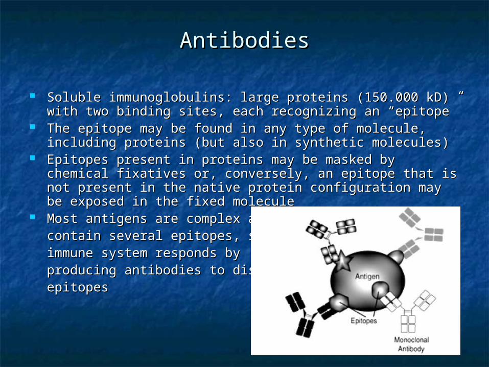
Soluble immunoglobulins: large proteins (150.000 kD) with Soluble immunoglobulins: large proteins (150.000 kD) with two binding sites, each recognizing an “epitope”two binding sites, each recognizing an “epitope”
The epitope may be found in any type of molecule, The epitope may be found in any type of molecule, including proteins (but also in synthetic molecules)including proteins (but also in synthetic molecules)
Epitopes present in proteins may be masked by chemical Epitopes present in proteins may be masked by chemical fixatives or, conversely, an epitope that is not present in fixatives or, conversely, an epitope that is not present in the native protein configuration may be exposed in the the native protein configuration may be exposed in the fixed moleculefixed molecule
Most antigens are complex and Most antigens are complex and contain several epitopes, so the contain several epitopes, so the immune system responds by immune system responds by producing antibodies to distinct producing antibodies to distinct epitopes epitopes
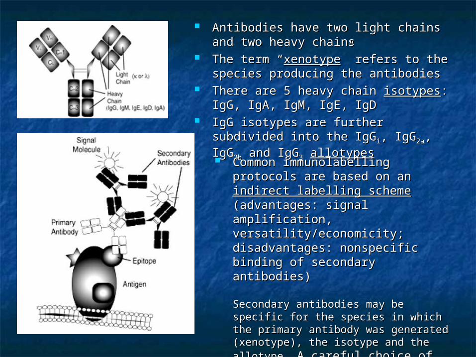
Antibodies have two light chains and Antibodies have two light chains and two heavy chainstwo heavy chains
The term “The term “xenotypexenotype” refers to the ” refers to the species producing the antibodiesspecies producing the antibodies
There are 5 heavy chain There are 5 heavy chain isotypesisotypes: IgG, : IgG, IgA, IgM, IgE, IgDIgA, IgM, IgE, IgD
IgG isotypes are further subdivided into IgG isotypes are further subdivided into the IgGthe IgG11, IgG, IgG2a2a, IgG, IgG2b2b and IgG and IgG33 allotypesallotypes
Common immunolabelling protocols Common immunolabelling protocols are based on an are based on an indirect labelling indirect labelling scheme scheme (advantages: signal (advantages: signal amplification, versatility/economicity; amplification, versatility/economicity; disadvantages: nonspecific binding of disadvantages: nonspecific binding of secondary antibodies)secondary antibodies)
Secondary antibodies may be specific for Secondary antibodies may be specific for the species in which the primary antibody the species in which the primary antibody was generated (xenotype), the isotype was generated (xenotype), the isotype and the allotype.and the allotype. A careful choice of A careful choice of secondary antibodies may thus help secondary antibodies may thus help to reduce nonspecific staining to reduce nonspecific staining considerablyconsiderably
AntiseraAntisera
A preparation containing the immunoglobulins A preparation containing the immunoglobulins from an animal’s serumfrom an animal’s serum
Each antiserum contains a wide array of Each antiserum contains a wide array of immunoglobulins, each recognizing distinct immunoglobulins, each recognizing distinct epitopes of the antigen used for the immunizationepitopes of the antigen used for the immunization
However, in fixed tissue, many if not all of these However, in fixed tissue, many if not all of these antigenic sites may be destroyedantigenic sites may be destroyed
In addition, among antisera that stain something In addition, among antisera that stain something in tissues, many may stain spurious antigens in tissues, many may stain spurious antigens ((cross-reactivitycross-reactivity), and only a few will recognize ), and only a few will recognize the original molecule (importance of adsorption the original molecule (importance of adsorption controls)controls)
Most antisera probably have only one or two clones of antibodies Most antisera probably have only one or two clones of antibodies that recognize the relevant antigen, the rest of the antibodies that recognize the relevant antigen, the rest of the antibodies being irrelevant (when not producing artifacts!). So, they being irrelevant (when not producing artifacts!). So, they functionally act as monoclonal antibodies!functionally act as monoclonal antibodies!Affinity purified antiseraAffinity purified antisera: they are purified by binding to the : they are purified by binding to the appropriate antigen. They are less prone to artifact and tend to appropriate antigen. They are less prone to artifact and tend to give lower background staininggive lower background staining
Affinity-purified AntiseraAffinity-purified Antisera
Monoclonal antibodiesMonoclonal antibodies
A single clone of immunoglobulins raised against a specific A single clone of immunoglobulins raised against a specific antigen (each B-cell produces antibodies which specifically antigen (each B-cell produces antibodies which specifically bind to a single epitope of the antigen)bind to a single epitope of the antigen)
Monoclonal antibodies are obtained from an hybridoma, which Monoclonal antibodies are obtained from an hybridoma, which is derived from the fusion of B-cells with neoplastic B-cells is derived from the fusion of B-cells with neoplastic B-cells isolated from a myeloma tumor (Kohler and Milstein, 1975)isolated from a myeloma tumor (Kohler and Milstein, 1975)
Hybridoma cells are grown either in culture or by injecting Hybridoma cells are grown either in culture or by injecting them intraperitoneally in a host animal. In this case, the host them intraperitoneally in a host animal. In this case, the host animal builds up a fluid (ascites), which contains a high animal builds up a fluid (ascites), which contains a high concentration of monoclonal antibodies. In either case, the concentration of monoclonal antibodies. In either case, the culture fluids or the ascites containing the antibodies are culture fluids or the ascites containing the antibodies are purified by precipitating the antibodies with protein Apurified by precipitating the antibodies with protein A
Even the purest monoclonal antibodies provide no assurance Even the purest monoclonal antibodies provide no assurance that the epitope recognized in tissue sections belongs to the that the epitope recognized in tissue sections belongs to the antigen used for the immunization (they require the same antigen used for the immunization (they require the same type of controls!)type of controls!)
Affinity of antibody-Affinity of antibody-antigen antigen
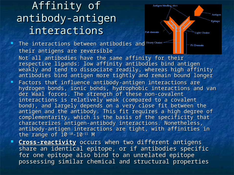
interactionsinteractions The interactions between antibodies and The interactions between antibodies and
their antigens are reversibletheir antigens are reversibleNot all antibodies have the same affinity for their respective Not all antibodies have the same affinity for their respective ligands: low affinity antibodies bind antigen weakly and tend to ligands: low affinity antibodies bind antigen weakly and tend to dissociate readily, whereas high affinity antibodies bind antigen dissociate readily, whereas high affinity antibodies bind antigen more tightly and remain bound longermore tightly and remain bound longer
Factors that influence antibody-antigen interactions are hydrogen Factors that influence antibody-antigen interactions are hydrogen bonds, ionic bonds, hydrophobic interactions and van der Waal bonds, ionic bonds, hydrophobic interactions and van der Waal forces. The strength of these non-covalent interactions is forces. The strength of these non-covalent interactions is relatively weak (compared to a covalent bond), and largely relatively weak (compared to a covalent bond), and largely depends on a very close fit between the antigen and the antibody. depends on a very close fit between the antigen and the antibody. This fit requires a high degree of complementarity, which is the This fit requires a high degree of complementarity, which is the basis of the specificity that characterizes antigen-antibody basis of the specificity that characterizes antigen-antibody interactions. Nonetheless, antibody-antigen interactions are tight, interactions. Nonetheless, antibody-antigen interactions are tight, with affinities in the range of 10with affinities in the range of 10-10-10-10-10-11-11 M M
Cross-reactivityCross-reactivity occurs when two different antigens share occurs when two different antigens share an identical epitope, or if antibodies specific for one epitope an identical epitope, or if antibodies specific for one epitope also bind to an unrelated epitope possessing similar also bind to an unrelated epitope possessing similar chemical and structural propertieschemical and structural properties
Adequate controls for Adequate controls for immunocytochemistryimmunocytochemistry
1.1. The knock-out testThe knock-out testStain tissue from a wild-type mouse and a KO mouse in which Stain tissue from a wild-type mouse and a KO mouse in which the antigen of interest has been eliminated by transgenic the antigen of interest has been eliminated by transgenic engineering. An even better choice is the use of inducible or engineering. An even better choice is the use of inducible or spatially-restricted deletion of the protein of interest, which spatially-restricted deletion of the protein of interest, which could minimize up or downregulation of other proteinscould minimize up or downregulation of other proteins
LimitationsLimitations
Applicable only in mice (requires additional controls in other species) Applicable only in mice (requires additional controls in other species)
The antigen recognized by the antibody may be present not on The antigen recognized by the antibody may be present not on molecules whose gene was deleted, but on another downstream molecules whose gene was deleted, but on another downstream molecule (in a metabolic or enzymatic pathway) molecule (in a metabolic or enzymatic pathway)
In many KO mice, the original protein is not entirely eliminated, and the In many KO mice, the original protein is not entirely eliminated, and the portion that remains may have no function, but still stain with the portion that remains may have no function, but still stain with the antibody antibody
Antibodies bind with low affinity to numerous tissue constituents, and Antibodies bind with low affinity to numerous tissue constituents, and even antibodies that are known to give a specific staining in normal even antibodies that are known to give a specific staining in normal conditions can give rise to non-specific signals in tissue obtained from conditions can give rise to non-specific signals in tissue obtained from KO miceKO mice
Adequate controls for Adequate controls for immunocytochemistryimmunocytochemistry
2.2. Western BlotWestern Blot
The antibody should recognize only one antigen in the The antibody should recognize only one antigen in the tissue and this must be of the appropriate molecular tissue and this must be of the appropriate molecular weight.weight.
LimitationsLimitations
Antibodies that recognize two or more bands may identify the Antibodies that recognize two or more bands may identify the same target molecule in different states of post-translational same target molecule in different states of post-translational modification (or in oligomeric forms). In that case, the use of modification (or in oligomeric forms). In that case, the use of such an antibody requires verification with other antibodies such an antibody requires verification with other antibodies raised against different parts of the same moleculeraised against different parts of the same molecule
Many antibodies that work well in WB do not recognize the Many antibodies that work well in WB do not recognize the target antigen after it has been distorted by the fixation process target antigen after it has been distorted by the fixation process
Adequate controls for Adequate controls for immunocytochemistryimmunocytochemistry
3.3. The adsorption testThe adsorption testThe antibody is pre-adsorbed with a The antibody is pre-adsorbed with a large large excessexcess of diluted antigen of diluted antigen
LimitationsLimitations
The antigen may contain an amino acid sequence that is The antigen may contain an amino acid sequence that is shared among a group of structurally-related proteins shared among a group of structurally-related proteins (may require adsorption against structurally related (may require adsorption against structurally related antigens) antigens)
Not applicable to monoclonal antibodies (they are Not applicable to monoclonal antibodies (they are screened for their binding to the target, and therefore will screened for their binding to the target, and therefore will always pass a preadsorption test, even if they stain always pass a preadsorption test, even if they stain something entirely different in tissue). This test is also something entirely different in tissue). This test is also meaningless for affinity-purified antibodies (for the same meaningless for affinity-purified antibodies (for the same reason)reason)
Adequate controls for Adequate controls for immunocytochemistryimmunocytochemistry
4.4. The dilution testThe dilution testThe antibody is diluted to the point where it just stains the the The antibody is diluted to the point where it just stains the the sites of interest (can be combined with adsorption tests)sites of interest (can be combined with adsorption tests)
5.5. DNA transfectionDNA transfectionDNA of the target protein (and possibly of protein isoforms) is DNA of the target protein (and possibly of protein isoforms) is transfected into cells that normally do not make that protein. transfected into cells that normally do not make that protein. However, this control does not prove that the antibody will However, this control does not prove that the antibody will onlyonly stain its target in the tissuestain its target in the tissue
6.6. Convergent dataConvergent dataa. Compare staining pattern with other, well-characterized a. Compare staining pattern with other, well-characterized antibodiesantibodiesb. Apply a combination of in situ hybridization and b. Apply a combination of in situ hybridization and immunocytochemistry (not always applicable)immunocytochemistry (not always applicable)c. Compare staining pattern of two or more antibodies raised c. Compare staining pattern of two or more antibodies raised against distinct epitopes on the same target proteinagainst distinct epitopes on the same target proteind. If the subcellular distribution of the target molecule is known, d. If the subcellular distribution of the target molecule is known, the staining pattern should conform to this distribution (not the staining pattern should conform to this distribution (not easily applicable to ubiquitous molecules). easily applicable to ubiquitous molecules).
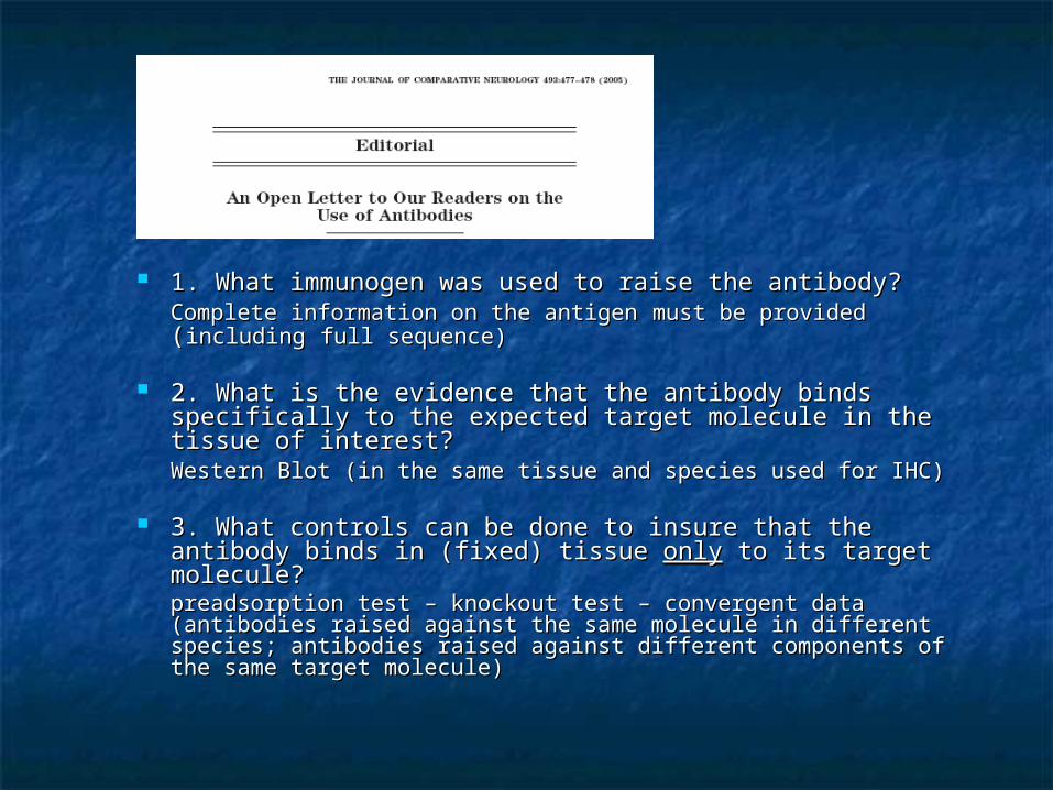
1. What immunogen was used to raise the antibody?1. What immunogen was used to raise the antibody?Complete information on the antigenComplete information on the antigen must be provided must be provided ((includingincluding full sequence)full sequence)
2. What is the evidence that the antibody binds 2. What is the evidence that the antibody binds specifically to the expected target molecule in the tissue specifically to the expected target molecule in the tissue of interest?of interest?Western Blot (in the same tissue and species used for IHC)Western Blot (in the same tissue and species used for IHC)
3. What controls can be done to insure that the antibody 3. What controls can be done to insure that the antibody binds in (fixed) tissue binds in (fixed) tissue onlyonly to its target molecule? to its target molecule?preadsorption test – knockout test – convergent data (antibodies preadsorption test – knockout test – convergent data (antibodies raised against the same molecule in different species; antibodies raised against the same molecule in different species; antibodies raised against different components of the same target raised against different components of the same target molecule)molecule)
Final notesFinal notesA single bleed from a rabbit is A single bleed from a rabbit is ~25 ml, half of which consists of ~25 ml, half of which consists of serum after the red blood cells have been eliminated. Each bleed serum after the red blood cells have been eliminated. Each bleed is essentially a unique combination of antibody clones. Hence, is essentially a unique combination of antibody clones. Hence, different batches of a polyclonal antiserum may have entirely different batches of a polyclonal antiserum may have entirely different staining properties (it is important to note the lot number different staining properties (it is important to note the lot number of antibodies, i.e. of antibodies, i.e. the code number for the animal that produced the code number for the animal that produced the antiserum and the bleedthe antiserum and the bleed). ).
Lack of staining for a molecule of interest cannot be interpreted Lack of staining for a molecule of interest cannot be interpreted as absence of that molecule (other possible causes are insufficient as absence of that molecule (other possible causes are insufficient affinity of the antibody, suboptimal tissue processing or IHC affinity of the antibody, suboptimal tissue processing or IHC protocol, epitope masking) protocol, epitope masking)
Antibodies are made against a globular, aqueous phase of a Antibodies are made against a globular, aqueous phase of a protein or peptide, and they recognize epitopes that are exposed protein or peptide, and they recognize epitopes that are exposed on the surface of that protein in the acqueous state. However, on the surface of that protein in the acqueous state. However, fixation is based on a chemical reaction that may change the fixation is based on a chemical reaction that may change the conformation of the molecule and mask the antigenic epitopes. In conformation of the molecule and mask the antigenic epitopes. In addition, some epitopes may be masked by other (associated) addition, some epitopes may be masked by other (associated) proteins. In other words, whether an antibody is capable of proteins. In other words, whether an antibody is capable of recognizing its epitope or not critically depends on the method recognizing its epitope or not critically depends on the method and also on the subcellular location of the proteinand also on the subcellular location of the protein
High variety of immunolabelling protocols, due to High variety of immunolabelling protocols, due to experimental conditions, antigen properties, antibody experimental conditions, antigen properties, antibody reactivity …reactivity …
For a successful immunostaining of an antigen in a tissue For a successful immunostaining of an antigen in a tissue section there must be:section there must be:
a.a. Retention of the subcellular distribution of the antigen (at the Retention of the subcellular distribution of the antigen (at the same sites that it occupied in the living organism) same sites that it occupied in the living organism) → fixation→ fixation
b.b. Permeability of tissue to the antibody molecules (cross-linking of Permeability of tissue to the antibody molecules (cross-linking of proteins by fixation impedes antibody penetration – weak fixation proteins by fixation impedes antibody penetration – weak fixation enhances penetration at the expense of structural preservation)enhances penetration at the expense of structural preservation)
c.c. The epitopes must be accessible to the primary antibody (cross-The epitopes must be accessible to the primary antibody (cross-linking due to the fixative is likely to mask epitopes → false linking due to the fixative is likely to mask epitopes → false negative results)negative results)
Preservation of structure and Preservation of structure and antigenicity antigenicity
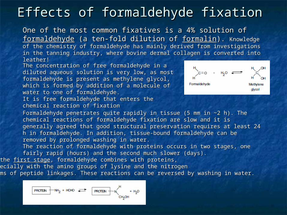
One of the most common fixatives is a 4% solution of One of the most common fixatives is a 4% solution of formaldehydeformaldehyde (a ten-fold dilution of (a ten-fold dilution of formalinformalin). ). Knowledge of the Knowledge of the chemistry of formaldehyde has mainly derived from investigations in the tanning chemistry of formaldehyde has mainly derived from investigations in the tanning industry, where bovine dermal collagen is converted into leather!industry, where bovine dermal collagen is converted into leather!
The concentration of free formaldehyde in a diluted The concentration of free formaldehyde in a diluted aqueous solution is very low, as most formaldehyde is aqueous solution is very low, as most formaldehyde is present as methylene glycol, which is formed by present as methylene glycol, which is formed by addition of a molecule of water to one of addition of a molecule of water to one of formaldehyde. formaldehyde. It is free formaldehyde that enters the chemical It is free formaldehyde that enters the chemical reaction of fixationreaction of fixationFormaldehyde penetrates quite rapidly in tissue (5 mm in Formaldehyde penetrates quite rapidly in tissue (5 mm in ~~2 h). The chemical 2 h). The chemical reactions of formaldehyde fixation are slow and it is generally agreed that good reactions of formaldehyde fixation are slow and it is generally agreed that good structural preservation requires at least 24 h in formaldehyde. In addition, tissue-structural preservation requires at least 24 h in formaldehyde. In addition, tissue-bound formaldehyde can be removed by prolonged washing in water. bound formaldehyde can be removed by prolonged washing in water. The reaction of formaldehyde with proteins occurs in two stages, one fairly rapid The reaction of formaldehyde with proteins occurs in two stages, one fairly rapid (hours) and the second much slower (days).(hours) and the second much slower (days).
In the In the first stagefirst stage, formaldehyde combines with proteins, , formaldehyde combines with proteins, especially with the amino groups of lysine and the nitrogen especially with the amino groups of lysine and the nitrogen atoms of peptide linkages. These reactions can be reversed by washing in water. atoms of peptide linkages. These reactions can be reversed by washing in water.
Effects of formaldehyde fixation Effects of formaldehyde fixation
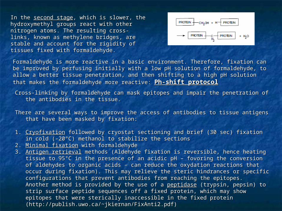
In the In the second stagesecond stage, which is slower, the , which is slower, the hydroxymethyl groups react with other nitrogen hydroxymethyl groups react with other nitrogen atoms. The resulting cross-links, known as atoms. The resulting cross-links, known as methylene bridges, are stable and account for methylene bridges, are stable and account for the rigidity of tissues fixed with formaldehyde.the rigidity of tissues fixed with formaldehyde.
Formaldehyde is more reactive in a basic environment. Therefore, fixation can be improved Formaldehyde is more reactive in a basic environment. Therefore, fixation can be improved by perfusing initially with a low pH solution of formaldehyde, to allow a better tissue by perfusing initially with a low pH solution of formaldehyde, to allow a better tissue penetration, and then shifting to a high pH solution that makes the formaldehyde more penetration, and then shifting to a high pH solution that makes the formaldehyde more reactive: reactive: Ph-shift protocolPh-shift protocol
Cross-linking by formaldehyde can mask epitopes and impair the penetration of the Cross-linking by formaldehyde can mask epitopes and impair the penetration of the antibodies in the tissue.antibodies in the tissue.
There are several ways to improve the access of antibodies to tissue antigens that have There are several ways to improve the access of antibodies to tissue antigens that have been masked by fixation:been masked by fixation:
1.1. CryofixationCryofixation followed by cryostat sectioning and brief (30 sec) fixation in cold (-20°C) followed by cryostat sectioning and brief (30 sec) fixation in cold (-20°C) methanol to stabilize the sectionsmethanol to stabilize the sections
2.2. Minimal fixationMinimal fixation with formaldehyde with formaldehyde3.3. Antigen retrievalAntigen retrieval methods (Aldehyde fixation is reversible, hence heating tissue to 95°C methods (Aldehyde fixation is reversible, hence heating tissue to 95°C
in the presence of an acidic pH – fovoring the conversion of aldehydes to organic acids in the presence of an acidic pH – fovoring the conversion of aldehydes to organic acids – can reduce the oxydation reactions that occur during fixation). This may relieve the – can reduce the oxydation reactions that occur during fixation). This may relieve the steric hindrances or specific configurations that prevent antibodies from reaching the steric hindrances or specific configurations that prevent antibodies from reaching the epitopes.epitopes.Another method is provided by the use of a Another method is provided by the use of a peptidasepeptidase (trypsin, pepsin) to strip surface (trypsin, pepsin) to strip surface peptide sequences off a fixed protein, which may show epitopes that were sterically peptide sequences off a fixed protein, which may show epitopes that were sterically inaccessible in the fixed protein (http://publish.uwo.ca/~jkiernan/FixAnti2.pdf)inaccessible in the fixed protein (http://publish.uwo.ca/~jkiernan/FixAnti2.pdf)
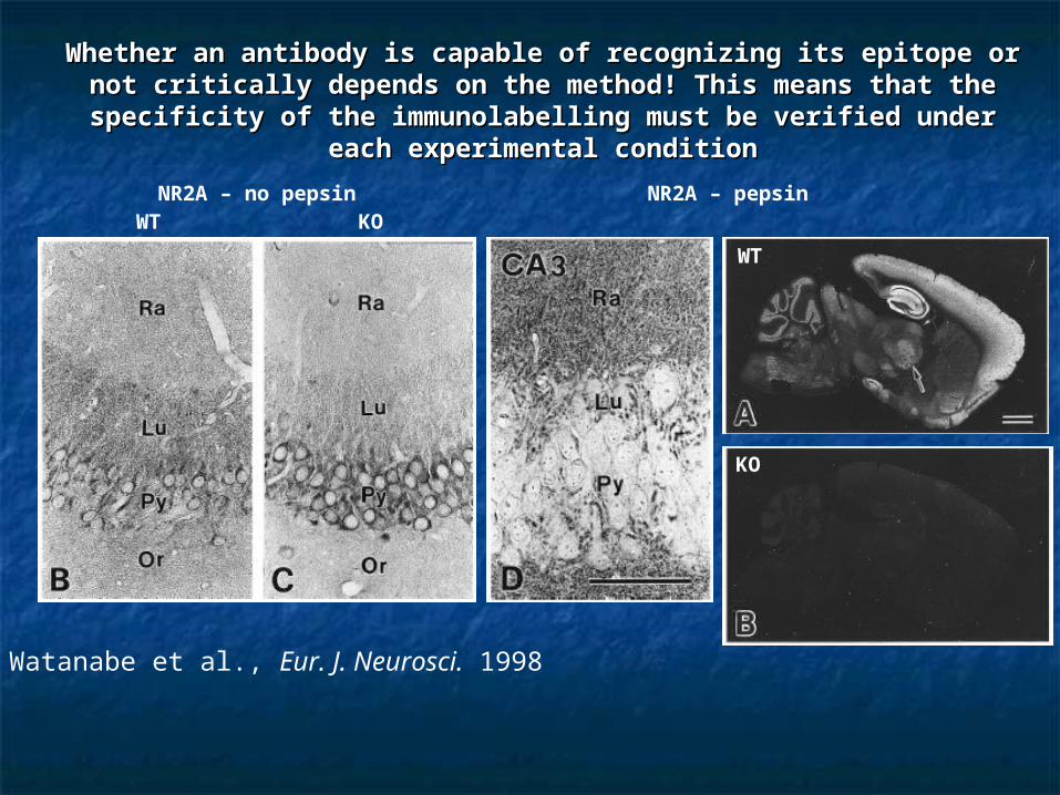
Whether an antibody is capable of recognizing its epitope or not Whether an antibody is capable of recognizing its epitope or not critically depends on the method! This means that the critically depends on the method! This means that the
specificity of the immunolabelling must be verified under each specificity of the immunolabelling must be verified under each experimental conditionexperimental condition
WT KONR2A – no pepsin
Watanabe et al., Eur. J. Neurosci. 1998
NR2A – pepsin
WT
KO

Giustetto et al., Neuroscience 1997
Sassoè-Pognetto and Ottersen, J. Neurosci. 2000
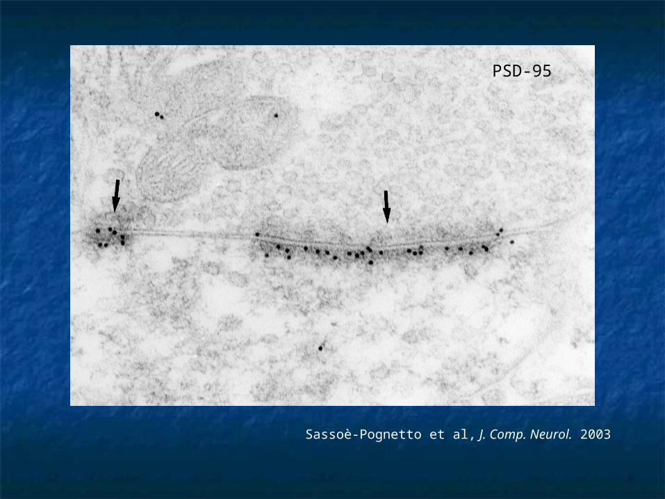
Sassoè-Pognetto et al, J. Comp. Neurol. 2003
PSD-95
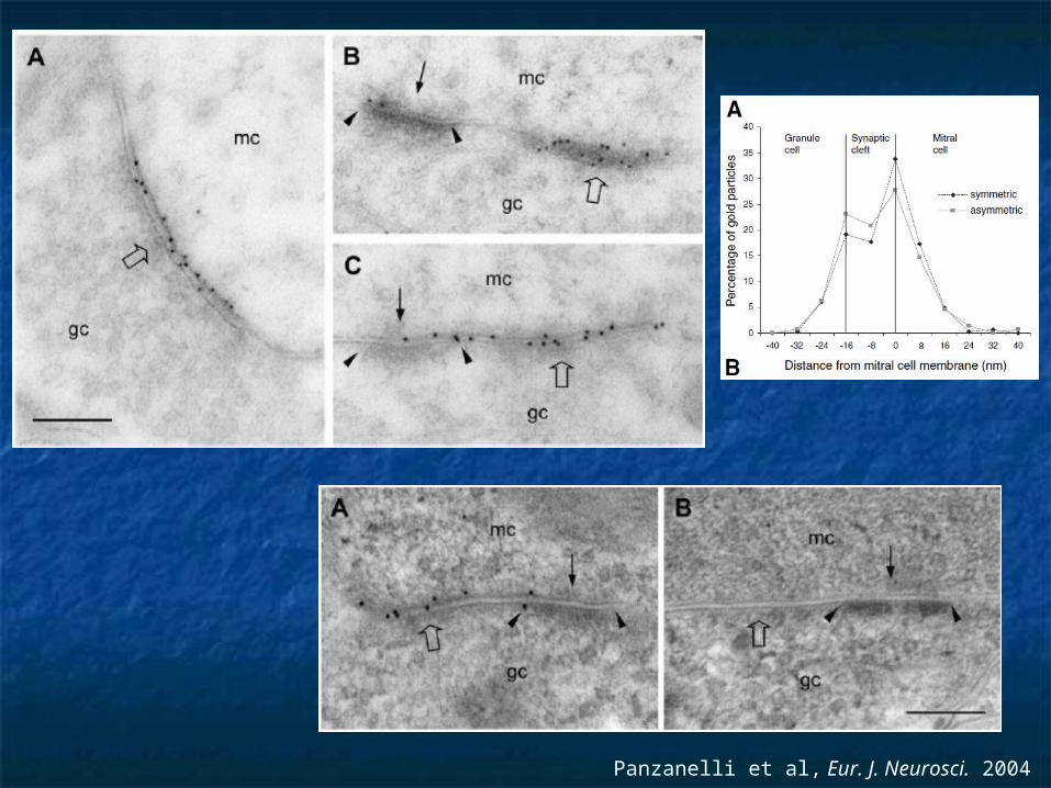
Panzanelli et al, Eur. J. Neurosci. 2004
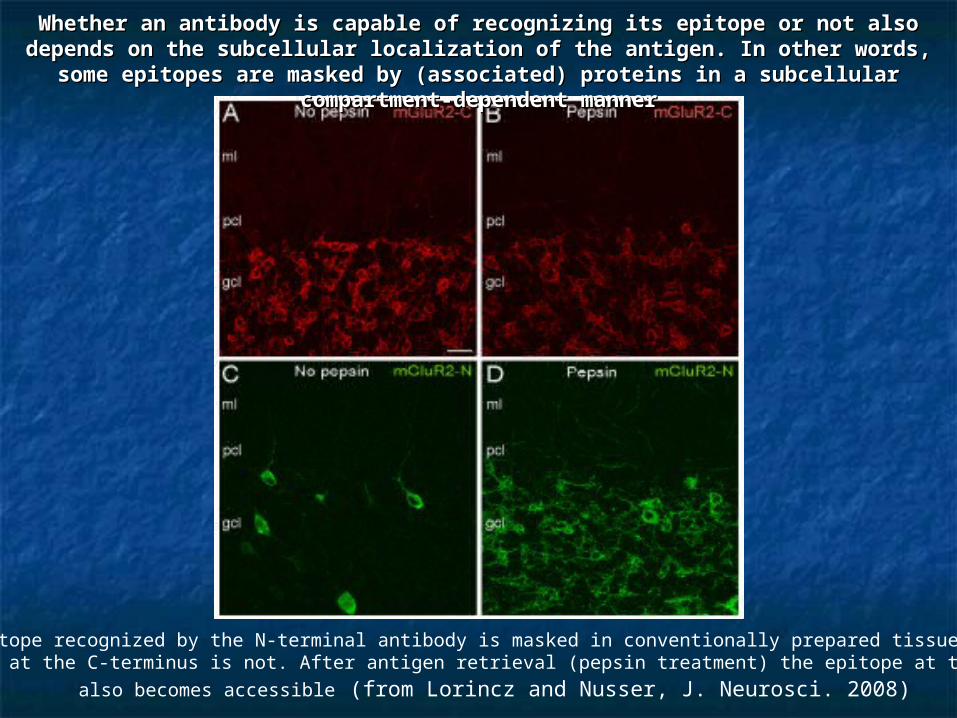
The epitope recognized by the N-terminal antibody is masked in conventionally prepared tissue, whereas the epitope at the C-terminus is not. After antigen retrieval (pepsin treatment) the epitope at the N-terminus
also becomes accessible (from Lorincz and Nusser, J. Neurosci. 2008)
Whether an antibody is capable of recognizing its epitope or not also Whether an antibody is capable of recognizing its epitope or not also depends on the subcellular localization of the antigen. In other words, some depends on the subcellular localization of the antigen. In other words, some epitopes are masked by (associated) proteins in a subcellular compartment-epitopes are masked by (associated) proteins in a subcellular compartment-
dependent mannerdependent manner

ReferencesReferences Saper CB and Sawchenko PE (2003) Magic peptides, magic antibodies: Saper CB and Sawchenko PE (2003) Magic peptides, magic antibodies:
guidelines for appropriate controls for immunocytochemistry. guidelines for appropriate controls for immunocytochemistry. J. Comp. J. Comp. Neurol.Neurol. 465:161-163. 465:161-163.
Saper CB (2005) An open letter to our readers on the use of antibodies. Saper CB (2005) An open letter to our readers on the use of antibodies. J. J. Comp. Neurol.Comp. Neurol. 493:477-478. 493:477-478.
Rhodes KJ and Trimmer JS (2006) Antibodies as valuable neuroscience tools Rhodes KJ and Trimmer JS (2006) Antibodies as valuable neuroscience tools versus reagents of mass distraction. versus reagents of mass distraction. J. Neurosci.J. Neurosci. 26:8017-8020. 26:8017-8020.
Schneider Gasser EM, Straub CJ, Panzanelli P, Weinmann O, Schneider Gasser EM, Straub CJ, Panzanelli P, Weinmann O, Sassoè-Sassoè-Pognetto MPognetto M, Fritschy JM (2006) Immunofluorescence in brain sections: , Fritschy JM (2006) Immunofluorescence in brain sections: simultaneous detection of pre- and postsynaptic proteins in identified simultaneous detection of pre- and postsynaptic proteins in identified neurons. neurons. Nat. ProtocolsNat. Protocols 1:1887-1897. 1:1887-1897.
Lorincz A and Nusser Z (2008) Specificity of immunoreactions: the Lorincz A and Nusser Z (2008) Specificity of immunoreactions: the importance of testing specificity in each method. importance of testing specificity in each method. J. Neurosci.J. Neurosci. 28:9083- 28:9083-9086.9086.
Fritschy JM (2008) Is my antibody staining specific? How to deal with Fritschy JM (2008) Is my antibody staining specific? How to deal with pitfalls of immunohistochemistry. pitfalls of immunohistochemistry. Eur. J. Neurosci.Eur. J. Neurosci. 28:2365-2370. 28:2365-2370.
Saper CB (2009) A guide to the perplexed on the specificity of antibodies. Saper CB (2009) A guide to the perplexed on the specificity of antibodies. J. J. Histochem. Cytochem.Histochem. Cytochem. 57:1-5. 57:1-5.
Illustration credit: Karl GarshaIllustration credit: Karl Garshahttp://www.itg.uiuc.edu/people/garsha/documents/Immunolabeling.pdf#search=%22garsha%20concepts%22http://www.itg.uiuc.edu/people/garsha/documents/Immunolabeling.pdf#search=%22garsha%20concepts%22