expression adhesion molecules resorption felineteeth: a
TRANSCRIPT

J Dent Res 75(9): 1650-1657, September, 1996
Expression of Adhesion Molecules duringTooth Resorption in Feline Teeth: A ModelSystem for Aggressive Osteoclastic ActivityY. Shigeyamal, T.K. Grove3, C. Strayhorn1, and M.J. Somermanl,2*
Departments of 1Periodontics/Prevention/Geriatrics and 2Pharmacology, School of Medicine, University of Michigan, 1011 N. University,Ann Arbor, Michigan 48109-1078; 3The Florida Veterinary Dental Clinic, 875 17th Street, Vero Beach, Florida 32960; *to whom correspondenceshould be addressed
Abstract. Tooth resorption, a common feline dentalproblem, is often initiated at the cemento-enamel junctionand hence is called cat 'neck' lesion. Studies havedemonstrated that osteoclasts/odontoclasts are increasedand activated at resorption sites, and that areas ofresorption are partly repaired by formation of tissuesresembling bone, cementum, and possibly dentin. However,the cellular/molecular mechanisms/factors involved inresorption and repair are unknown. In this study of tissuesfrom cats with 'neck' lesions, we used specific antibodiesand immunohistochemical analyses to examine adhesionmolecules associated with mineralized tissues, bonesialoprotein (BSP) and osteopontin (OPN), and a cell-surfacereceptor linked with these molecules, avl3 for theirlocalization in these lesions. In addition, to determinegeneral cellular activity during repair, we performed in situhybridization using a type I collagen riboprobe.
Results showed OPN localized to resorption fronts andreversal lines, while BSP was localized to reversal lines.However, some osteoclasts and odontoblasts "sat" onmineralized surfaces not associated with OPN. The cell-surface receptor, oXv3, was localized to surfaces ofosteoclasts/odontoclasts. Type I collagen mRNA wasexpressed where osteoblasts attempted to repairmineralized tissue. In contrast, odontoblasts did not expressmRNA for type I collagen. This study suggests thatosteoclastic resorption is the predominant activity in 'neck'lesions and that this activity was accompanied, at least inpart, by increased concentrations of OPN and an associatedintegrin, aLv3, at resorption sites. Lack of collagenexpression by odontoblasts indicates that odontoblasts donot play an active role in attempts at repair.
Key words: av 3 bone sialoprotein, odontoclasts,osteoclasts, osteopontin.
Received May 4, 1995; Accepted February 1, 1996
Introduction
Extensive tooth resorption is common in domesticated cats(Coles, 1990). Beginning at age 4, about 40% of cats areaffected with an average of 2.3 lesions per affected cat(Harvey, 1992). Other species rarely develop this lesion(Okuda and Harvey, 1992a). The lesion is characterized byresorption beginning at the cervical region of the tooth, thecemento-enamel junction (Fig. 1). As a result, this lesion isoften referred to as a 'neck' lesion or 'cervical line' lesion.With disease progression, internal resorption of toothstructures is seen. Affected cats may experience severedental hypersensitivity, tooth fracture, tooth loss bycomplete subgingival resorption, a variety of aversive socialbehaviors, and eating disorders. Because of their clinicalsimilarity to dental caries, these lesions were originallyclassified as such (Fig. 1). Early histologic studies (Schneckand Osborn, 1976) revealed osteoclast/odontoclastresorption in 'neck' lesions, and these investigatorsconcluded that such lesions were a type of periodontaldisease. A similar lesion in humans, external resorption,begins slightly apical to the cemento-enamel junction andlooks, radiographically, like caries associated withxerostomia (Regezi and Sciubba, 1989). The cause of thehuman lesion is unknown. The lesion progresses naturallyto extensive destruction and tooth loss. Although theseresorptive lesions share many similarities, when the twospecies are compared, the rate of destruction seems muchfaster in cats than in humans. Additionally, in cats, thelesions occur frequently and often in multiple teeth. Thesecharacteristics as well as the accessible contained nature ofthe lesion make the cat a good model for the study ofaggressive osteoclastic behavior.
Histologically, it has been reported that dental tissuesresorbed as a consequence of disease are repaired in part byformation of mineralized tissues resembling bone,cementum, and/or dentin (osteodentin) (Reichart et al.,1984; Okuda and Harvey, 1992a). Although significantrepair may occur, it rarely results in reformation ofcomplete structural integrity. Importantly, immuno-histochemical assays indicated the presence of two
1650

Adhesion Molecules during Tootlh Resorption
Figure 1. 1hotograph of cat 'neck' lesion. (a) Clinical view, lower molar. Large arrow points to cervical lesion. Note extensioni of the lesioninto the pulp chamber, resulting in an appearance similar to that of a caries lesion (small arrow). (b) Radiograph of cat 'neck' lesion (largearrow) and caries (small arrow) in molar shown in (a).
cytokines, IL-1 and IL-6. Both of these interleukins areconsidered to function in recruitment and activation ofosteoclasts/odontoclasts locally, prior to root resorption(Okuda and Harvey, 1992b). Also, 'odontoclastic' cells fromthese lesions stain positive for tartrate-resistant acidphosphatase (TRAP), an enzyme associated with osteoclasts(Okuda and Harvey, 1992b).We hypothesized that specific adhesion molecules and
their receptors play an active role in regulating rootresorption and repair. Strong evidence exists that adhesionmolecules direct cell behavior and differentiation duringreplacement of lost tissues (Albelda and Buck, 1990). Westudied two adhesion molecules important in mineralizedtissues, bone sialoprotein (BSP) (for review, see Fisher, 1992;Sodek et al., 1992) and osteopontin (OPN) (for review, seeDenhardt and Guo, 1993; Patarca et al., 1993; Denhardt andChambers, 1994), and an integrin linked with both BSP andOPN, Uj33 (Reinholt et al., 1990; Helfrich et al., 1992; Ross etal., 1993). Osteopontin is a phosphorylated glycoprotein richin sialic acid. It was originally isolated from the extracellularmatrix of bone and was later identified in several othertissues and cells, including transformed cell lines and fluids.Accumulating evidence indicating that OPN modulatesosteoclast function by regulating Ca2" flux, controlling nitricoxide production and promoting osteoclast adhesion,supports a role for this adhesion molecule as a regulator of'neck' lesions. Importantly, 033 integrin is found in highconcentrations on the cell surfaces of osteoclasts. OPN mayalso act as an inhibitor of crystal formation (Shiraga et al.,1992; Boskey et al., 1993; Hunter et al., 1994) and play aprotective role by recruiting macrophages to a site ofinfection (Patarca et al., 1993). In contrast to OPN, thephosphorylated glycoprotein, BSP, appears to be restrictedto mineralized tissues. It is most abundant in bone,cementum, and hypertrophic cartilage, but has also beenidentified in dentin, cartilage, and decidua. BSP couldregulate mineralization (Sodek et al., 1992), possibly by
functioning as a nucleator (Hunter and Goldberg, 1994). Weexpected that OPN and (xv[3 integrin would associate withosteoclasts present in cat 'neck' lesions and that both OPNand BSP would be found in proximal repairative tissue.
Materials and methods
Tissue preparationsTissues selected for analysis were removed for hutmaniereasons from 14 cats, aged from 7 to 13 years, with clinicalsigns of extensive lesions in the hard structure of the teeth atthe gingival margin. Extractions were performed at aveterinary practice (Dr. Keith Grove, Vero Beach, FL), andthe procedures were reviewed and approved by the AnimalCare Committee, University of Michigan. With the catsunder general anesthesia, diseased teeth and surroundingtissues were removed with a high-speed carbide bur.Samples were immediately placed in Bouin's fixative (0.9',picric acid, 9%, v/v formaldehyde, 59% acetic acid;Polysciences, Warrington, PA) and kept there overnight.The use of Bouin's fixative and other procedures describedwere based on our previous studies indicating that mouseand monkey tissues maintain both antigenicity andmolecular integrity for OPN and BSI' with this fixative(MacNeil et al., 1994, and personal communication). Next,samples were demineralized in acetic acid/formal saline[AFS; 4'S, formaldehyde in 10",, acetic acid and 0.85',sodium chloride (NaCl)]. After approximately two monthsof demineralization, the tissues were embedded in paraffin,sectioned (7 pm in thickness), and placed on slides pre-coated with TES (3-aminopropyltriethoxysilane, SigmaChemical, St. Louis, MO).
Immunohistochemical techniquesAntibodies: BSP-Two antibodies were tested initially: rabbit
j Dent Res 75(9) 1996
1652 Sliigeyania et al.
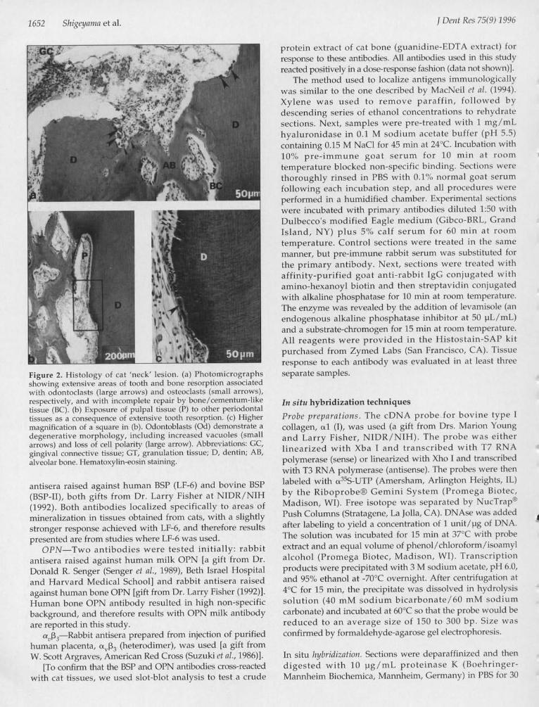
'9'^' ''9'#Figure 2. Histology of cat 'neck' lesion. (a) Photomicrographsshowing extensive areas of tooth and bone resorption associatedwith odontoclasts (large arrows) and osteoclasts (small arrows),respectively, and with incomplete repair by bone/cementum-liketissue (BC). (b) Exposure of pulpal tissue (P) to other periodontaltissues as a consequence of extensive tooth resorption. (c) Highermagnification of a square in (b). Odontoblasts (Gd) demonstrate adegenerative morphology, including increased vacuoles (smallarrows) and loss of cell polarity (large arrow). Abbreviations: GC,gingival connective tissue; GT, granulation tissue; D, dentin; AB,alveolar bone. Hematoxylin-eosin staining.
antisera raised against human BSP (LF-6) and bovine BSP(BSP-II), both gifts from Dr. Larry Fisher at NIDR/NIH(1992). Both antibodies localized specifically to areas ofmineralization in tissues obtained from cats, with a slightlystronger response achieved with LF-6, and therefore resultspresented are from studies where LF-6 was used.OPN-Two antibodies were tested initially: rabbit
antisera raised against human milk OPN [a gift from Dr.Donald R. Senger (Senger ct al., 1989), Beth Israel Hospitaland Harvard Medical School] and rabbit antisera raisedagainst human bone OPN [gift from Dr. Larry Fisher (1992)].Human bone OPN antibody resulted in high non-specificbackground, and therefore results with OPN milk antibodyare reported in this study.
a,,3,-Rabbit antisera prepared from injection of purifiedhuman placenta, o,,P, (heterodimer), was used [a gift fromW. Scott Argraves, American Red Cross (Suzuki et al., 1986)].
[To confirm that the BSP and OPN antibodies cross-reactedwith cat tissues, we used slot-blot analysis to test a crude
proteini extract of cat bone (guanidiin-EDTA extract) forresponse to these antibodies. All antibodies used in this studiyreacted positively in a dose-response fashion (data not shown)].
The method used to localize antigens immunologicallywas similar to the one described by MacNeil ct al. (1994).Xylene was used to remove paraffin, followed bydescending series of ethanol concentrations to rehyd ratesections. Next, samples were pre-treated witlh I mg/mLhyaluronidase in 0.1 M sodium acetate buffer (pH 5.5)containing 0.15 M NaCl for 45 min at 24"C. IncuLbationi with10"', pre-immune goat serum for 10 mni at roomtemperature blocked non-specific binding. Sections werethoroughly rinsed in I'BS with 0.1%U nor-miial goat serumfollowing each incubationi step, and all proceduL-es wer-eperformed in a humidified chamber. Experimental sectionswere incubated with primary antibodies diluted 1:50 witlDulbecco's modified Eagle medium (Gibco-BRL, GCrandIsland, NY) plus 5%,O calf serum for 60 min at room
temperature. Control sections were treated in the samemanner, but pre-immune rabbit serum was substitLted forthe primary antibody. Next, sections were treated withaffinity-purified goat anti-rabbit IgG conjugated withamino-hexanoyl biotin and then streptavidin conjugatedwith alkaline phosphatase for 10 min at room temperature.The enzyme was revealed by the addition of levamisole (anendogenous alkaline phosphatase inhibitor at 50 pL/mL)and a substrate-chromogen for 15 min at room temperatUre.All reagents were provided in the Histostain-SAP kitpurchased from Zymed Labs (San Francisco, CA). Tissueresponse to each antibody was evaluated in at least threeseparate samples.
In situ hybridization techniques
Probe preparations. The cDNA probe for bovine type I
collagen, o.1 (I), was used (a gift from Drs. Marion Youngand Larry Fisher, NIDR/NIH). The probe was eitherlinearized with Xba I and transcribed with T7 RNApolymerase (sense) or linearized with Xho I and transcribedwith T3 RNA polymerase (antisense). The probes were thenlabeled with a3&5S-UTP (Amersham, Arlington Heights, IL)by the RiboprobeD Gemini System (Promega Biotec,Madison, WI). Free isotope was separated by NucTrap"Push Columns (Stratagene, La Jolla, CA). DNAse was addedafter labeling to yield a concentration of 1 unit/pg of DNA.The solution was incubated for 15 min at 37°C with probeextract and an equal volume of phenol/chloroform/isoamnylalcohol (Promega Biotec, Madison, WI). Transcriptionproducts were precipitated with 3 M sodium acetate, pH 6.0),and 95%O ethanol at -70°C overnight. After centrifugationi at4°C for 15 min, the precipitate was dissolved in hydrolysissolution (40 mM sodium bicarbonate/6() mM sodiumcarbonate) and incubated at 60(C so that the probe would bereduced to an average size of 150 to 300 bp. Size wasconfirmed by formaldehyde-agarose gel electrophoresis.
In situ hlybridization. Sections were deparaffinized and thendigested with 10 pg/mL proteinase K (Boehringer-Mannheim Biochemica, Mannheim, Germany) in I'BS for 30
I Dent Res 75(9) 1996
Adhesion Moleciules during Tooth Resorption
AA#
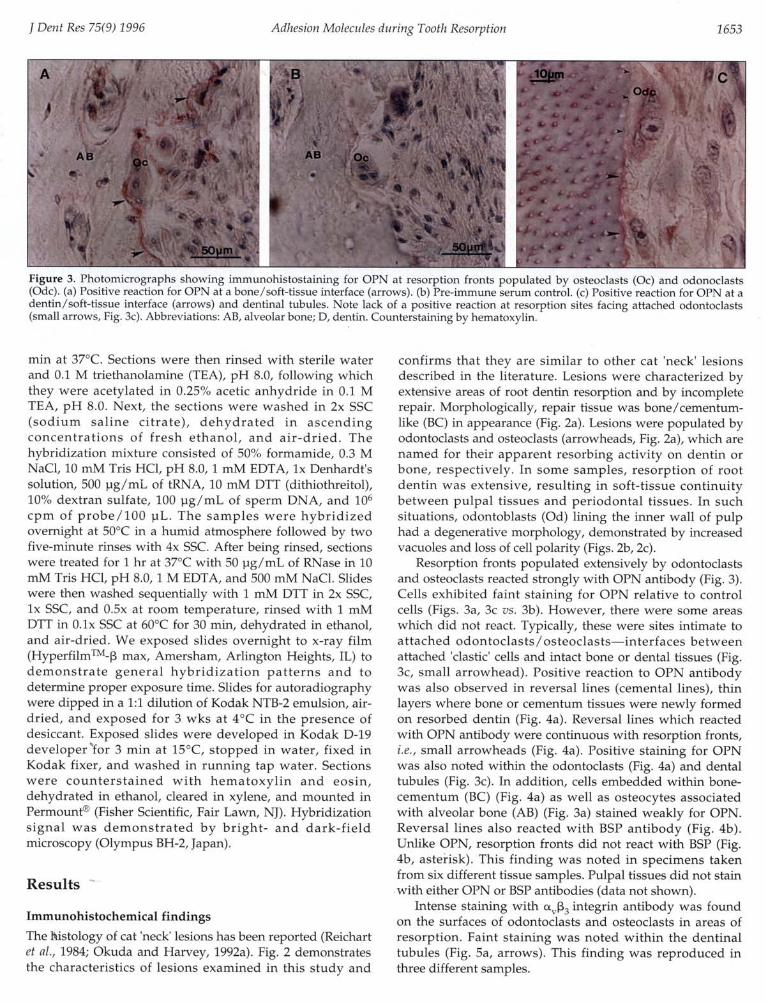
Figure 3. Photomicrographs showing immunohistostaining for OPN at resorption fronts populated by osteoclasts (0c) and odonoclasts(Odc). (a) Positive reaction for OPN at a bone/soft-tissue interface (arrows). (b) Pre-immune serum control. (c) Positive reaction for OPN at adentin/soft-tissue interface (arrows) and dentinal tubules. Note lack of a positive reaction at resorption sites facing attached odontoclasts(small arrows, Fig. 3c). Abbreviations: AB, alveolar bone; D, dentin. Counterstaining by hematoxylin.
min at 37°C. Sections were then rinsed with sterile waterand 0.1 M triethanolamine (TEA), pH 8.0, following whichthey were acetylated in 0.25% acetic anhydride in 0.1 MTEA, pH 8.0. Next, the sections were washed in 2x SSC(sodium saline citrate), dehydrated in ascendingconcentrations of fresh ethanol, and air-dried. Thehybridization mixture consisted of 50% formamide, 0.3 MNaCl, 10 mM Tris HCl, pH 8.0, 1 mM EDTA, Ix Denhardt'ssolution, 500 pg/mL of tRNA, 10 mM DTT (dithiothreitol),10% dextran sulfate, 100 pg/mL of sperm DNA, and 101cpm of probe/100 pL. The samples were hybridizedovernight at 50°C in a humid atmosphere followed by twofive-minute rinses with 4x SSC. After being rinsed, sectionswere treated for 1 hr at 37°C with 50 pg/mL of RNase in 10mM Tris HCl, pH 8.0, 1 M EDTA, and 500 mM NaCl. Slideswere then washed sequentially with 1 mM DTT in 2x SSC,Ix SSC, and 0.5x at room temperature, rinsed with 1 mMDTT in 0.Ix SSC at 60°C for 30 min, dehydrated in ethanol,and air-dried. We exposed slides overnight to x-ray film(HyperfilmTM-3 max, Amersham, Arlington Heights, IL) todemonstrate general hybridization patterns and todetermine proper exposure time. Slides for autoradiographywere dipped in a 1:1 dilution of Kodak NTB-2 emulsion, air-dried, and exposed for 3 wks at 4°C in the presence ofdesiccant. Exposed slides were developed in Kodak D-19developer for 3 min at 15°C, stopped in water, fixed inKodak fixer, and washed in running tap water. Sectionswere counterstained with hematoxylin and eosin,dehydrated in ethanol, cleared in xylene, and mounted inPermount' (Fisher Scientific, Fair Lawn, NJ). Hybridizationsignal was demonstrated by bright- and dark-fieldmicroscopy (Olympus BH-2, Japan).
Results
Immunohistochemical findingsThe histology of cat 'neck' lesions has been reported (Reichartet al., 1984; Okuda and Harvey, 1992a). Fig. 2 demonstratesthe characteristics of lesions examined in this study and
confirms that they are similar to other cat 'neck' lesionsdescribed in the literature. Lesions were characterized byextensive areas of root dentin resorption and by incompleterepair. Morphologically, repair tissue was bone/cementum-like (BC) in appearance (Fig. 2a). Lesions were populated byodontoclasts and osteoclasts (arrowheads, Fig. 2a), which arenamed for their apparent resorbing activity on dentin orbone, respectively. In some samples, resorption of rootdentin was extensive, resulting in soft-tissue continuitybetween pulpal tissues and periodontal tissues. In suchsituations, odontoblasts (Od) lining the inner wall of pulphad a degenerative morphology, demonstrated by increasedvacuoles and loss of cell polarity (Figs. 2b, 2c).
Resorption fronts populated extensively by odontoclastsand osteoclasts reacted strongly with OPN antibody (Fig. 3).Cells exhibited faint staining for OPN relative to controlcells (Figs. 3a, 3c vs. 3b). However, there were some areaswhich did not react. Typically, these were sites intimate toattached odontoclasts/osteoclasts-interfaces betweenattached 'clastic' cells and intact bone or dental tissues (Fig.3c, small arrowhead). Positive reaction to OPN antibodywas also observed in reversal lines (cemental lines), thinlayers where bone or cementum tissues were newly formedon resorbed dentin (Fig. 4a). Reversal lines which reactedwith OPN antibody were continuous with resorption fronts,i.e., small arrowheads (Fig. 4a). Positive staining for OPNwas also noted within the odontoclasts (Fig. 4a) and dentaltubules (Fig. 3c). In addition, cells embedded within bone-cementum (BC) (Fig. 4a) as well as osteocytes associatedwith alveolar bone (AB) (Fig. 3a) stained weakly for OPN.Reversal lines also reacted with BSP antibody (Fig. 4b).Unlike OPN, resorption fronts did not react with BSP (Fig.4b, asterisk). This finding was noted in specimens takenfrom six different tissue samples. Pulpal tissues did not stainwith either OPN or BSP antibodies (data not shown).
Intense staining with Ucv,P3 integrin antibody was foundon the surfaces of odontoclasts and osteoclasts in areas ofresorption. Faint staining was noted within the dentinaltubules (Fig. 5a, arrows). This finding was reproduced inthree different samples.
j Deiit Res 75(9) 1996 1 653

1654 Sliigeyaina et al.
a°-'u0, ": S iFigure 4. Localization of OPN and BSP at reversal lines and resorption fronts. (a) Photomicrograph showing positive immunohistostaining forOPN at resorption fronts (small arrow) and reversal lines (large arrovvs). (b) Immunohistostaming for BSP showing positive reaction at reversallines (arrows). No positive reaction was noted at resorption fronts (*). (c) Pre-immune control Abbreviations: C, cementum; D, dentin; BC,bone/cementum-like tissue; Odc, odontoclast. Bar = 50 pm. Counterstaining by hematoxvlin Results shown were taken from one tissue sample.
In situ hybridization findingsSignificantly, type I collagen mRNA was not expressed inodontoblasts (Od) lining the inner wall of pulp exposed as aconsequence of resorption (Figs. 6a, 6b, 6c). However,'osteoblast-like' cells involved in repair of hard tissuesdemonstrated strong type I collagen expression at the RNAlevel (Figs. 6a, 6c).
DiscussionReported cases of cat 'neck' lesions have increased, but thecause of these lesions and the reason for an increase in lesionnumbers have not been established. The studies describedhere focused on the pathology of this 'disease' at the cellularand molecular levels. Histological results from this studyconfirm that extensive damage occurs by resorption of bothcoronal and apical dentin. Advanced lesions extended intothe pulp, resulting in degeneration of odontoblasts.Attempts at repair, where dentin had been resorbed, wereobserved frequently. Reichart et al. (1984) called this newlyformed tissue "reparative cementum" or "osteodentin-likematerial". In contrast, Okuda and Harvey (1992a) suggestedthat this reparative tissue was significantly different fromdentin: Reparative tissue did not contain dentinal tubules,cells involved in repair did not appear to be derived frompulpal tissue, and the histologic appearance of repairedtissue resembled that of bone. Their rationale for suggestingthat it was bone-like rather than cementum-like was basedon stain density, the presence of marrow-like soft tissue, andlamellar patterning. Results reported here support theconcept that the reparative tissue is bone- or cementum-likerather than dentinal. However, a distinction between boneor cementum was not possible. Our conclusion was based
on the histologic appearance and expression of type Icollagen. Type I collagen mRNA was not expressed stronglyby odontoblasts lining the pulp wall. In contrast, type Icollagen mRNA was expressed by cells lining the endostealsurface of bone, suggesting that newly formed mineralizedtissue was initiated, in part, by osteoblasts fromsurrounding alveolar bone.
As an initial attempt to understand the mechanisms andfactors promoting osteoclast activity, we elected to determinewhether OPN, an adhesion molecule that has been inplicatedas having a role in promoting osteoclast activity (for review,see Denhardt and Guo, 1993), was localized to regionsassociated with resorptive activities in tissues obtained fromcats with 'neck' lesions. In addition, we determined whetherofU , an integrin found in high concentrations on the cellsurfaces of osteoclasts, could be identified in resorptive cellsin tissue samples from these same lesions. Importantly, sinceone of the ligands associated with a\. I3 is OPN, we were ableto determine, by using serial sections, the association of a, 33with OPN. Furthermore, using serial sections from the samelesion, we also examined whether BSP was associated withresorptive and/or reparative processes in these lesions. BSP,a mineralized-tissue-specific protein, has also been identifiedas a ligand for ac3,; thus, differences/similarities in thelocalization of these three proteins would provide insightinto the possible mechanisms regulatingosteoclast/odontoclast and osteoblast activity duringdestruction and repair, respectively. As discussed below, thespecific regions to which these molecules were localizedhave allowed us to propose several theories as to thefactors/mechanisms controlling these aggressive lesions,resulting in a promising new direction for our studies toconfirm our suggested hypotheses. OPN and BSP were
f Dciit Rcs 75(9) 1996

Adhesion Moleciules durig ToothResorption6
Figure 5. Localization of a [3 at resorption fronts. Photomicrographof immunohistostaining for a, f3 showing positive reaction byodontoclasts (Odc) at resoiption fronts (arrows). Bar = 50 pm. D,dentin. Counterstaining by hematoxylin. Results shovwn were takenfrom the same tissue sample as shown in Fig. 4.
localized to reversal lines, e.g., sites which separatereparative tissue from intact dentin. Localization of OPN andBSP to mineral/mineral and mineral/soft tissue interfacesduring development and in mature tissues has been reportedpreviously (Chen et al., 1993; McKee and Nanci, 1996). Here,we demonstrated that OPN and BSP accumulate at reversallines during repair. Reversal lines may indicate transitionsbetween resorption and formation for mineralized tissues. Inaddition, there is some evidence that specific growth factors,e.g., transforming growth factors [ (TGF-[s) and/or bonemorphogenetic proteins (BMPs), implicated in the regulationof osteoblast-osteoclast homeostasis, accumulate at reversallines (Oguro and Ozawa, 1988; Kingsley, 1994). Thus, severalmolecules localized to reversal lines may play importantroles in maintaining mineralized tissue homeostasis.A strong relationship between OPN and osteoclasts was
noted. Additionally, a, [33 was prominent on 'clastic' cellsurfaces in areas of resorption and was closely associatedwith surfaces where OPN reacted strongly (Fig. 5, serialsections), suggesting a functional link between a,[3 andOPN in cat 'neck' lesions. This possibility is supported byother studies which indicate that OPN interacts with thea,[3 receptor on osteoclasts and promotes osteoclastbinding to resorptive sites (Reinholt et al., 1990). However,the exact interaction between a, [3 and OPN on such cellsneeds to be clarified, especially since many other adhesionmolecules are linked with a, [3. on cell surfaces (Ross et al.,1993), with reportedly different cell signals depending onthe associated ligand (for review, see Denhardt and Guo,1993). Furthermore, while consensus exists that an OPN-Ua,[33 reaction is important for osteoclast attachment to bone(Ross et al., 1993), it has not yet been established whetherthese two molecules co-localize to specific areas on theosteoclast surface (Lakkakorpi et al., 1991). Lakkakorpi et /l.(1991) agree with other researchers in stating that both a,,
4
S~
Figure 6. in situ hybridization tor type I collagen mRNA. (a) Brightfield image of antisense probe indicating positive expression for type Icollagen by osteoblasts (0b), with absence of expression byodontoblasts (Gd). (b) Sense probe. (c) Antisense probe; highermagnification of white square outlined in (a). Note intense expressionfor type I collagen by osteoblasts (Gb), black grains in bright-field,with lack of expression by odontoblasts (Gd). Abbreviations: D,dentin, BC, bone/cementum-like tissue; Gd, odontoblasts; Ob,osteoblasts. Counterstaining by hematoxylin and eosi1.
and [3 subunits are important for osteoclast activity;however, they failed to demonstrate the localization of UaO[33to the sealing zone. This led them to conclude that UA,3 maynot be critical for final sealing zone attachment of theosteoclasts to bone surfaces, as required for creation of anisolated local micro-environment where resorption activitycan occur. At the light microscopic level, we were not able todetermine if (a, [3 was associated with sealing zones onosteoclasts. However, OPN was localized to the regions ofosteoclasts demonstrating a, [3 receptors, i.e., resorptionsites, whereas BSP was not identified in these regions. Inspite of this association, we noted that OPN did not localizeto sites where "clastic" cells appeared to be sitting directlyon bone. Thus, it is possible that OPN, through one of itstarget receptors, e.Cg., a, [3., triggers cell signals as yet to beidentified, resulting in osteoclast adhesion and subsequentactivation. Evidence from other studies supports ourconclusion that OPN may have a critical role in theresorptive phase of bone remodeling, while BSP may play amore significant role in the initiation of mineralization (forreview, see Bianco et al., 1991; Sodek et al., 1992; Denhardtand Guo, 1993). However, other factors must be considered,
J Deiit Res 75(9) 1996 1 655

1656 Shigeyama et al. J Dent Res 75(9) 1996
especially since other studies have identified weak stainingfor BSP in osteoclasts from human bones (Bianco et al., 1991)and the synthesis of BSP by osteoclast-like cells (FLG 29.1;Masi et al., 1995). Thus, BSP cannot be ruled out as amolecule important for osteoclast activity. Indeed, it ispossible that there was a loss of protein duringdecalcification and/or that the plane of sectioning may haveresulted in variability in staining for proteins at reversallines and resorption fronts, and in odontoclasts, osteoclasts,and osteocytes. An alternative explanation for this finding isthat cells are at different stages of functional activity. Suchvariation in OPN staining in osteoclasts (Maeda et al., 1994)as well as staining for other proteins in cells has beenreported previously, and these results have been attributedto differences in cell activity.
These results provide several possibilities as to afunction(s) of OPN in aggressive osteoclast resorption asobserved in cat 'neck' lesions: (1) Increased concentrations ofOPN produced locally in response to inflammatory agentspromote migration and/or attachment of "clastic" cells tothese areas; (2) increased concentrations of OPN locallyresult in up-regulation of 0'133 cell-surface receptors onosteoclasts with subsequent local increase in osteoclasts;and/or (3) increased concentrations of OPN, producedlocally, serve a protective role, i.e., inhibit programed celldeath in osteoclasts. This latter hypothesis is supported byseveral findings. Tumor cells producing high levels of OPNhave the capacity to defend themselves against mediators ofcell death. This has been attributed to the ability of OPN toeffect expression of specific genes/proteins that are noxiousto cells, such as nitrous oxide synthetase, and to inhibitkilling of tumor cells by activated macrophages andendothelial cells (Denhardt and Chambers, 1994). Ourstudies to date, demonstrating that OPN localizes to areasassociated with resorptive activity, have prompted us topropose that OPN protects osteoclasts from cell death,resulting in "uncoupling" of osteoblast-osteoclasthomeostasis in favor of osteoclast resorptive activity. Futurestudies have been planned to address this hypothesis.
AcknowledgmentsThe authors thank Jessica Madow and Mary Schmidt forassistance in the preparation of the manuscript. This workwas supported by Nestec Ltd. (Friskies Research).
ReferencesAlbelda SM, Buck CA (1990). Integrins and other cell adhesion
molecules. FASEB J 4:2868-2880.Bianco P, Fisher LW, Young MF, Termine JD, Robey PG (1991).
Expression of bone sialoprotein (BSP) in developinghuman tissues. Calcif Tissue Int 49:421-426.
Boskey AL, Maresca M, Ullrich W, Doty SB, Butler WT, PrinceCW (1993). Osteopontin-hydroxyapatite interactions invitro: inhibition of hydroxyapatite formation and growth ina gelatin-gel. Bone Miner 22:147-159.
Chen J, McCulloch C, Sodek J (1993). Bone sialoprotein in
developing porcine dental tissues: cellular expression andcomparison of tissue localization with osteopontin andosteonectin. Arch Oral Biol 38:241-249.
Coles S (1990). The prevalence of buccal cervical rootresorptions in Australian cats. I Vet Dent 7:14-16.
Denhardt DT, Chambers AF (1994). Overcoming obstacles tometastasis-defenses against host defenses: Osteopontin(OPN) as a shield against attack by cytotoxic host cells. lCell Biochem 56:48-51.
Denhardt DT, Guo X (1993). Osteopontin: a protein with diversefunctions. FASEB J 7:1475-1482.
Fisher LW (1992). Structure/function studies of thesialoglycoproteins and proteoglycans of bone: It is still theearly days. In: Chemistry and biology of mineralizedtissues. Slavkin H, Price P, editors. Amsterdam-New York:Excerpta Medica, pp. 177-187.
Harvey CE (1992). Epidemiology of periodontal conditions indog and cats. Proceedings of the Sixth Annual VeterinaryDental Forum, the American Veterinary Dental Collegeand the Academy of Veterinary Dentistry, Nov 13-15, 1992,Las Vegas, Nevada. Nabisco Foods Company, pp. 45-46.
Helfrich MH, Nesbitt SA, Dorey EL, Horton MA (1992). Ratosteoclasts adhere to a wide range of RGD (Arg-Gly-Asp)peptide-containing proteins, including the bonesialoproteins and fibronectin, via a 13 integrin. J Bone MinerRes 7:335-343.
Hunter GK, Goldberg HA (1994). Modulation of crystalformation by bone phosphoproteins: role of glutamic acid-rich sequences in the nucleation of hydroxyapatite by bonesialoprotein. Biochem J 302:175-179.
Hunter GK, Kyle CL, Goldberg HA (1994). Modulation ofcrystal formation by bone phosphoproteins: structuralspecificity of the osteopontin-mediated inhibition ofhydroxyapatite formation. Biochem 1 300:723-728.
Kingsley DM (1994). The TGF 1 superfamily: new members,new receptors, and new genetic tests of function indifferent organisms. Gene Dev 8:133-146.
Lakkakorpi PT, Horton MA, Helfrich MH, Karhukorpi E-K,Vaananen HK (1991). Vitronectin receptor has a role inbone resorption but does not mediate tight sealing zoneattachment of osteoclasts to the bone surface. l Cell Biol115:1179-1186.
MacNeil RL, Sheng N, Strayhom C, Fisher LW, Somerman MJ(1994). Bone sialoprotein is localized to the root surfaceduring cementogenesis. J Bone Miner Res 9:1597-1606.
Maeda H, Kukita T, Akamine A, Kukita A, Iijima T (1994).Localization of osteopontin in resorption lacunae formedby osteoclast-like cells: a study by a novel monoclonalantibody which recognizes rat osteopontin. Histochem102:247-254.
Masi L, Brandi ML, Robey PG, Crescioli C, Calvo JC, Bemabei P, etal. (1995). Biosynthesis of bone sialoprotein by a humanosteoclast-like cell line (FLG 29.1). I Bone Miner Res 10:187-196.
McKee MD, Nanci A (1996). Osteopontin at mineralized tissueinterfaces in bone, teeth and osseointegrated implants:Ultrastructural distribution and implications formineralized tissue formation, turnover and repair. MicroscRes Technique 33:141-164.
Oguro I, Ozawa H (1988). The histochemical localization of acidphosphatase activity in BMU. J Bone Min Metab 6:44-49.

Adhesion Molecules during Tooth Resorption
Okuda A, Harvey CE (1992a). Etiopathogenesis of feline dentalresorptive lesions. Vet Clin North Am Small Anim Pract22:1385-1404.
Okuda A, Harvey CE (1992b). Immunohistochemicaldistributions of interleukins as possible stimulators ofodontoclastic resorption activity in feline dental resorptivelesions. Proceedings of the Sixth Annual Veterinary DentalForum, the American Veterinary Dental College and theAcademy of Veterinary Dentistry, Nov 13-15, 1992, LasVegas, Nevada. Nabisco Foods Company, pp. 41-43.
Patarca R, Saavedra RA, Cantor H (1993). Molecular andcellular basis of genetic resistance to bacterial infection: Therole of the early T-lymphocyte activation-1/osteopontingene. Crit Rev Immunol 13:225-246.
Regezi JA, Sciubba JJ (1989). Abnormalities of dental pulp:Internal resorption. In: Oral pathology: Clinical-pathologiccorrelations. Philadelphia: W.B. Saunders Co., pp. 483-484.
Reichart PA, Durr U-M, Triadan H, Vickendey G (1984).Periodontal disease in the domestic cat. A histopathologicstudy. J Periodont Res 19:67-75.
Reinholt FP, Hultenby K, Oldberg A, Heinegard D (1990).Osteopontin-a possible anchor of osteoclasts to bone. ProcNatl Acad Sci USA 87:4473-4475.
Ross FP, Chappel J, Alvarez JI, Sander D, Butler WT, Farach-Carson MC, et al. (1993). Interactions between the bone
matrix proteins osteopontin and bone sialoprotein and theosteoclast integrin caA3 potentiate bone resorption. J BiolChem 268:9901-9907.
Schneck GW, Osborn JW (1976). Neck lesions in the teeth ofcats. Vet Rec 99:100.
Senger DG, Perruzzi CA, Papadopoulos A, Tenen DG (1989).Purification of a human milk protein closely similar totumor-secreted phosphoproteins and osteopontin. BiochemBiophys Acta 996:43-48.
Shiraga H, Min W, VanDusen WJ, Clayman MD, Miner D,Terrell CH, et al. (1992). Inhibition of calcium oxalatecrystal growth in vitro by uropontin: another member ofthe aspartic acid-rich protein superfamily. Proc Natl AcadSci USA 89:426-430.
Sodek J, Chen J, Kasugai S, Nagata T, Zhang Q, McKee MD, etal. (1992). Elucidating the functions of bone sialoproteinand osteopontin in bone formation. In: Chemistry andbiology of mineralized tissues. Slavkin H, Price P, editors.Amsterdam-New York: Excerpta Medica, pp. 297-307.
Suzuki S, Argraves WS, Pytela R, Arai H, Krusius T,Pierschbacher MD, et al. (1986). cDNA and amino acidsequences of the cell adhesion protein receptor recognizingvitronectin reveal a transmembrane domain andhomologies with other adhesion protein receptors. ProcNatl Acad Sci USA 83:8614-8618.
f Dent Res 75(9) 1996 1657