expression of sox9 mis , and dmrt1 in the dependent...
TRANSCRIPT

PATTERNS & PHENOTYPES
Expression of Sox9, Mis, and Dmrt1 in theGonad of a Species With Temperature-Dependent Sex DeterminationChristina Shoemaker, Mary Ramsey, Joanna Queen, and David Crews*
Sex determination in vertebrates, the process of forming an ovary or testis from a bipotential gonad, can beinitiated by genetic or environmental factors. Elements of the downstream molecular pathways underlyingthese different sex-determining mechanisms have been evolutionarily conserved. We find the first evidencethat Sox9 expression is preferentially organized in the testis early in the temperature-sensitive period in aspecies with temperature-dependent sex determination (Trachemys scripta). This pattern occurs beforesexually dimorphic Mis expression and in a temporal hierarchy that is similar to mammals. Furthermore,we extend previous findings that Dmrt1 expression at early stages of sex determination has a dimorphicpattern consistent with a possible upstream role in determining the fate of the bipotential gonad.Developmental Dynamics 236:1055–1063, 2007. © 2007 Wiley-Liss, Inc.
Key words: temperature-dependent sex determination; reptile; Sox9; Mis; Dmrt1; testis differentiation
Accepted 10 January 2007
INTRODUCTIONIn vertebrates, the bipotential gonadfirst forms as a thickening of ventrome-dial mesonephric tissue. These genitalridges have the potential to be directedtoward either ovarian or testicular fatesand go through a second phase of devel-opment wherein sex is determined. Inthe third phase of gonadal develop-ment, sexual fate is committed and dif-ferentiation into a testis or an ovaryoccurs. In organisms exhibiting geno-typic sex determination (GSD), includ-
ing mammals, birds, and some reptiles,genetic factors determine the sexualfate of the initially bipotential gonad. Inother vertebrates, environmental fac-tors direct sexual development, such asin temperature-dependent sex determi-nation (TSD) found in all crocodiliansand many turtles. While the initial up-stream factor determining gonadal sexdiffers radically between TSD and GSD,many of the same genes are involved inthe downstream process of gonad differ-entiation.
In organisms with TSD, genes in-volved in early phases of sex-deter-mining of the bipotential gonad areexpected to be expressed in a dimor-phic manner before or early in thetemperature-sensitive period (TSP),whereas genes more integral to down-stream testis or ovary differentiationshould be expressed dimorphically af-ter the TSP. To clarify their location inthe temperature-dependent sex-deter-mining molecular network, we exam-ined the expression of Sox9, Mis, and
ABBREVIATIONS MPT male-producing temperature FPT female-producing temperature TSP temperature-sensitive period TSD tem-perature-dependent sex determination GSD genetic sex determination Mis Mullerian-inhibiting substance Dmrt1 doublesex mab3-relatedtranscription factor 1 Sox9 SRY-like HMG-box 9 Mis-R2 Mis receptor type II PP1 protein phosphatase type I RT-PCR reverse-transcriptase polymerase chain reaction qPCR quantitative real-time PCR ISH in situ hybridization
Department of Integrative Biology, University of Texas at Austin, Austin, TexasGrant sponsor: National Science Foundation; Grant number: IBN 200001269.*Correspondence to: David Crews, Department of Integrative Biology, University of Texas at Austin, Austin, TX 78712.E-mail: [email protected]
DOI 10.1002/dvdy.21096Published online 26 February 2007 in Wiley InterScience (www.interscience.wiley.com).
DEVELOPMENTAL DYNAMICS 236:1055–1063, 2007
© 2007 Wiley-Liss, Inc.

Dmrt1 in the red-eared slider turtle,Trachemys scripta. In this species, thegonad is sensitive to the effect of tem-perature during the middle third ofembryonic development, and afterthis window closes, the gonad becomescommitted to an ovarian or testicularfate (Bull et al., 1990; Wibbels et al.,1991). The timing of this window in T.scripta lasts from approximately stage14 (Greenbaum staging series)through stage 18 at a female-produc-ing temperature (FPT) and throughstage 19 at a male-producing temper-ature (MPT; Wibbels et al., 1991;Greenbaum, 2002). Cooler incubationtemperatures (25–27°C) produce allmale hatchlings and warmer temper-atures (31–35°C) result in all femalehatchlings, with varying sex ratiosproduced by temperatures in between(Wibbels et al., 1991). Shifting eggsduring the TSP from one end of thetemperature spectrum to the other re-directs gonadal development, result-ing in 100% sex reversal (Crews et al.,1994).
Analysis of human patients withcampomelic dysplasia, roughly twothirds of whom develop as XY females,revealed the importance of the Sry-related gene Sox9 (SRY-like HMG-box9) in the molecular sex-determiningcascade (Foster et al., 1994). Exten-sive studies in the mouse have sinceshown Sox9 to be both necessary andsufficient to cause the determinationand differentiation of a testis (Vidal etal., 2001). In both humans and mice,SOX9 interacts directly with SF1 (Ste-roidogenic factor 1) to up-regulate theexpression of Mullerian-inhibitingsubstance (Mis or anti-Mullerian hor-mone, Amh; de Santa Barbara et al.,1998; Arango et al., 1999). Mis, amember of the transforming growthfactor-! (TGF-!) superfamily, is thefirst factor secreted by differentiatedSertoli cells in the testis, and causesthe regression of the Mullerian ducts,anlagen which otherwise develop intothe uterus, cervix, and fallopian tubesin females (Behringer et al., 1990).
The regulatory relationship be-tween Sox9 and Mis in mammals hasnot been strictly conserved acrossphyla or mechanisms of sex determi-nation. In the chicken, another verte-brate with GSD, sexually dimorphicexpression of Mis in the gonad pre-cedes Sox9, and the relationship be-
tween the two has yet to be fully char-acterized (Oreal et al., 1998; Smith etal., 1999b). Expression patterns inspecies with TSD have thus far ap-peared similar to those in chicken. Inthe American alligator, Alligator mis-sissippiensis, dimorphic expression ofMis occurs in the middle of the TSP,preceding the onset of testis-specificSox9 (Western et al., 1999). Similarly,expression of Sox9 does not becometestis-specific until the end of the TSPin both the leopard gecko and the Ol-ive Ridley sea turtle, Lepidochelys oli-vacea (Moreno-Mendoza et al., 1999;Valleley et al., 2001), although Mishas not yet been characterized inthese species. In the red-eared sliderturtle, Mis expression has also beenshown to be male-specific by the mid-dle of the TSP (Takada et al., 2004).Furthermore, Sox9 expression ap-pears in one study to be testis-specificafter the TSP (Spotila et al., 1998),while in another study, reverse-tran-scriptase polymerase chain reaction(RT-PCR) showed comparable levelsof expression in both sexes at allstages examined (Kettlewell et al.,2000).
Continued evidence of molecularconservation between sex-determin-ing mechanisms across phyla camewith the discovery of a mammaliangene, Dmrt1, that possesses func-tional similarity to sex-determininggenes in both nematodes (mab-3) andflies (doublesex; Shen and Hodgkin,1988; Burtis and Baker, 1989). Loss ofDmrt1 is thought to be responsible forthe male-to-female sex reversal seenin XY humans with chromosome 9 de-letions (Flejter et al., 1998), andDmrt1 has since been studied in a va-riety of vertebrates. Although it seemsto play a downstream role in testisdifferentiation in mammals (Raymondet al., 1999), it has been proposed to bea master sex-determining gene in bothchicken and medaka (Nanda et al.,1999; Matsuda et al., 2002). In organ-isms with TSD, testis-specific expres-sion of Dmrt1 early in the TSP hasbeen demonstrated in the Olive Ridleysea turtle (Torres-Maldonaldo et al.,2002). In the American alligator, go-nadal expression of Dmrt1 before andduring the TSP is also greater inmales, but increases through develop-ment in both sexes, raising questionsabout its function (Smith et al.,
1999a). In T. scripta, dimorphic ex-pression during the TSP has been re-ported, beginning at either stage 15(Kettlewell et al., 2000) or stage 17(Murdock and Wibbels, 2003).
To clarify the nature of the generegulatory network underlying testisdevelopment in organisms with TSD,we analyzed the expression patternsof Sox9, Mis, and Dmrt1 by whole-mount in situ hybridization (ISH) andquantitative real-time polymerasechain reaction (qPCR) during the pe-riod of sex determination and differ-entiation in T. scripta. Whereas theexpression of all three of these geneshas been reported as male-specific, thelocalization of expression patternsand the temporal hierarchy of expres-sion in relation to each other have notbeen detailed. We visualize and com-pare localized expression patterns ofeach gene at the earliest stage of bipo-tential gonad formation (stage 15), inthe middle and at the end of the TSP(stages 17 and 19), as well as at twostages of gonad differentiation (stages21 and 23). We find the first evidencein an organism with TSD that Sox9 isexpressed in a testis-specific mannerearly in the TSP, before the onset oftestis-specific Mis expression. Fur-thermore, we support previous find-ings and confirm in T. scripta thatDmrt1 shows sexually dimorphic ex-pression at the beginning of the TSP,in a pattern that is consistent with apossible upstream role in testis deter-mination.
RESULTS AND DISCUSSIONSox9 Expression IsDimorphic Near theBeginning of the TSPPrevious reports have shown that lev-els of Sox9 expression in T. scripta aresimilar at MPT and FPT during theTSP, and only become higher in thetestis during differentiation (Spotilaet al., 1998). Our data are consistentwith this report, and we also find evi-dence of an earlier dimorphism in thelocalized organization of this tran-scription factor. Early in the TSP,Sox9 expression at MPT occurs inclusters of cells surrounding othernonexpressing cells. We suggest thatthese clustered cells are perhaps pre-sumptive Sertoli cells of the develop-
1056 SHOEMAKER ET AL.
ing seminiferous tubules. While levelsof Sox9 expression at FPT are similar,transcript localization is diffuse andappears unorganized throughout thegonad. It is not unlikely that this dif-ferential localization corresponds to afunctional dimorphism early in thesex-determining period. Thus, we findthe first evidence of Sox9 expression
in an organism with TSD that sug-gests similarity to the pattern seen inmammals. These findings are consis-tent with a possible upstream role forSox9 in TSD, although this was notinvestigated in the current study andwarrants future attention.
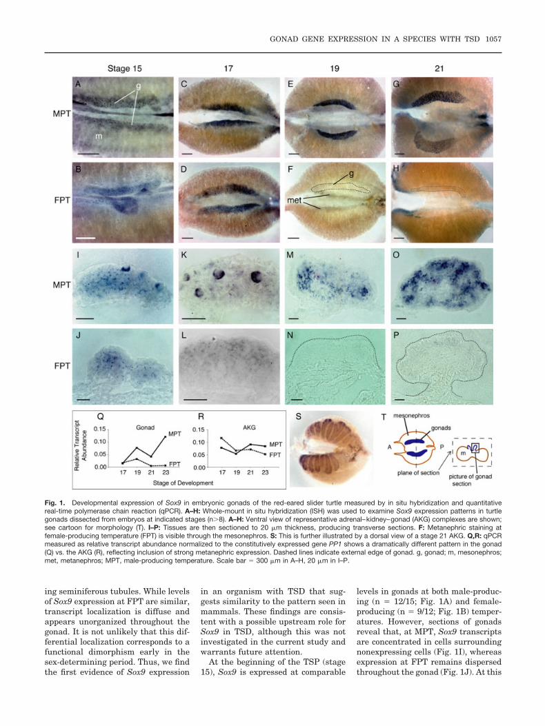
At the beginning of the TSP (stage15), Sox9 is expressed at comparable
levels in gonads at both male-produc-ing (n " 12/15; Fig. 1A) and female-producing (n " 9/12; Fig. 1B) temper-atures. However, sections of gonadsreveal that, at MPT, Sox9 transcriptsare concentrated in cells surroundingnonexpressing cells (Fig. 1I), whereasexpression at FPT remains dispersedthroughout the gonad (Fig. 1J). At this
Fig. 1. Developmental expression of Sox9 in embryonic gonads of the red-eared slider turtle measured by in situ hybridization and quantitativereal-time polymerase chain reaction (qPCR). A–H: Whole-mount in situ hybridization (ISH) was used to examine Sox9 expression patterns in turtlegonads dissected from embryos at indicated stages (n#8). A–H: Ventral view of representative adrenal–kidney–gonad (AKG) complexes are shown;see cartoon for morphology (T). I–P: Tissues are then sectioned to 20 $m thickness, producing transverse sections. F: Metanephric staining atfemale-producing temperature (FPT) is visible through the mesonephros. S: This is further illustrated by a dorsal view of a stage 21 AKG. Q,R: qPCRmeasured as relative transcript abundance normalized to the constitutively expressed gene PP1 shows a dramatically different pattern in the gonad(Q) vs. the AKG (R), reflecting inclusion of strong metanephric expression. Dashed lines indicate external edge of gonad. g, gonad; m, mesonephros;met, metanephros; MPT, male-producing temperature. Scale bar " 300 $m in A–H, 20 $m in I–P.
GONAD GENE EXPRESSION IN A SPECIES WITH TSD 1057

stage at MPT, Sox9 is also expressedin dorsal metanephric tissue in half ofthe embryos examined (n " 7/15; datanot shown).
As the sex-determining periodprogresses and temperature exerts itseffect (stage 17), organized Sox9 ex-pression persists at MPT (n " 15/16;Fig. 1C,K). There is strong staining indorsal metanephric tissue and theposterior tip of the mesonephros, al-though it is unclear if posterior meso-nephros is actually a contributingsource of gonadal cells (Fig. 1C). Inaddition, approximately half of MPTembryos show staining in developingWolffian ducts (n " 7/16; data notshown). Although Sox9 expression atthis stage is retained in gonads at FPTin comparable levels to gonads at MPT(n " 8/11; Fig. 1C,D), it still occurs ina diffuse pattern spread throughoutthe gonad (Fig. 1L). Metanephric ex-pression is also detected in most em-bryos at FPT (n " 7/11), but Wolffianduct expression is not. In situ data areconfirmed by qPCR studies in which to-tal RNA was extracted from one sampleper sex/stage. Each sample was com-posed of either pooled gonads or pooledadrenal–kidney–gonad (AKG) dis-sected from at least 20 embryos. Thisprocedure was technically possible be-ginning at stage 17, and Sox9 expres-sion is seen at comparable levels atboth MPT and FPT at this stage (Fig.1Q).
At a stage when sexual fate is com-mitted at an MPT but remains some-what reversible at an FPT (stage 19),Sox9 expression increases in virtuallyall gonads developing at MPT (n "7/8; Fig. 1E,Q) in a pattern that issuggestive of Sertoli cells organizinginto sex cords (Fig. 1M). Expression isdown-regulated in all FPT gonads ex-amined by ISH (n " 0/12; Fig. 1F,N),but is still detectable by qPCR, a dis-crepancy we are investigating (Fig.1Q). Expression is maintained in dor-sal metanephric tissue in some em-bryos at both MPT (n " 4/8) and FPT(n " 7/12), while Wolffian duct expres-sion disappears from males.
During testis differentiation (stages21 and 23), Sox9 is strongly expressedin all gonads examined developing atMPT (n " 13/13 and 12/12, respec-tively; Fig. 1G,Q). In these gonads, ex-pression continues in an increasinglyreticulated pattern, probably corre-
sponding to the expansion of primitivesex cords and their subsequent trans-formation into seminiferous tubules(Fig. 1O). In contrast, only a few cellsweakly expressing Sox9 were ob-served in several differentiating ova-ries at stages 21 and 23 (n " 3/12 and5/12, respectively; Fig. 1H,P,Q). Inboth sexes, metanephric expressionremains strong in the beginning of go-nad differentiation (stage 21, n " 9/13at MPT, n " 11/12 at FPT; Fig. 1S)and then diminishes to a faint level(stage 23, n " 9/12 at MPT, n " 3/12at FPT).
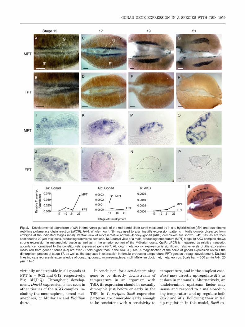
Dimorphic Mis ExpressionOccurs Midway Through theTSPIt has been shown in T. scripta thatMis expression is undetectable in thebipotential gonad, is up-regulated indeveloping testes during the TSP andbecomes increasingly stronger as sexdetermination occurs and differentia-tion begins (Takada et al., 2004). Ourdata are consistent with these find-ings as well, and further reveal that,in contrast to Sox9, both the level ofMis expression as well as its cellularorganization appear similar in MPTand FPT gonads early in development.At the start of the TSP, Mis tran-scripts are organized in similar clus-ters of cells in both sexes. It is notuntil after the sex-determining period,and after Sox9 patterns are dimor-phic, that Mis is up-regulated inmales and down-regulated in females.These results are consistent with arole for Sox9 upstream of Mis, al-though a regulatory relationship be-tween them in this species remains tobe shown.
Mis expression is virtually equiva-lent at both male- and female-produc-ing temperatures at the earliest stageof gonad formation (stage 15; n "10/12 at both MPT and FPT; Fig.2A,B,I,J). As the sex-determining pe-riod progresses (stage 17), Mis expres-sion is up-regulated in the gonads ofembryos incubating at MPT (n " 16/16; Fig. 2C,K) and persists but isdown-regulated at FPT (n " 11/12;Fig. 2D,L,Qb). As sexual fate is com-mitted and gonad differentiation oc-curs (stages 19, 21, and 23), develop-ing testes strongly express Mis in apattern similar to Sox9, possibly in
the preSertoli cells of nascent seminif-erous tubules (Fig. 2E,M,G,O,Q). Ex-pression is also seen at MPT in dorsalmetanephric tissue, as well as pro-gressively in a cranial to caudal wavein the Mullerian ducts (Fig. 2S). Incontrast, expression at FPT at stage19 is undetectable in half of the go-nads examined and faint in the rest(n " 7/14; Fig. 2F,N,Q). In later stagesof ovarian differentiation (stages 21and 23), expression at FPT falls toundetectable levels (n " 0/12 and0/12, respectively; Fig. 2H,P,Q). Al-though qPCR levels at FPT appearclose to zero in all stages examined,this finding is an artifact produced bythe extremely high expression levelsseen later at MPT (see Fig. 2Qa,Qb).
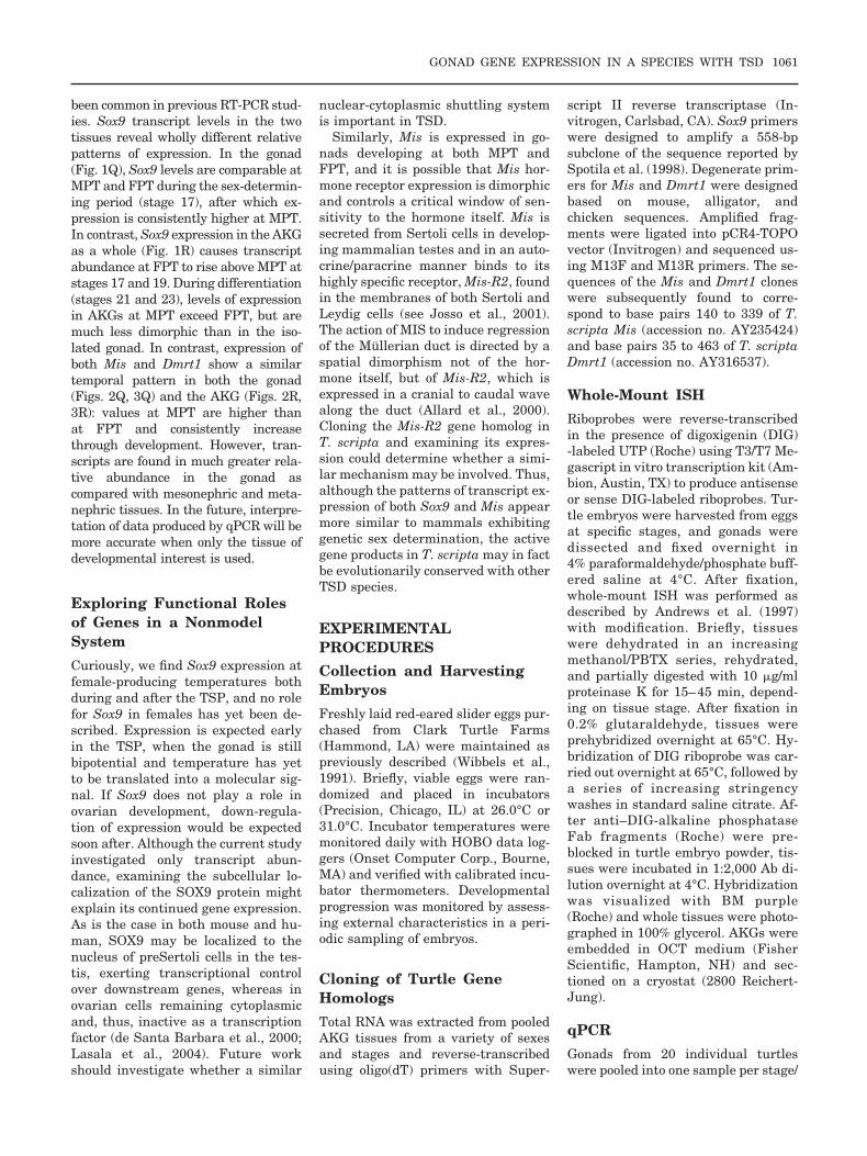
Dmrt1 Expression IsConsistent With a PossibleUpstream Role in T. scriptaWe used whole-mount ISH to confirmprevious reports that Dmrt1 expres-sion is dimorphic before sex is deter-mined. At the beginning of the TSP(stage 15), all embryos developing atMPT show punctate Dmrt1 expressionin the gonad (n " 12/12; Fig. 3A,I),while half of FPT embryos showweaker, diffuse gonadal expression(n " 6/12; Fig. 3B,J). At stage 17, ex-pression of Dmrt1 continues in gonadsat MPT (n " 14/14; Fig. 3C,K) and isdetected in only a very few cells ingonads at FPT (n " 2/9; Fig. 3D,L).Up-regulation of expression at MPT islocalized in clusters of cells surround-ing nonexpressing cells (Fig. 3K).qPCR confirms a difference in Dmrt1transcript levels at this stage, and al-though expression in both sexes is lowoverall, relative transcript abundanceis three times higher at MPT than atFPT (Fig. 3Q).
By stage 19, Dmrt1 expression isupregulated further at MPT (n " 7/7;Fig. 3E,Q). Expression at FPT de-clines in all ISH gonads examined(n " 0/10; Fig. 3F,N), but is still de-tectable by the more sensitive methodof qPCR (Fig. 3Q). Sectioned gonadsreveal continued Dmrt1 expression incells organized in what are possiblydeveloping sex cords of the testis (Fig.3M). Finally, during differentiation(stages 21 and 23), expression at MPTcontinues to increase (n " 12/12 and8/8, respectively; Fig. 3G,O,Q) and is
1058 SHOEMAKER ET AL.
virtually undetectable in all gonads atFPT (n " 0/12 and 0/12, respectively;Fig. 3H,P,Q). Throughout develop-ment, Dmrt1 expression is not seen inother tissues of the AKG complex, in-cluding the mesonephros, dorsal met-anephros, or Mullerian and Wolffianducts.
In conclusion, for a sex-determininggene to be directly downstream oftemperature in an organism withTSD, its expression should be sexuallydimorphic just before or early in theTSP. In T. scripta, Sox9 expressionpatterns are dimorphic early enoughto be consistent with a sensitivity to
temperature, and in the simplest case,Sox9 may directly up-regulate Mis asit does in mammals. Alternatively, anundetermined upstream factor maysense and respond to a male-produc-ing temperature and up-regulate bothSox9 and Mis. Following their initialup-regulation in this model, Sox9 ex-
Fig. 2. Developmental expression of Mis in embryonic gonads of the red-eared slider turtle measured by in situ hybridization (ISH) and quantitativereal-time polymerase chain reaction (qPCR). A–H: Whole-mount ISH was used to examine Mis expression patterns in turtle gonads dissected fromembryos at the indicated stages (n#8). Ventral view of representative adrenal–kidney–gonad (AKG) complexes are shown. I–P: Tissues are thensectioned to 20 $m thickness, producing transverse sections. S: A dorsal view of a male-producing temperature (MPT) stage 19 AKG complex showsstrong expression in metanephric tissue as well as in the anterior portion of the Mullerian ducts. Qa,R: qPCR is measured as relative transcriptabundance normalized to the constitutively expressed gene PP1. Although metanephric expression is significant, relative levels of Mis expressionmeasured from gonad tissues (Qa) are over 20-fold higher than in the AKG (R). Qb: A magnification of the scale of gonad expression reveals thedimorphism present at stage 17, as well as the decrease in expression in female-producing temperature (FPT) gonads through development. Dashedlines indicate represents external edge of gonad. g, gonad; m, mesonephros; mull, Mullerian duct; met, metanephros. Scale bar " 300 $m in A–H, 20$m in I–P.
GONAD GENE EXPRESSION IN A SPECIES WITH TSD 1059

pression in preSertoli cells may thendirect their differentiation and orga-nize and maintain expression of Mis.Furthermore, our findings of the spa-tial and temporal patterns of Dmrt1expression are also consistent withthe possibility that Dmrt1 could be atarget of temperature, although thiswas not directly tested in the current
study. To gain insight into these pos-sibilities, and shed light on the hier-archy of genes in the regulatory net-work underlying TSD, we arecurrently investigating the responseof these genes to sex-reversing shiftsin temperature and elimination oftranscript in the gonad in an in vitroorgan culture system.
Tissue-Specific qPCR
The qPCR was performed on pooledAKG complexes as well as pooled go-nads. Comparing relative expressionlevels between the two RNA sourcesreveals the problems associated with re-taining extraneous tissue when exam-ining sex-determining genes, as has
Fig. 3. Developmental expression of Dmrt1 in embryonic gonads of the red-eared slider turtle measured by in situ hybridization (ISH) and quantitativereal-time polymerase chain reaction (qPCR). A–H: Whole-mount ISH was used to examine Dmrt1 expression patterns in turtle gonads dissected fromembryos at the indicated stages (n#8). Ventral view of representative adrenal-kidney-gonad complexes are shown. I–P: Tissues are then sectionedto 20 $m thickness, producing transverse sections. Q,R: qPCR is measured as relative transcript abundance normalized to the constitutivelyexpressed gene PP1. Dashed lines indicate external edge of gonad. g, gonad; m, mesonephros; MPT, male-producing temperature; FPT, female-producing temperature; AKG, adrenal–kidney–gonad complex. Scale bar " 300 $m in A–H; 20 $m in I–P.
1060 SHOEMAKER ET AL.
been common in previous RT-PCR stud-ies. Sox9 transcript levels in the twotissues reveal wholly different relativepatterns of expression. In the gonad(Fig. 1Q), Sox9 levels are comparable atMPT and FPT during the sex-determin-ing period (stage 17), after which ex-pression is consistently higher at MPT.In contrast, Sox9 expression in the AKGas a whole (Fig. 1R) causes transcriptabundance at FPT to rise above MPT atstages 17 and 19. During differentiation(stages 21 and 23), levels of expressionin AKGs at MPT exceed FPT, but aremuch less dimorphic than in the iso-lated gonad. In contrast, expression ofboth Mis and Dmrt1 show a similartemporal pattern in both the gonad(Figs. 2Q, 3Q) and the AKG (Figs. 2R,3R): values at MPT are higher thanat FPT and consistently increasethrough development. However, tran-scripts are found in much greater rela-tive abundance in the gonad ascompared with mesonephric and meta-nephric tissues. In the future, interpre-tation of data produced by qPCR will bemore accurate when only the tissue ofdevelopmental interest is used.
Exploring Functional Rolesof Genes in a NonmodelSystemCuriously, we find Sox9 expression atfemale-producing temperatures bothduring and after the TSP, and no rolefor Sox9 in females has yet been de-scribed. Expression is expected earlyin the TSP, when the gonad is stillbipotential and temperature has yetto be translated into a molecular sig-nal. If Sox9 does not play a role inovarian development, down-regula-tion of expression would be expectedsoon after. Although the current studyinvestigated only transcript abun-dance, examining the subcellular lo-calization of the SOX9 protein mightexplain its continued gene expression.As is the case in both mouse and hu-man, SOX9 may be localized to thenucleus of preSertoli cells in the tes-tis, exerting transcriptional controlover downstream genes, whereas inovarian cells remaining cytoplasmicand, thus, inactive as a transcriptionfactor (de Santa Barbara et al., 2000;Lasala et al., 2004). Future workshould investigate whether a similar
nuclear-cytoplasmic shuttling systemis important in TSD.
Similarly, Mis is expressed in go-nads developing at both MPT andFPT, and it is possible that Mis hor-mone receptor expression is dimorphicand controls a critical window of sen-sitivity to the hormone itself. Mis issecreted from Sertoli cells in develop-ing mammalian testes and in an auto-crine/paracrine manner binds to itshighly specific receptor, Mis-R2, foundin the membranes of both Sertoli andLeydig cells (see Josso et al., 2001).The action of MIS to induce regressionof the Mullerian duct is directed by aspatial dimorphism not of the hor-mone itself, but of Mis-R2, which isexpressed in a cranial to caudal wavealong the duct (Allard et al., 2000).Cloning the Mis-R2 gene homolog inT. scripta and examining its expres-sion could determine whether a simi-lar mechanism may be involved. Thus,although the patterns of transcript ex-pression of both Sox9 and Mis appearmore similar to mammals exhibitinggenetic sex determination, the activegene products in T. scripta may in factbe evolutionarily conserved with otherTSD species.
EXPERIMENTALPROCEDURESCollection and HarvestingEmbryosFreshly laid red-eared slider eggs pur-chased from Clark Turtle Farms(Hammond, LA) were maintained aspreviously described (Wibbels et al.,1991). Briefly, viable eggs were ran-domized and placed in incubators(Precision, Chicago, IL) at 26.0°C or31.0°C. Incubator temperatures weremonitored daily with HOBO data log-gers (Onset Computer Corp., Bourne,MA) and verified with calibrated incu-bator thermometers. Developmentalprogression was monitored by assess-ing external characteristics in a peri-odic sampling of embryos.
Cloning of Turtle GeneHomologsTotal RNA was extracted from pooledAKG tissues from a variety of sexesand stages and reverse-transcribedusing oligo(dT) primers with Super-
script II reverse transcriptase (In-vitrogen, Carlsbad, CA). Sox9 primerswere designed to amplify a 558-bpsubclone of the sequence reported bySpotila et al. (1998). Degenerate prim-ers for Mis and Dmrt1 were designedbased on mouse, alligator, andchicken sequences. Amplified frag-ments were ligated into pCR4-TOPOvector (Invitrogen) and sequenced us-ing M13F and M13R primers. The se-quences of the Mis and Dmrt1 cloneswere subsequently found to corre-spond to base pairs 140 to 339 of T.scripta Mis (accession no. AY235424)and base pairs 35 to 463 of T. scriptaDmrt1 (accession no. AY316537).
Whole-Mount ISHRiboprobes were reverse-transcribedin the presence of digoxigenin (DIG)-labeled UTP (Roche) using T3/T7 Me-gascript in vitro transcription kit (Am-bion, Austin, TX) to produce antisenseor sense DIG-labeled riboprobes. Tur-tle embryos were harvested from eggsat specific stages, and gonads weredissected and fixed overnight in4% paraformaldehyde/phosphate buff-ered saline at 4°C. After fixation,whole-mount ISH was performed asdescribed by Andrews et al. (1997)with modification. Briefly, tissueswere dehydrated in an increasingmethanol/PBTX series, rehydrated,and partially digested with 10 $g/mlproteinase K for 15–45 min, depend-ing on tissue stage. After fixation in0.2% glutaraldehyde, tissues wereprehybridized overnight at 65°C. Hy-bridization of DIG riboprobe was car-ried out overnight at 65°C, followed bya series of increasing stringencywashes in standard saline citrate. Af-ter anti–DIG-alkaline phosphataseFab fragments (Roche) were pre-blocked in turtle embryo powder, tis-sues were incubated in 1:2,000 Ab di-lution overnight at 4°C. Hybridizationwas visualized with BM purple(Roche) and whole tissues were photo-graphed in 100% glycerol. AKGs wereembedded in OCT medium (FisherScientific, Hampton, NH) and sec-tioned on a cryostat (2800 Reichert-Jung).
qPCRGonads from 20 individual turtleswere pooled into one sample per stage/
GONAD GENE EXPRESSION IN A SPECIES WITH TSD 1061

sex for total RNA extractions usingthe RNAgents Total RNA IsolationKit (Promega, Madison, WI). TotalRNA was also extracted from pooledAKG complexes at each stage/sex forcomparison. Total RNA was treatedwith Turbo DNA-free DNase I (Am-bion, Austin, TX) and reverse-tran-scribed using the SuperScript First-Strand Synthesis for RT-PCR system(Invitrogen) with both oligo-(dT) andrandom hexamers. Relative gene ex-pression levels were quantified usingSYBR Green I dye (Invitrogen) and anABI PRISM 7900HT real-time PCRcycler (ABI SDS 2.2.1 software). Allsamples were run in triplicate, andgene-specific PCR efficiencies werecalculated from gene-specific standardcurves. Relative transcript abundancecorrected for PCR efficiency was nor-malized to expression of PP1, a consti-tutively expressed transcript acrossboth stage and sex (Muller et al., 2002;Simon, 2003). Primers used to assaygene expression were designed acrossexon boundaries where possible usingMacVector (Accelrys, San Diego, CA),and specificity was verified by agarosegel electrophoresis. Primers were asfollows: Mis forward 5%-CGG CTACTC CTC CCA CAC G-3%, reverse 5%-CCT GGC TGG AGT ATT TGA CGG-3%; Dmrt1 forward 5%-CAA CTA CTCCCA ATA CCA GAT GGC-3%, reverse5%-GGC TTC GCA GGC TGT TTT TC-3%; Sox9 forward 5%-CCT GCC CTTCTG GTT CCG-3%, reverse 5%-TCCTCG TCC CTC TCT TTC TTC AG-3%;PP1 forward 5%-CAG CAG ACC CTGAGA ACT TCT TCC TGC TG-3%, re-verse 5%-GCG CCT CTT GCA CTCATC AT-3%.
ACKNOWLEDGMENTSWe express our thanks to Raymond S.Porter, without whom this manuscriptwould not have been possible. We alsothank three anonymous reviewersfrom whom we received invaluablecomments. The work was assisted byDr. James Skipper, Sarah Withy-combe, and Sebastian Partesotti andwas funded by a National ScienceFoundation grant awarded to D.Crews and Graduate Research Fel-lowships through The University ofTexas at Austin awarded to C. Shoe-maker.
REFERENCES
Allard S, Adin P, Gouedard L, di ClementeN, Josso N, Orgebin-Crist M, Picard JY,Xavier F. 2000. Molecular mechanismsof hormone-mediated Mullerian duct re-gression: involvement of beta-catenin.Development 127:3349–3360.
Andrews J, Smith C, Sinclair A. 1997. Sitesof estrogen receptor and aromatas ex-pression in the chicken embryo. GenComp Endo 108:182–190.
Arango NA, Lovell-Badge R, Behringer RR.1999. Targeted mutagenesis of the en-dogenous mouse Mis gene promoter: invivo definition of genetic pathways ofvertebrate sexual development. Cell 99:409–419.
Behringer RR, Cate RL, Froelick GJ,Palmiter RD, Brinster RL. 1990. Abnor-mal sexual development in transgenicmice chronically expressing Mullerian-inhibiting substance. Nature 345:167–170.
Bull JJ, Wibbels T, Crews D. 1990. Sex-determining potencies vary among fe-male incubation temperatures in a tur-tle. J Exp Zool 256:339–341.
Burtis K, Baker B. 1989. Drosophila dou-blesex gene controls somatic sexual dif-ferentiation by producing alternativelyspliced mRNAs encoding related sex-spe-cific polypeptides. Cell 56:997–1010.
Crews D, Bergeron JM, Bull JJ, Flores D,Tousignant A, Skipper JK, Wibbels T.1994. Temperature-dependent sex deter-mination in reptiles: proximate mecha-nisms, ultimate outcomes, and practicalapplications. Dev Genet 15:297–312.
de Santa Barbara P, Bonneaud N, BoizetB, Desclozeaux M, Moniot B, Sudbeck P,Scherer G, Poulat G, Berta P. 1998. Di-rect interaction of SRY-related proteinSOX9 and steroidogenic factor 1 regu-lates transcription of the human anti-Mullerian hormone gene. Mol Cell Biol18:6653–6665.
de Santa Barbara P, Moniot B, Poulat F,Berta P. 2000. Expression and subcellu-lar localization of SF1, SOX9, WT1 andAMH proteins during early human tes-ticular development. Dev Dyn 217:293–298.
Flejter W, Fergestad J, Gorski J, VarvillT, Chandrasekharappa S. 1998. A geneinvolved in XY sex reversal is locatedon chromosome 9, distal to markerD9S1779. Am J Hum Genet 63:794 –802.
Foster JW, Dominguez-Steglich MA, GuioliS, Kwok C, Weller P, Weissenbach J,Mansour S, Young I, Goodfellow PN,Brook J, Schafer A. 1994. Campomelicdysplasia and autosomal sex reversalcaused by mutations in SRY-relatedgene. Nature 372:525–530.
Greenbaum E. 2002. A standardized seriesof embryonic stages for the emydid turtleTrachemys scripta. Can J Zool 80:1350–1370.
Josso N, di Clemente N, Gouedard L. 2001.Anti-Mullerian hormone and its recep-tors. Mol Cell Endocrinol 179:25–32.
Kettlewell JR, Raymond CS, Zarkower D.2000. Temperature-dependent expres-sion of turtle Dmrt1 prior to sexual dif-ferentiation. Genesis 26:174–178.
Lasala C, Carre-Eusebe D, Picard J, Rey R.2004. Subcellular and molecular mecha-nisms regulating anti-Mullerian hor-mone gene expression in mammalianand nonmammalian species. DNA CellBiol 23:572–585.
Matsuda M, Nagahama Y, Shinomiya A,Sato T, Matsuda C, Kobayashi T, Mor-rey C, Shibata N, Asakawa S, ShimizuN, Hori H, Hamaguchi S, Sakaizumi M.2002. DMY is a Y-specific DM-domaingene required for male development inthe medaka fish. Nature 417:559 –563.
Moreno-Mendoza N, Harley V, Merchant-Larios H. 1999. Differential expressionof SOX9 in gonads of the sea turtle Lepi-dochelys olivacea at male- or female-pro-moting temperatures. J Exp Zool 284:705–710.
Muller P, Janovjak H, Miserez A, Dobbie Z.2002. Processing of gene expression datagenerated by quantitative real-time RT-PCR. Biotechniques 32:1372–1379.
Murdock C, Wibbels T. 2003. Expression ofDmrt1 in a turtle with temperature-de-pendent sex determination. CytogenetGenome Res 101:302–308.
Nanda I, Shan Z, Schartl M, Burt D,Koehler M, Nothwang H, Grutzner F,Paton I, Windsor D, Dunn I, Engel W,Staeheli P, Mizuno S, Haaf T, Schmid M.1999. 300 million years of conserved syn-teny between chicken Z and human chro-mosome 9. Nat Genet 21:258–259.
Oreal E, Pieau C, Mattei M-G, Josso N,Picard J-Y, Carre-Eusebe D, Magre S.1998. Early expression of AMH inchicken embryonic gonads precedes tes-ticular SOX9 expression. Dev Dyn 212:522–532.
Raymond CS, Parker E, Kettlewell JR,Brown LG, Page DC, Kusz K, JaruzelskaJ, Reinberg Y, Flejter WL, Bardwell VJ,Hirsch B, Zarkower D. 1999. A region ofthe human chromosome 9p required fortestis development contains two genesrelated to known sexual regulators. HumMol Genet 8:989–996.
Shen M, Hodgkin J. 1988. mab-3, a generequired for sex-specific yolk protein ex-pression and a male-specific lineage in C.elegans. Cell 54:1019–1031.
Simon P. 2003. Q-Gene: processing quanti-tative real-time RT-PCR data. Bioinfor-matics 19:1439–1440.
Smith C, McClive PJ, Western PS, ReedKJ, Sinclair AH. 1999a. Conservationof a sex determining gene. Nature 402:601–602.
Smith C, Smith M, Sinclair AH. 1999b. Geneexpression during gonadogenesis in thechicken embryo. Gene 234:395–402.
Spotila LD, Spotila JR, Hall S. 1998. Se-quence and expression analysis of Wt1and Sox9 in the red-eared slider turtle,Trachemys scripta. J Exp Zool 281:417–427.
Takada S, DiNapoli L, Capel B, KoopmanP. 2004. Sox8 is expressed at similar lev-els in gonads of both sexes during the sex
1062 SHOEMAKER ET AL.

determining period in turtles. Dev Dyn231:387–395.
Torres-Maldonado L, Piedra AL, Moreno-Mendoza N, Valencia AM, Martinez AM,Merchant-Larios H. 2002. Expressionprofiles of Dax1, Dmrt1, and Sox9 duringtemperature sex determination in go-nads of the sea turtle Lepidochelys oliva-cea. Gen Comp Endocrinol 129:20–26.
Valleley E, Cartwright E, Croft N,Markham A, Colettta L. 2001. Charac-terisation and expression of Sox9 in theleopard gecko, Eublepharis macularius.J Exp Zool 291:85–91.
Vidal VPI, Chaboissier M-C, de Rooij DG,Schedl A. 2001. Sox9 induces testis de-velopment in XX transgenic mice. NatGenet 28:216–217.
Western PS, Harry JL, Graves JAM, Sin-clair AH. 1999. Temperature-dependentsex determination in the American alli-gator: Amh precedes Sox9 expression.Dev Dyn 216:411–419.
Wibbels T, Bull JJ, Crews D. 1991. Chro-nology and morphology of temperature-dependent sex determination. J Exp Zool260:371–381.
GONAD GENE EXPRESSION IN A SPECIES WITH TSD 1063