expression of the extracellular fatty acid binding protein (ex-fabp) during muscle fiber formationin...
TRANSCRIPT

Expression of the Extracellular Fatty Acid Binding Protein (Ex-FABP)during Muscle Fiber Formation in Vivo and in Vitro
Chiara Gentili,* Silvia Cermelli,* Carlo Tacchetti,† Giulio Cossu,‡Ranieri Cancedda,*,§ and Fiorella Descalzi Cancedda*,¶,1
*Istituto Nazionale per la Ricerca sul Cancro, Centro di Biotecnologie Avanzate, Genoa; †Istituto di Anatomia Umana Normale,Universita’ di Genova, Genoa; ‡Dipartimento di Istologia ed Embriologia Medica, Universita’ di Roma La Sapienza, Rome;
§Dipartimento di Oncologia Clinica e Sperimentale, Universita’ di Genova, Genoa; and ¶Centro di Endocrinologiaed Oncologia Sperimentale, Consiglio Nazionale delle Ricerche, Naples, Italy
We report that Ex-FABP, an extracellular proteinbelonging to the lipocalin family and involved in theextracellular transport of long-chain fatty acids, isexpressed in the forming myotubes both in vivo andin vitro. The presence of the protein and of themRNA was observed in newly formed myotubes atearly stages of chick embryo development by immu-nohistochemistry and by in situ hybridization. Atlater stages of development myofibers still expressedboth the mRNA and the protein. Ex-FABP expressionwas observed also in the developing myocardiumand the muscular layer of large blood vessels. Inagreement with these findings, an initial expressionof the mRNA and protein secretion by culturedchicken myoblasts were observed only after the on-set of myoblast fusion. Double-immunofluorescencestaining of these cultured cells revealed thatmultinucleate myotubes were stained by antibodiesdirected against both the Ex-FABP and the sarco-meric myosin, whereas immature myotubes and sin-gle myoblasts were not. When added to culturedmyoblasts, antibodies against the Ex-FABP induceda strong enhancement of the production of the sameprotein. In all experiments some cell sufferance anda transient impairment of myotube formation werealso observed. The finding that the continuous re-moval of the Ex-FABP from the culture medium ofmyoblasts, due to the formation of immune com-plexes, resulted in an overproduction of the proteinsuggests a feedback (autocrine) control during myo-tube differentiation and maturation. We proposethat the requirement for increased transport andmetabolism of free fatty acid released from the mem-brane phospholipids and storage lipids, mediated byEx-FABP, may be essential during differentiation ofmultinucleated myotubes or that an increased localdemand of fatty acids and metabolites may act as a
local hormone in tissues differentiating and under-going morphogenesis. © 1998 Academic Press
Key Words: myogenesis; lipocalin; fatty acid.
INTRODUCTION
Intracellular fatty acid binding proteins (FABPs) havebeen studied extensively over the years. So far, at leasteight proteins have been described (heart, adipocyte,myelin, intestine, liver, epidermis, testis, and brain).These fatty acid binding proteins belong to a largerfamily of intracellular lipid binding proteins, mostprobably sharing a common ancestral origin [1–5]. AllFABPs share a tertiary structure constituted by 10 anti-parallel b strands arranged as two orthogonal b sheets.This structure results in a pocket for the bound fatty acid,covered on one side by two short a helices [1]. The possi-ble role of these proteins is still questioned. Most intra-cellular FABPs have been identified in cells character-ized by a high lipid metabolism, and their role in lipidmetabolism has been suggested, possibly binding andtransporting long-chain fatty acids in the cytoplasm [6].Roles in signal transduction, growth, and differentiationhave also been proposed [7–9]. The heart FABP (H-FABP) is the most widely distributed and has been ob-served in heart [10,11], skeletal muscle [12,13], andmammary gland [8]. All of these tissues use fatty acids asthe main source of energy [14,15]. Notably, skeletal mus-cle and heart are also characterized by a high palmitateoxidation capacity [13].
Little is yet known about the extracellular transportof fatty acids. It has been reported that, in extracellu-lar fluids, fatty acids are bound to albumin; until re-cently, specific extracellular binding and transport pro-teins for fatty acids were unknown. We have recentlydescribed [16] that Ch21, a lipocalin isolated fromchick embryo hypertrophic cartilage [17–20], was in-deed specifically binding fatty acids. Long-chain unsat-urated fatty acids are preferentially bound, while other
1 To whom reprint requests should be addressed at Centro diBiotecnologie Avanzate, Largo Rosanna Benzi, 10, I-16132 Genoa,Italy. Fax: 139 10 5737405. E-mail: [email protected].
4100014-4827/98 $25.00Copyright © 1998 by Academic PressAll rights of reproduction in any form reserved.
EXPERIMENTAL CELL RESEARCH 242, 410–418 (1998)ARTICLE NO. EX984098
hydrophobic molecules, i.e., retinoic acid, progesterone,prostaglandins, and long-chain alcohols and alde-hydes, are not. Based on these findings we have re-named the Ch21 as extracellular fatty acid bindingprotein (Ex-FABP).
Lipocalins are a family of small extracellular pro-teins; despite sequence dissimilarities, they share ahighly conserved tertiary structure consisting of twosets of four antiparallel b strands, arranged in twoorthogonally stranded b sheets, enclosing an internalhydrophobic ligand binding site. Together with intra-cellular FABPs and avidins, another family of ligandbinding proteins, lipocalins belong to calycins, an over-all structural superfamily.
In this paper we show the expression of the Ex-FABPprotein and messenger RNA in chick embryo formingskeletal muscle fibers and developing heart and bloodvessels, as determined by immunohistochemistry andby in situ hybridization studies, and the secretion oflarge amounts of metabolically labeled Ex-FABP bycultured myotubes. Furthermore, we show that theaddition of affinity-purified anti-Ex-FABP polyclonalantibodies to the culture medium enhanced the syn-thesis and the secretion of Ex-FABP by early myo-tubes, determined cell damage, ranging from minimalto extreme, and delayed muscle fiber formation. Thepossible role of Ex-FABP in developing skeletal muscleis discussed.
MATERIALS AND METHODS
Cell culture. Cultured myoblasts were obtained from limbs of 11-to 12-day old chick embryos, as previously described [21]. Briefly,isolated tissues were digested with 0.05% trypsin (Gibco BRL Inc.,Grand Island, NY) in phosphate-buffered saline (PBS) at 37°C. Afterproteolytic digestion, tissues were fragmented by repeated pipetting,debris were removed by filtration through a sterile nylon gauze, andcells were collected by centrifugation. The cell suspension was firstplated in tissue culture dishes for 30 min in order to reduce thenumber of contaminating fibroblasts, which adhere more rapidly tothe plastic, and was then plated on collagen-coated dishes. Theculture was grown in Dulbecco’s minimum essential medium supple-mented with 15% horse serum and 5% chick embryo extract.
In some experiments affinity-purified rabbit antibodies directedagainst the recombinant Ex-FABP [16] were added to the culturemedium at a concentration of 100 mg/ml. Antibodies were addeddaily; culture medium was changed every other day. In order toinactivate serum complement, in these experiments serum and em-bryo extract were heated at 56°C for 40 min before being added to theculture medium as a supplement.
The cell line EAHy 926, used as control, is described in Edgell etal. [22].
Cell culture labeling. Culture dishes were washed with PBS,incubated at 37°C for 2 h with methionine-free medium, and labeledfor 2 h with [35S]methionine (100 mCi/ml) in the presence of ascorbicacid (100 mg/ml) as previously described [17].
Organ culture labeling of chicken heart. After extensive washingwith PBS to remove all blood cells and stripping of surroundingmembranes, chicken hearts of 12-day embryos (38-HH stage) werecut into small pieces. The pieces were additionally washed, incu-bated in standard culture medium containing 0.1% bovine serumalbumin overnight, and subsequently labeled for 5 h with [35S]me-thionine (100 mCi/ml) in methionine-free medium.
Immunohistochemistry and immunofluorescence. For immuno-histochemical localization of Ex-FABP antigen in the embryonictissues, whole chicken embryos from the 29-, 30-, and 40-HH stagesand limbs from the 36- and 40-HH stages were embedded in paraffin.Serial sections (5 mm) were made, dewaxed, and treated with meth-anol:H2O2 (49:1) for 30 min to inhibit endogenous peroxidases. Sec-tions were then treated with 1 mg/ml hyaluronidase in PBS for 20min at 37°C and, after washing with PBS, incubated with goat serumfor 20 min to reduce nonspecific binding of the secondary antibody.The specific antibody was added for 1 h at room temperature. Afterseveral washings with PBS, sections were challenged with biotinyl-ated goat anti-rabbit IgG (Dako, etc.) and peroxidase-conjugatedegg-white avidin (Dako). Sections were then washed with PBS and50 mM Na acetate, pH 5, and the peroxidase activity was visualizedduring 15 min in the dark at room temperature by enzymatic mod-ification of the 3-amino-9-ethylcarbazole substratum (3-amino-9-eth-ylcarbazole 0.4% in dimethylformamide:50 mM Na acetate, pH5:30% H2O2; 100:900:1). Sections were then counterstained withHarris’ hematoxylin and mounted with Gel/mount from BiomedaCorp.(Foster City, CA). Slides were observed and photographed witha Leica-DM microscope.
When antigen localization was performed by immunofluorescence,at the indicated culture time, the cultured cells were fixed withethanol:acetone (1:1 v/v) at 220°C for 10 min and washed with PBS.After treatment with hyaluronidase and goat serum, the cells wereincubated with anti-myosin monoclonal antibody MF20 or with anti-Ex-FABP polyclonal antibody for 1 h at room temperature, followedby fluorescein-labeled anti-mouse IgG or rhodamine-labeled anti-rabbit IgG (H1L; Jackson Immunoresearch Laboratories, Inc.).When double-immunofluorescence was performed, fluorescein- andrhodamine-labeled IgG were added simultaneously.
Antiserum directed against chicken Ex-FABP was obtained in ourlaboratory by injecting a rabbit with Ex-FABP purified from baculo-virus-infected cell medium [16]. The antiserum was affinity purifiedon recombinant Ex-FABP bound to Sepharose 4B–CNBr (PharmaciaLKB Biotechnology, Uppsala, Sweden). MF20 is an anti-myosinheavy-chain monoclonal antibody described by Tajbakhsh et al. [23].
In situ hybridization. Sections (4 mm) of paraffin-embedded chickembryo tibiae (29, 36, and 40 HH) were dewaxed and treated sequen-tially with: (i) 0.2 N HCl, (ii) 20 mg/ml proteinase K in 50 mMTris–HCl, pH 7.6, 5 mM EDTA for 7 min at room temperature, (iii)4% paraformaldehyde in PBS for 15 min, and (iv) 1/400 (vol/vol)acetic anhydride in 0.2 M triethanolamine–HCl (pH 8.0) for 10 min.Hybridization of tissue sections was performed in a hybridizationmixture containing 50% deionized formamide, 10% dextran sulfate,0.3 M NaCl, 10 mM Tris–HCl, pH 7.6, 5 mM EDTA, 5 mg/ml polya-
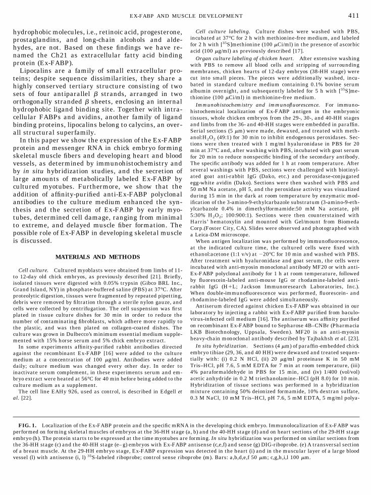
FIG. 1. Localization of the Ex-FABP protein and the specific mRNA in the developing chick embryo. Immunolocalization of Ex-FABP wasperformed on forming skeletal muscles of embryos at the 36-HH stage (a, b) and the 40-HH stage (d) and on heart sections of the 29-HH stageembryo (h). The protein starts to be expressed at the time myotubes are forming. In situ hybridization was performed on similar sections fromthe 36-HH stage (c) and the 40-HH stage (e–g) embryos with Ex-FABP antisense (c,e,f) and sense (g) DIG-riboprobe. (e) A transversal sectionof a breast muscle. At the 29-HH embryo stage, Ex-FABP expression was detected in the heart (i) and in the muscular layer of a large bloodvessel (l) with antisense (i, l) 35S-labeled riboprobe; control sense riboprobe (m). Bars: a,b,d,e,f 50 mm; c,g,h,i,l 100 mm.
411EX-FABP AND MUSCLE DEVELOPMENT

412 GENTILI ET AL.

denylic acid, 0.5 mg/ml tRNA, and 20 mM DTT. To the mix 1/100volume of 25 mM a-S-thio-ATP (Boehringer) and 1 3 105 cpm/ml ofthe appropriate labeled probe were added. After hybridization over-night at 50°C, slides were rinsed in FSM (50% formamide, 23 SSC,20 mM b-mercaptoethanol) at 60°C for 30 min twice and then rinsedfor 10 min in STE (43 SSC, 20 mM Tris–HCl, pH 7.6, 1 mM EDTA).Nonhybridized transcripts were digested with 10 mg/ml RNase A for30 min at 37°C. Sections were washed, dehydrated, and dried in air.Slides were then dipped in NTB-2 emulsion (diluted 1:1 in water)and exposed for 2 weeks at 4°C; then they were developed, fixed, andmounted with DPX mounting medium.
After hybridization with DIG RNA probes, the transcripts weredetected using a DIG nucleic acid detection kit (Boehringher Mann-heim, Germany) in which the specific transcripts were observed withanti-digoxigenin antibody conjugated to alkaline phoshatase. Slideswere washed several times with 100 mM Tris–HCl, pH 9.5, 100 mMNaCl, 50 mM MgCl2 and then immersed in the color-developmentsolution, 0.3 mg/ml nitroblue tetrazolium, 0.15 mg/ml of 5-bromo-4chloro-3-indolyl phosphate, and 125 nM levamisole. Color develop-ment was stopped by immersion of the slides in 0.1 M NaCl, 0.1 MTris, 5 mM MgCl2. Sections were weakly counterstained with nu-clear fast red.
For probe preparation, we used a 620-bp cDNA clone (pDR20)which included most of a coding sequence for the Ex-FABP protein[20]. Probes, labeled with [35S]UTP (to a specific activity of 108
cpm/mg), were synthesized using T7 or T3 polymerase. When nonra-
dioactive in situ hybridization was performed, probes were labeledwith DIG-UTP (0.5 ng/ml).
Northern blot analysis. Total RNA was extracted from cells usingthe guanidinium thiocyanate method [24]. Poly(A) RNA was selectedby two cycles of chromatography on oligo(dT)-cellulose [25]. ForNorthern analysis, the RNA was electrophoresed through 1% aga-rose gels in the presence of formaldehyde and blotted by capillarytransfer onto Hybond-N membranes (Amersham International,Buckinghamshire, UK). Blot prehybridizations were performed at65°C for 30 min in 333 mM NaH2PO4, pH 7.2, 6.66% SDS, and 250mg/ml denatured salmon sperm DNA. Blot hybridization was per-formed at 65°C for 18 h in the same solution containing 106 cpm/mlof denatured 32P-labeled probe (insert of pDR20 [20]). After hybrid-ization blots were washed twice at 65°C for 30 min in 0.2% SDS, 23SSPE and one time at 65°C for 30 min in 0.2 SDS, 0.23 SSPE.Autoradiography was performed at 280°C with hyperscreen andhyperfilm from Amersham.
RESULTS
Ex-FABP in Developing Skeletal Muscle and Heart
Immunohistochemistry and in situ hybridizationwere performed on developing skeletal muscle and
FIG. 5. Coexpression of Ex-FABP and heavy-chain myosin in myoblasts after 10 days in culture. Double immunofluorescence wasperformed as described under Materials and Methods. (a) Interferential contrast; (b) unrelated control antibody; (c) anti-MF-20 (anti-myosinheavy chain); (d) anti-ExFABP. The expression of Ex-FABP is observed when myotubes are formed at the same time as myosin is synthesized.Bar: 5 mm.
413EX-FABP AND MUSCLE DEVELOPMENT

heart (Fig. 1). At embryo stage 36 HH, mesenchymalcells showed no detectable levels of Ex-FABP proteinand the relative mRNA. Expression of the protein andexpression of the mRNA were observed in newlyformed myotubes (Figs. 1a–1c) and became prominentin well-formed myofibers at 40 HH (Figs. 1d–1g). Ex-FABP mRNA and protein were observed also in themyocardium of early developing heart (stage 29 HH)(panels h and i) and in the muscular layer of largeblood vessels (panels l and m).
Furthermore, additional evidence of Ex-FABP ex-pression by developing myocardium came from themetabolic labeling of small pieces of embryonic heart.After 5 h of labeling with [35S]methionine, the proteinsreleased into the culture medium were analyzed bySDS–PAGE under reducing and nonreducing condi-tions (Fig. 2). Moreover, a protein immunoprecipitatedby specific antibodies raised against the chondrocyte
Ex-FABP showed the same electrophoretic migrationof the Ex-FABP that was synthesized by cultured chickchondrocytes.
Ex-FABP Is Developmentally Regulated in ChickenMyoblast Cultures
Cultured chick myoblasts undergo myogenic differ-entiation in vitro. Proliferating myoblasts become post-mitotic, fuse to form multinucleate myotubes, and ex-press a number of muscle-specific proteins, includingthe muscle-specific myosins. Cultures of in vitro differ-entiating chicken myoblasts were labeled with[35S]methionine at different time intervals from day 2to day 13, when mature highly contracting myotubeswere present. Labeled proteins released into the cul-ture medium were analyzed by SDS–PAGE. As shownin Fig. 3, cells started to express Ex-FABP at day 8; theexpression increased with time and reached the maxi-mal level at day 12. A similar behavior was observed inall cell cultures examined, although the timing of theinitial appearance of the protein varied from one cul-ture to another (from day 5 to day 9; see also Fig. 6).Identification of the protein was based on immunopre-cipitation by specific antibodies and its electrophoreticcomigration with the Ex-FABP purified from chondro-cyte medium (not shown).
When the poly(A)-enriched RNA extracted from cul-tured myoblasts was analyzed by Northern blot with aprobe specific for the chondrocyte Ex-FABP mRNA, asingle band with the same electrophoretic migration ofthe chondrocyte mRNA was observed (Fig. 4).
Double-immunofluorescence staining of the cultureswas also performed (Fig. 5). In the 10-day culture, weobserved that multinucleate myotubes were positivelystained by antibodies against the Ex-FABP (Fig. 5D);the same myotubes were also stained by antibodiesrecognizing the sarcomeric myosin (Fig. 5C). Immaturemyotubes were not stained by both antibodies.
Antibodies against Ex-FABP Interfere with theExpression of the Protein in Cultured Myoblasts
Cultures of myoblasts were supplemented with af-finity-purified antibodies directed against the recombi-nant Ex-FABP protein. Antibodies were added to theculture medium daily from the first day. At differenttime intervals, antibody-supplemented and control un-supplemented cell cultures were metabolically labeledwith [35S]methionine; the labeled proteins releasedinto the culture medium were subsequently analyzedby SDS–PAGE (Fig. 6). Ex-FABP was already detect-able at day 5 in control culture and its expression washighly enhanced in antibody-treated cultures. At day 7,Ex-FABP production was still lower in untreated cul-tures than in antibody-treated cultures, in which aheavy floculate was detected in the medium. The media
FIG. 2. Analysis of proteins released from heart of 12-day oldchicken embryo. Small pieces of tissue were labeled with [35S]methi-onine and analyzed by 15% SDS–PAGE under reducing and unre-ducing conditions. Lane 1, medium from cultured hypertrophic con-drocytes (control); lane 2, medium from cultured pieces of heart; lane3, immunoprecipitation of sample of lane 2 with anti-Ex-FABP an-tibodies. Arrows refer to molecular mass markers (from top to bot-tom: 220, 97, 66, 46, 30, 14 kDa).
414 GENTILI ET AL.

from both cultures were collected and centrifuged at12,000g for 20 min. The precipitates, clearly detectableonly from the medium of the antibody-supplementedculture, were washed and Western blot analyzed (Fig.7). Ex-FABP was identified only in the precipitate fromthe medium of antibody-treated cultures.
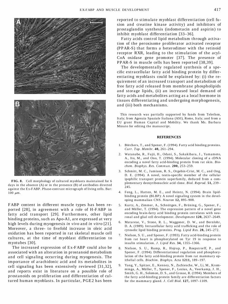
In six different experiments, the added antibodiesinduced increased synthesis of Ex-FABP; however,they also induced some cell sufferance and a delay ofmyotube formation. The extent of the morphologicalchanges ranged from dramatic (as in the culture shownin Fig. 8) to minimal. Addition of the anti-Ex-FABPantibodies to cultures of unrelated cells, as the EAHy926 endothelial cells, did not have any effect on theculture morphology and behavior.
DISCUSSION
Here we report that Ex-FABP, the first describedextracellular lipocalin able to selectively bind fatty ac-ids, previously found in chick hypertrophic cartilageand granulocytes [20] is also expressed in vivo and in
FIG. 3. Proteins secreted by in vitro differentiating chicken myoblasts. Aliquots of [35S]methionine-labeled culture medium frommyoblasts were analyzed on 15% SDS–PAGE under reducing conditions. Numbers on the top refer to culture days. In lanes 5 and 12, aliquotsof labeled culture medium from 5- and 12-day cultures were immunoprecipitated with anti-Ex-FABP affinity-purified antibodies. Arrowsrefer to molecular mass markers as in Fig. 2.
FIG. 4. Northern blot analysis of Ex-FABP mRNA. Total RNAwas extracted from myoblasts on the 14th day of culture (lane 1) andfrom cultured hypertrophic chondrocytes (lane 2). About 5 mg ofpoly(A) was loaded on each lane. Probe was the cDNA insert ofpDr20.
415EX-FABP AND MUSCLE DEVELOPMENT

vitro in developing skeletal muscle at the time of myo-tube formation and in mature myofibers and in vivo inthe embryonic myocardium. In particular, we observedthat in vitro-cultured myogenic cells expressed Ex-FABP after fusion and the protein accumulated inlarge multinucleated myotubes. It is worth noting thatcardiac muscle cells do not undergo cell fusion duringdevelopment.
To evaluate the functional role of Ex-FABP in thedifferentiation of myoblasts to myotubes, we used atissue culture model system allowing the in vitro de-velopment of myotubes from myoblasts. The cells werecultured in the presence of antibodies against the Ex-FABP. The continuous removal of the protein from theculture medium of proliferating myoblasts, as a conse-quence of the formation of immunoglobulin–proteincomplexes, resulted in an overproduction of the pro-tein, a delay in myotube formation, and varying de-grees of cell sufferance. These findings suggest that thesynthesis of Ex-FABP in muscle cells is regulated by a
feedback mechanism and that it plays a role in myo-blast differentiation via autocrine signaling.
It should be noted that, in the initial stage of themyogenesis, when blood vessels are not yet formed inthe region, a protein able to bind and transport fattyacids and functioning with autocrine and/or paracrinemechanisms should most likely be synthesized locally.Similarly, during endochondral bone formation, theEx-FABP is synthesized by hypertrophic chondrocytesbefore blood vessel invasion is observed [18,19].
The active synthesis of specialized cellular mem-branes occurring during the maturation of myofibersmay explain the expression of a fatty acid carrier pro-tein during myogenesis. The potential relevance offatty acid transport was previously suggested by thefinding of an intracellular heart-type fatty acid bindingprotein (H-FABP), expressed in heart and skeletalmuscle [13,26] and in skeletal muscle cell lines [27]. Acorrelation between fatty acid oxidation rate and H-
FIG. 7. Identification of Ex-FABP in the immunoprecipitates.Precipitates present in the medium of myoblast at 7-day culture,both supplemented and not supplemented with antibodies againstEx-FABP, were run on 15% SDS–PAGE under unreducing condi-tions, blotted on a nitrocellulose filter, and challenged with anti-Ex-FABP affinity-purified antibodies. Ex-FABP purified protein (lane 1);unsupplemented culture of myoblasts (lane 2); supplemented cultureof myoblasts (lane 3). Immunoblotting was performed with affinity-purified antibodies against Ex-FABP (B) or without addition of spe-cific antibodies (control, A). Arrows refer to molecular mass markersas in Fig. 2.
FIG. 6. Metabolically [35S]methionine-labeled protein releasedfrom myoblasts grown in the presence and in the absence of Ex-FABP affinity-purified antibodies (100 mg/ml). Number on the toprefers to culture day. Electrophoresis was performed on 15% SDS–PAGE under reducing conditions. Arrows refer to molecular massmarkers as in Fig. 2.
416 GENTILI ET AL.
FABP content in different muscle types has been re-ported [28], in agreement with a role of H-FABP infatty acid transport [29]. Furthermore, other lipidbinding proteins, such as Apo-A1, are expressed at veryhigh levels during myogenesis in vivo and in vitro [21].Moreover, a three- to fivefold increase in oleic acidoxidation has been reported in rat skeletal muscle cellcultures, at the time of myoblast differentiation tomyotubes [30].
The increased expression of Ex-FABP could also beassociated with an alteration in prostanoid metabolismand cell signaling occurring during myogenesis. Theimportance of arachidonic acid and its metabolites incell signaling has been extensively reviewed [31,32],and reports exist in literature on a possible role ofprostanoids on proliferation and differentiation of cul-tured human myoblasts. In particular, PGE2 has been
reported to stimulate myoblast differentiation (cell fu-sion and creatine kinase activity) and inhibitors ofprostaglandin synthesis (indometacin and aspirin) toinhibit myoblast differentiation [33–36].
Fatty acids control lipid metabolism through activa-tion of the peroxisome proliferator activated receptor(PPAR-S) that forms a heterodimer with the retinoidreceptor RXR, leading to the stimulation of the acyl-CoA oxidase gene promoter [37]. The presence ofPPAR-S in muscle cells has been reported [38,39].
The developmentally regulated synthesis of a spe-cific extracellular fatty acid binding protein by differ-entiating myoblasts could be explained by: (i) the re-quirement of an increased transport and metabolism offree fatty acid released from membrane phospholipidsand storage lipids, (ii) an increased local demand offatty acids and metabolites acting as a local hormone intissues differentiating and undergoing morphogenesis,and (iii) both mechanisms.
This research was partially supported by funds from Telethon,Italy; from Agenzia Spaziale Italiana (ASI), Rome, Italy; and from aEU grant Human Capital and Mobility. We thank Ms. BarbaraMinuto for editing the manuscript.
REFERENCES
1. Borchers, T., and Spener, F. (1994). Fatty acid binding proteins.Curr. Top. Membr. 40, 261–294.
2. Watanabe, R., Fujii, H., Odani, S., Sakakibara, J., Yamamoto,A., Ito, M., and Ono, T. (1994). Molecular cloning of a cDNAencoding a novel fatty acid-binding protein from rat skin. Bio-chem. Biophys. Res. Commun. 200, 253–259.
3. Schmitt, M. C., Jamison, R. S., Orgebin-Crist, M. C., and Ong,D. E. (1994). A novel, testis-specific member of the cellularipophilic transport protein superfamily, deduced from a com-plementary deoxyribonucleic acid clone. Biol. Reprod. 51, 239–245.
4. Feng, L., Hatten, M. E., and Heintz, N. (1994). Brain lipid-binding protein (BLBP): A novel signaling system in the devel-oping mammalian CNS. Neuron 12, 895–908.
5. Kurtz, A., Zimmer, A., Schnutgen, F., Bruning, G., Spener, F.,and Muller, T. (1994). The expression pattern of a novel geneencoding brain-fatty acid binding protein correlates with neu-ronal and glial cell development. Development 120, 2637–2649.
6. Matarese, V., Stone, R. L., Waggoner, D. W., and Bernlohr,D. A. (1989). Intracellular fatty acid trafficking and the role ofcytosolic lipid binding proteins. Prog. Lipid Res. 28, 245–272.
7. Nielson, S. U., and Spener, F. (1993). Fatty acid-binding proteinfrom rat heart is phosphorylated on Tyr 19 in response toinsulin stimulation. J. Lipid Res. 34, 1355–1366.
8. Nielson, S. U., Rump, R., Hojrup, P., Roepstrorff, P., andSpener, F. (1994). Differentiational regulation and phosphory-lation of the fatty acid-binding protein from rat mammary ep-ithelial cells. Biochim. Biophys. Acta 1211, 189–197.
9. Yang, Y., Spitzer, E., Kenney, N., Zschiesche, W., Li, M., Krom-minga, A., Muller, T., Spener, F., Lezius, A., Veerkamp, J. H.,Smith, G. H., Solomon, D. S., and Grosse, R. (1994). Members ofthe fatty acid-binding protein family are differentiation factorsfor the mammary gland. J. Cell Biol. 127, 1097–1109.
FIG. 8. Cell morphology of cultured myoblasts maintained for 6days in the absence (A) or in the presence (B) of antibodies directedagainst the Ex-FABP. Phase-contrast micrograph of living cells. Bar:100 mm.
417EX-FABP AND MUSCLE DEVELOPMENT

10. Borchers, T., Unteberg, C., Rudel, H., Robenek, H., and Spener,F. (1989). Subcellular distribution of cardiac fatty acid-bindingprotein in bovine heart muscle and quantitation with an en-zyme-linked immunosorbent assay. Biochim. Biophys. Acta1002, 54–61.
11. Crisman, T. S., Claffey, K. P., Saouaf, R., Hanspai, J., andBrecher, P. (1987). Measurement of rat heart fatty acid bindingprotein by ELISA. Tissue distribution, developmental changesand subcellular distribution. J. Mol. Cell. Cardiol. 19, 423–431.
12. Paulussen, R. J., Geelen, M. J., Beynen, A. C., and Veerkamp,J. H. (1989). Immunochemical quantitation of fatty acid bindingproteins. Tissue and intracellular distribution, postnatal devel-opment and influence of physiological conditions on rat heartand liver FABP. Biochim. Biophys. Acta 1001, 201–209.
13. Peeters, R. A., in’t-Groen, M. A., and Veerkamp, J. H. (1989).The fatty acid binding protein from human skeletal muscle.Arch. Biochem. Biophys. 274, 556–563.
14. Felig, P., and Wahren, J. (1975). Fuel homeostasis in exercise.N. Engl. J. Med. 293, 1078–1084.
15. Lopaschuk, G. D., and Saddik, M. (1992). The relative contribu-tion of glucose and fatty acids to ATP production in hearts reper-fused following ischemia. Mol. Cell. Biochem. 116, 111–116.
16. Descalzi Cancedda, F., Malpeli, M., Gentili, C., Di Marzo, V.,Bet, P., Carlevaro, M., Cermelli, S., and Cancedda, R. (1996).The developmentally regulated avian Ch21 lipocalin is an ex-tracelular fatty acid-binding protein. J. Biol. Chem. 271,20163–20169.
17. Descalzi Cancedda, F., Manduca, P., Tacchetti, C., Fossa, P.,Quarto, R., and Cancedda, R. (1988). Developmentally regu-lated synthesis of a low molecular weight protein (Ch 21) bydifferentiating chondrocytes. J. Cell Biol. 107, 2455–2463.
18. Manduca, P., Descalzi Cancedda, F., Tacchetti, C., Quarto, R.,Fossa, P., and Cancedda, R. (1989). Synthesis and secretion ofCh 21 protein in embryonic chick skeletal tissues. Eur. J. CellBiol. 50, 154–161.
19. Descalzi Cancedda, F., Dozin, B., Rossi, F., Molina, F., Can-cedda, R., Negri, A., and Ronchi, S. (1990). The Ch 21 protein,developmentally regulated in chick embryo, belongs to the su-perfamily of lipophilic molecule carrier proteins. J. Biol. Chem.265, 19060–19064.
20. Dozin, B., Descalzi, F., Briata, P., Hayashy, M., Gentili, C.,Hayashi, K., Quarto, R., and Cancedda, R. (1992). Expression,regulation, and tissue distribution of the Ch 21 protein duringchicken embryogenesis. J. Biol. Chem. 267, 2979–2985.
21. Ferrari, S., Battini, R., and Cossu, G. (1990). Differentiationdependant expression of Apolipoprotein AI in chicken myogeniccells in culture. Dev. Biol. 140, 430–436.
22. Edgell, C. J. S., McDonald, C. C., and Graham, J. B. (1983).Permanent cell line expressing human factor VIII-related anti-gen established by hybridization. Proc. Natl. Acad. Sci. USA 80,3734–3737.
23. Tajbakhsh, S., Vivarelli, E., Cusella De Angelis, G., Rocancourt,D., Buckingham, M., and Cossu, G. (1994). A population ofmyogenic cells derived from the mouse neural tube. Neuron 13,813–821.
24. Chomczynski, P., and Sacchi, N. (1987). Single-step method ofRNA isolation by acid guanidium thycyanate-phenol-chloro-form extraction. Anal. Biochem. 162, 156–159.
25. Krystosek, A., Cawthon, M. L., and Kabat, D. (1975). Improvedmethods for purification and assay of eukariotic messengerribonucleic acids and ribosomes. Quantitative analysis of theirinteraction in a fractionated reticulocyte cell-free system.J. Biol. Chem. 250, 6077–6084.
26. Carey, J. O., Neufer, P. D., Farrar, R. P., Veerkamp, J. H., andDohm, G. L. (1994). Transcriptional regulation of muscle fattyacid-binding protein. Biochem. J. 298, 613–617.
27. Rump, R., Buhlmann, C., Borchers, T., and Spener, F. (1996).Differentiation-dependent expression of heart type fatty acid-binding protein in C2C12 muscle cells. Eur. J. Cell Biol. 69,135–142.
28. Veerkamp, J. H., and van Moerkerk, H. T. (1993). Fatty acid-binding protein and its relation to fatty acid oxidation. Mol. CellBiochem. 123, 101–106.
29. Glatz, J. F. C., Vork, M. N., Cistola, D. P., and van der Vusse,G. J. (1993). Cytoplasmic fatty acid binding protein: Signifi-cance for intracellular transport of fatty acids and putative roleon signal transduction pathways. Prostaglandins LeukotrienesEssent. Fatty Acids 48, 33–41.
30. Sauro, V. S., and Strickland, K. P. (1987). Changes in oleic acidoxidation and incorporation into lipids of differentiating L6myoblasts cultured in normal or fatty acid-supplementedgrowth medium. Biochem. J. 24, 743–748.
31. Piomelli, D. (1993). Arachidonic acid in cell signaling. Curr.Opin. Cell Biol. 5, 274–280.
32. Di Marzo, V. (1995). Arachidonic acid and eicosanoids as tar-gets and effectors in second messenger interactions. Prosta-glandins Leukotrienes Essent. Fatty Acids 53, 239–254.
33. Entwistle, A., Curtis, D. H., and Zalin, R. J. (1986). Myoblastfusion is regulated by a prostanoid of the one serie inde-pendently of a rise in cyclic AMP. J. Cell Biol. 103, 857–866.
34. Zalin, R. J. (1987). The role of hormones and prostaglandins inthe in vitro proliferation and differentiation of human myo-blasts. Exp. Cell Res. 172, 265–281.
35. Elgendy, H., and Hausman, R. E. (1990). Prostaglandin-depen-dant phosphatidylinositol signaling during embryonic chickmyogenesis. Cell Differ. Dev. 32, 109–115.
36. McLennan, I. S. (1991). E and F alpha series prostaglandins indeveloping muscles. Prostaglandins Leukotrienes Essent. FattyAcids 43, 77–82.
37. Keller, H., Dreyer, C., Medin, J., Mahfoudi, A., Ozato, K., andWahli, W. (1993). Fatty acids and retinoids control lipid metab-olism through activation of peroxisome proliferator-activatedreceptor-retinoid X receptor heterodimers. Proc. Natl. Acad.Sci. USA 90, 2160–2164.
38. Park, K. S., Ciaraldi, T. P., Abramscarter, L., Mudaliar, S.,Nikoulina, S. E., and Henry, R. R. (1997). PPAR gamma geneexpression is elevated in skeletal muscle of OBESE and type IIdiabetic subjects. Diabetes 46, 1230–1234.
39. Elbrecht, A., Chen, Y. L., Cullinan, C. A., Hayes, N., Leibowitz,M. D., Moller, D. E., and Berger, J. (1996). Molecular cloningexpression and characterization of human peroxisome prolifer-ation activated receptors gamma 1 and gamma 2. Biochem.Biophys. Res. Commun. 224, 431–437.
Received December 2, 1997Revised version received February 20, 1998
418 GENTILI ET AL.