expression patterns of innate immunity-related genes in
TRANSCRIPT
385https://www.ejast.org
Journal of Animal Science and Technology
RESEARCH ARTICLEJ Anim Sci Technol 2020;62(3):385-395https://doi.org/10.5187/jast.2020.62.3.385 pISSN 2672-0191 eISSN 2055-0391
Expression patterns of innate immunity-related genes in response to polyinosinic:polycytidylic acid (poly[I:C]) stimulation in DF-1 chicken fibroblast cellsHyun-Jun Jang1,2 and Ki-Duk Song1,2*1Department of Animal Biotechnology, Jeonbuk National University, Jeonju 54896, Korea2The Animal Molecular Genetics and Breeding Center, Jeonbuk National University, Jeonju 54896, Korea
AbstractPolyinosinic:polycytidylic acid (poly[I:C]) can stimulate Toll-like receptor 3 (TLR3) signaling pathways. In this study, DF-1 cells were treated with poly(I:C) at various concentrations and time points to examine the comparative expression patterns of innate immune response genes. The viability of DF-1 cells decreased from 77.41% to 38.68% when cells were treated different dose of poly(I:C) from 0.1 µg/mL to 100 µg/mL for 24 h respectively. The expres-sions of TLR3, TLR4, TLR7, TLR15, TLR21, IL1B, and IL10 were increased in dose- and time-dependent manners by poly(I:C) treatment. On the contrary, the expression patterns of interferon regulatory factors 7 (IRF7), Jun proto-oncogene, AP-1 transcription factor subunit (JUN), Nuclear Factor Kappa B Subunit 1 (NF-κB1), and IL8L2 were varied; IRF7 and IL8L2 were increasingly expressed whereas the expressions of JUN and NF-κB1 were decreased in a dose-dependent manner after they were early induced. In time-dependent analysis, IRF7 expression was significantly upregulated from 3 h to 24 h, whereas JUN and NF-κB1 expres-sions settled down from 6 h to 24 h after poly(I:C) treatment although they were induced at early time from 1 h to 3 h. Poly(I:C) treatment rapidly increased the expression of IL8L2 from 3 h to 6 h with a plateau at 6 h and then the expression of IL8L2 was dramatically decreased until 24 h after poly(I:C) treatment although the expression level was still higher than the non-treated control. These results may provide the basis for understanding host response to viral infection and its mimicry system in chickens.Keywords: Poly(I:C), TLR, Immune response, Chicken, Gene expression
INTRODUCTIONInnate immunity is the first defense line against various pathogens through sensing pathogens, elimi-nating them, and activating adaptive immune response [1]. In sensing pathogens, nucleic acids (NAs) that are originated from pathogenic bacteria and viruses are recognized by innate immune receptor sig-naling, which are mediated by pattern recognition receptors (PRRs) including toll-like receptors (TLRs),
Received: Feb 13, 2020Revised: Feb 29, 2020Accepted: Mar 3, 2020
*Corresponding authorKi-Duk SongDepartment of Animal Biotechnology, Jeonbuk National University, Jeonju 54896, Korea.Tel: +82-63-219-5523E-mail: [email protected]
Copyright © 2020 Korean Society of Animal Sciences and Technology.This is an Open Access article distributed under the terms of the Creative Commons Attribution Non-Commercial License (http://creativecommons.org/licenses/by-nc/4.0/) which permits unrestricted non-commercial use, distribution, and reproduction in any medium, provided the original work is properly cited.
ORCIDHyun-Jun Janghttps://orcid.org/0000-0003-2906-7543Ki-Duk Songhttps://orcid.org/0000-0003-2827-0873
Competing interestsNo potential conflict of interest relevant to this article was reported.
Funding sourcesThis research was supported by Basic Science Research Program through the National Research Foundation of Korea (NRF) funded by the Ministry of Education (NRF-2016R1D1A1B04935092) and by grants from the Next-Generation

Immune gene expressions of TLR3 stimulated chicken cells
386 | https://www.ejast.org https://doi.org/10.5187/jast.2020.62.3.385
retinoic acid inducible gene I (RIG-I), melanoma differentiation-associated protein 5 (MDA5), and laboratory of genetics and physiology 2 (LGP2) [2,3]. Among them, TLR3, TLR7/8, TLR9, and TLR13 of TLRs are known as nucleic acid NA-sensing TLRs. They primarily exist in endosome and respond to double-stranded RNA (dsRNA), single-stranded RNA (ssRNA), single-stranded DNA, and bacterial ribosomal RNA respectively [4,5]. RIG-I, MDA5, and LGP2 are cytosolic NA receptors which detect dsRNA. RIG-I primarily responds to 5’-triphosphorylated blunt-ended RNA or dsRNA produced during RNA virus infections and MDA5 responds to long dsRNA [2]. LGP2 also seems to enhance initial MDA5-RNA interaction [6]. Complex with cognate PRRs and their ligands leads to the engagements of myeloid differentiation primary response 88 (MYD88), Toll/IL-1R homologous region (TIR) domain-containing adapter-inducing interferon-β (TRIF), or mitochondrial antiviral-signaling protein (MAVS). It activates transcription factors (TFs) such as interferon regulatory factor3 (IRF3), IRF7, nuclear factor kappa B (NF-κB), and activating pro-tein 1 (AP-1) (ATF2/JUN) by orchestrating a combination of multi-protein complexes. The TFs induce to express inflammatory cytokines, chemokines and type I interferons [7–12].
Among the NA-sensing TLRs, chickens have obvious orthologues of TLR3 and TLR7 while TLR8 has been disrupted by the insertion of a large CR1 repeat [13]. TLR9 and TLR13 were also absent [8,14]. In addition, TLR15 and TLR21 uniquely existed in chickens compared to human and mouse [15,16]. Chicken TLR21 has recently been shown to recognize CpG motifs, suggesting a functional homologue to mammalian TLR9 [17] whereas an virus-related agonist for TLR15 re-mains unknown [18,19]. MDA5 and LGP2 are also present in chicken genome and their function seems to be similar to mammals whereas RIG-I is obviously absent [20,21]. It has been suggested that the lack of RIG-I caused a susceptibility for zoonotic RNA virus such as avian influenza in chickens [22]. Even if immune responses to NA have been comparatively well characterized in chickens, the precise mechanism remains to be elucidated.
Polyinosinic:polycytidylic acid (poly[I:C]), viral like dsRNA, has generally been used to mimic NA-sensing responses of the innate immune system. Poly(I:C) is recognized by TLR3 and MDA-5, activate various TFs such as IRFs and NF-κB, and stimulates various cytokines and chemokine, IFNs and costimulatory factors in various species [10,11,23–26]. Poly(I:C) exhibited a toxicity in various tissues and cells [27,28]. Especially, the viability of chicken embryonic fibroblasts (CEFs) reduced to about 80% and below 50% with 1,000 µg/mL of poly(I:C) for 24 h and 72 h respectively and it suggested that poly(I:C) induced apoptosis of CEFs through the activation of caspase-3 and -8 by TNFRSF8 [29]. In addition, DF-1 cells, chicken fibroblast cell line modulated IRF7-related immune signaling pathways responding to poly(I:C) [30]. In this regard, chicken fibroblasts includ-ing DF-1 are a useful model to study in vitro immune responses which are stimulated by poly(I:C).
In this study, we examined the expression patterns of innate immune signaling-related genes such as canonical and non-canonical TLRs, the related TFs, cytokines, and immune-related effector molecules in chickens after poly(I:C) treatment. Our results could contribute to understanding the gene expression which is involved in NA-sensing and the related responses in chicken cells.
MATERIALS AND METHODSCell culture and poly(I:C) treatmentDF-1 chicken fibroblast cell lines were obtained from the American Type Culture Collection (Rockville, MD, USA) and maintained in the Dulbecco’s modified Eagle’s medium with 10% fetal bovine serum (Biowest, Nuaillé, France). DF-1 cells were cultured at 37℃ in 5% CO2 incubator. Poly(I:C) was purchased from Invivogen (San Diego, CA, USA) and was stocked according to the manufacturer’s instruction and all poly(I:C) treatment was maintained under the culture condition
BioGreen 21 Program (No. PJ01324201, PJPJ01315101), Rural Development Administration, Korea).
AcknowledgementsNot applicable.
Availability of data and materialNot applicable.
Authors’ contributionsConceptualization: Song KD.Formal analysis: Jang HJ.Methodology: Jang HJ.Investigation: Jang HJ.Writing - original draft: Jang HJ.Writing - review & editing: Song KD.
Ethics approval and consent to participateThis manuscript does not require IRB/IACUC approval because there are no human and animal participants.

https://doi.org/10.5187/jast.2020.62.3.385 https://www.ejast.org | 387
Jang and Song
of DF-1 cells.
Cell viability assayCell viability assays were performed using tetrazolium compound based CellTiter 96® AQueous One Solution Cell Proliferation (MTS) assay (Promega, Madison, WI, USA). MTS assay was then performed according to the manufacturer’s instruction at 24 h after treatment at indicated concen-trations of poly(I:C).
RNA extraction and quantitative RT-PCRRNAs were isolated from DF-1 cells using RNA extraction kit (Invitrogen, CA, USA). For quanti-tative reverse transcription-polymerase chain reaction (qRT-PCR), 1 µg of total RNA was used for cDNA synthesis with Rever Tra Ace-α- first strand cDNA Synthesis Kit (Toyobo, Osaka, Japan). Sequence-specific primers (Table 1) were designed using the Primer-BLAST program (https://www.ncbi.nlm.nih.gov/tools/primer-blast/index.cgi?LINK_LOC=BlastHome). qRT- PCR was performed using the iCycler real-time PCR detection system (Bio-Rad, Hercules, CA, USA) and SYBR Green (Bio-Rad, Hercules, CA, USA). Non-template wells without cDNA were included as negative controls. Each sample was tested in triplicate. The PCR conditions were 95℃ for 3 min, followed by 40 cycles at 95℃ for 10 s and 60℃ for 30 s, using a melting curve program (increasing temperature from 65℃ to 95℃ at a rate of 0.5℃ per 5 s) and continuous fluorescence measure-
Table 1. Lists of primers used to perform qRT-PCRTarget gene
(accession number of NCBI)Primer type
5’ to 3’ Sequence
TLR3 (422720) Forward CCATTTTGAAGGGTGGAGAA
Reverse CCTGCTTCGAAGTCTCGTTC
TLR4 (417241) Forward TTCCAAGCACCAGATAGCAACATC
Reverse ACGGGTCACAGAAGAACTTAGGG
TLR7 (418638) Forward TTCTGGCCACAGATGTGACC
Reverse CCTTCAACTTGGCAGTGCAG
TLR15 (421219) Forward GTTCTCTCTCCCAGTTTTGTAAATAGC
Reverse GTGGTTCATTGGTTGTTTTTAGGAC
TLR21 (415623) Forward CAACAGACTGCTGGAGGTGA
Reverse TGCAGCTTCAGGTCGTACAG
IRF7 (396330) Forward GAGGATCCGGCCAAATGGAA
Reverse CCAAATCGTGGTGGTTGAGC
JUN (424673) Forward CCCGGTGTATGCCAATCTCA
Reverse CTCCTGCGACTCCATGTCAA
NF-κB1 (395587) Forward AGAAAAGCTGGGTCTTGGCA
Reverse CCATCTGTGTCAAAGCAGCG
IL1B (395196) Forward GGATTCTGAGCACACCACAGT
Reverse TCTGGTTGATGTCGAAGATGTC
IL8L2 (396495) Forward CCAAGCACACCTCTCTTCCA
Reverse GCAAGGTAGGACGCTGGTAA
IL10 (428264) Forward AGCAGATCAAGGAGACGTTC
Reverse ATCAGCAGGTACTCCTCGAT
GAPDH (374193) Forward TGCTGCCCAGAACATCATCC
Reverse ACGGCAGGTCAGGTCAACAA
Immune gene expressions of TLR3 stimulated chicken cells
388 | https://www.ejast.org https://doi.org/10.5187/jast.2020.62.3.385
ment. The qRT-PCR data were normalized relative to the expression of GAPDH and calculated using the 2 ∆∆Ct method, where ∆∆Ct = (Ct of the target gene – Ct of GAPDH) treatment – (Ct of the target gene – Ct of GAPDH) control [31].
Statistical analysisStatistical significance (p < 0.05, p < 0.01, p < 0.001) of apparent differences in gene expression after poly(I:C) treatment was assessed by ANOVA and Tukey’s multiple comparison test (GraphPad Prism 5.01, San Diego, CA, USA).
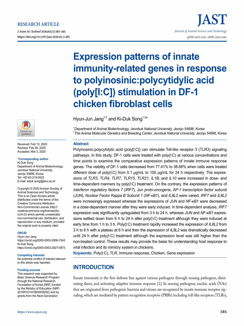
RESULTS AND DISCUSSIONViability test of DF-1 cells in various concentrations of poly(I:C)In this study, poly(I:C) treatment with different doses from 0.1 µg/mL to 100 µg/mL for 24 h decreased the viability of DF-1 cells (chicken fibroblasts cell line) by 77.41%, 57.63%, 56.28%, 46.69%, 43.06%, 43.19%, 44.22%, 43.32%, 38.9%, 39.19%, 38.25%, 38.1%, 36.85%, 37.73%, 38.42%, 37.17%, and 38.68% respectively, compared to the non-treated control. The statistical anal-ysis showed significant difference at all the treated concentrations except the concentration of 0.1 µg/mL, compared to the non-treated control and no difference among the cell viabilities from 0.5 µg/mL to 100 µg/mL poly(I:C) (p < 0.05) (Fig. 1). These results suggested that poly(I:C) rapidly affected on the cell viability from 0.5 µg/mL and this effect was saturated from 0.5 µg/mL to 100 µg/mL. Thus, we supposed that DF-1 cells could be much more sensitive to poly(I:C) than primary cultured CEFs.
Fig. 1
Fig. 1. The viability and morphology of DF-1 cells in the poly(I:C)-treated conditions with various concentrations of poly(I:C) for 24 h. The statistical analysis was performed to assess statistical significance between each concentration and the non-treated control. Error bars were expressed as SEM *p < 0.05, **p < 0. 01, ***p < 0.001.

https://doi.org/10.5187/jast.2020.62.3.385 https://www.ejast.org | 389
Jang and Song
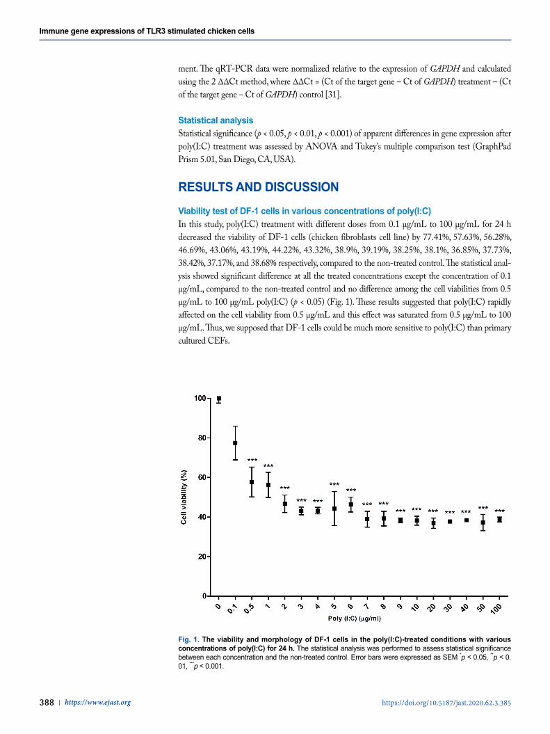
Dose- and time-dependent expression patterns of TLRs by poly(I:C) treatment TLR3 and TLR7 are known as NA-sensing TLRs while the function of TLR4 is associated with the recognition of endotoxins molecules, in particular lipopolysaccharide from gram-negative bac-teria [13,32]. Recently, the several studies have shown that TLR3, 4, and 7 mediated the responses to the viral-associated PAMPs such as poly(I:C), F protein of Respiratory Syncytial Virus (RSV), and imidazoquinolines, antiviral therapeutic compounds, respectively [33–38]. In addition, it has been reported that selective activation of TLR3/4-IRF3 pathway was associated with potential inhibition of viral replication [39]. TLR15, an avian-specific TLR, has been reported to be induced by salmonella, mycoplasma, and even Marek’s Disease Virus (MDV) [16,18,19,40]; however, the specifically virus-associated agonist was still unknown [41]. Instead of mammalian TLR9 which was missing from the chicken genome, chicken TLR21 acted as a functional homologue to the mammalian TLR9 to recognize CpG [17]. Poly(I:C) and CpG ODN (CpG-motif containing oligodeoxydinucleotide) synergized the expression of pro-inflammatory cytokines and chemokines and the production of nitric oxide in chicken monocytes [42,43].
To investigate chicken TLRs expressions in response to poly(I:C) treatment, the expressions of chicken TLRs were analyzed dose and time-dependently. From the analysis, the expressions of TLR3, 4, 7, 15, and 21 were significantly induced at the poly(I:C) concentrations of 5 µg/mL and 10 µg/mL for 24 h (Fig. 2A). In addition, the expression levels of TLR3, 4, 7, 15, and 21 were sig-nificantly increased with 10 µg/mL poly(I:C) at 12 h and 24 h after poly(I:C) treatment (Fig. 2B). Therefore, we suggested that poly(I:C) was directly targeted at these TLRs in DF-1 cells to stimu-late immune responses.
Dose- and time-dependent expression patterns of TLR signaling-associated tran-scription factors (TFs) by poly(I:C) treatmentTLRs which recognize their ligands activated conserved TFs including AP-1, NF-κB, and IRFs through the interplay of complex TLR signaling pathways [44–47]. Among the AP-1 family, JUN that was a target protein of c-Jun N-terminal kinase ( JNK) was regarded as a key factor in TLR signaling [47]. Among NF-κB protein complex, NF-κB1 (also known as p50) was known to have DNA binding activity for the promoter region of its target genes [48]. Among IRFs, IRF3 and IRF7 were activated by various ligands, such as poly(I:C), LPS, and virus infection and mainly controlled type-I IFN expression [49]. In mammalian, type I IFNs-mediated signaling pathways were dependent on the stimulus and the responding cell types. TLR signaling pathways associated with type I IFN, TLR3 and TLR4 induced type I IFN production in various cell types in a manner dependent on TIR-domain-containing adaptor protein inducing IFNβ (TRIF) whereas TLR7, TLR8 and TLR9 induced type I IFN production in dendritic cells via a pathway dependent on MYD88. Eventually they can activate some common signaling molecules including TNF recep-tor-associated factor 3 (TRAF3) and IRF3 and IRF7 [49,50]. Additionally, poly(I:C) treatment increased IRF7 and type-I IFN (IFNA) in DF-1 cells [25].
To reveal TFs which are associated with TLR signaling responded to poly(I:C), the expressions of IRF7, JUN, and NF-κB1 were analyzed in DF-1 cells at different doses of poly(I:C) and time points. From the dose-dependent treatment, IRF7 and NF-κB1 expressions were significantly in-creased at 5 µg/mL and 10 µg/mL and 5 µg/mL of the poly(I:C) treatment for 24 h, respectively. Whereas the expression of JUN was significantly decreased at 1 µg/mL, 5 µg/mL, and 10 µg/mL of poly(I:C) for 24 h (Fig. 3A). When the expressions of IRF7, JUN, and NF-κB1 were analyzed with 10 µg/mL poly(I:C) according to time course, the expression of IRF7 steadily increased from 3 h to 24 h after poly(I:C) treatment. JUN and NF-κB1 expressions were commonly increased from 1 h to 3 h after poly(I:C) treatment, but were decreased from 6 h to 24 h after poly(I:C) treatment
Immune gene expressions of TLR3 stimulated chicken cells
390 | https://www.ejast.org https://doi.org/10.5187/jast.2020.62.3.385
Fig.2
A
B
Fig. 2. Dose- and time-dependent expression patterns of TLRs by poly(I:C) treatment. The expressions of TLR3, 4, 7, 15, and 21 in DF-1 cells were analyzed in poly(I:C)-treated conditions with concentrations of 0, 0.1, 1, 5, and 10 µg/mL for 24 h (A) and with concentration of 10 µg/mL for 1, 3, 6, 12, and 24 h (B). The statistical analysis was performed to assess statistical significance between each treated condition and the non-treated control. Error bars were expressed as SEM *p < 0.05, **p < 0.01, ***p < 0.001.

https://doi.org/10.5187/jast.2020.62.3.385 https://www.ejast.org | 391
Jang and Song
(Fig. 3B). These results suggested that TLR3 stimulation by poly(I:C) induced IRF7 transcription, whereas the expressions of JUN and NF-κB1 were gradually decreased and maintained to the ground state although they were rapidly induced within 1 h after the poly(I:C) treatment. Thus, we speculated that poly(I:C) may mainly induce immune-effector genes by IRF7-mediated signaling pathway after the recognition by TLRs such as TLR3, 4, 7, 15, and 21 in 24 h after the treatment while direct or indirect pathways may exist to acutely induce JUN and NF-κB1. The further study is necessary to prove the activation of TLR pathway-mediated TFs.
Dose- and time-dependent expression patterns of immune-related effector mole-cules by poly(I:C) treatmentFrom TLRs recognizing their ligands, the activated TFs can induce a variety of interferons, cy-tokines and chemokines [9,39]. During the immune responses, cytokine and chemokine families acted as extracellular molecular regulators which mediated immune cell recruitment and partici-pated in complex intracellular signaling processes [9]. Among them, IL1B belonging to IL1 family
Fig. 3
A
B
Fig. 3. Dose- and time-dependent expression patterns of TLR signaling-associated transcription factors (TFs) by poly(I:C) treatment. The expressions of IRF7, JUN, and NF-κB1 in DF-1 cells were analyzed in poly(I:C)-treated conditions with concentrations of 0, 0.1, 1, 5, and 10 µg/mL for 24 h (A) and with concentration of 10 µg/mL for 1, 3, 6, 12, and 24 h (B). The statistical analysis was performed to assess statistical significance between each treated condition and the non-treated control. Error bars were expressed as SEM *p < 0.05, **p < 0.01, ***p < 0.001.

Immune gene expressions of TLR3 stimulated chicken cells
392 | https://www.ejast.org https://doi.org/10.5187/jast.2020.62.3.385
and IL10 have been known as a pro-inflammatory and an anti-inflammatory cytokine respectively. These cytokines were induced by viral infections [9,51–53]. IL8, a critical inflammatory chemokine was also upregulated by various viral infection in human epithelial cells [54].
To examine whether the expressions of immune-related effector genes are affected by poly(I:C) treatment, IL1B, IL8L2 (chicken IL8-like 2), and IL10 expressions were analyzed after the poly(I:C) treatment at different dose and time points . From the analysis, the expressions of IL1B, IL8L2, and IL10 were significantly increased by poly(I:C) treatments from 5 µg/mL to 10 µg/mL for 24 h (Fig. 4A). In time-dependent analysis, the expressions of IL1B, and IL10 were significantly increased from 12 h to 24 h after the poly(I:C) treatment (Fig. 4B). Unlike IL1B and IL10, the expression of IL8L2 showed the rapid increase at 3 h after the poly(I:C) treatment and reached to the plateau at 6 h after the poly(I:C) treatment. In addition, it was continuously decreased from 12 h to 24 h after the poly(I:C) treatment compared to the expression of IL8L2 at 6 h after the poly(I:C) treatment although the expressions of IL8L2 at 12 h and 24 h after the poly(I:C) treatment were still higher
Fig. 4
A
B
Fig. 4. Dose- and time-dependent expression patterns of immune-related effector molecules by poly(I:C) treatment. The expressions of IL1B, IL8L2, and IL10 in DF-1 cells were analyzed in poly(I:C)-treated conditions with concentrations of 0, 0.1, 1, 5, and 10 µg/mL for 24 h (A) and with concentration of 10 µg/mL for 1, 3, 6, 12, and 24 h (B). The statistical analysis was performed to assess statistical significance between each treated condition and the non-treated control. Error bars were expressed as SEM *p < 0.05, **p < 0.01, ***p < 0.001.

https://doi.org/10.5187/jast.2020.62.3.385 https://www.ejast.org | 393
Jang and Song
than the non-treated control (Fig. 4B). This result suggested that the inductions of IL1B, IL8L2, and IL10 in DF-1 cells could be mediated by TLR-signaling pathways. In addition, IL8L2 could more sensitively respond to poly(I:C) and be inhibited by other feedback systems compared to IL1B and IL10.
Conclusively, we suggested the distinct TLR signaling pathways which responded to poly(I:C) in chicken-originated cell line (DF-1) compared to mammalian TLRs for NA-sensing and their signaling pathways. Our results could contribute to understanding NA-sensing and subsequent im-mune signaling pathways in chicken cells.
REFERENCES1. Riera Romo M, Pérez-Martinez D, Castillo Ferrer C. Innate immunity in vertebrates: an over-
view. Immunology. 2016;148:125-39.2. Thompson MR, Kaminski JJ, Kurt-Jones EA, Fitzgerald KA. Pattern recognition receptors and
the innate immune response to viral infection. Viruses. 2011;3:920-40.3. Roers A, Hiller B, Hornung V. Recognition of endogenous nucleic acids by the innate immune
system. Immunity. 2016;44:739-54.4. Majer O, Liu B, Barton GM. Nucleic acid-sensing TLRs: trafficking and regulation. Curr
Opin Immunol. 2017;44:26-33.5. Oldenburg M, Krüger A, Ferstl R, Kaufmann A, Nees G, Sigmund A, et al. TLR13 recog-
nizes bacterial 23S rRNA devoid of erythromycin resistance-forming modification. Science. 2012;337:1111-5.
6. Bruns AM, Leser GP, Lamb RA, Horvath CM. The innate immune sensor LGP2 activates antiviral signaling by regulating MDA5-RNA interaction and filament assembly. Mol Cell. 2014;55:771-81.
7. Werling D, Jungi TW. TOLL-like receptors linking innate and adaptive immune response. Vet Immunol Immunopathol. 2003;91:1-12.
8. Kaiser P. Advances in avian immunology--prospects for disease control: a review. Avian Pathol. 2010;39:309-24.
9. Turner MD, Nedjai B, Hurst T, Pennington DJ. Cytokines and chemokines: at the cross-roads of cell signalling and inflammatory disease. Biochim Biophys Acta Mol Cell Res. 2014;1843:2563-82.
10. Kawai T, Akira S. Toll-like receptor and RIG-I-like receptor signaling. Ann N Y Acad Sci. 2008;1143:1-20.
11. Kawai T, Akira S. Antiviral signaling through pattern recognition receptors. J Biochem. 2007;141:137-45.
12. Randall RE, Goodbourn S. Interferons and viruses: an interplay between induction, signalling, antiviral responses and virus countermeasures. J Gen Virol. 2008;89:1-47.
13. Philbin VJ, Iqbal M, Boyd Y, Goodchild MJ, Beal RK, Bumstead N, et al. Identification and characterization of a functional, alternatively spliced Toll-like receptor 7 (TLR7) and genomic disruption of TLR8 in chickens. Immunology. 2005;114:507-21.
14. Temperley ND, Berlin S, Paton IR, Griffin DK, Burt DW. Evolution of the chicken Toll-like receptor gene family: a story of gene gain and gene loss. BMC Genomics. 2008;9:62.
15. Roach JC, Glusman G, Rowen L, Kaur A, Purcell MK, Smith KD, et al. The evolution of ver-tebrate Toll-like receptors. Proc Natl Acad Sci USA. 2005;102:9577-82.
16. Higgs R, Cormican P, Cahalane S, Allan B, Lloyd AT, Meade K, et al. Induction of a novel chicken toll-like receptor following Salmonella enterica serovar typhimurium infection. Infect

Immune gene expressions of TLR3 stimulated chicken cells
394 | https://www.ejast.org https://doi.org/10.5187/jast.2020.62.3.385
Immun. 2006;74:1692-8.17. Brownlie R, Allan B. Avian toll-like receptors. Cell Tissue Res. 2011;343:121-30.18. Nerren JR, He H, Genovese K, Kogut MH. Expression of the avian-specific toll-like receptor
15 in chicken heterophils is mediated by gram-negative and gram-positive bacteria, but not TLR agonists. Vet Immunol Immunopathol. 2010;136:151-6.
19. Oven I, Resman Rus K, Dušanić D, Benčina D, Keeler Jr CL, Narat M. Diacylated lipopep-tide from Mycoplasma synoviae mediates TLR15 induced innate immune responses. Vet Res. 2013;44:99.
20. Liniger M, Summerfield A, Zimmer G, McCullough KC, Ruggli N. Chicken cells sense in-fluenza a virus infection through MDA5 and CARDIF signaling involving LGP2. J Virol. 2012;86:705-17.
21. Karpala AJ, Stewart C, McKay J, Lowenthal JW, Bean AG. Characterization of chick-en Mda5 activity: regulation of IFN-β in the absence of RIG-I functionality. J Immunol. 2011;186:5397-405.
22. Barber MR, Aldridge Jr JR, Webster RG, Magor KE. Association of RIG-I with innate im-munity of ducks to influenza. Proc Natl Acad Sci USA. 2010;107:5913-8.
23. Wu J, Huang S, Zhao X, Chen M, Lin Y, Xia Y, et al. Poly(I:C) treatment leads to interfer-on-dependent clearance of hepatitis B virus in a hydrodynamic injection mouse model. J Virol. 2014;88:10421-31.
24. Fortier ME, Kent S, Ashdown H, Poole S, Boksa P, Luheshi GN. The viral mimic, polyinos-inic:polycytidylic acid, induces fever in rats via an interleukin-1-dependent mechanism. Am J Physiol Regul Integr Comp Physiol. 2004;287:R759-66.
25. Kim TH, Zhou H. Correction: functional analysis of chicken IRF7 in response to dsRNA an-alog poly(I:C) by integrating verexpression and knockdown. PLOS ONE. 2015;10:e0137672.
26. Liu J, Cao D, Liu Y, Li Z, Si Y, Wang Z, et al. Expression and functional analysis of recep-tor-interacting serine/threonine kinase 2 (RIP2) in Japanese flounder (Paralichthys olivaceus). Fish Shellfish Immunol. 2018;75:327-35.
27. Bianchi F, Pretto S, Tagliabue E, Balsari A, Sfondrini L. Exploiting poly(I:C) to induce cancer cell apoptosis. Cancer Biol Ther. 2017;18:747-56.
28. Pyo S, Gangemi JD, Ghaffar A, Mayer EP. Poly I:C-induced antiviral and cytotoxic activities are mediated by different mechanisms. Int J Immunopharmacol. 1993;15:477-86.
29. Lv Y, Bao E. Apoptosis induced in chicken embryo fibroblasts in vitro by a polyinosinic:poly-cytidylic acid copolymer. Toxicol In Vitro. 2009;23:1360-4.
30. Kim TH, Zhou H. Functional analysis of chicken IRF7 in response to dsRNA analog poly(I:C) by integrating overexpression and knockdown. PLOS ONE. 2015;10:e0133450.
31. Livak KJ, Schmittgen TD. Analysis of relative gene expression data using real-time quantitative PCR and the 2-∆∆ CT method. Methods. 2001;25:402-8.
32. Molteni M, Gemma S, Rossetti C. The role of toll-like receptor 4 in infectious and noninfec-tious inflammation. Mediators Inflamm. 2016;2016:6978936.
33. Alexopoulou L, Holt AC, Medzhitov R, Flavell RA. Recognition of double-stranded RNA and activation of NF-kappaB by toll-like receptor 3. Nature. 2001;413:732-8.
34. Hemmi H, Kaisho T, Takeuchi O, Sato S, Sanjo H, Hoshino K, et al. Small anti-viral com-pounds activate immune cells via the TLR7 MyD88-dependent signaling pathway. Nat Immu-nol. 2002;3:196-200.
35. Kopp EB, Medzhitov R. The toll-receptor family and control of innate immunity. Curr Opin Immunol. 1999;11:13-8.
36. Kurt-Jones EA, Popova L, Kwinn L, Haynes LM, Jones LP, Tripp RA, et al. Pattern recogni-

https://doi.org/10.5187/jast.2020.62.3.385 https://www.ejast.org | 395
Jang and Song
tion receptors TLR4 and CD14 mediate response to respiratory syncytial virus. Nat Immunol. 2000;1:398-401.
37. Takeuchi O, Akira S. Toll-like receptors; their physiological role and signal transduction sys-tem. Int Immunopharmacol. 2001;1:625-35.
38. Takeuchi O, Hoshino K, Kawai T, Sanjo H, Takada H, Ogawa T, et al. Differential roles of TLR2 and TLR4 in recognition of gram-negative and gram-positive bacterial cell wall compo-nents. Immunity. 1999;11:443-51.
39. Doyle S, Vaidya S, O’Connell R, Dadgostar H, Dempsey P, Wu T, et al. IRF3 mediates a TLR3/TLR4-specific antiviral gene program. Immunity. 2002;17:251-63.
40. Yang QL, Chen H, Wei P. Marek’s disease virus can infect chicken brain microglia and pro-mote the transcription of toll-like receptor 15 and 1LB genes. Bing Du Xue Bao. 2011;27:18-25.
41. Chen S, Cheng A, Wang M. Innate sensing of viruses by pattern recognition receptors in birds. Vet Res. 2013;44:82.
42. He H, MacKinnon KM, Genovese KJ, Kogut MH. CpG oligodeoxynucleotide and dou-ble-stranded RNA synergize to enhance nitric oxide production and mRNA expression of inducible nitric oxide synthase, pro-inflammatory cytokines and chemokines in chicken mono-cytes. Innate Immun. 2011;17:137-44.
43. He H, Genovese KJ, Nisbet DJ, Kogut MH. Synergy of CpG oligodeoxynucleotide and dou-ble-stranded RNA (poly I:C) on nitric oxide induction in chicken peripheral blood monocytes. Mol Immunol. 2007;44:3234-42.
44. Karin M, Greten FR. NF-kappaB: linking inflammation and immunity to cancer development and progression. Nat Rev Immunol. 2005;5:749-59.
45. Theofilopoulos AN, Baccala R, Beutler B, Kono DH. Type I interferons (α/β) in immunity and autoimmunity. Annu Rev Immunol. 2005;23:307-36.
46. Shaulian E, Karin M. AP-1 as a regulator of cell life and death. Nat Cell Biol. 2002;4:E131-6.47. Kawai T, Akira S. TLR signaling. Cell Death Differ. 2006;13:816-25.48. Blank V, Kourilsky P, Israёl A. Cytoplasmic retention, DNA binding and processing of the
NF-kappa B p50 precursor are controlled by a small region in its C-terminus. EMBO J. 1991;10:4159-67.
49. González-Navajas JM, Lee J, David M, Raz E. Immunomodulatory functions of type I inter-ferons. Nat Rev Immunol. 2012;12:125-35.
50. Hacker H, Redecke V, Blagoev B, Kratchmarova I, Hsu LC, Wang GG, et al. Specificity in toll-like receptor signalling through distinct effector functions of TRAF3 and TRAF6. Nature. 2006;439:204-7.
51. Pirhonen J, Sareneva T, Kurimoto M, Julkunen I, Matikainen S. Virus infection activates IL-1 β and IL-18 production in human macrophages by a caspase-1-dependent pathway. J Immu-nol. 1999;162:7322-9.
52. Rojas JM, Avia M, Martin V, Sevilla N. IL-10: a multifunctional cytokine in viral infections. J Immunol Res. 2017;2017:6104054.
53. Chen IY, Ichinohe T. Response of host inflammasomes to viral infection. Trends Microbiol. 2015;23:55-63.
54. Chun YH, Park JY, Lee H, Kim HS, Won S, Joe HJ, et al. Rhinovirus-infected epithelial cells produce more IL-8 and rANTES compared with other respiratory viruses. Allergy Asthma Immunol Res. 2013;5:216-23.