expressionof the - pnas · infectionofmonkeykidneycellswithansv40temperature-sen-sitive early...
TRANSCRIPT
Proc. Nati. Acad. Sci. USAVol. 78, No. 4, pp. 2606-2610, April 1981Microbiology
Expression of the hepatitis B virus surface antigen gene in cellculture by using a simian virus 40 vector
(recombinant DNA/animal virus/gene expression/antigen characterization)
ANN M. MORIARTY*, BILL H. HOYER*, J. WAI-KUO SHIH*, JOHN L. GERIN*, AND DEAN H. HAMERtf*Division of Molecular Virology and Immunology, Georgetown University Medical Center, Rockville, Maryland 20852; and tDNA Recombinant Unit, NationalInstitute of Allergy and Infectious Diseases, National Institutes of Health, Bethesda, Maryland 20205
Communicated by Robert M. Chanock, December 29, 1980
ABSTRACT We have constructed a simian virus 40 recom-binant carrying a fragment of DNA from hepatitis B virus. Cul-tured monkey kidney cells infected with this recombinant producehepatitis B surface antigen. The antigen is excreted into the cul-ture medium as 22-nm particles with the same physical properties,antigenic composition, and constituent polypeptides as those foundin the sera of patients with type B hepatitis.
At least half of the world population shows evidence of past orpresent infection by hepatitis B virus (HBV), and the approxi-mately 200 million carriers in the world are at serious risk ofchronic liver disease and, possibly, primary liver cancer. Theclassic marker for chronic infection by this virus is the surfaceantigen HBsAg which circulates in the serum of HBV carriersin three forms: 22-nm spherical particles, 22-nm filaments ofvarious lengths, and the 42-nm spherical form known as theDane particle. The 22-nm particles and filaments are subviralforms containing two predominant polypeptides, with apparentmolecular weights of about 23,000 and 29,000, together withseveral minor polypeptides of larger size (1, 2). The two pre-dominant species, which are probably identical except that thelarger is glycosylated, carry both the group (a) and the subtype(d/y) antigenic determinants of HBsAg (3). The Dane particle,which represents the infectious virion, consists of a lipoproteincoat (HBsAg) surrounding an internal core particle which con-tains a DNA polymerase and the 3200-base pair (bp) circularDNA genome. The 22-nm particle is the predominant form inthe sera of chronic carriers and circulates at concentrations ashigh as 100-200 ,ug/ml.
Characterization ofthe life cycle and biology ofHBV has beenhampered by its narrow host range, which is restricted to hu-mans and a few other primates, and by its inability to grow incultured cells. Recently, however, several groups have suc-ceeded in cloning the viral genome in Escherichia coli phageA (4) and plasmid vectors (5, 6) and in determining its primarystructure (7-9). This has allowed the identification of a contin-uous 892-bp sequence that could encode surface antigen (7), a549-bp sequence that may specify the core antigen (8), and sev-eral additional open sequences of unknown function (9).
Although the DNA sequence provides crucial structural in-formation, it clearly is not sufficient to establish all of the HBVgene products or to indicate how these products interact duringinfection of the target cell. For this purpose it would be usefulto develop a system for introducing defined portions ofthe viralgenome into cultured cells. Simian virus 40 (SV40), a small DNAtumor virus that can lytically infect cultured monkey cells, pro-vides a useful vector for this purpose. In this paper we describethe construction and propagation of a SV40 recombinant car-
rying a 1350-bp fragment ofHBV DNA that includes the struc-tural sequences for surface antigen. We show that monkey kid-ney cells infected with this recombinant synthesize surfaceantigen that is excreted into the culture medium as 22-nm par-ticles. These results set an upper limit on the amount of HBVgenetic information required for 22-nm particle formation anddemonstrate the feasibility ofusing SV40 recombinants to studyHBV gene expression in cultured primate cells.
MATERIALS AND METHODS
The following methods have been described: general proce-dures for the construction of recombinant plasmids and viruses(10); growth of African green monkey kidney cells and propa-gation of virus stocks (11); preparation of plasmid (12) and in-tracellular SV40 DNA (13); analysis of DNA by restriction en-donuclease cleavage (11) and agarose gel electrophoresis (14,15); and transformation of EK2 Escherichia coli strain HB101(16). Restriction endonucleases and T4 DNA ligase were pur-chased from Bethesda Research Laboratories (Rockville, MD)and reaction conditions were according to the supplier.
The source ofHBV DNA was plasma, subtype adw, from anHBsAg-positive donor. Dane particles were purified by themethod ofRobinson (17) and incubated in the endogenous DNApolymerase reaction (18) with all four deoxynucleotide triphos-phates prior to DNA extraction.
The 22-nm form of HBsAg was purified from the plasma ofchronic carriers as described (19). Hyperimmune guinea pigantiserum to HBsAg/ad (V801-502-058) was from Research Re-sources Branch (National Institute of Allergy and InfectiousDiseases) and monospecific antibodies to the HBs/a and HBs/d determinants were prepared from this serum by affinity chro-matography (3). Fluorescein isothiocyanate-conjugated rabbitanti-guinea pig IgG was obtained from Cappel Laboratories(Cochranville, PA). Radioimmunoassays for hepatitis B coreantigen (20), 8 antigen (21), and e antigen (HBeAg test kit, Ab-bott Laboratories) have been described. HBsAg was detectedby the Ausria II radioimmunoassay (Abbott Laboratories) andquantitated by a parallel-line assay with a known standard (BoBHBsAg/adw vaccine, reference lot 1, 40 jig/ml). The d/y sub-type of HBsAg was determined by the competition radioim-munoassay method of Hoofnagle (22).
All experiments requiring physical containment level P3were performed at the certified P3 facility of the GeorgetownUniversity Division of Molecular Virology and Immunology
Abbreviations: SV40, simian virus 40; HBV, hepatitis B virus; HBsAg,hepatitis B surface antigen; SVHBV, SV40-hepatitis B virus recombi-nant; SVHBV-HBsAg, hepatitis B surface antigen produced by cellsinfected with SVHBV; bp, base pair(s).t Present address: Laboratory of Biochemistry, National Cancer Insti-tute, National Institutes of Health, Bethesda, MD 20205.
2606
The publication costs of this article were defrayed in part by page chargepayment. This article must therefore be hereby marked "advertise-ment" in accordance with 18 U. S. C. §1734 solely to indicate this fact.

Proc. Natl. Acad. Sci. USA 78 (1981) 2607
(Rockville, MD) according to the National Institutes of Healthrecombinant DNA research guidelines.
SV40 RESULTSConstruction and Propagation of the SV40-HBV Recombi-
Step B Inant. The SV40-HBV recombinant described here carries a1350-bp fragment ofHBV DNA, representing about 40% of theHBV genome, inserted into the late gene region of SV40. Thefirst step in the construction of this recombinant was to amplifythe HBV genome by cloning it in an E. coli plasmid vector (Fig.
'BR322-SV40 1). Dane particles were purified from the serum of a chronicHBsAg carrier, subtype adw, and the partially single-strandedviral genome was repaired by an endogenous DNA polymerasereaction. Two fragments, 1350 and 1850 bp, were obtained aftercleavage of this DNA with BamHI. Partial digestion with
Step C BamHI generated a full HBV genome which was ligated toBamHI-cleaved plasmid pBR322 DNA and cloned in E. coli.From the published sequence data (7-9) we anticipated that
the HBsAg coding sequence would be located within the 1350-bp BamHI fragment. This fragment was purified by electro-phoresis, subcloned in pBR322, isolated, ligated to a BamHI-cleaved pBR322-SV40 vector plasmid, and recloned in E.coli. Hae II digestion of the resultant pBR322-SV40-HBV"double recombinant" plasmid which retained only the 1350-bpHBV fragment removed all but 143 bp of the pBR322 DNA andyielded ahomogenous preparation of4950-bp SV40-HBV linearrecombinant molecules. These molecules retained the SV40origin of DNA replication and the complete SV40 early generegion but lacked most of the SV40 late gene region and hencewere defective. Nevertheless, they could be packaged intoSV40 coats and propagated as virions by making a mixed DNAinfection ofmonkey kidney cells with an SV40 temperature-sen-sitive early gene mutant (SV40tsA239) as helper. This mixed in-fection was performed at the nonpermissive temperature (39°C)to ensure that progeny virions would be produced only by cellsdoubly infected with the SV40-HBV recombinant, which sup-plies functional SV40 early gene products, and with the helper,
1350 Lanes supplies all ofthe required SV40 late gene products (11, 25, 26).To determine if the SV40-HBV recombinant was encapsi-
1 dated into SV40 virions, we took advantage of the fact that only2 those genomes incorporated into viral particles during the orig-
inal DNA infection will be transferred and replicated in a sub-3 sequent viral infection (27). Accordingly, we infected a fresh
culture of monkey cells with the virus stock from the DNA in-fection, prepared intracellular viral DNA 3 days later, and ex-
5 amined it by restriction endonuclease cleavage and agarose gelelectrophoresis (Fig. 1). This showed that the stock containedapproximately 75% helper genomes and 5% SV40-HBV recom-binant genomes retaining the complete 1350-bp HBV fragment.
-HBV recombinant. The remaining 20% of the DNA was found in a heterogeneousigese -witgof HBV collection of genomes with lengths ranging from about 3000 to
0°C for 10 min, ex- 4900 bp; although not examined in detail, these molecules con-suspended in 20 Al tained no more HBV genetic information than present in the
5200 34,00SlS~~1
a}~~~~~~~~~~~~~~~~~~~~~~~~~~~~~~~~~~~~~~~~~~~~~~~~~~~~~~~~~~.
5200l t4750
Cells
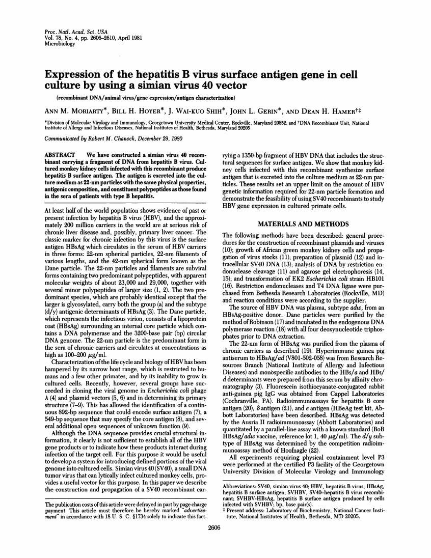
FIG. 1. Construction and analysis ofthe SV4O-(Upper) Step A. A 120-Iu reaction mixture containDNA and 1.4 j.g of plasmid pBR322 DNA was dilof BamHI for 2 hr at 37°C, heat inactivated at 71tracted with CHC13, precipitated with ethanol, rej
of ligation assay buffer, and treated with 4 units ofT4 DNA ligase for5 hr at 14°C. This mixture was used to transform E. coli HB101, andapBR322-HBV clone was identified by colony hybridization (23). DNAfrom this clone was cleaved with BamHI and the 1350 bp fragmentcontaining the HBsAg coding region was purified by preparative elec-trophoresis through a 0.8% agarose gel (15); subcloned in pBR322 andreisolated by electrophoresis. Step B. pBR322-SV40 was prepared, as
described (24), by cleavage with BamHI/EcoRI, treatment with ligase,and cloning in E. coli. Step C. pBR322-HBV was then ligated toBamHI-cleaved pBR322-SV40 DNA and cloned in E. coli as describedabove. Step D. Hae II cleavage of the resulting pBR322-SV4O-HBV"double recombinant" DNA generated the 4950-bp linear SVHBVDNA fragment that was used to infect monkey kidney cells. Cleavagesites: X, BamHI; e EcoRP; o,Hae II. (Lower) Ethidium bromide-stained1% agarose gel containing intracellular viral DNA from cells infected
with the SVHBV plus SV40tsA239 stock (lanes 1 and 2), plasmidpBR322-SV4O-HBV DNA (lane 3), and purified SV40 DNA (lanes 4and 5). The numbers above represent the lengths of linear molecules;the numbers below refer to the lengths of covalently closed circularDNAs. The uncleaved SVHBV plus SV40-tsA239 DNA (lane 2) showsa predominant band ofhelper DNA at 5200 bp, a band ofSVHBV DNAat 4750 bp, and a heterogeneous collection of-shorter DNAs; the moreslowly migrating bands represent nicked circular and host cell DNA.BamHI cleavage of this DNA (lane 1) generated a predominant 5200-bp band of helper DNA, a 3400-bp band of SV40 vector DNA, a 1350-bpband ofinserted HBVDNA that comigrated with its authentic coun-terpart from BamHI-cleaved pBR322-SV40-HBV DNA (lane 3), andseveral other bands of unknown origin.
Bam HI
+
Microbiology: Moriarty et al.
2608 Microbiology: Moriarty et al.
original subeloned 1350-bp fragment. We refer to this complexmixture of virus as "SVHBV."Monkey Kidney Cells Infected with SVHBV Synthesize
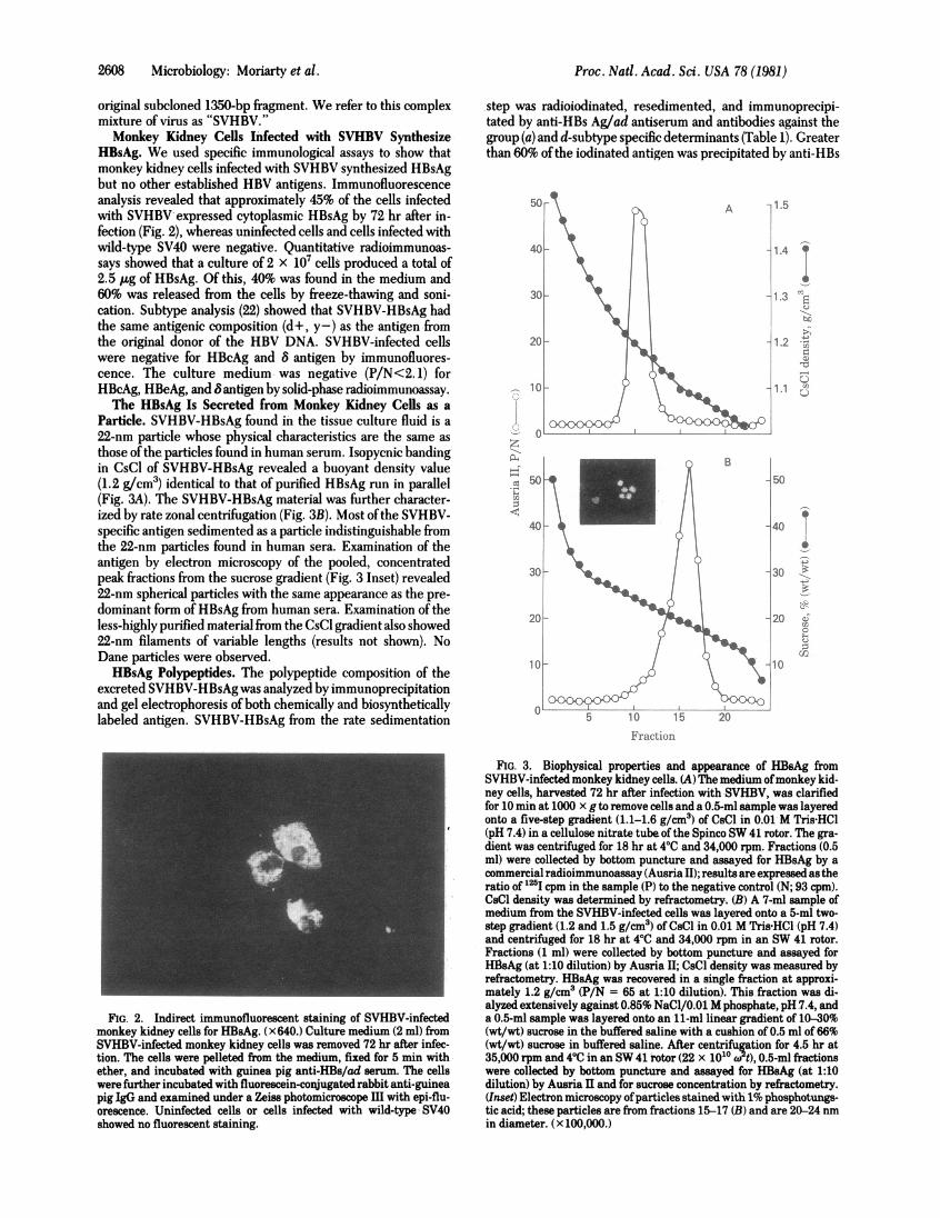
HBsAg. We used specific immunological assays to show thatmonkey kidney cells infected with SVHBV synthesized HBsAgbut no other established HBV antigens. Immunofluorescenceanalysis revealed that approximately 45% of the cells infectedwith SVHBV expressed cytoplasmic HBsAg by 72 hr after in-fection (Fig. 2), whereas uninfected cells and cells infected withwild-type SV40 were negative. Quantitative radioimmunoas-says showed that a culture of 2 x 10⁷ cells produced a total of2.5 ,ug of fBsAg. Of this, 40% was found in the medium and60% was released from the cells by freeze-thawing and soni-cation. Subtype analysis (22) showed that SVHBV-HBsAg hadthe same antigenic composition (d+, y -) as the antigen fromthe original donor of the HBV DNA. SVHBV-infected cellswere negative for HBcAg and 8 antigen by immunofluores-cence. The culture medium was negative (P/N<2. 1) forHBcAg, HBeAg, and B antigen by solid-phase radioimmunoassay.The HBsAg Is Secreted from Monkey Kidney Cells as a
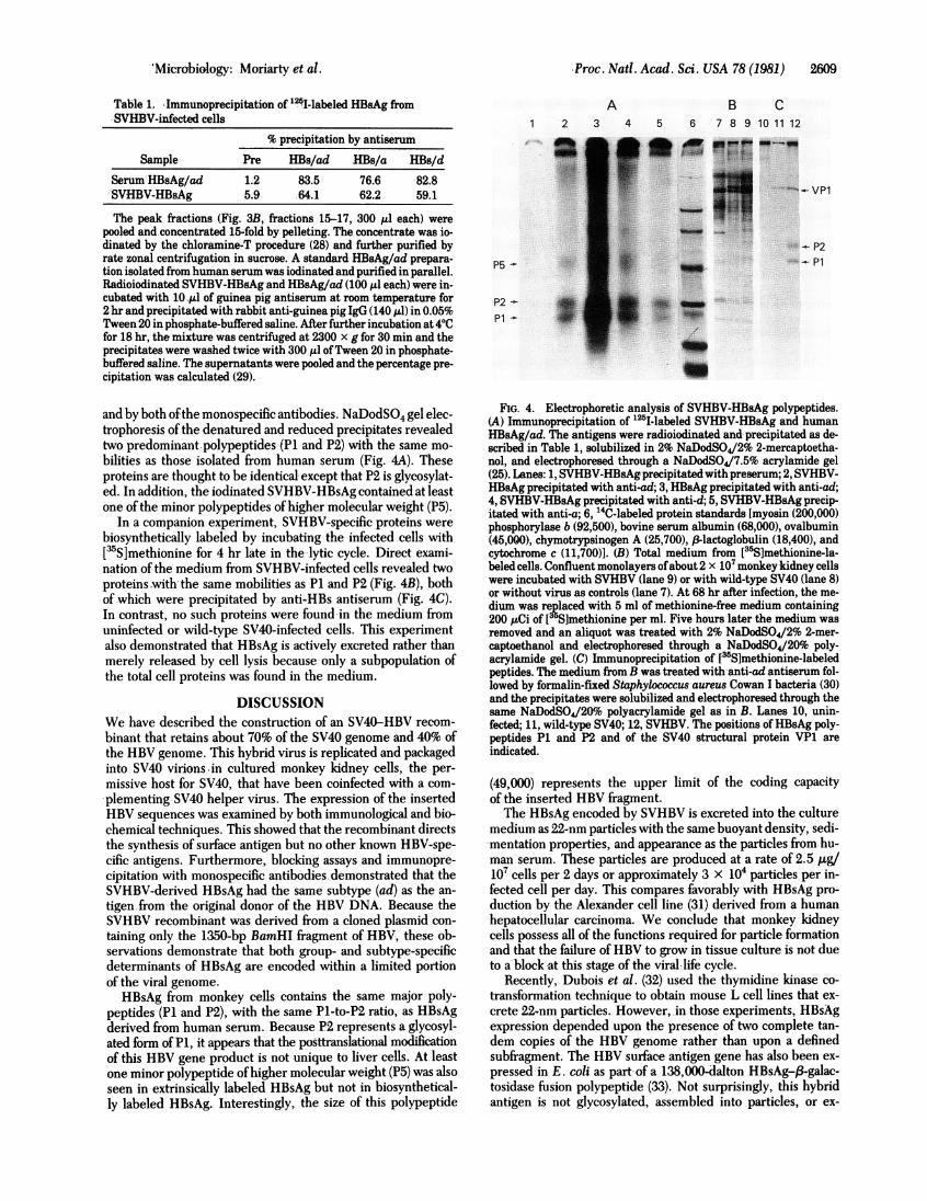
Particle. SVHBV-HBsAg found in the tissue culture fluid is a22-nm particle whose physical characteristics are the same asthose of the particles found in human serum. Isopycnic bandingin CsCl of SVHBV-HBsAg revealed a buoyant density value(1.2 g/cm3) identical to that of purified HBsAg run in parallel(Fig. 3A). The SVHBV-HBsAg material was further character-ized by rate zonal centrifugation (Fig. 3B). Most ofthe SVHBV-specific antigen sedimented as a particle indistinguishable fromthe 22-nm particles found in human sera. Examination of theantigen by electron microscopy of the pooled, concentratedpeak fractions from the sucrose gradient (Fig. 3 Inset) revealed22-nm spherical particles with the same appearance as the pre-dominant form of HBsAg from human sera. Examination of theless-highly purified material from the CsCl gradient also showed22-nm filaments of variable lengths (results not shown). NoDane particles were observed.HBsAg Polypeptides. The polypeptide composition of the
excreted SVHBV-HBsAg was analyzed by immunoprecipitationand gel electrophoresis of both chemically and biosyntheticallylabeled antigen. SVHBV-HBsAg from the rate sedimentation
step was radioiodinated, resedimented, and immunoprecipi-tated by anti-HBs Ag/ad antiserum and antibodies against thegroup (a) and d-subtype specific determinants (Table 1). Greaterthan 60% ofthe iodinated antigen was precipitated by anti-HBs
p1.5
.L.
z
1.4 t.i
1.3vX
I1.2 -Ii rim
-r_
1.1 :,-
50
Fraction
FIG. 2. Indirect immunofluorescent staining of SVHBV-infectedmonkey kidney cells for HBsAg. (x640.) Culture medium (2 ml) fromSVHBV-infected monkey kidney cells was removed 72 hr after infec-tion. The cells were pelleted from the medium, fixed for 5 min withether, and incubated with guinea pig anti-HBs/ad serum. The cellswere further incubated with fluorescein-conjugated rabbit anti-guineapig IgG and examined under a Zeiss photomicroscope III with epi-flu-orescence. Uninfected cells or cells infected with wild-type- SV40showed no fluorescent staining.
FIG. 3. Biophysical properties and appearance of HBsAg fromSVHBV-infected monkey kidney cells. (A) The medium ofmonkey kid-ney cells, harvested 72 hr after infection with SVHBV, was clarifiedfor 10 min at 1000 x g to remove cells and a 0.5-ml sample was layeredonto a five-step gradient (1.1-1.6 g/cm3) of CsCl in 0.01 M Tris HCl(pH 7.4) in a cellulose nitrate tube ofthe Spinco SW 41 rotor. The gra-dient was centrifuged for 18 hr at 40C and 34,000 rpm. Fractions (0.5ml) were collected by bottom puncture and assayed for HBsAg by acommercial radioimmunoassay (Ausria II); results are expressed as theratio of 125I cpm in the sample (P) to the negative control (N; 93 cpm).CsCl density was determined by refractometry. (B) A 7-ml sample ofmedium from the SVHBV-infected cells was layered onto a 5-ml two-step gradient (1.2 and 1.5 g/cm3) of CsCl in 0.01 M Tris HCl (pH 7.4)and centrifuged for 18 hr at 40C and 34,000 rpm in an SW 41 rotor.Fractions (1 ml) were collected by bottom puncture and assayed forHlsAg (at 1:10 dilution) by Ausria II; CsCl density was measured byrefractometry. HBsAg was recovered in a single fraction at approxi-mately 1.2 g/cm3 (P/N = 65 at 1:10 dilution). This fraction was di-alyzed extensively against 0.85% NaCl/0.01 M phosphate, pH 7.4, anda 0.5-ml sample was layered onto an 11-ml linear gradient of 10-30%(wt/wt) sucrose in the buffered saline with a cushion of 0.5 ml of 66%(wt/wt) sucrose in buffered saline. After centrifugation for 4.5 hr at35,000 rpm and 40C in an SW 41 rotor (22 x 1010 w t), 0.5-ml fractionswere collected by bottom puncture and assayed for HBsAg (at 1:10dilution) by Ausria II and for sucrose concentration by refractometry.(Inset) Electron microscopy ofparticles stained with 1% phosphotungs-tic acid; these particles are from fractions 15-17 (B) and are 20-24 nmin diameter. (x 100,000.)
Proc. Natl. Acad. Sci. USA 78 (1981)
!--
MProc. Natl. Acad. Sci. USA 78 (1981) 2609
Table 1. Immunoprecipitation of 1251-labeled HBsAg fromSVHBV-infected cells
% precipitation by antiserum
Sample Pre HBs/ad HBs/a HBs/d
Serum HBsAg/ad 1.2 83.5 76.6 82.8SVHBV-HBsAg 5.9 64.1 62.2 59.1
The peak fractions (Fig. 3B, fractions 15-17, 300 1d each) werepooled and concentrated 15-fold by pelleting. The concentrate was io-dinated by the chloramine-T procedure (28) and further purified byrate zonal centrifugation in sucrose. A standard HBsAg/ad prepara-tion isolated from human serum was iodinated and purified in parallel.Radioiodinated SVHBV-HBgAg and HBsAg/ad (100 ,ul each) were in-cubated with -10,Al of guinea pig antiserum at room temperature for2 hr and precipitated with rabbit anti-guinea pig IgG (140 Ad) in 0.05%Tween 20 in phosphate-buffered saline. After further incubation at 4VCfor 18 hr, the mixture was centrifuged at 2300 x g for 30 min and theprecipitates were washed twice with 300 tul ofTween 20 in phosphate-buffered saline. The supernatants were pooled and the percentage pre-cipitation was calculated (29).
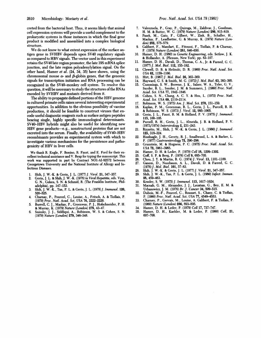
and by both ofthe monospecific antibodies. NaDodSO4 gel elec-trophoresis of the denatured and reduced precipitates revealedtwo predominant polypeptides (P1 and P2) with the same mo-bilities as those isolated from human serum (Fig. 4A). Theseproteins are thought to be identical except that P2 is glycosylat-ed. In addition, the iodinated SVHBV-HBsAg contained at leastone of the minor polypeptides of higher molecular weight (P5).
In a companion experiment, SVHBV-specific proteins werebiosynthetically labeled by incubating the infected cells with[3S]methionine for 4 hr late in the 'lytic cycle. Direct exami-nation of the medium from SVHBV-infected cells revealed twoproteins with the same mobilities as P1 and P2 (Fig. 4B), bothof which were precipitated by anti-HBs antiserum (Fig. 4C).In contrast, no such proteins were found in the medium fromuninfected or wild-type SV40-infected cells. This experimentalso demonstrated that HBsAg is actively excreted rather thanmerely released by cell lysis because only a subpopulation ofthe total cell proteins was found in the medium.
DISCUSSIONWe have described the construction of an SV40-HBV recom-binant that retains about 70% of the SV40 genome and 40% ofthe HBV genome. This hybrid virus is replicated and packagedinto SV40 virions.in cultured monkey kidney cells, the per-missive host for SV40, that have been coinfected with a com-plementing SV40 helper virus. The expression of the insertedHBV sequences was examined by both immunological and bio-chemical techniques. This showed that the recombinant directsthe synthesis of surface antigen but no other known HBV-spe-cific antigens. Furthermore, blocking assays and immunopre-cipitation with monospecific antibodies demonstrated that theSVHBV-derived HBsAg had the same subtype (ad) as the an-tigen .from the original donor of the HBV DNA. Because theSVHBV recombinant was derived from a cloned plasmid con-taining only the 1350-bp BamHI fragment of HBV, these ob-servations demonstrate that both group- and subtype-specificdeterminants of HBsAg are encoded within a limited portionof the viral genome.HBsAg from monkey cells contains the same major poly-
peptides (P1 and P2), with the same Pl-to-P2 ratio, as HBsAgderived from human serum. Because P2 represents a glycosyl-ated form ofP1, it appears that the posttranslational modificationof this HBV gene product is not unique to liver cells. At leastone minor polypeptide ofhigher molecular weight (P5) was alsoseen in extrinsically labeled HBsAg but not in biosynthetical-ly labeled HBsAg. Interestingly, the size of this polypeptide
A B C1 2 3 4 5 6 7 8 9 10 11 12
SS
P5- -
P1'P_
- VP1
P2P1
FIG. 4. Electrophoretic analysis of SVHBV-HBsAg polypeptides.(A) Immunoprecipitation of "25I-labeled SVHBV-HBsAg and humanHBsAg/ad. The antigens were radioiodinated and precipitated as de-scribed in Table 1, solubilized in 2% NaDodSO4/2% 2-mercaptoetha-nol, and electrophoresed through a NaDodSO4/7.5% acrylamide gel(25). Lanes: 1, SVHBV-HBsAg precipitated with preserum; 2, SVHBV-HBsAg precipitated with anti-ad; 3, HBsAg precipitated with anti-ad;4, SVHBV-HBsAg precipitated with anti-d; 5, SVHBV-HBsAg precip-itated with anti-a; 6, 14C-labeled protein standards [myosin (200,000)phosphorylase b (92,500), bovine serum albumin (68,000), ovalbumin(45,000), chymotrypsinogen A (25,700), 3-lactoglobulin (18,400), andcytochrome c (11,700)]. (B) Total medium from [35S]methionine-la-beled cells. Confluent monolayers ofabout 2 x 10⁷ monkey kidney cellswere incubated with SVHBV (lane 9) or with wild-type SV40 (lane 8)or without virus as controls (lane 7). At 68 hr after infection, the me-dium was replaced with 5 ml of methionine-free medium containing200 ,uCi of [35S]methionine per ml. Five hours later the medium wasremoved and an aliquot was treated with 2% NaDodSO4/2% 2-mer-captoethanol and electrophoresed through a NaDodSO4/20% poly-acrylamide gel. (C) Immunoprecipitation of [35S~methionine-labeledpeptides. The medium -from B was treated with anti-ad antiserum fol-lowed by formalin-fixed Staphylococcus aureus Cowan I bacteria (30)and the precipitates were solubilized and electrophoresed through thesame NaDodSO4/20o polyacrylamide gel as in B. Lanes 10, unin-fected; 11, wild-type SV40; 12, SVHBV. The positions ofHBsAg poly-peptides P1 and P2 and of the SV40 structural protein VP1 areindicated.
(49,000) represents the upper limit of the coding capacityof the inserted HBV fragment.The HBsAg encoded by SVHBV is excreted into the culture
medium as 22-nm particles with the same buoyant density, sedi-mentation properties, and appearance as the particles from hu-man serum. These particles are produced at a rate of 2.5 Ag/107 cells per 2 days or approximately 3 x 10⁴ particles per in-fected cell per day. This compares favorably with HBsAg pro-duction by the Alexander cell line (31) derived from a humanhepatocellular carcinoma. We conclude that monkey kidneycells possess all of the functions required for particle formationand that the failure of HBV to grow in tissue culture is not dueto a' block at this stage of the viral life cycle.
Recently, Dubois et al. (32) used the thymidine kinase co-transformation technique to obtain mouse L cell lines that ex-crete 22,nm particles. However, in those experiments, HBsAgexpression depended upon the presence of two complete tan-dem copies of the HBV genome rather than upon a definedsubfragment. The HBV surface antigen gene has also been ex-pressed in E. coli as part of a 138,000-dalton HBsAg-f3-galac-tosidase fusion polypeptide (33). Not surprisingly, this hybridantigen is not glycosylated, assembled into particles, or ex-
.'Micrabiology: Moriarty et al.
2610 Microbiology: Moriarty et al.
creted from the bacterial host. Thus, it seems likely that animalcell expression systems will provide a useful complement to theprokaryotic systems in those instances in which the final geneproduct is modified and assembled into a complex biologicalstructure.We do not know to what extent expression ofthe surface an-
tigen gene in SVHBV depends upon SV40 regulatory signalsas compared to HBV signals. The vector used in this experimentretains the SV40 late region promoter, the late 19S mRNA splicejunction, and the late region polyadenylylation signal. On theother hand, Hamer et al. (24, 34, 35) have shown, using thechromosomal mouse a- and 81-globin genes, that the genomicsignals for transcription initiation and RNA processing can berecognized in the SV4O-monkey cell system. To resolve thisquestion, it will be necessary to study the structures ofthe RNAsencoded by SVHBV andrmutants derived from it.The ability to propagate defined portions ofthe HBV genome
in cultured primate cells raises several interesting experimentalopportunities. In addition to the obvious possibility of vaccineproduction, it should be feasible to construct viruses that en-code useful diagnostic reagents such as surface antigen peptidesbearing single, highly specific immunological determinants.SV40-HBV hybrids might also be useful for identifying newHBV gene products-e.g., nonstructural proteins that are notexcreted into the serum. Finally, the availability of SV40-HBVrecombinants provides an experimental system with which toinvestigate various mechanisms for the persistence and patho-genicity of HBV in liver cells.We thank R. Engle, F. Bonino, R. Faust, and E. Ford for their ex-
cellent technical assistance and V. Reap for typing the manuscript. Thiswork was supported in part by Contract NO1-AI-82572 betweenGeorgetown University and the National Institute of Allergy and In-fectious Diseases.
1. Shih, J. W.-K. & Gerin, J. L. (1977) J. Virol. 21, 347-357.2. Gerin, J. L. & Shih, J. W.-K. (1978) in Viral Hepatitis, eds. Vyas,
G. N., Cohen, S. N. & Schmid, R. (The Franklin Institute, Phil-adelphia), pp. 147-153.
3. Shih, J. W.-K., Tan, P. L. & Gerin, J. L. (1978) J. Immunol. 120,520-525.
4. Charnay, P., Pourcel, C., Louise, A., Fritsch, A. & Tiollais, P.(1979) Proc. Natl. Acad. Sci. USA 76, 2222-2226.
5. Burrell, C. J., Mackay, P., Greenway, P. J., Hofschneider, P. H.& Murray, K. (1979) Nature (London) 279, 43-47.
6. Sninsky, J. J., Siddiqui, A., Robinson, W. S. & Cohen, S. N.(1979) Nature (London) 279, 346-3.
7. Valenzuela, P., Gray, P., Quiroga, M., Zaldivar, J., Goodman,H. M. & Rutter, W. G. (1979) Nature (London) 280, 815-819.
8. Pasek, M., Gato, F., Gilbert, W., Zink, B., Schaller, H.,Mackay, P., Leadbetter, G. & Murray, K. (1979) Nature (Lon-don) 282, 575-579.
9. Galibert, F., Mandart, E., Fitoussi, F., Tiollais, P. & Charnay,P. (1979) Nature (London) 281, 646-650.
10. Hamer, D. H. (1980) in Genetic Engineering, eds. Setlow, J. K.& Hollander, A. (Plenum, New York), pp. 83-107.
11. Hamer, D. H., Davali, D., Thomas, C. A., Jr. & Fareed, G. C.(1977)J. Mol. Biol. 112, 155-182.
12. Clewell, D. B. & Helinski, D. R. (1969) Proc. Nati. Acad. Sci.USA 62, 1159-1166.
13. Hirt, B. (1967) J. Mol. Biol. 26, 365-369.14. Hayward, G. S. & Smith, M. G. (1972)J. Mol. Biol. 63, 383-395.15. Cummings, S. W., Browne, J. K., Salser, W. A., Tyler, G. V.,
Snyder, R. L., Smolec, J. M. & Summers, J. (1980) Proc. Natl.Acad. Sci. USA 77, 1842-1846.
16. Cohen, S. N., Chang, A. C. Y. & Hsu, L. (1972) Proc. Nati.Acad. Sci. USA 69, 2110-2114.
17. Robinson, W. S. (1975) Am. J. Med. Sci. 270, 151-159.18. Kaplan, P. M., Greenman, R. L., Gerin, J. L., Purcell, R. H.
& Robinson, W. S. (1973)J. Virol. 12, 995-1005.19. Gerin, J. L., Faust, R. M. & Holland, P. V. (1975)J. Immunol.
115, 100-105.20. Purcell, R. H., Gerin, J. L., Almeida, J. B. & Holland, P. V.
(1973/1974) Intervirology 2, 231-243.21. Rizzetto, M., Shih, J. W.-K. & Gerin; J. L. (1980)J. Immunol.
125, 318-324.22. Hoofnagle, J. H., Gerety, R. J., Smallwood, L. A. & Barker, L.
F. (1977) Gastroenterology 72, 290-296.23. Grunstein, M. & Hogness, P. C. (1975) Proc. Natl. Acad. Sci.
USA 72, 3961-3965.24. Hamer, D. H. & Leder, P. (1979) Cell 18, 1299-1302.25. Goff, S. P. & Berg, P. (1976) Cell 9, 695-705.26. Chou, J. Y. & Martin, R. G. (1974)J. Virol. 13, 1101-1109.27. Ganem, D., Nussbaum, A. L., Davoli, D. & Fareed, G. C.
(1976)J. Mol. Biol. 101, 57-83.28. Shih, J. W.-K. & Gerin, J. L. (1977) J. Virol. 21, 347-357.29. Shih, J. W.-K., Tan, P. L. & Gerin, J. L. (1980) Infect. Immun.
28, 459-463.30. Kessler, S. W. (1975)J. Immunol. 115, 1617-1624.31. Macnab, G. M., Alexander, J. J., Lecatsas, G., Bey, E. M. &
Urbanowicz, J. M. (1976) Br. J. Cancer 34, 509-515.32. Dubois, M.-F., Pourcel, C., Rousset, S., Chany, C. & Tiollais,
P. (1980) Proc. Natl. Acad. Sci. USA 77, 4549-4553.33. Charney, P., Gervais, M., Louise, A. Galibert, F. & Tiollais, P.
(1980) Nature (London) 286, 893-895.34. Hamer, D. H. & Leder, P. (1979) Cell 17, 737-747.35. Hamerj D. H., Kaehler, M. & Leder, P. (1980) Cell, 21,
697-708.
Proc. Natl. Acad. Sci. USA 78'(1981)