extended report a homeostatic function of cxcr2 signalling
TRANSCRIPT
EXTENDED REPORT
A homeostatic function of CXCR2 signallingin articular cartilageJoanna Sherwood,1,2 Jessica Bertrand,2 Giovanna Nalesso,1 Blandine Poulet,3
Andrew Pitsillides,4 Laura Brandolini,5 Alexandra Karystinou,6 Cosimo De Bari,6
Frank P Luyten,7 Costantino Pitzalis,1 Thomas Pap,2 Francesco Dell’Accio1
Handling editor Tore K Kvien
▸ Additional material ispublished online only. To viewplease visit the journal online(http://dx.doi.org/10.1136/annrheumdis-2014-205546).
For numbered affiliations seeend of article.
Correspondence toProfessor Francesco Dell’Accio,Centre for ExperimentalMedicine and Rheumatology,William Harvey ResearchInstitute, Barts and the LondonSchool of Medicine andDentistry, Queen MaryUniversity of London, UK;[email protected]
TP and FDA shared co-seniorauthorship.
Received 11 March 2014Revised 12 June 2014Accepted 20 July 2014Published Online First18 August 2014
To cite: Sherwood J,Bertrand J, Nalesso G, et al.Ann Rheum Dis2015;74:2207–2215.
ABSTRACTObjective ELR+ CXC chemokines are heparin-bindingcytokines signalling through the CXCR1 and CXCR2receptors. ELR+ CXC chemokines have been associatedwith inflammatory arthritis due to their capacity toattract inflammatory cells. Here, we describe anunsuspected physiological function of these molecules inarticular cartilage homeostasis.Methods Chemokine receptors and ligands weredetected by immunohistochemistry, western blotting andRT-PCR. Osteoarthritis was induced in wild-type andCXCR2−/− mice by destabilisation of the medialmeniscus (DMM). CXCR1/2 signalling was inhibitedin vitro using blocking antibodies or siRNA. Chondrocytephenotype was analysed using Alcian blue staining,RT-PCR and western blotting. AKT phosphorylation andSOX9 expression were upregulated using constitutivelyactive AKT or SOX9 plasmids. Apoptosis was detected byterminal deoxynucleotidyl transferase dUTP nick endlabelling (TUNEL) assay.Results CXCL6 was expressed in healthy cartilage andwas retained through binding to heparan sulfateproteoglycans. CXCR2−/− mice developed more severeosteoarthritis than wild types following DMM, withincreased chondrocyte apoptosis. Disruption of CXCR1/2in human and CXCR2 signalling in mouse chondrocytesled to a decrease in extracellular matrix production,reduced expression of chondrocyte differentiationmarkers and increased chondrocyte apoptosis. CXCR2-dependent chondrocyte homeostasis was mediated byAKT signalling since forced expression of constitutivelyactive AKT rescued the expression of phenotypic markersand the apoptosis induced by CXCR2 blockade.Conclusions Our study demonstrates an importantphysiological role for CXCR1/2 signalling in maintainingcartilage homeostasis and suggests that the loss of ELR+CXC chemokines during cartilage breakdown inosteoarthritis contributes to the characteristic loss ofchondrocyte phenotypic stability.
INTRODUCTIONOsteoarthritis (OA) is a leading cause of chronicdisability, characterised by the breakdown of thearticular cartilage, for which we have no cure.Whereas in inflammatory arthritides inflammationis the main driver of tissue destruction, in OA,mechanical factors are the main drivers of cartilagebreakdown while a low degree of synovitis detectedonly in a subset of patients may have an ancillaryrole.1
Metalloproteinase and aggrecanase-mediatedextracellular matrix (ECM) degradation, and chon-drocyte apoptosis all contribute to cartilage break-down and are initially compensated by chondrocyteproliferation and upregulation of SOX9, which dir-ectly regulates the synthesis of major ECM compo-nents including aggrecan and type II collagen.2–4
When such compensatory mechanisms are impairedor insufficient, cartilage breakdown progresses andultimately leads to joint failure. Supporting thehomeostatic response of cartilage can slow down oreven revert cartilage degeneration in animalmodels.5 6
Enclosed within the cartilage matrix, chondro-cytes are not known to migrate in physiologicalconditions. In spite of this, however, chondrocytesexpress several chemokine receptors, includingCXCR1 and CXCR2 and their cognate ligands thathave been extensively studied in the context ofarthritis.7 8
ELR+ CXC chemokines are chemotactic cytokinescharacterised by their glutamic acid-leucine-arginine(ELR+) motif. The chemokine receptor CXCR2binds the human CXC chemokine ligands CXCL1,CXCL2, CXCL3, CXCL5, CXCL6, CXCL7 andCXCL8 while CXCR1 binds only to CXCL6and CXCL8.9 Mice lack Cxcl8 and express only onegene that shows homology to the human CXCL5and CXCL6 (hereafter referred to as mouseCXCL6). Although a putative murine homologue ofhuman CXCR1 has been identified,10 mouseCXCR2 is considered the main ELR+ CXC chemo-kine receptor because its function cannot be compen-sated in neutrophil chemotaxis and woundhealing.11–13 CXCR1 and CXCR2 activate intracellu-lar calcium and lead to activation of the Pi3K/AKTsignalling pathway.14 15
Although the biology and expression in vivo ofCXCR1 and CXCR2 in intact normal humanarticular cartilage has not been reported, theexpression of these receptors and various chemo-kines have been characterised in isolated arthriticchondrocytes.7 8 ELR+ CXC chemokines havebeen studied and targeted in inflammatory arthritisbecause of their capacity to attract inflammatorycells. CXCL8 was shown to stimulate, in vitro, theproduction of inflammatory mediators, metallopro-teinases and the induction of hypertrophy andcalcification.16
Here, we report a novel, unsuspected homeo-static role for CXCR1/2 signalling in the articularcartilage where ELR+ CXC chemokines are
Open AccessScan to access more
free content
Basic and translational research
Sherwood J, et al. Ann Rheum Dis 2015;74:2207–2215. doi:10.1136/annrheumdis-2014-205546 2207
on January 25, 2022 by guest. Protected by copyright.
http://ard.bmj.com
/A
nn Rheum
Dis: first published as 10.1136/annrheum
dis-2014-205546 on 18 August 2014. D
ownloaded from
on January 25, 2022 by guest. P
rotected by copyright.http://ard.bm
j.com/
Ann R
heum D
is: first published as 10.1136/annrheumdis-2014-205546 on 18 A
ugust 2014. Dow
nloaded from
retained in the ECM through binding to GAGs andcell-autonomously support chondrocyte viability and differenti-ation through AKT-dependent SOX9 expression. We suggestthat disruption of CXCR1/2 signalling is an important event inosteoarthritis, resulting in the loss of chondrocyte phenotypicstability and promoting OA-like changes.
MATERIALS AND METHODSMice000.651 BALB/cJ and C.129S2(B6)-Cxcr2tm1Mwm/J CXCR2(−/−) mice (BALB/cJ background) were obtained from TheJackson Laboratory and maintained in pathogen-free conditions.The Animal Use Committee for the University of Münster(Landesamt für Natur, Umwelt und Verbraucherschutz, approvalnumber 84-02.04.2012.A189) approved all mouse procedures.
Destabilisation of the medial meniscusTen-week-old BALB/C or CXCR2−/− male mice received desta-bilisation of the medial meniscus (DMM) and sham surgery tothe contralateral limb as described.17
After 8 weeks, mice were killed and the joints were processedas previously described.18 A minimum of five sections per jointwere stained using toluidine blue for histological analysis andOsteoarthritis Research Society International (OARSI) scoringfor osteoarthritis severity by two independent investigators.
Cartilage harvest, chondrocyte isolation and cultureFull-thickness human articular cartilage was obtained from thefemoral condyles of patients undergoing knee joint replacementsurgery (ethics approval from the East London & The CityEthics Committee 3). Normal articular cartilage was obtainedfrom postmortem and trauma surgery donors. Chondrocyteswere isolated and cultured as previously described.19 Mousecostal chondrocytes were obtained from the ribcages of BALB/Cwild-type and CXCR2−/− mice.
Micromass culture of primary chondrocytes or JJ012 cellsand quantification of glycosaminoglycan content were per-formed as described.20 The ATDC5 cell line was differentiatedin monolayer culture for 14 days in Dulbecco’s modified Eagle’smedium (DMEM) supplemented with 10 mg/mL human insulinas described21 before use.
CXCR blockade using blocking antibodiesChondrocyte culture medium was replaced with DMEM supple-mented with 1% heat-inactivated fetal bovine serum. After 24 h,CXCR1 and CXCR2 blocking antibodies (R&D systems) orisotype-matched negative control antibodies (Dako) were addedat a total concentration of 10 μg/mL. Chondrocytes were thencultured for 4 days before phenotypic analysis.
siRNA transfectionHuman CXCR1 and CXCR2, or mouse CXCR2, were knockeddown using Stealth siRNA (Life Technologies), used at a totalconcentration of 20 nM in complete DMEM using jetPRIMEtransfection reagent (Polyplus) according to the manufacturer’sinstructions. A Stealth RNAi negative control duplex of mediumguanine–cytosine (GC) content (Life Technologies) was used asa negative control.
Western blot analysisCell lysates were run on a 10% Tris-glycine gel (LifeTechnologies) and transferred onto nitrocellulose membrane(GE Healthcare). Primary antibodies used were rabbit anti-mousepAKT (ser473) (Cell Signaling) 1:200 dilution, rabbit anti-
mouse AKT (Cell Signaling) 1:500 dilution or rabbit anti-mouseCXCL6 (Biorbyt) 1:200 dilution in blocking solution at 4°Covernight. For more detailed protocols, see online supplemen-tary methods.
Immunofluorescence analysis of mouse and human cartilageHuman cartilage explant and decalcified mouse knee joint sec-tions were deparaffinised and the subsequent steps includingblocking and pepsin antigen retrieval were performed as previ-ously described.22 Human sections and monolayer cells wereanalysed using (anti-CXCR1, -CXCR2 or CXCL6 (R&D))primary antibodies followed by Cy2 conjugated goat anti-mouseIgG secondary antibodies ( Jackson ImmunoResearch). Mouseknee sections were incubated with rabbit anti-mouse -GCP2(Biorbyt), -CXCR2 (R&D), -Col X (Abcam) or anti-Col II(Chemicon), followed by chicken anti-rabbit Alexa Fluor 488 orchicken anti-rabbit Alexa Fluor 594 secondary antibodies (LifeTechnologies). Mouse isotype control IgG (Dako) or rabbit IgG(Abcam) were used as control primary antibodies.
Apoptosis was detected by terminal deoxynucleotidyl trans-ferase dUTP nick end labelling (TUNEL) assay (Roche) accord-ing to the manufacturer’s instruction.
Images were acquired at 22°C by either Leica DM5500 QConfocal microscope using 40× magnification/0.75 numericalaperture, or Olympus BX61 microscope with a fixed exposureusing either 10×/0.4 or 20×/0.7 objective lenses using Cell-P soft-ware. Images were enhanced using Adobe Photoshop for betterrendering without altering relationship of target to control images.
Cartilage explant digestionHip caps from 4-week-old BALB/C wild-type mice were decellu-larised by repeated freeze thawing. Hip caps were incubatedovernight at 37°C either in phosphate buffered saline (PBS) orin PBS containing 5 mU/mL heparitinase (Seikagaku). Total pro-teins were precipitated from supernatants using trichloroaceticacid and assessed by western blotting.
Total RNA extraction and real-time RT-PCRRNA extraction and gene expression analysis were performed aspreviously described,19 with additional primers found in onlinesupplementary table S1.
caAKT and SOX9 plasmid transfectionMouse primary chondrocytes and ATDC5 cells were transfectedin monolayer using jetPRIME (Polyplus) according to the manu-facturer’s instructions. caAKT (Addgene plasmid 10841)23 wasused to constitutively activate AKT signalling, and a SOX9plasmid was used to overexpress SOX9.24
Statistical analysisData are presented as means±SEM. According to data distribu-tion, parametric (Student t test) or non-parametric (Mann–Whitney) tests were performed using GraphPad Prism Software,V.5.0c (GraphPad Software Inc, San Diego, USA), with p<0.05determining the primary level of significance.
RESULTSCXCR1/2 and their ligand CXCL6 are expressed in adultarticular cartilageIn vitro, primary adult human articular chondrocytes (AHAC)expressed the CXC chemokine receptors CXCR1 and CXCR2at mRNA (figure 1A) and protein levels (figure 1B). We con-firmed the expression of CXCR1 and CXCR2 within nativehuman articular cartilage using immunohistochemistry showing
Basic and translational research
2208 Sherwood J, et al. Ann Rheum Dis 2015;74:2207–2215. doi:10.1136/annrheumdis-2014-205546
on January 25, 2022 by guest. Protected by copyright.
http://ard.bmj.com
/A
nn Rheum
Dis: first published as 10.1136/annrheum
dis-2014-205546 on 18 August 2014. D
ownloaded from

that CXCR1 and CXCR2 were expressed in normal and osteo-arthritic cartilage (figure 1C). CXCR2, the main functionalmurine ELR+ CXC chemokine receptor,12 13 was expressed innormal mouse articular cartilage (figure 1D).
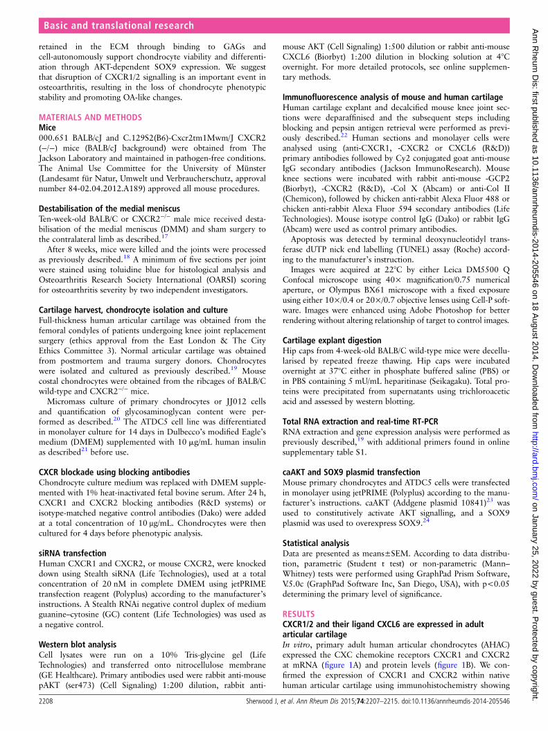
The CXCR1/2 ligand CXCL6 had a striking and specificexpression pattern in normal cartilage tissue, conserved acrossthe mouse and human species (figure 2A, B). In humans,CXCL6 was found within the chondrocyte territorial matrix ofarticular cartilage from healthy donors; however, it could nolonger be detected in the matrix of early osteoarthritic cartilage(figure 2A, B). This pattern was confirmed in the mouse, whereCXCL6 was present within the articular cartilage matrix ofunchallenged BALB/C mice, but was reduced 8 weeks followingDMM surgery (figure 2C, D). In contrast, CXCL8 in humanand CXCL1 in mouse were hardly detectable at protein level inhealthy or OA cartilage (see online supplementary figure S1).
If high levels of CXCL6 are produced by healthy chondro-cytes, what mechanism prevents them from attracting
inflammatory cells in physiological conditions? ELR+ CXC che-mokines are known to bind to heparan sulfate proteoglycans(HSPGs),25–27 therefore we hypothesised that binding to HSPGswithin the avascular cartilage could retain them within the ECMand avoid their availability to inflammatory cells. To establishwhether CXCL6 is retained within the cartilage ECM throughbinding to HSPGs, mouse cartilage explants were decellularisedthrough freeze-thawing and were incubated overnight in thepresence or absence of heparitinase. In keeping with its postu-lated binding to HSPGs, CXCL6 was subsequently retrievedfrom the supernatants of heparitinase digested explants but notfrom control undigested explants (figure 2E).
In vitro, in addition to CXCR1 and CXCR2 (figure 1),AHAC expressed CXCL6 (figure 2F) and CXCL8 (online sup-plementary figure 1D) mRNA at early passage, but not follow-ing serial passaging. Serial passaging is known to be associatedwith the loss of chondrocyte phenotypic markers—a processknown as ‘dedifferentiation’28–30—and of their capacity to form
Figure 1 ELR+ CXC chemokinereceptors are expressed in human andmouse articular cartilage. (A)Semi-quantitative RT-PCR for CXCR1and CXCR2 in human primarychondrocytes cultured in monolayer.(B) Confocal microscopic image ofimmunofluorescence staining forCXCR1 and CXCR2 (green) in humanprimary articular chondrocytes culturedin monolayer, with propidium iodide(red) staining the nuclei. Scale bar,20 μm. (C) Immunofluorescencestaining for CXCR1 and CXCR2 (green)in normal and osteoarthritic articularcartilage, with propidium iodide (red)staining the nuclei. Scale bar, 20 μm.(D) Immunofluorescence staining forCXCR2 (green) in wild type andCXCR2−/− mouse articular cartilage,with propidium iodide (red) stainingthe nuclei. Scale bar, 100 μm.
Basic and translational research
Sherwood J, et al. Ann Rheum Dis 2015;74:2207–2215. doi:10.1136/annrheumdis-2014-205546 2209
on January 25, 2022 by guest. Protected by copyright.
http://ard.bmj.com
/A
nn Rheum
Dis: first published as 10.1136/annrheum
dis-2014-205546 on 18 August 2014. D
ownloaded from
cartilage in vivo.28 Conversely, chondrogenic differentiation ofthe mouse chondrocytic cell line ATDC5 using insulin (figure2G) resulted in the upregulation of CXCL6 (figure 2H). Takentogether, these data suggested that ELR+ CXC chemokine sig-nalling may have a physiological function in chondrocytehomeostasis.
CXCR2 deficiency results in more severe osteoarthritisfollowing surgically induced joint instabilityTo investigate whether ELR+ CXC chemokine signalling has aphysiological function in vivo, we compared the outcome ofexperimental OA in CXCR2-deficient mice and wild-type con-trols following DMM surgery. CXCR2-deficient mice, which, at
the time of surgery did not display histological features of OAand expressed collagens type II and type X at levels comparableto wild-type controls (see online supplementary figure S2),exhibited increased OA-like cartilage breakdown inDMM-operated joints in comparison to wild-type controls(figure 3A, B). In keeping with OA features, CXCR2-deficientmice also displayed lower expression of collagen type II withinthe articular cartilage (figure 3C, D), accompanied by increasedexpression of the chondrocyte hypertrophy marker collagentype X (figure 3E, F). These results confirm that CXCR2 signal-ling supports articular cartilage homeostasis in vivo in condi-tions of challenge and demonstrate that its disruption isassociated with more severe OA.
Figure 2 CXCL6 is present in healthy articular cartilage and its expression is associated with chondrocyte differentiation. (A) Immunofluorescencestaining for CXCL6 (green) in normal and early osteoarthritis (moderate Mankin score) articular cartilage. Nuclei are stained using propidium iodide(red). Scale bar, 100 μm. (B) Densitometric quantification of CXCL6 staining (n=3). (C) Immunofluorescence staining for CXCL6 (red) in mousearticular cartilage of sham-operated control and destabilisation of the medial meniscus (DMM) operated mice, with 40,6-diamidino-2-phenylindolestaining the nuclei. Scale bar, 100 μm. (D) Densitometric quantification of CXCL6 staining (n=4). (E) Western blot analysis of CXCL6 release intosupernatant from vehicle control or heparitinase treated, freeze-thawed wild-type mouse hip caps. (F) Real-time RT-PCR for CXCL6 mRNA in earlyand late passage human articular chondrocytes (n=3), ***p<0.001 by paired t test. (G) Alcian blue staining and spectrophotometric quantificationof ATDC5 cell micromasses differentiated for 14 days using insulin (n=6). (H) Real-time RT-PCR quantification of CXCL6 mRNA expression in ATDC5cells following 14 days of culture in either control or insulin supplemented differentiation medium (n=6) **p<0.01, ****p<0.0001.
Basic and translational research
2210 Sherwood J, et al. Ann Rheum Dis 2015;74:2207–2215. doi:10.1136/annrheumdis-2014-205546
on January 25, 2022 by guest. Protected by copyright.
http://ard.bmj.com
/A
nn Rheum
Dis: first published as 10.1136/annrheum
dis-2014-205546 on 18 August 2014. D
ownloaded from

CXCR1/2 signalling is required for maintenance of articularchondrocyte phenotypic stability in a cell-autonomousfashionOA is a disease of the whole joint and several mechanisms, bothwithin and outside of cartilage, contribute to its pathogenesis.To investigate whether the homeostatic effects of CXCR2 signal-ling on chondrocytes are cell-autonomous, we investigatedwhether disruption of CXCR2 signalling in vitro in humanchondrocytes resulted in phenotypic changes. Specific simultan-eous inhibition of CXCR1 and CXCR2 using blocking anti-bodies in human primary chondrocytes (figure 4A), or bysiRNA in the JJ012 human chondrosarcoma cell line (figure4B), resulted in reduced ECM production in micromass cul-tures. Accordingly, treatment of human primary articular chon-drocytes with anti-CXCR1 and CXCR2 blocking antibodiesresulted in the downregulation of SOX9, COL2A1 and aggrecanmRNA in comparison to those treated with an isotype-matchedIgG negative control (figure 4C–E).
In keeping with these data, freshly isolated costal chondro-cytes from CXCR2-deficient mice displayed reduced sulfatedproteoglycan accumulation compared with wild-type controlswhen cultured in micromass (figure 4F) and expressed signifi-cantly less SOX9 and COL2A1 mRNA (figure 4G, H).
Therefore, cell-autonomous CXCR1/2 signalling in human, andCXCR2 signalling in mouse, are required for the maintenanceof chondrocyte phenotypic stability.
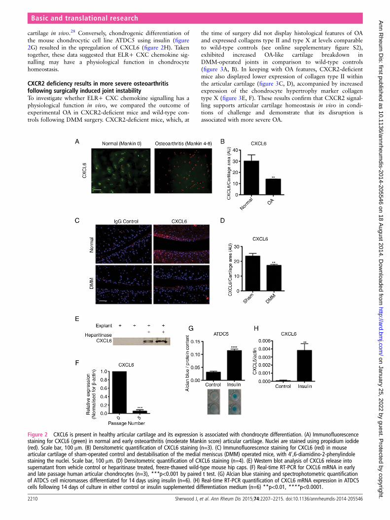
CXCR2-dependant modulation of the chondrocytephenotype is AKT dependentWe then asked the question of what molecular mechanism linksCXCR2 signalling to the maintenance of SOX9 expression andrelated phenotypic stability. It is known that AKT mediates bothchemotactic CXCR2 signalling in neutrophils31 32 and anabolicIGF-1 signalling in human chondrocytes.33 Therefore, we testedthe hypothesis that CXCR2 signalling supports SOX9 expres-sion via AKTactivation.
CXCL6 dose-dependently induced AKT phosphorylationin mouse primary chondrocytes (figure 5A). Less AKT phos-phorylation was detected by western blotting in cell lysatesobtained from CXCR2-deficient mouse costal chondrocytes incomparison to wild-type controls (figure 5B). In vivo, less phos-phorylated AKT was detected in the articular cartilage ofCXCR2-deficient mice compared with wild-type controls(figure 5C). In keeping with the decreased CXCL6 levels in OA,AKT phosphorylation was also decreased following DMM inwild-type mice (see online supplementary figure S3).
Figure 3 CXCR2 is required for articular cartilage homeostasis. (A) Toluidine blue staining for wild-type and CXCR2−/− mouse articular cartilage8 weeks following destabilisation of the medial meniscus (DMM) surgery. Scale bar, 100 μm. (B) Osteoarthritis Research Society International scoreof osteoarthritis-like changes and cartilage breakdown in BALB/C wild-type and CXCR2−/− mice 8 weeks following DMM surgery (n=10). MT, medialtibial plateau; MF, medial femoral head, LT, lateral tibial plateau; LF, lateral femoral head. Statistical comparison using Mann–Whitney U test. (C)Type II collagen immunofluorescence staining of wild-type and CXCR2−/− mouse articular cartilage 8 weeks following DMM. Nuclei arecounterstained with 40,6-diamidino-2-phenylindole (DAPI). Scale bar, 100 μm. (D) Densitometric quantification of type II collagen staining in articularcartilage following DMM (n=4). (E) Type X collagen immunofluorescence staining of wild-type and CXCR2−/− mouse articular cartilage 8 weeksfollowing DMM. Nuclei are counterstained with DAPI. Scale bar, 100 μm. (F) Densitometric quantification of ColX staining in articular cartilagefollowing DMM (n=4) *p<0.05, **p<0.01, ***p<0.001.
Basic and translational research
Sherwood J, et al. Ann Rheum Dis 2015;74:2207–2215. doi:10.1136/annrheumdis-2014-205546 2211
on January 25, 2022 by guest. Protected by copyright.
http://ard.bmj.com
/A
nn Rheum
Dis: first published as 10.1136/annrheum
dis-2014-205546 on 18 August 2014. D
ownloaded from

To assess whether CXCR2-induced AKT phosphorylation isrequired for chondrocyte differentiation or whether it is simplyassociated with it, we tested whether rescuing AKT activity alsorescued the differentiation of CXCR2-deficient chondrocytes.Chondrocytes from wild-type and CXCR2-deficient mice weretransfected with a constitutively active AKT (caAKT)-expressionplasmid or empty plasmid as control. As expected, Sox9 mRNAexpression was reduced in CXCR2-deficient chondrocytes com-pared with wild-type chondrocytes; however, SOX9 wasrescued to levels comparable to those of wild-type cells follow-ing transfection with caAKT (figure 5D). The same pattern wasobserved for type II collagen (figure 5E). Transfection ofCXCR2-deficient chondrocytes with a SOX9 expressingplasmid also resulted in the rescue of COL2A1 mRNA levels tolevels comparable to that of wild-type cells (figure 5F).
Similarly to CXCR2 deficiency, SOX9 deficiency in adult chon-drocytes does not result in spontaneous OA34 but makes chondro-cytes more prone to apoptosis,35 36 a process that has beendemonstrated to drive cartilage loss during osteoarthritis.37 38
Therefore, we tested whether CXCR2 disruption results in
increased chondrocyte apoptosis in an AKT-dependent manner.First, at 8 weeks following DMM, CXCR2-deficient mice dis-played significantly greater chondrocyte apoptosis withinthe superficial cartilage layers compared with wild-type mice(figure 6A, B). Although much lower than after DMM, thenumber of apoptotic cells in the superficial layer of control jointsof CXCR2-deficient mice was significantly higher than in wild-type controls (see online supplementary figure S4). Second,siRNA-mediated knockdown of CXCR2 in the chondrogenicATDC5 cells resulted in increased spontaneous apoptosis com-pared with scrambled siRNA control. Overexpression of caAKT,however, prevented the increase of apoptosis induced by the silen-cing of CXCR2 (figure 6C, D). Taken together, these data suggestthat CXCR2 signalling protects chondrocytes from apoptosis inconditions of challenge by supporting AKT phosphorylation andSOX9 expression.
DISCUSSIONIn this study, we discovered that CXCL6 is expressed by articu-lar chondrocytes in physiological conditions and is retained
Figure 4 Disruption of CXCR1/2 signalling results in chondrocyte de-differentiation and reduced extracellular matrix production in acell-autonomous fashion. (A) Alcian blue staining and spectrophotometric quantification for sulphated proteoglycan content of human articularchondrocyte micromass cultures 4 days following treatment with either CXCR1 and CXCR2 blocking antibodies or a non-specific IgG isotype control(n=4). (B) Spectrophotometric quantification of Alcian blue staining of JJ012 micromass cultures 4 days following CXCR1 and CXCR2 siRNAtransfection compared with scrambled siRNA-treated control (n=4). (C–E) Real-time RT-PCR analysis of chondrocyte phenotype marker genes SOX9,COL2A1 and aggrecan expression in human primary chondrocytes 4 days following treatment with CXCR1 and CXCR2 blocking antibodies incomparison to non-specific IgG isotype control (n=4). (F) Alcian blue staining and spectrophotometric quantification of wild-type and CXCR2−/−
mouse costal chondrocytes cultured for 7 days in micromass (n=8). (G, H) Real-time RT-PCR analysis of SOX9 and COL2A1 mRNA expression offreshly isolated costal chondrocytes from wild-type and CXCR2−/− mice (n=4). *p<0.05, ***p<0.001, ****p<0.0001.
Basic and translational research
2212 Sherwood J, et al. Ann Rheum Dis 2015;74:2207–2215. doi:10.1136/annrheumdis-2014-205546
on January 25, 2022 by guest. Protected by copyright.
http://ard.bmj.com
/A
nn Rheum
Dis: first published as 10.1136/annrheum
dis-2014-205546 on 18 August 2014. D
ownloaded from
locally in the cartilage matrix to contribute to the phenotypicstability and functional homeostasis by supporting SOX9 expres-sion in an AKT-dependent manner. Disruption of CXCR2 sig-nalling resulted, in vivo, in increased susceptibility toinstability-induced OA, and in vitro, in loss of differentiationmarkers and ECM production.
Although no significant infiltration of inflammatory cells wasdetected 8 weeks following DMM in either CXCR2−/− or wild-type mice (see online supplementary figure S5), this experimen-tal set-up does not allow us to assess whether additionalCXCR2 functions in cells other than chondrocytes contributedto the phenotype.
Our findings that CXCR1/2 signalling supports cartilagehomeostasis are not at odds with the well-established pathogenicrole of ELR+ CXC chemokines in arthritis.7 8 39–43 In physio-logical conditions, a tight regulation of their expression,together with their matrix binding through HSPGs, allows forthe restriction of their signalling domain to the avascular chon-drocyte pericellular matrix, away from the reach of inflamma-tory cells. In arthritis, ECM breakdown, together with theupregulation of multiple chemokines, including CXCL8,42
would result in excessive and ectopic activation of chemokine
signalling in the joint with pathological consequences, whilesimultaneously depriving chondrocytes of homeostatic local che-mokine signalling (figure 6E).
Interestingly, other chemokine families have been linked tophysiological and even developmental roles outside of inflam-mation including several developmental processes44 45 and thehomeostasis of the haematopoietic system.46 This suggests thatcompartmentalisation of chemokine signalling in specific tissuecontexts plays an important role in defining their function, andthat in specific situations, the disruption of such compartmental-isation, rather than the expression of the chemokines them-selves, may be pathogenic.
CXCR2-deficient mice did not develop spontaneous OA, buttheir phenotype was elicited after joint destabilisation. IfCXCR1/2 signalling supports SOX9 expression, why did wenot observe spontaneous osteoarthritis in CXCR2-deficientmice? In this respect it is interesting to notice that, althoughSOX9 is essential for embryonic chondrogenesis,4 its disrup-tion in adulthood did not result in spontaneous OA;34
however, its absence from differentiated chondrocytes madethem susceptible to apoptosis.35 36 Therefore, SOX9 is strictlyrequired for chondrogenesis, but, once chondrocytes are
Figure 5 CXCR2 modulation of the articular chondrocyte phenotype is mediated by AKT. (A) Western blot of phospho-AKT (ser473) in wild-typemouse chondrocytes following 30 min incubation with recombinant mouse CXCL6. (B) Western blot comparison of phospho-AKT in freshly isolatedchondrocytes from wild-type and CXCR2−/− mice. (C) Immunofluorescence staining for pAKT in mouse articular cartilage of unchallenged wild-typeand CXCR2−/− mice, nuclei are stained with 40,6-diamidino-2-phenylindole. Scale bar, 100 μm. (D, E) Real-time RT-PCR analysis of SOX9 andCOL2A1 mRNA expression of wild type and CXCR2−/− early passage mouse chondrocytes 24 h following transfection with either a control emptyplasmid or constitutively active AKT (caAKT) expressing plasmid. (F) Real-time RT-PCR analysis of COL2A1 mRNA expression of wild-type andCXCR2−/− mouse chondrocytes 24 h following transfection with either a control empty plasmid or a SOX9 expressing plasmid, *p<0.05, **p<0.01,***p<0.001.
Basic and translational research
Sherwood J, et al. Ann Rheum Dis 2015;74:2207–2215. doi:10.1136/annrheumdis-2014-205546 2213
on January 25, 2022 by guest. Protected by copyright.
http://ard.bmj.com
/A
nn Rheum
Dis: first published as 10.1136/annrheum
dis-2014-205546 on 18 August 2014. D
ownloaded from

differentiated, it becomes only required in conditions ofchallenge. A second consideration is that, since SOX9 is upre-gulated following cartilage damage,38 47 the baseline expres-sion of SOX9 is sufficient to support cartilage homeostasis inphysiological conditions, but is insufficient when, after cartil-age damage, SOX9 upregulation is required.
It is interesting to note that the baseline phosphorylation ofAKT was reduced upon inhibition of CXCR2 signalling. Since,in chondrocytes, AKT phosphorylation mediates IGF1 signal-ling, which is a potent homeostatic signal supporting chondro-cyte differentiation and ECM production,48 this suggests acertain level of interaction between these two signalling path-ways. The hierarchy of such interactions is yet to be determined.
The dual role of ELR+ CXC chemokines, homeostatic inhealthy cartilage and pathogenic in arthritis, represents animportant pharmacological challenge and yet an opportunity forthe development of targeted strategies for cytokine blockade thatpreserve homeostatic mechanisms, while efficiently targeting thesynovial and systemic compartments. The heterogeneity ofmechanisms of cartilage damage in different subsets of patients islikely to require personalised therapeutic intervention, addressingindividual disease mechanisms. We believe that further knowl-edge of three aspects of chemokine biology will be key to achiev-ing this therapeutic goal: first, the mechanism by which signallingdomains are defined and restricted; second, the role of ligandand receptor specificity in fine-tuning the regulation of chemo-kine signalling; and finally, the identification of suitable deliverysystems to target intervention to specific tissue compartments.
Author affiliations1Centre for Experimental Medicine and Rheumatology, William Harvey ResearchInstitute, Barts and the London School of Medicine and Dentistry, Queen MaryUniversity of London, London, UK2Institute of Experimental Musculoskeletal Medicine, University Hospital Muenster,Muenster, Germany3Division of Medicine, Centre for Rheumatology and Connective Tissue Disease, UCL,London, UK4Department of Veterinary Basic Sciences, Royal Veterinary College, University ofLondon, Royal College Street, London, UK5Dompé S.P.A., L’Aquila, Italy6Institute of Medical Sciences, University of Aberdeen, UK7Skeletal Biology and Engineering Research Center, KU Leuven, Belgium
Correction notice This article has been corrected since it was published OnlineFirst. The Acknowledgements and Funding sections have been corrected.
Acknowledgements We thank Professor Joel Block (Chicago) for providing theJJ012 cell line and Dr Simon Tew (Liverpool) for providing the SOX9 expressingplasmid.
Contributors FD, TP and JS incepted and designed the experiments, analysed thedata and prepared the manuscript. Experimental work was performed by JS, withcontributions from JB and AK. JB, GN, CDeB, FL and CP contributed to experimentaldesign and writing of the manuscript. JB provided human cartilage samples. LB, BPand AP contributed to the analysis of CXCR2 knockout mice.
Funding This work was funded by Arthritis Research UK (grants 17859, 17971,19654), INNOCHEM EU FP6 (grant LSHB-CT-2005-51867), MRC (MR/K013076/1)and the William Harvey Research Foundation.
Competing interests LB is an employee of Dompe S.P.A. Francesco Dell’Accio,CDeB and FPL are co-inventors in a patent application on the use of chemokines toenhance cartilage stability.
Provenance and peer review Not commissioned; externally peer reviewed.
Figure 6 Disruption of CXCR2 signalling results in increased chondrocyte apoptosis in an AKT-dependent manner. (A) Terminal deoxynucleotidyltransferase dUTP nick end labelling (TUNEL) staining of wild-type and CXCR2−/− articular cartilage 8 weeks following destabilisation of the medialmeniscus surgery. Scale bar, 100 μm. (B) Quantification of TUNEL-positive chondrocytes in superficial and deep zones of articular cartilage ofwild-type and CXCR2−/− mice (n=5). (C) TUNEL staining of monolayer differentiated ATDC5 24 h following co-transfection with either scrambledcontrol or CXCR2 siRNA along with either a control or caAKT expressing plasmid. Scale bar, 100 μm. (D) Quantification of TUNEL-positive ATDC5cells following siRNA and plasmid transfection (n=3) **p<0.01, ***p<0.001. (E) In healthy articular cartilage, CXCL6 is expressed by chondrocytesand retained within the extracellular matrix (ECM) by HSPGs where it is available and required for signalling via CXCR1 and CXCR2 on nearbychondrocytes for the maintenance of their phenotypic stability. During osteoarthritis, mechanical and inflammatory injury leads to the breakdown ofHSPGs within the ECM, leading to the release of CXCL6. This not only results in the release of CXCL6 from the articular cartilage, but disrupts thecell-autonomous ELR+ CXC chemokine signalling mechanism required for chondrocyte homeostasis.
Basic and translational research
2214 Sherwood J, et al. Ann Rheum Dis 2015;74:2207–2215. doi:10.1136/annrheumdis-2014-205546
on January 25, 2022 by guest. Protected by copyright.
http://ard.bmj.com
/A
nn Rheum
Dis: first published as 10.1136/annrheum
dis-2014-205546 on 18 August 2014. D
ownloaded from
Open Access This is an Open Access article distributed in accordance with theterms of the Creative Commons Attribution (CC BY 3.0) license, which permitsothers to distribute, remix, adapt and build upon this work, for commercial use,provided the original work is properly cited. See: http://creativecommons.org/licenses/by/3.0/
REFERENCES1 Goldring MB, Otero M. Inflammation in osteoarthritis. Curr Opin Rheumatol
2011;23:471–8.2 Han Y, Lefebvre V. L-Sox5 and Sox6 drive expression of the aggrecan gene in
cartilage by securing binding of Sox9 to a far-upstream enhancer. Mol Cell Biol2008;28:4999–5013.
3 Lefebvre V, Huang W, Harley VR, et al. SOX9 is a potent activator of thechondrocyte-specific enhancer of the pro alpha1(II) collagen gene. Mol Cell Biol1997;17:2336–46.
4 Bi W, Deng JM, Zhang Z, et al. Sox9 is required for cartilage formation. Nat Genet1999;22:85–9.
5 Sampson ER, Hilton MJ, Tian Y, et al. Teriparatide as a chondroregenerative therapyfor injury-induced osteoarthritis. SciTranslMed 2011;3:101ra93.
6 Johnson K, Zhu S, Tremblay MS, et al. A stem cell-based approach to cartilagerepair. Science 2012;336:717–21.
7 Coelho FM, Pinho V, Amaral FA, et al. The chemokine receptors CXCR1/CXCR2modulate antigen-induced arthritis by regulating adhesion of neutrophils to thesynovial microvasculature. Arthritis Rheum 2008;58:2329–37.
8 Borzì RM, Mazzetti I, Cattini L, et al. Human chondrocytes express functionalchemokine receptors and release matrix-degrading enzymes in response to C-X-Cand C-C chemokines. Arthritis Rheum 2000;43:1734–41.
9 Murphy PM, Baggiolini M, Charo IF, et al. International union of pharmacology.XXII. Nomenclature for chemokine receptors. Pharmacol Rev 2000;52:145–76.http://www.ncbi.nlm.nih.gov/pubmed/10699158 (accessed 11 Sep 2013).
10 Fan X, Patera AC, Pong-Kennedy A, et al. Murine CXCR1 is a functional receptorfor GCP-2/CXCL6 and interleukin-8/CXCL8. JBiolChem 2007;282:11658–66.
11 Devalaraja RM, Nanney LB, Du J, et al. Delayed wound healing in CXCR2 knockoutmice. J Invest Dermatol 2000;115:234–44.
12 Cacalano G, Lee J, Kikly K, et al. Neutrophil and B cell expansion in mice that lackthe murine IL-8 receptor homolog. Science 1994;265:682–4. http://www.ncbi.nlm.nih.gov/pubmed/8036519 (accessed 11 Sep 2013).
13 Lee J, Cacalano G, Camerato T, et al. Chemokine binding and activities mediated bythe mouse IL-8 receptor. J Immunol 1995;155:2158–64.
14 Thelen M. Dancing to the tune of chemokines. Nat Immunol 2001;2:129–34.15 Bacon K, Baggiolini M, Broxmeyer H, et al. Chemokine/chemokine receptor
nomenclature. J Interferon Cytokine Res 2002;22:1067–8.16 Merz D, Liu R, Johnson K, et al. IL-8/CXCL8 and growth-related oncogene alpha/
CXCL1 induce chondrocyte hypertrophic differentiation. J Immunol 2003;171:4406–15.
17 Glasson SS, Blanchet TJ, Morris EA. The surgical destabilization of the medialmeniscus (DMM) model of osteoarthritis in the 129/SvEv mouse. Osteoarthr Cartil2007;15:1061–9.
18 Eltawil NM, De Bari C, Achan P, et al. A novel in vivo murine model of cartilageregeneration. Age and strain-dependent outcome after joint surface injury.Osteoarthr Cartil 2009;17:695–704.
19 Nalesso G, Sherwood J, Bertrand J, et al. WNT-3A modulates articular chondrocytephenotype by activating both canonical and noncanonical pathways. J Cell Biol2011;193:551–64.
20 De Bari C, Dell’Accio F, Luyten FP. Human periosteum-derived cells maintainphenotypic stability and chondrogenic potential throughout expansion regardless ofdonor age. Arthritis Rheum 2001;44:85–95.
21 Muramatsu S, Wakabayashi M, Ohno T, et al. Functional gene screening systemidentified TRPV4 as a regulator of chondrogenic differentiation. J Biol Chem2007;282:32158–67.
22 Dell’accio F, De Bari C, Eltawil NM, et al. Identification of the molecular response ofarticular cartilage to injury, by microarray screening: Wnt-16 expression andsignaling after injury and in osteoarthritis. Arthritis Rheum 2008;58:1410–21.
23 Takeuchi F. Akt, a pleckstrin homology domain containing kinase, is activatedprimarily by phosphorylation. J Biol Chem 1996;271:21920–6.
24 Ikeda T, Kamekura S, Mabuchi A, et al. The combination of SOX5, SOX6, and SOX9(the SOX trio) provides signals sufficient for induction of permanent cartilage.Arthritis Rheum 2004;50:3561–73.
25 Middleton J, Neil S, Wintle J, et al. Transcytosis and surface presentation of IL-8 byvenular endothelial cells. Cell 1997;91:385–95.
26 Parish CR. The role of heparan sulphate in inflammation. Nat Rev Immunol2006;6:633–43.
27 Webb LM, Ehrengruber MU, Clark-Lewis I, et al. Binding to heparan sulfate orheparin enhances neutrophil responses to interleukin 8. Proc Natl Acad Sci USA1993;90:7158–62.
28 Dell’Accio F, De Bari C, Luyten FP. Molecular markers predictive of the capacity ofexpanded human articular chondrocytes to form stable cartilage in vivo. ArthritisRheum 2001;44:1608–19.
29 Dell’Accio F, Bari CD, Luyten FP, et al. Microenvironment and phenotypic stabilityspecify tissue formation by human articular cartilage-derived cells in vivo. Exp CellRes 2003;287:16–27.
30 Benya PD, Shaffer JD. Dedifferentiated chondrocytes reexpress the differentiatedcollagen phenotype when cultured in agarose gels. Cell 1982;30:215–24.
31 Lane HC, Anand AR, Ganju RK. Cbl and Akt regulate CXCL8-induced and CXCR1-and CXCR2-mediated chemotaxis. Int Immunol 2006;18:1315–25.
32 Lindemann O, Umlauf D, Frank S, et al. TRPC6 regulates CXCR2-mediatedchemotaxis of murine neutrophils. J Immunol 2013;190:5496–505.
33 Yin W, Park JI, Loeser RF. Oxidative stress inhibits insulin-like growth factor-Iinduction of chondrocyte proteoglycan synthesis through differential regulation ofphosphatidylinositol 3-Kinase-Akt and MEK-ERK MAPK signaling pathways.J BiolChem 2009;284:31972–81.
34 Henry SP, Liang S, Akdemir KC, et al. The postnatal role of Sox9 in cartilage.J bone Miner Res Off J Am Soc Bone Miner Res 2012;27:2511–25.
35 Dy P, Wang W, Bhattaram P, et al. Sox9 Directs Hypertrophic Maturation andBlocks Osteoblast Differentiation of Growth Plate Chondrocytes. Dev Cell2012;22:597–609.
36 Ikegami D, Akiyama H, Suzuki A, et al. Sox9 sustains chondrocyte survival andhypertrophy in part through Pik3ca-Akt pathways. Development 2011;138:1507–19.
37 Thomas CM, Fuller CJ, Whittles CE, et al. Chondrocyte death by apoptosis isassociated with cartilage matrix degradation. Osteoarthr Cartil 2007;15:27–34.
38 Aigner T, Fundel K, Saas J, et al. Large-scale gene expression profiling reveals majorpathogenetic pathways of cartilage degeneration in osteoarthritis. Arthritis Rheum2006;54:3533–44.
39 Jacobs JP, Ortiz-Lopez A, Campbell JJ, et al. Deficiency of CXCR2, but not otherchemokine receptors, attenuates autoantibody-mediated arthritis in a murine model.Arthritis Rheum 2010;62:1921–32.
40 Haringman JJ, Tak PP. Chemokine blockade: a new era in the treatment ofrheumatoid arthritis? Arthritis Res Ther 2004;6:93–7.
41 Barsante MM, Cunha TM, Allegretti M, et al. Blockade of the chemokine receptorCXCR2 ameliorates adjuvant-induced arthritis in rats. Br J Pharmacol2008;153:992–1002.
42 Borzi RM, Mazzetti I, Macor S, et al. Flow cytometric analysis of intracellularchemokines in chondrocytes in vivo: constitutive expression and enhancement inosteoarthritis and rheumatoid arthritis. FEBS Lett 1999;455:238–42.
43 Endo H, Akahoshi T, Nishimura A, et al. Experimental arthritis induced by continuousinfusion of IL-8 into rabbit knee joints. Clin Exp Immunol 1994;96:31–5.
44 Longobardi L, Li T, Myers TJ, et al. TGF-β type II receptor/MCP-5 axis: at thecrossroad between joint and growth plate development. Dev Cell 2012;23:71–81.
45 Kunwar PS, Siekhaus DE, Lehmann R. In vivo migration: a germ cell perspective.Annu Rev Cell Dev Biol 2006;22:237–65.
46 Heissig B, Hattori K, Dias S, et al. Recruitment of stem and progenitor cells from thebone marrow niche requires mmp-9 mediated release of kit-ligand. Cell2002;109:625–37.
47 Sato T, Konomi K, Yamasaki S, et al. Comparative analysis of gene expressionprofiles in intact and damaged regions of human osteoarthritic cartilage. ArthritisRheum 2006;54:808–17.
48 Cravero JD, Carlson CS, Im H-J, et al. Increased expression of the Akt/PKB inhibitorTRB3 in osteoarthritic chondrocytes inhibits insulin-like growth factor 1-mediatedcell survival and proteoglycan synthesis. Arthritis Rheum 2009;60:492–500.
Basic and translational research
Sherwood J, et al. Ann Rheum Dis 2015;74:2207–2215. doi:10.1136/annrheumdis-2014-205546 2215
on January 25, 2022 by guest. Protected by copyright.
http://ard.bmj.com
/A
nn Rheum
Dis: first published as 10.1136/annrheum
dis-2014-205546 on 18 August 2014. D
ownloaded from

Supplementary Methods
Western blot analysis
Cells were lysed in RIPA buffer containing PhosSTOP protease inhibitor
cocktail tablets (Roche) and Complete Mini EDTA–free phosphatase inhibitor
cocktail tablets (Roche). 30μl of total cell lysates were run on a 10% Tris-glycine
gel (Life Technologies) and transferred onto nitrocellulose membrane (GE
Healthcare). Membranes were blocked for 3 hours in 5% BSA, 0.1% Tween20
PBS solution, then treated with rabbit anti-mouse pAKT (ser473) (Cell Signaling)
1:200 dilution, rabbit anti-mouse AKT (Cell Signaling) 1:500 dilution, or rabbit
anti-mouse CXCL6 (Biorbyt) 1:200 dilution in blocking solution at 4°C overnight.
Protein bands were detected using horseradish peroxidase conjugated
secondary antibodies and chemiluminescent substrates (ECL Western Blotting
Detection Reagents, Amersham).
Immunohistochemical analysis of mouse cartilage
Decalcified mouse knee joint sections were deparaffinized and
dehydrated in xylene and 100% ethanol. Sections were blocked in 10% H2O2 in
methanol, washed in PBS and digested in trypsin for 10 minutes. After further
washing, sections were blocked in 10% horse serum in PBS for 10 minutes. The
sections were incubated at 4°C overnight with Ly6B.2 primary antibody (AbD
Serotec), 1:2000 dilution. After washing in PBS, sections were incubated for 30
minutes at room temperature with biotinylated goat anti-rat IgG (Vector
Laboratories), 1:200 dilution, washed again and incubated for 30 minutes at
room temperature with the Vectastain ABC peroxidase complex (Vector
Laboratories) according to the manufacturer’s instructions. After further
washing, the staining was developed using DAB substrate (Vector Laboratories)
according to the manufacturer’s instructions and slides were mounted in DPX.

Additional immunofluorescence staining of human and mouse
articular cartilage
Human cartilage paraffin sections were deparaffinised and stained as
described in the Materials and Methods sections of the manuscript, using the
following antibodies. CXCL8 staining was performed using mouse anti-human
CXCL8 primary antibody (R&D), followed by Cy2 conjugated goat anti-mouse IgG
secondary antibody (Jackson ImmunoResearch). Mouse CXCL1 staining was
performed using rat anti-mouse CXCL1 primary antibody (R&D), followed by
Alexa Fluor 488 goat anti-rat IgG secondary antibody (Life Technologies).
Safranin Orange staining
Human or mouse cartilage paraffin sections were deparaffinised as described in
Materials and Methods. Sections were incubated with 0.2% Safranin Orange in
acetate buffer for 13 minutes at room temperature, washed in distilled water,
100% ethanol and xylene and mounted in DPX.

Supplementary figures
Supplementary Figure 1. (A) Immunofluorescence staining comparison of CXCL8
(green) in healthy cartilage and preserved areas of OA cartilage. Nuclei are stained using
propidium iodide. Scale bar, 100µm. (B) Safranin orange staining of healthy articular
cartilage and preserved areas of OA articular cartilage. Scale bar, 100µm. (C)
Immunofluorescence staining of mouse CXCL1 (green) in articular cartilage from sham
and DMM operated mouse articular cartilage. Nuclei are stained using DAPI (blue).
Scale bar, 100µm. (D) Real time RT-PCR for CXCL8 mRNA in early and late passage
human articular chondrocytes (n = 3), **P < 0.01 by paired t-test.
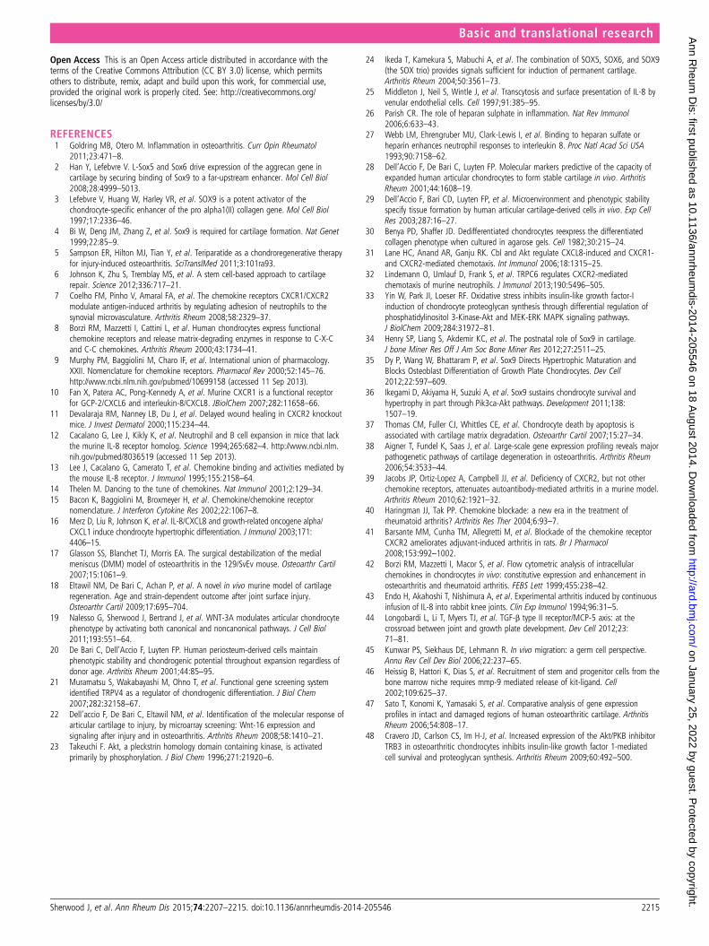
Supplementary Figure 2. (A) Histological comparison of 10 week old CXCR2- /- mouse
articular cartilage to wild type controls stained using Safranin orange. Scale bar, 200µm.
(B, C) Immunofluorescence staining and quantification of type II collagen and type X
collagen (green) in unchallenged wild type and CXCR2-/- articular cartilage. Nuclei are
stained using DAPI (blue). Scale bar, 100µm.
Supplementary Figure 3. Immunofluorescence staining of phospho-AKT (473) (red) in
wild type sham operated control and DMM operated knees 8 weeks following surgery.
Nuclei are stained using DAPI (blue). Scale bar, 100µm.
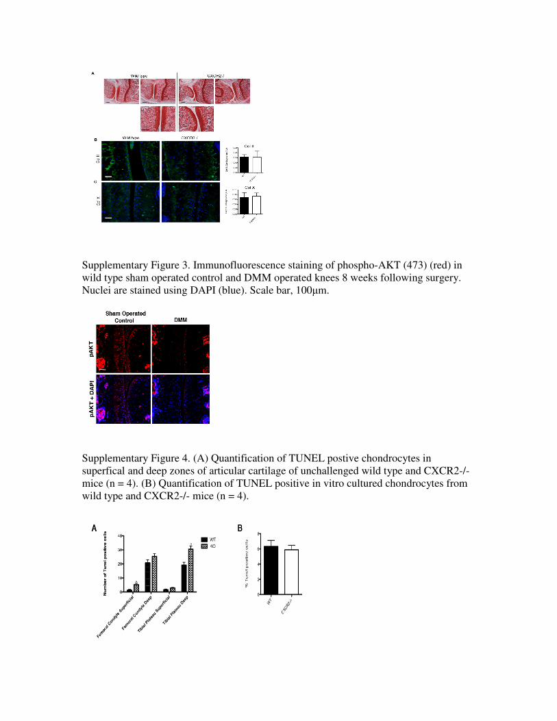
Supplementary Figure 4. (A) Quantification of TUNEL postive chondrocytes in
superfical and deep zones of articular cartilage of unchallenged wild type and CXCR2-/-
mice (n = 4). (B) Quantification of TUNEL positive in vitro cultured chondrocytes from
wild type and CXCR2-/- mice (n = 4).

Supplementary Figure 5. (A) Immunohistochemical staining for Ly-6B.2 in hind paws of
hTNFtg mutant mice. Scale bar, 100µm (B) Immunohistochemical staining for Ly-6B.2
in knee joints of wild type and CXCR2-/- mice 8 weeks following DMM surgery. Scale
bar, 100µm.