extended report autophagy is activated in systemic...
TRANSCRIPT
EXTENDED REPORT
Autophagy is activated in systemic lupuserythematosus and required for plasmablastdevelopmentAlexander J Clarke,1 Ursula Ellinghaus,1 Andrea Cortini,1 Amanda Stranks,2
Anna Katharina Simon,2 Marina Botto,3 Timothy J Vyse1
Handling editor Tore K Kvien
▸ Additional material ispublished online only. To viewplease visit the journal online(http://dx.doi.org/10.1136/annrheumdis-2013-204343).1Medical and MolecularGenetics and Division ofImmunology, Infection, andInflammatory Disease, King’sCollege London, London, UK2Nuffield Department ofClinical Medicine andTranslational ImmunologyLaboratory, NIHR BRC,University of Oxford, Oxford,UK3Department of Medicine,Centre for Complement andInflammation Research,Imperial College London,London, UK
Correspondence toProfessor Timothy J Vyse,Medical and MolecularGenetics and Division ofImmunology, Infection, andInflammatory Disease, King’sCollege London, 7th Floor,Tower Wing, Guy’s Hospital,Great Maze Pond,London SE1 9RT, UK;[email protected]
Received 24 July 2013Revised 30 November 2013Accepted 15 December 2013Published Online First13 January 2014
To cite: Clarke AJ,Ellinghaus U, Cortini A,et al. Ann Rheum Dis2015;74:912–920.
ABSTRACTBackground Autophagy has emerged as a criticalhomeostatic mechanism in T lymphocytes, influencingproliferation and differentiation. Autophagy in B cells hasbeen less studied, but genetic deficiency causesimpairment of early and late developmental stagesObjectives To explore the role of autophagy in thepathogenesis of human and murine lupus, a disease inwhich B cells are critical effectors of pathology.Methods Autophagy was assessed using multipletechniques in NZB/W and control mice, and in patientswith systemic lupus erythematosus (SLE) compared tohealthy controls. We evaluated the phenotype of theB cell compartment in Vav-Atg7−/− mice in vivo, andexamined human and murine plasmablast formationfollowing inhibition of autophagy.Results We found activation of autophagy in earlydevelopmental and transitional stages of B celldevelopment in a lupus mouse model even beforedisease onset, and which progressively increased withage. In human disease, again autophagy was activatedcompared with healthy controls, principally in naïve Bcells. B cells isolated from Vav-Atg7F/F mice failed toeffectively differentiate into plasma cells followingstimulation in vitro. Similarly, human B cells stimulatedin the presence of autophagy inhibition did notdifferentiate into plasmablasts.Conclusions Our data suggest activation of autophagyis a mechanism for survival of autoreactive B cells, andalso demonstrate that it is required for plasmablastdifferentiation, processes that induce significant cellularstress. The implication of autophagy in two majorpathogenic pathways in SLE suggests the potential touse inhibition of autophagy as a novel treatment targetin this frequently severe autoimmune disease.
INTRODUCTIONAutophagy is a highly conserved mechanism for thesurvival of cells during times of metabolic stress.Autophagosomes form at points of contact betweenthe endoplasmic reticulum (ER) and mitochondria,and two ubiquitin-like conjugation systems (Atg12and Atg8/LC3) extend a double-membraned autop-hagosome to sequester a portion of cytoplasm,which then fuses with the lysosome degradationpathway.1 2
Its role as a key factor in lymphocyte biology hasemerged only over the last few years.3 4 Autophagyis required for the normal development of B and Tlymphocyte populations,5–8 provides metabolic
support to proliferating lymphocytes,9 10 and isactivated on B cell receptor (BCR) signalling in theabsence of co-stimulation.11 12
Systemic lupus erythematosus (SLE) is a poten-tially fatal autoimmune disease characterised byfailure of multiple tolerance checkpoints, leading tothe escape and proliferation of autoreactive Bcells,11 13 through mechanisms which are poorlyunderstood.Indirect evidence for the role of autophagy in
SLE comes from genetic association studies impli-cating variants in the region of the autophagy geneATG5,14 15 which affect its expression in B lym-phocytes,16 17 and a therapeutic benefit in MRL/lprmice from the peptide P140, which modulatesautophagy.3 4
To date, autophagy in SLE T cells has been exam-ined and found to be activated in the naïve CD4+
T cell compartment by Alessandri et al,5–8 and thetotal CD4+ population by Gros et al.9 10 However,autophagy in B cells, which are of fundamentalimportance in SLE, the hallmark of which is theproduction of pathogenic high affinity autoanti-bodies,11 12 has not yet been studied in humanautoimmune disease.In this study, we demonstrate enhanced autop-
hagy in murine and human lupus B cells, and thatautophagy is required for B cell survival and matur-ation. Intervention in autophagy provides a poten-tially new therapeutic avenue for SLE.
MATERIALS AND METHODSMiceFemale NZB/WF1 (NZB/W) and C57BL/6 (B6)mice were obtained from Charles River, UK.Atg7Flox/Flox mice (a kind gift from MaasakiKomatsu, Tokyo Metropolitan Institute of MedicalScience, Japan) were crossed with Vav-iCre (a kindgift from D Kioussis, London, UK) to obtainVav-iCre; Atg7Flox/Flox.11 13
PatientsA total of 43 consecutive patients meeting therevised American College of Rheumatology (ACR)criteria for the definition of SLE14 15 were recruitedfrom hospitals of King’s Health Partners followinginformed consent and with ethical approval. Themean age of the patients was 36, with a male:female (M:F) ratio of 0.12. Patients were excludedif they had received rituximab therapy within thelast year, or intravenous methylprednisolone within
Open AccessScan to access more
free content
Basic and translational research
912 Clarke AJ, et al. Ann Rheum Dis 2015;74:912–920. doi:10.1136/annrheumdis-2013-204343
on 1 July 2018 by guest. Protected by copyright.
http://ard.bmj.com
/A
nn Rheum
Dis: first published as 10.1136/annrheum
dis-2013-204343 on 13 January 2014. Dow
nloaded from
the preceding month. SELENA-SLEDAI scores were calcu-lated.18 Healthy controls were recruited from King’s CollegeLondon. The mean age of the controls was 33 years, with anM:F ratio of 0.18. We found no statistical association betweenage or sex and autophagy measures in the control group. Patientcharacteristics are listed in online supplementary table S1.
Cell isolation and cultureHuman peripheral blood mononuclear cells were separatedfrom whole blood using Ficoll density centrifugation. CD19+ Bcells were isolated using negative selection (DynaBeadsUntouched B cell Isolation Kit, Invitrogen, UK), with routinepurity >95%. Cell culture was performed in RPMI 1640(Invitrogen, UK) supplemented with 10% fetal calf serum(FCS), 2 mM L-glutamine and 100 U/mL penicillin/streptomycin(all from Invitrogen, UK). For human plasmablast differenti-ation, B cells were stimulated with ODN2006 (5 μM)(Invivogen, USA), IL-10 (50 ng/mL) and IL-15 (10 ng/mL)(PeproTech, USA), and monoclonal mouse anti-CD40 L (1 μg/mL) (clone 82111, R&D Systems). Alternatively, human B cellswere stimulated with goat anti-IgM F(ab)2 fragment (5 μg/mL)(Stratech, USA), monoclonal mouse anti-CD40 L (1 μg/mL), orrecombinant human interferon-α2a (5000 U/mL) (Peprotech).Murine B cells were isolated by negative selection (Miltenyi,Germany), with a routine purity of >95%, and stimulated withlipopolysaccharide 10 μg/mL (0111:B4) (Sigma, UK) and IL-4(10 ng/mL). Autophagy was inhibited by 3-methyladenine(5 mM), bafilomycin A1 (100 nM) or chloroquine (10 μM) (allSigma, UK).
Flow cytometryIn longitudinal experiments, cytometer settings were standar-dised using BD Cytometer Setup and Tracking Beads (BDBiosciences). Flow cytometry was performed on BD Canto II orFortessa instruments. Cell viability was assessed with Live/DeadGreen (Invitrogen, UK), or Fixable Viability Dye (eBioscience, UK).
Intracellular staining for p62 was performed following fix-ation and permeabilisation with BD Fix/Perm kit I (BDBiosciences). In human experiments, mouse monoclonalanti-p62-Alexa Fluor 647 (1 μg/mL) (clone 5F2, MBL, Japan)was used. Mouse cells were incubated with monoclonal rabbitanti-p62 1:500 (clone D10E10) (Cell Signaling Technology,USA), washed, and then incubated with DyLight 649-goat anti-rabbit secondary antibody (Abcam, UK).
For mitochondrial staining, cells were incubated in full mediaat 37°C for 30 min in the presence of 100 nM MitotrackerDeep Red FM and 100 nM Mitotracker Green (both Invitrogen,UK). Cells were washed and then surface stained.
Cell proliferation was measured by staining cells at room tem-perature for 10 min with 1 μM carboxyfluorescein diacetate suc-cinimidyl ester (CFSE) (eBioscience, UK). Annexin V stainingwas performed using Annexin V-PE (eBioscience, UK).
Multispectral imaging flow cytometryFor each sample, 5×10⁶ cells were used. Cells were stained forviability with Fixable Viability Dye (eBioscience, UK), then Fcreceptors were blocked (mouse: anti-CD16/CD32 (clone 9);human: Fc Receptor Binding Inhibitor, both from eBioscience,UK). Cells were surface stained using antibodies listed above,then fixed and permeabilised with the BD Fix/Perm kit I (BDBiosciences). For mouse cells, samples were blocked with 10%goat serum, incubated with rabbit polyclonal anti-LC3 1:400(Novus Bio, USA), washed, and incubated with Alexa Fluor488-goat anti-rabbit secondary antibody (Invitrogen, UK). For
human cells, samples were incubated with mouse monoclonalanti-LC3-FITC 1:400 (clone 2E6) (Enzo Life Sciences) andmouse monoclonal anti-active capase-3-PE (C92-605, BDBioscience). Multispectral imaging flow cytometry (MIFC) wasperformed on an Amnis ImageStreamX instrument. Up to2.5×105 images were acquired per sample. Cells were gated onaspect ratio to include only singlets, and the gradientroot-mean-square feature to include focused cells. Non-viableand apoptotic cells were excluded from analysis based on signalintensity. Using a spot count mask based on the FITC channelof the instrument, the number of LC3+ punctae per cell werequantified.
Autophagosomotropic dye stainingUp to 1×10⁶ cells were incubated in RPMI (supplemented with10% FCS and 2 mM L-glutamine but not antibiotics) at 37°Cfor 30 min in the presence of 0.25 μL/mL Cyto-ID GreenAutophagy Detection Reagent (Enzo, UK). Cells were subse-quently analysed by flow cytometry. Data are expressed as themean fluorescence intensity of CytoID divided by the meanforward scatter of the cells, to correct for difference in cellsizes.
ELISAMouse immunoglobulins were detected using an immunoglobu-lin isotyping kit (eBioscience, UK), according to standardtechniques.
StatisticsStatistics were calculated using either GraphPad Prism 5, or theR package. Student’s t test, or analysis of variance with Tukey’spost-test was used as appropriate. Regression modelling of medi-cation use and autophagosome count was performed with R. Resultswere considered significant if p<0.05.
RESULTSAutophagy is increased in the B lymphocytes of the NZB/WF1 murine lupus modelThe New Zealand (black×white) F1 hybrid mouse (NZB/W)develops spontaneous autoimmune disease by 12 weeks of age,which shares many characteristics with human SLE, such as theproduction of high-affinity anti-dsDNA IgG antibodies, glomer-ulonephritis and female sex bias.19–21 We used MIFC to quan-tify the number of LC3+ autophagosomes in CD19+ B cells in13-week-old female NZB/W mice compared with C57BL/6 (B6)controls of similar age and sex. In this technique, followingimmunofluorescent intracellular staining of endogenous LC3,high-resolution images of >100 000 individual cells in flowwere captured and the number of LC3+ positive punctae (spotcount) were calculated, following background subtraction.Figure 1A shows representative images of B cells with high andlow numbers of LC3+ spots, and 1B typical distributions ofspot count between the mice. There were significantly moreautophagosomes (LC3+ punctae) in splenic CD19+ B cells inNZB/W mice compared with controls (figure 1C).
To quantify autophagosomes by an alternative technique, weused the novel amphiphilic autophagosome tracer dye CytoID,which co-localises with LC3 and has negligible non-specificstaining of lysosomes.22 There was an increase in CytoID uptakein splenic total CD19+ B cell populations, but no difference insplenic CD4+ T cells (figure 1D,E). Autoimmunity in the NZB/W mouse model is age dependent, with autoantibodies develop-ing after 8 weeks of age.19 To determine whether autophagywas differentially elevated in young, pre-disease mice, we
Basic and translational research
Clarke AJ, et al. Ann Rheum Dis 2015;74:912–920. doi:10.1136/annrheumdis-2013-204343 913
on 1 July 2018 by guest. Protected by copyright.
http://ard.bmj.com
/A
nn Rheum
Dis: first published as 10.1136/annrheum
dis-2013-204343 on 13 January 2014. Dow
nloaded from
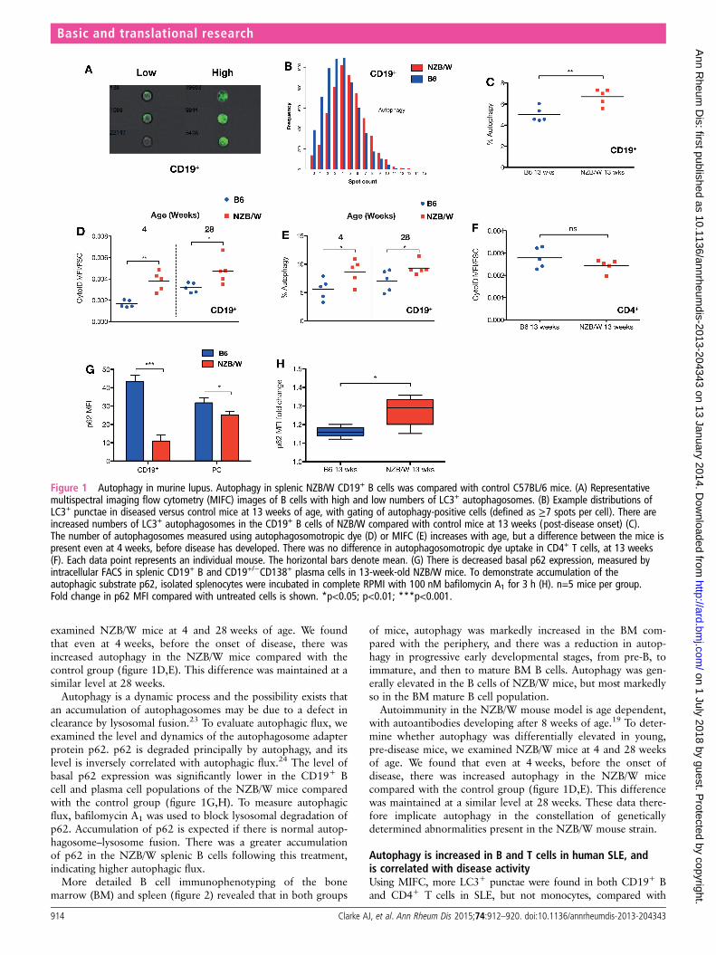
examined NZB/W mice at 4 and 28 weeks of age. We foundthat even at 4 weeks, before the onset of disease, there wasincreased autophagy in the NZB/W mice compared with thecontrol group (figure 1D,E). This difference was maintained at asimilar level at 28 weeks.
Autophagy is a dynamic process and the possibility exists thatan accumulation of autophagosomes may be due to a defect inclearance by lysosomal fusion.23 To evaluate autophagic flux, weexamined the level and dynamics of the autophagosome adapterprotein p62. p62 is degraded principally by autophagy, and itslevel is inversely correlated with autophagic flux.24 The level ofbasal p62 expression was significantly lower in the CD19+ Bcell and plasma cell populations of the NZB/W mice comparedwith the control group (figure 1G,H). To measure autophagicflux, bafilomycin A1 was used to block lysosomal degradation ofp62. Accumulation of p62 is expected if there is normal autop-hagosome–lysosome fusion. There was a greater accumulationof p62 in the NZB/W splenic B cells following this treatment,indicating higher autophagic flux.
More detailed B cell immunophenotyping of the bonemarrow (BM) and spleen (figure 2) revealed that in both groups
of mice, autophagy was markedly increased in the BM com-pared with the periphery, and there was a reduction in autop-hagy in progressive early developmental stages, from pre-B, toimmature, and then to mature BM B cells. Autophagy was gen-erally elevated in the B cells of NZB/W mice, but most markedlyso in the BM mature B cell population.
Autoimmunity in the NZB/W mouse model is age dependent,with autoantibodies developing after 8 weeks of age.19 To deter-mine whether autophagy was differentially elevated in young,pre-disease mice, we examined NZB/W mice at 4 and 28 weeksof age. We found that even at 4 weeks, before the onset ofdisease, there was increased autophagy in the NZB/W micecompared with the control group (figure 1D,E). This differencewas maintained at a similar level at 28 weeks. These data there-fore implicate autophagy in the constellation of geneticallydetermined abnormalities present in the NZB/W mouse strain.
Autophagy is increased in B and T cells in human SLE, andis correlated with disease activityUsing MIFC, more LC3+ punctae were found in both CD19+ Band CD4+ T cells in SLE, but not monocytes, compared with
Figure 1 Autophagy in murine lupus. Autophagy in splenic NZB/W CD19+ B cells was compared with control C57BL/6 mice. (A) Representativemultispectral imaging flow cytometry (MIFC) images of B cells with high and low numbers of LC3+ autophagosomes. (B) Example distributions ofLC3+ punctae in diseased versus control mice at 13 weeks of age, with gating of autophagy-positive cells (defined as ≥7 spots per cell). There areincreased numbers of LC3+ autophagosomes in the CD19+ B cells of NZB/W compared with control mice at 13 weeks (post-disease onset) (C).The number of autophagosomes measured using autophagosomotropic dye (D) or MIFC (E) increases with age, but a difference between the mice ispresent even at 4 weeks, before disease has developed. There was no difference in autophagosomotropic dye uptake in CD4+ T cells, at 13 weeks(F). Each data point represents an individual mouse. The horizontal bars denote mean. (G) There is decreased basal p62 expression, measured byintracellular FACS in splenic CD19+ B and CD19+/−CD138+ plasma cells in 13-week-old NZB/W mice. To demonstrate accumulation of theautophagic substrate p62, isolated splenocytes were incubated in complete RPMI with 100 nM bafilomycin A1 for 3 h (H). n=5 mice per group.Fold change in p62 MFI compared with untreated cells is shown. *p<0.05; p<0.01; ***p<0.001.
Basic and translational research
914 Clarke AJ, et al. Ann Rheum Dis 2015;74:912–920. doi:10.1136/annrheumdis-2013-204343
on 1 July 2018 by guest. Protected by copyright.
http://ard.bmj.com
/A
nn Rheum
Dis: first published as 10.1136/annrheum
dis-2013-204343 on 13 January 2014. Dow
nloaded from

healthy control subjects (figure 3A–C). The B cell LC3-BDI+
punctal count was positively correlated with the SELENA-SLEDAIdisease activity score (figure 3D). A potentially confounding factor
could be the use of hydroxychloroquine, which might increaseautophagosome count by blocking fusion with lysosomes, howeverthere was no significant correlation between LC3-BDI and use ofthis drug (p=0.52). Similarly, we found no association with spe-cific immunosuppressant medication use, although there was atrend towards correlation with prednisolone dose (r=0.39,p=0.08). To more directly demonstrate intact autophagic flux inSLE, isolated CD19+ B cells were incubated with chloroquine,an alternative lysosomal acidification inhibitor, and LC3 punctaemeasured by MIFC. There was a significant increase in autopha-gosome number following this, indicating active autophagic flux(figure 3E). Finally, to confirm intact autophagosome–lysosomefusion, we examined co-localisation between lysosomes andautophagosomes in CD19+ B cells using MIFC (see online sup-plementary figure S1).25 There was no fusion defect in SLEpatients compared with controls, but a non-statistically signifi-cant trend towards increased co-localisation, supporting theinhibitor data.
Analysis of total CD19+ B cells using CytoID confirmed theincrease in autophagosome load in SLE (figure 4A). When ana-lysed by B cell subset (CD19+CD27− naïve, CD19+CD27+
memory and CD19+/−CD27++ plasmablast) we found that inboth healthy controls and patients, dye uptake was greatest innaïve B cells, with lower levels in memory B and plasmablasts(figure 4C). Autophagosome density was significantly higher innaïve B cells, with lesser increases in memory B and plasmablasts.p62 levels, which are inversely correlated with autophagy,mirrored these results, demonstrating higher turnover of thisautophagy substrate in naïve B cells in both controls and cases, but
Figure 2 Autophagosome density in mouse B cell subsets.Distribution of CytoID MFI in B cell subsets from bone marrow (BM)and spleens of 13-week-old NZB/W mice and age matched controls.Pre-B (CD19+IgM−IgD−), immature B (BM Imm, CD19+IgD−IgM+),mature B (BM Mat, CD19+IgD+IgM+), T1 (CD19+IgM+
IgD−CD23−CD21−), T2 (CD19+IgM+ IgD+CD23+CD21+), marginal zone(MZ, CD19+IgM+IgD−CD23−CD21hi), follicular (FO, CD19+IgM−IgD+
CD23hiCD21+), and BM plasma cell (CD19+/−CD138+) subsets areillustrated. n=5 mice per group. Box and whisker plots denote maximumand minimum, IQR, and median. *p<0.05; **p<0.01; ***p<0.001.
Figure 3 Autophagy in systemic lupus erythematosus (SLE) patients versus healthy controls. (A–C) Representative images of CD19+ B cells,CD4+ T cells, and CD14+ monocytes with high and low numbers of LC3+ punctae, and comparison with healthy controls. CD19+ B cell LC3+ punctacount is correlated with SELENA-SLEDAI disease activity index (D). (E) Increased autophagic flux in SLE patients. Isolated CD19+ B cells from patientswere incubated with 10 μM chloroquine in complete RPMI media for 2 h, and then analysed by multispectral imaging flow cytometry. Each pointrepresents one individual patient or healthy donor. **p<0.01; ***p<0.001.
Basic and translational research
Clarke AJ, et al. Ann Rheum Dis 2015;74:912–920. doi:10.1136/annrheumdis-2013-204343 915
on 1 July 2018 by guest. Protected by copyright.
http://ard.bmj.com
/A
nn Rheum
Dis: first published as 10.1136/annrheum
dis-2013-204343 on 13 January 2014. Dow
nloaded from
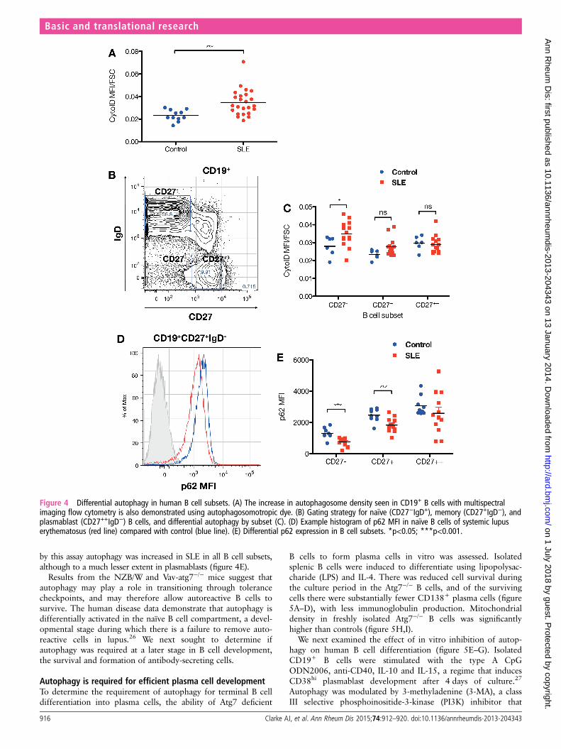
by this assay autophagy was increased in SLE in all B cell subsets,although to a much lesser extent in plasmablasts (figure 4E).
Results from the NZB/W and Vav-atg7−/− mice suggest thatautophagy may play a role in transitioning through tolerancecheckpoints, and may therefore allow autoreactive B cells tosurvive. The human disease data demonstrate that autophagy isdifferentially activated in the naïve B cell compartment, a devel-opmental stage during which there is a failure to remove auto-reactive cells in lupus.26 We next sought to determine ifautophagy was required at a later stage in B cell development,the survival and formation of antibody-secreting cells.
Autophagy is required for efficient plasma cell developmentTo determine the requirement of autophagy for terminal B celldifferentiation into plasma cells, the ability of Atg7 deficient
B cells to form plasma cells in vitro was assessed. Isolatedsplenic B cells were induced to differentiate using lipopolysac-charide (LPS) and IL-4. There was reduced cell survival duringthe culture period in the Atg7−/− B cells, and of the survivingcells there were substantially fewer CD138+ plasma cells (figure5A–D), with less immunoglobulin production. Mitochondrialdensity in freshly isolated Atg7−/− B cells was significantlyhigher than controls (figure 5H,I).
We next examined the effect of in vitro inhibition of autop-hagy on human B cell differentiation (figure 5E–G). IsolatedCD19+ B cells were stimulated with the type A CpGODN2006, anti-CD40, IL-10 and IL-15, a regime that inducesCD38hi plasmablast development after 4 days of culture.27
Autophagy was modulated by 3-methyladenine (3-MA), a classIII selective phosphoinositide-3-kinase (PI3K) inhibitor that
Figure 4 Differential autophagy in human B cell subsets. (A) The increase in autophagosome density seen in CD19+ B cells with multispectralimaging flow cytometry is also demonstrated using autophagosomotropic dye. (B) Gating strategy for naïve (CD27−IgD+), memory (CD27+IgD−), andplasmablast (CD27++IgD−) B cells, and differential autophagy by subset (C). (D) Example histogram of p62 MFI in naïve B cells of systemic lupuserythematosus (red line) compared with control (blue line). (E) Differential p62 expression in B cell subsets. *p<0.05; ***p<0.001.
Basic and translational research
916 Clarke AJ, et al. Ann Rheum Dis 2015;74:912–920. doi:10.1136/annrheumdis-2013-204343
on 1 July 2018 by guest. Protected by copyright.
http://ard.bmj.com
/A
nn Rheum
Dis: first published as 10.1136/annrheum
dis-2013-204343 on 13 January 2014. Dow
nloaded from
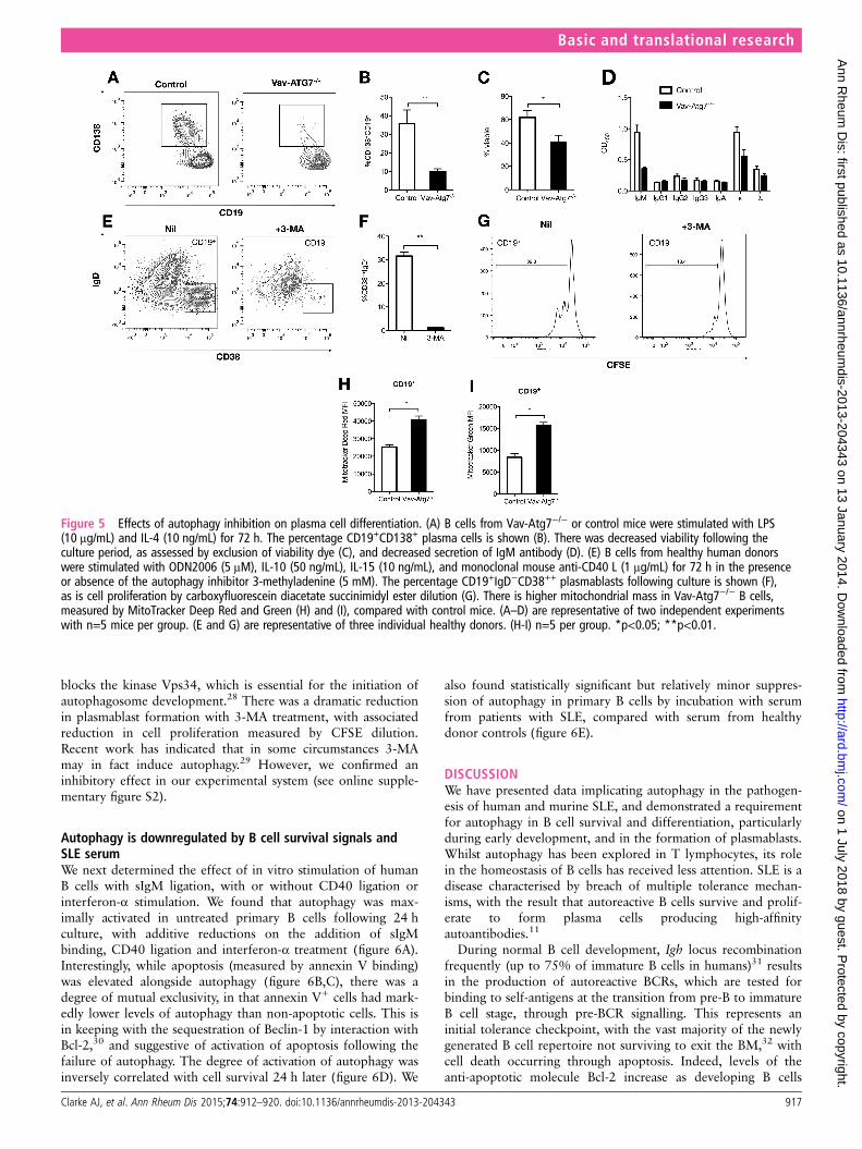
blocks the kinase Vps34, which is essential for the initiation ofautophagosome development.28 There was a dramatic reductionin plasmablast formation with 3-MA treatment, with associatedreduction in cell proliferation measured by CFSE dilution.Recent work has indicated that in some circumstances 3-MAmay in fact induce autophagy.29 However, we confirmed aninhibitory effect in our experimental system (see online supple-mentary figure S2).
Autophagy is downregulated by B cell survival signals andSLE serumWe next determined the effect of in vitro stimulation of humanB cells with sIgM ligation, with or without CD40 ligation orinterferon-α stimulation. We found that autophagy was max-imally activated in untreated primary B cells following 24 hculture, with additive reductions on the addition of sIgMbinding, CD40 ligation and interferon-α treatment (figure 6A).Interestingly, while apoptosis (measured by annexin V binding)was elevated alongside autophagy (figure 6B,C), there was adegree of mutual exclusivity, in that annexin V+ cells had mark-edly lower levels of autophagy than non-apoptotic cells. This isin keeping with the sequestration of Beclin-1 by interaction withBcl-2,30 and suggestive of activation of apoptosis following thefailure of autophagy. The degree of activation of autophagy wasinversely correlated with cell survival 24 h later (figure 6D). We
also found statistically significant but relatively minor suppres-sion of autophagy in primary B cells by incubation with serumfrom patients with SLE, compared with serum from healthydonor controls (figure 6E).
DISCUSSIONWe have presented data implicating autophagy in the pathogen-esis of human and murine SLE, and demonstrated a requirementfor autophagy in B cell survival and differentiation, particularlyduring early development, and in the formation of plasmablasts.Whilst autophagy has been explored in T lymphocytes, its rolein the homeostasis of B cells has received less attention. SLE is adisease characterised by breach of multiple tolerance mechan-isms, with the result that autoreactive B cells survive and prolif-erate to form plasma cells producing high-affinityautoantibodies.11
During normal B cell development, Igh locus recombinationfrequently (up to 75% of immature B cells in humans)31 resultsin the production of autoreactive BCRs, which are tested forbinding to self-antigens at the transition from pre-B to immatureB cell stage, through pre-BCR signalling. This represents aninitial tolerance checkpoint, with the vast majority of the newlygenerated B cell repertoire not surviving to exit the BM,32 withcell death occurring through apoptosis. Indeed, levels of theanti-apoptotic molecule Bcl-2 increase as developing B cells
Figure 5 Effects of autophagy inhibition on plasma cell differentiation. (A) B cells from Vav-Atg7−/− or control mice were stimulated with LPS(10 μg/mL) and IL-4 (10 ng/mL) for 72 h. The percentage CD19+CD138+ plasma cells is shown (B). There was decreased viability following theculture period, as assessed by exclusion of viability dye (C), and decreased secretion of IgM antibody (D). (E) B cells from healthy human donorswere stimulated with ODN2006 (5 μM), IL-10 (50 ng/mL), IL-15 (10 ng/mL), and monoclonal mouse anti-CD40 L (1 μg/mL) for 72 h in the presenceor absence of the autophagy inhibitor 3-methyladenine (5 mM). The percentage CD19+IgD−CD38++ plasmablasts following culture is shown (F),as is cell proliferation by carboxyfluorescein diacetate succinimidyl ester dilution (G). There is higher mitochondrial mass in Vav-Atg7−/− B cells,measured by MitoTracker Deep Red and Green (H) and (I), compared with control mice. (A–D) are representative of two independent experimentswith n=5 mice per group. (E and G) are representative of three individual healthy donors. (H-I) n=5 per group. *p<0.05; **p<0.01.
Basic and translational research
Clarke AJ, et al. Ann Rheum Dis 2015;74:912–920. doi:10.1136/annrheumdis-2013-204343 917
on 1 July 2018 by guest. Protected by copyright.
http://ard.bmj.com
/A
nn Rheum
Dis: first published as 10.1136/annrheum
dis-2013-204343 on 13 January 2014. Dow
nloaded from

transition from immature to mature stages.33 Bcl-2 negativelyregulates autophagy through its interaction with Beclin-1,30 andB cell developmental stages with low Bcl-2 expression coincidewith higher levels of autophagy.
However, the resting, mature peripheral B cell pool in autop-hagy deficiency is largely normal. Autophagy may therefore beactivated during early B cell development as a means to survivepro-apoptotic stimuli associated with the generation of a self-reactive or otherwise dysfunctional BCR.
We found that in NZB/W mice, autophagy was maximallyincreased compared with B6 control mice during early B celldevelopment, at the pre-B to mature B cell stage.
Analysis of autophagy in peripheral B cells of patients withSLE demonstrated maximal activation in naïve B cells, whichencounter a tolerance checkpoint following egress from the BM,which has been shown to be defective in SLE.26 We thereforepropose that enhanced autophagy at this stage may allow B
cells with autoreactive BCRs to escape physiological deletion.Stimulation of human B cells in vitro demonstrates that autop-hagy is activated in the absence of survival signals, but isreduced additively with BCR stimulation, CD40 ligation andinterferon-α. We found a degree of mutual exclusivity betweenautophagy and apoptosis, suggesting activation of pro-grammed cell death if autophagy failed. These results supportprevious observations that autophagy is induced in B cells inthe absence of co-stimulation, a situation that leads to celldeath.12
Interestingly, we found an age independent increase in autop-hagy in the B cells of the lupus prone NZB/W F1 strain, withsignificantly more autophagy than the control B6 strain even ata young age, 4 weeks, before the onset of disease.19 NZB/Wmice have a genetically determined defect in B cell activation,with excessive polyclonal IgM production from shortly afterbirth, and impaired tolerance induction.20 21 The function of
Figure 6 Effect of in vitro stimulation of human B cells on autophagy. (A) Human B cells were isolated and stimulated with combinations ofanti-IgM, anti-CD40 and interferon-α, then stained with CytoID. (B) Annexin V staining in B cells, cultured with and without anti-IgM, and CytoIDuptake in cells grouped as annexin V positive or negative (C). (D) Correlation between autophagy activation and cell viability, as assessed byviability dye exclusion. (E) Effect of B cell culture in healthy control or systemic lupus erythematosus patient serum on autophagy. Isolated B cellswere cultured in RPMI supplemented with 10% human serum for 4 h, then stained with CytoID. Data illustrate the effects of serum from threepatients and three healthy controls. Panels A–E are representative of three or more independent experiments. *p<0.05; **p<0.01; ***p<0.001.
Basic and translational research
918 Clarke AJ, et al. Ann Rheum Dis 2015;74:912–920. doi:10.1136/annrheumdis-2013-204343
on 1 July 2018 by guest. Protected by copyright.
http://ard.bmj.com
/A
nn Rheum
Dis: first published as 10.1136/annrheum
dis-2013-204343 on 13 January 2014. Dow
nloaded from

autophagy activation in these mice, as with the human SLEdata, may represent an attempt by autoreactive B cells to survivedeletion. However, to what extent autophagy is required fordisease development is an outstanding question.
We also demonstrated an important role for autophagy inplasmablast differentiation, in both Atg7−/− and human B cells.Our results confirm similar observations in Atg5−/− models,6 7
with the advantage of knockout of a gene without known func-tions outside of autophagy. We found moderately decreased via-bility in Atg7−/− B cells following stimulation, but a markedfailure of differentiation into plasma cells, associated withreduced secretion of IgM. The transition from resting B cell toplasma cell, capable of secretion of large quantities of immuno-globulin, generates intense metabolic stress, and is dependent onthe induction of the unfolded protein response, triggered fromthe ER.34 35 The ER is expanded in autophagy deficiency,7 36
and the enhanced ER stress associated with this may be inhibi-tory to plasma cell differentiation.37 Similarly, ineffective clear-ance of defective mitochondria by impaired mitophagypredisposes cells to apoptosis,38 and this may represent anotherexplanation for our findings. In SLE, the plasmablast populationis often markedly expanded, and is correlated with disease activ-ity.39 Pharmacological inhibition of autophagy restricts humanplasmablast differentiation in vitro, as was seen with murineAtg7−/− cells.
Autophagy therefore presents a potential therapeutic target inSLE, and may be a clinical relevant mechanism of action of thecommonly used immunomodulatory anti-malarial hydroxychlor-oquine, which is an inhibitor of autophagy by raising lysosomalpH and therefore preventing autophagosome–lysosomefusion.40 Similarly, many pharmaceuticals approved in theEuropean Union and USA, and in regular clinical use for alter-native indications, inhibit autophagy and may therefore be noveltreatments for SLE.41
Acknowledgements The authors would like to thank Dr P Gordon and Prof DD’Cruz for assistance with sample collection.
Contributors AJC: designed and performed experiments; UE, AC and AS:performed experiments; MB and AKS: provided reagents and technical support; AJCand TJV: wrote the manuscript.
Funding We acknowledge financial support from the Department of Health via theNational Institute for Health Research (NIHR) comprehensive Biomedical ResearchCentre award to Guy’s and St Thomas’ NHS Foundation Trust in partnership withKing’s College London and King’s College Hospital NHS Foundation Trust. This workwas supported by a Wellcome Trust Clinical Research Fellowship to AJC(WT091013MA).
Competing interests None.
Patient consent Obtained.
Ethics approval London Multicentre Ethics Commitee.
Provenance and peer review Not commissioned; externally peer reviewed.
Data sharing statement We can provide raw MIFC data on request.
Open Access This is an Open Access article distributed in accordance with theterms of the Creative Commons Attribution (CC BY 3.0) license, which permitsothers to distribute, remix, adapt and build upon this work, for commercial use,provided the original work is properly cited. See: http://creativecommons.org/licenses/by/3.0/
REFERENCES1 Fernandez DR, Telarico T, Bonilla E, et al. Activation of mammalian target of
rapamycin controls the loss of TCR in lupus T cells through HRES-1/Rab4-regulatedlysosomal degradation. J Immunol 2009;182:2063–73.
2 Levine B, Kroemer G. Autophagy in the pathogenesis of disease. Cell2008;132:27–42.
3 Page N, Gros F, Schall N, et al. HSC70 blockade by the therapeutic peptide P140affects autophagic processes and endogenous MHCII presentation in murine lupus.Ann Rheum Dis 2011;70:837–43.
4 Levine B, Mizushima N, Virgin HW. Autophagy in immunity and inflammation.Nature 2011;469:323–35.
5 Alessandri C, Barbati C, Vacirca D, et al. T lymphocytes from patients with systemiclupus erythematosus are resistant to induction of autophagy. FASEB J2012;26:4722–32.
6 Conway KL, Kuballa P, Khor B, et al. ATG5 regulates plasma cell differentiation.Autophagy 2013;9:528–37.
7 Pengo N, Scolari M, Oliva L, et al. Plasma cells require autophagy for sustainableimmunoglobulin production. Nat Immunol 2013;14:298–305.
8 Miller BC, Zhao Z, Stephenson LM,, et al The autophagy gene ATG5 plays anessential role in B lymphocyte development. Autophagy 2008;4:309–14.
9 Gros F, Arnold J, Page N,, et al Macroautophagy is deregulated in murine andhuman lupus T lymphocytes. Autophagy 2012;8:1113–23.
10 Hubbard VM, Valdor R, Patel B, et al. Macroautophagy regulates energymetabolism during effector T cell activation. J Immunol 2010;185:7349–57.
11 Liu Z, Davidson A. Taming lupus—a new understanding of pathogenesis is leadingto clinical advances. Nat Medicine 2012;18:871–82.
12 Watanabe K, Ichinose S, Hayashizaki K, et al. Induction of autophagy by B cellantigen receptor stimulation and its inhibition by costimulation. Biochem BiophysRes Commun 2008;374:274–81.
13 Mortensen M, Ferguson DJP, Edelmann M, et al. Loss of autophagy in erythroidcells leads to defective removal of mitochondria and severe anemia in vivo. PNAS2010;107:832–7.
14 Tan EM, Cohen AS, Fries JF, et al. The 1982 revised criteria for the classification ofsystemic lupus erythematosus. Arthritis Rheum 1982;25:1271–7.
15 Gateva V, Sandling JK, Hom G, et al. A large-scale replication study identifiesTNIP1, PRDM1, JAZF1, UHRF1BP1 and IL10 as risk loci for systemic lupuserythematosus. Nat Genet Nature Publishing Group 2009;41:1228–33.
16 Rubinsztein DC, Codogno P, Levine B. Autophagy modulation as a potentialtherapeutic target for diverse diseases. Nat Rev Drug Discov 2012;11:709–30.
17 Zhou XJ, Lu XL, Lv JC, et al. Genetic association of PRDM1-ATG5 intergenic regionand autophagy with systemic lupus erythematosus in a Chinese population. AnnRheum Dis 2011;70:1330–7.
18 Petri M, Kim MY, Kalunian KC, et al. Combined oral contraceptives in women withsystemic lupus erythematosus. N Engl J Med 2005;353:2550–8.
19 Andrews BS, Eisenberg RA, Theofilopoulos AN, et al. Spontaneous murinelupus-like syndromes. Clinical and immunopathological manifestations in severalstrains. J Exp Med 1978;148:1198–215.
20 Reininger L, Radaszkiewicz T, Kosco M, et al. Development of autoimmune diseasein SCID mice populated with long-term “in vitro” proliferating (NZB×NZW)F1 pre-Bcells. J Exp Med 1992;176:1343–53.
21 Reininger L, Winkler TH, Kalberer CP, et al. Intrinsic B cell defects in NZB and NZWmice contribute to systemic lupus erythematosus in (NZB×NZW)F1 mice. J Exp Med1996;184:853–61.
22 Lee JS, Lee GM. Monitoring of autophagy in Chinese hamster ovary cells using flowcytometry. METHODS. Elsevier Inc, 2011:1–8.
23 Mizushima N, Yoshimori T, Levine B. Methods in mammalian autophagy research.Cell 2010;140:313–26.
24 Komatsu M, Waguri S, Koike M, et al. Homeostatic levels of p62 controlcytoplasmic inclusion body formation in autophagy-deficient mice. Cell2007;131:1149–63.
25 Phadwal K, Alegre-Abarrategui J, Watson AS, et al. A novel method for autophagydetection in primary cells: impaired levels of macroautophagy in immunosenescentT cells. Autophagy 2012;8:677–89.
26 Yurasov S. Defective B cell tolerance checkpoints in systemic lupus erythematosus.J Exp Med 2005;201:703–11.
27 Jourdan M, Caraux A, De Vos J, et al. An in vitro model of differentiation ofmemory B cells into plasmablasts and plasma cells including detailed phenotypicand molecular characterization. Blood 2009;114:5173–81.
28 Seglen PO, Gordon PB. 3-Methyladenine: specific inhibitor of autophagic/lysosomalprotein degradation in isolated rat hepatocytes. Proc Natl Acad Sci USA1982;79:1889–92.
29 Wu YT, Tan HL, Shui G, et al. Dual role of 3-Methyladenine in modulation ofautophagy via different temporal patterns of inhibition on class I and IIIphosphoinositide 3-Kinase. J Biol Chem 2010;285:10850–61.
30 Pattingre S, Tassa A, Qu X, et al. Bcl-2 antiapoptotic proteins inhibit beclin1-dependent autophagy. Cell 2005;122:927–39.
31 Wardemann H, Yurasov S, Schaefer A, et al. Predominant autoantibody productionby early human B cell precursors. Science 2003;301:1374–7.
32 Rolink AG, Brocker T, Bluethmann H, et al. Mutations affecting either generation orsurvival of cells influence the pool size of mature B cells. Immunity1999;10:619–28.
33 Merino R, Ding L, Veis DJ, et al. Developmental regulation of the Bcl-2 protein andsusceptibility to cell death in B lymphocytes. EMBO J 1994;13:683–91.
Basic and translational research
Clarke AJ, et al. Ann Rheum Dis 2015;74:912–920. doi:10.1136/annrheumdis-2013-204343 919
on 1 July 2018 by guest. Protected by copyright.
http://ard.bmj.com
/A
nn Rheum
Dis: first published as 10.1136/annrheum
dis-2013-204343 on 13 January 2014. Dow
nloaded from
34 Gass JN. Activation of an unfolded protein response during differentiation ofantibody-secreting B cells. J Biol Chem 2002;277:49047–54.
35 Goldfinger M, Shmuel M, Benhamron S, et al. Protein synthesis in plasma cells isregulated by crosstalk between endoplasmic reticulum stress and mTOR signaling.Eur J Immunol 2010;41:491–502.
36 Jia W, Pua HH, Li QJ, et al. Autophagy regulates endoplasmic reticulumhomeostasis and calcium mobilization in T lymphocytes. J Immunol2011;186:1564–74.
37 Ron D, Walter P. Signal integration in the endoplasmic reticulum unfolded proteinresponse. Nat Rev Mol Cell Biol 2007;8:519–29.
38 Green DR, Galluzzi L, Kroemer G. Mitochondria and the autophagy-inflammation-cell death axis in organismal aging. Science 2011;333:1109–12.
39 Odendahl M, Jacobi A, Hansen A, et al. Disturbed peripheral B lymphocytehomeostasis in systemic lupus erythematosus. J Immunol 2000;165:5970–9.
40 van Loosdregt J, Spreafico R, Rossetti M, et al. Hydroxychloroquine preferentiallyinduces apoptosis of CD45RO+ effector T cells by inhibiting autophagy: a possiblemechanism for therapeutic modulation of T cells. J Allergy Clin Immunol2013;131:1443-6.e1.
41 Rubinsztein DC, Gestwicki JE, Murphy LO, et al. Potential therapeutic applicationsof autophagy. Nat Rev Drug Discov 2007;6:304–12.
Basic and translational research
920 Clarke AJ, et al. Ann Rheum Dis 2015;74:912–920. doi:10.1136/annrheumdis-2013-204343
on 1 July 2018 by guest. Protected by copyright.
http://ard.bmj.com
/A
nn Rheum
Dis: first published as 10.1136/annrheum
dis-2013-204343 on 13 January 2014. Dow
nloaded from