extensive involvement of autophagy in alzheimer’s … 2005 jnen.pdf · extensive involvement of...
TRANSCRIPT

Ralph A. Nixon, M.D., Ph.D.
1
Extensive Involvement of Autophagy In Alzheimer’s Disease: An Immuno-
Electron Microscopy Study
Ralph A. Nixon1,2,3*, Jerzy Wegiel3,4, Asok Kumar1,2, Wai Haung Yu 1,2, Corrinne Peterhoff 1,
Anne Cataldo5,1, Ana Maria Cuervo6
1 Center for Dementia Research, Nathan Kline Institute for Psychiatric Research, Orangeburg,
New York
2 Departments of Psychiatry and 3Cell Biology, New York University School of Medicine, New
York, New York
4 Department of Pathological Neurobiology, New York State Institute for Basic Research in
Developmental Disabilities, Staten Island, New York
5 Laboratory for Molecular Neuropathology, Mailman Research Center, McLean Hospital,
Belmont, Massachusetts
6 Albert Einstein College of Medicine Department of Anatomy and Structural Biology, Marion
Bessin Liver Research Center, Bronx, New York
*Corresponding author and reprint requests: Ralph A. Nixon, M.D., Ph.D., Nathan Kline
Institute, New York University School of Medicine, 140 Old Orangeburg Road, Orangeburg, NY
10962 Tel: (845) 398-5423; Fax: (845) 398-5422; e-mail: [email protected]
This research was supported by the Alzheimer Association (USA) (TLL-99-1877) (RAN), NIH
AG17617 (RAN) and AG021904 (AMC), and by the Howard Hughes Medical Institutes (AMC)
and a post-doctoral fellowship from the Canadian Institutes of Health Research (WH Yu).

Ralph A. Nixon, M.D., Ph.D.
2
ABSTRACTThe accumulation of lysosomes and their hydrolases within neurons is a well-established
neuropathological feature of Alzheimer’s disease (AD). Here, we show that lysosomal
pathology in AD brain involves extensive alterations of macroautophagy, an inducible pathway
for the turnover of intracellular constituents, including organelles. Using immunogold labeling
with compartmental markers and electron microscopy on neocortical biopsies from AD brain, we
unequivocally identified autophagosomes and other pre-lysosomal autophagic vacuoles (AVs),
which were morphologically and biochemically similar to AVs highly purified from mouse liver.
AVs were uncommon in brains devoid of AD pathology, but were abundant in AD brains
particularly, within neuritic processes, including synaptic terminals. In dystrophic neurites,
autophagosomes, multivesicular bodies, multilamellar bodies and cathepsin-containing
autophagolysosomes were the predominant organelles and accumulated in large numbers. These
compartments were distinguishable from lysosomes and lysosomal dense bodies, previously
shown also to be abundant in dystrophic neurites. Autophagy was evident in the perikarya of
affected neurons, particularly in those with neurofibrillary pathology where it was associated
with a relative depletion of mitochondria and other organelles. These observations provide the
first evidence that macroautophagy is extensively involved in the neurodegenerative/regenerative
process in AD. The striking accumulations of immature AV forms in dystrophic neurites suggest
that the transport of AVs and their maturation to lysosomes may be impaired, thereby impeding
the suspected neuroprotective functions of autophagy.
Key words: Lysosomes, neurodegeneration, amyloid, apoptosis, necrosis.
Ralph A. Nixon, M.D., Ph.D.
3
INTRODUCTION
Alzheimer’s disease (AD) is characterized by the presence of intraneuronal
neurofibrillary tangles, β-amyloid-containing neuritic plaques, and the loss of specific
populations of neurons. Neuron death is preceded by retrograde degeneration of synaptic
terminals, axons and dendrites, which may evolve over many years [1-4]. This degeneration is
accompanied by attempts by neurons to repair and regenerate neuritic processes [5-7], which
yields a highly characteristic pattern of enlarged dystrophic neurites. The molecular basis for
this degenerative process and ultimate neuronal death is still poorly understood.
Growing attention has focused on proteases in both the early survival responses of
neurons in AD and as agents of neurodegeneration, resulting either from inappropriate activation
of proteases or from defective proteolysis, which allows buildup of toxic molecules. Widespread
activation of calpains in neurons results in cleavage of key structural proteins and promotes
cytoskeletal hyperphosphorylation by activating protein kinases, leading to neurofibrillary
degeneration [8-10]. Certain caspases are activated at low levels in vulnerable neuronal
populations in the absence of a complete morphological or biochemical pattern of neuronal
apoptosis [11, 12]. A decline in proteasome-mediated turnover of proteins during aging and AD
[13, 14] is one factor that increases reliance on the lysosomal system, the principal alternative to
the proteasome for intracellular turnover. Indeed, the synthesis of lysosomal system
components, including cathepsins, is markedly upregulated early and progressively in AD [15-
18]. The function of the endocytic pathway, one major route to the lysosome, is altered before
any other known pathologies in AD brain [19, 20], and this dysfunction may promote β-amyloid
peptide generation [20-23] and reduce neuronal survival [24]. Autophagy, the other major
pathway to lysosomes, has received limited attention in relation to AD [25], although its
Ralph A. Nixon, M.D., Ph.D.
4
importance as a mechanism for removing defective organelles and potentially toxic proteins [26,
27] and as a determinant of cell survival [28] in various disease settings is becoming appreciated
[29].
Autophagy is a tightly regulated process [30, 31], which is induced by nutritional or
trophic deprivation under conditions of cell stress to provide substrates for energy or new
synthesis by turning over non-essential cytoplasmic constituents, including organelles [32, 33].
Autophagy is initiated when a region of cytoplasm and organelles within the cells is sequestered
within a double membrane-limited vacuole, the autophagosome [34, 35]. Autophagosomes
mature to single membrane phagolysosomes [30, 36-38] and become autolysosomes when they
become acidified and acquire proteolytic enzymes by fusing with late endosomes or lysosomes
[36, 38]. Materials internalized by endocytosis also enter the autophagic pathway when
endosomes fuse with autophagosomes [39, 40]. The term autophagic vacuole (AV) is used here
to refer to any of these pre-lysosomal compartments.
Autophagy is active during development to support major changes in cell size and
morphology [25, 41]. Autophagy may also act as a surveillance system in stressed or injured
cells to remove damaged mitochondria and other organelles that have the potential to trigger
apoptosis [42-44]. On the other hand, auto-digestion by acutely upregulating autophagy is a
form of programmed cell death that is distinct from apoptosis, but shares some of its features [45,
46]. Autophagic cell death has become more frequently recognized as a caspase-independent
form of apoptosis and several cathepsins have been shown to initiate or mediate aspects of
apoptotic and necrotic of cell death in various pathological settings [15, 29]. Autophagic
vacuoles have recently been observed in experimental neurodegenerative states [47-50], and in
Ralph A. Nixon, M.D., Ph.D.
5
dying striatal neurons in Parkinson disease [51], although information about the extent to which
autophagy is involved in neurodegeneration and its pathogenic significance is limited [15, 25].
To investigate the possible involvement of autophagy in AD, we analyzed well-preserved
biopsy specimens of neocortex from AD and non-AD control brains which allowed us, for the
first time, to identify and extensively characterize autophagosomes and related autophagic
vacuoles and to differentiate them from other lysosome-related compartments known to
accumulate in neurons in AD brain [52-54]. Our results indicate that the lysosomal system
mobilization previously shown to occur in AD involves, in part, the induction of
macroautophagy in affected neurons, which is particularly evident within neuritic processes,
including synaptic regions. Moreover, AV proliferation is unexpectedly robust in dystrophic
neurites and includes grossly abnormal accumulations of immature forms of AVs such as
autophagosomes, suggesting that, while autophagy may be induced in AD, AV transport and
maturation are also impaired in affected neurons. These observations have implications for
neurodegenerative mechanisms and β-amyloidogenesis in AD.
Ralph A. Nixon, M.D., Ph.D.
6
MATERIALS AND METHODS
Acquisition of human biopsy brain and post-embedding for electron microscopy
Cortical biopsy specimens were studied from 7 patients with AD (aged 71-86 years) as
previously described [55]. Cortical biopsies free of plaques and tangles from 3 subjects (aged
67-72 years) were also examined [55]. Clinicopathologic characteristics of these cases are
described in Table 1. Tissue was fixed in 3% glutaraldehyde / 0.1 M phosphate buffer, pH 7.4,
and postfixed in 1% osmium tetroxide in Sorensen’s phosphate buffer. After dehydration in
ethyl alcohol, the tissue was embedded in Epon (EMS, Fort Washington, PA). Tissue blocks
were cut serially into ultrathin (0.06µm) sections. Ultrathin sections were stained with uranyl
acetate and lead citrate.
Semi-quantitative analysis of AVs in AD brain
For quantitative studies, the numbers of AVs in neuronal perikarya were counted from
electron micrographs at 8000x magnification. Neurons were selected for quantitative analyses
from cortical lamina III. Every fourth neuron with its nucleus present in the cross-section was
photographed within each sector of the entire EM grid. To capture additional neurons from the
same brain, every fourth ultra-thin section was used for quantification to avoid selecting the
same neuron twice. Glial cells were excluded on the basis of their nuclear morphology and
chromatin patterns.
Immunoelectron microscopy
Ultrathin sections from epon blocks were placed on nickel grids, air-dried, and etched
briefly with 1% sodium meta-periodate in PBS followed by washing in filtered double-distilled
deionized water and incubated in 1% BSA in PBS for two hours. Sections were incubated
overnight in primary antibody (calnexin 1:250 or protein disulfide isomerase (PDI) 1:200), both

Ralph A. Nixon, M.D., Ph.D.
7
from Stressgen (Victoria, BC, Canada); cathepsin D (1:20) from Dako (Carpinteria, CA) in a
humidified chamber at 4°C, washed several times in PBS, and incubated with 5-20nm gold
conjugated secondary antibody (Anti-mouse IgG or Anti-rabbit IgG; Amersham Biosciences,
Piscataway, NJ (1:50)) in PBS for 2 hr at room temperature. In negative control experiments,
primary antibody was substituted with normal rabbit serum or normal mouse serum depending
upon the primary antibody used (polyclonal or monoclonal). Grids were washed again and
briefly stained with uranyl acetate and lead citrate before being examined with a Philips CM 10
electron microscope.
Isolation of autophagic vacuoles from mouse liver and post-embedding for electron microscopy
To isolate liver AVs, three C57BL/6 mice were starved for 18 hours prior to sacrifice
[56]. Livers were harvested, minced and homogenized using a polytron teflon homogenizer and
separated by differential centrifugation to produce enriched fractions containing cytoplasm, AVs,
lysosomes, mitochondria, and a pellet containing the nuclear fraction and (up to 30%) unbroken
cells (PNP). A cytosol fraction was obtained by separating the supernatant from the
AV/lysosome/mitochondrial pellet by centrifugation at 100,000 x g for 1h at 4°C. Subsequently,
lysosomes were separated from two AV fractions using a metrizamide discontinuous gradient
(AV10 – 10% metrizamide and AV20 – 20% metrizamide). Autophagic vacuoles were fixed in
cold 4% paraformaldehyde and 0.25% glutaraldehyde in 0.1 M sodium cacodylate buffer (pH
7.2) overnight at 4°C. Following fixation, isolates were washed (3X) in cacodylate buffer, post-
fixed in 1% osmium tetroxide and progressively dehydrated in ethanol. The sample was then
embedded and polymerized in Epon 812 following infiltration. Sections were cut and mounted
onto copper grids that were examined using Philips CM 10 electron microscope.
Ralph A. Nixon, M.D., Ph.D.
8
RESULTS
Ultrastructure of autophagy pathology in AD cortex
In AD brain, cathepsin D (cat D) and many other lysosomal proteases in active form have
been shown to be concentrated in dystrophic neurites, which are frequently, but not exclusively,
associated with senile plaques [57, 58]. Our ultrastructural analyses of the neuropil in
neocortical biopsies from individuals with AD revealed many neurites in which the normal
cytoplasmic content of the enlarged process was almost completely replaced by vesicular
organelles with varying structural features (Fig. 1a). The range of morphologies (Fig. 1a,d,e)
was strikingly similar to that observed in highly purified populations of liver AVs isolated by
metrizamide density gradient centrifugation from mice starved for 18 h to induce AV
accumulation [56, 59] (Fig. 1b,c). Previous studies [60] have shown that these purified AV
fractions were highly enriched in the autophagosome markers, light chain 3 (LC3) [61, 62] and
rab24 [63] and were distinct from lysosomes. The morphologies and composition of vesicles
that accumulated in affected dystrophic neurites (e.g., in Fig. 1 and Fig. 2) corresponded to those
of the vesicular compartments of the autophagic pathway. Many vesicles met standard
morphologic criteria for the immature and mature autophagosomes [37, 64], including a size
>0.5 µm in diameter, a double-limiting membrane (immature), and the presence within a single
vacuole of multiple membranous structures from mitochondria, Golgi, or endoplasmic reticulum,
in addition to amorphous electron dense material. Multiple small double membrane-bound AVs
were often contained within a larger AV (Fig. 1d), suggesting that autophagy in these neurites
involves a continual process of AV consolidation. The vesicular contents of affected neurites, as
well as the preparations of highly enriched AVs from liver, also often contained single
membrane-limited multivesicular bodies containing light or dense amorphous material.
Ralph A. Nixon, M.D., Ph.D.
9
Multilamellar bodies, which are autophagic in origin [65], also commonly appeared in some
neurites (Fig. 1e, Fig. 2 inset). In regions of neuropil distant from β-amyloid deposits, frequent
dystrophic neurites were present next to relatively normal-appearing neurites. While many of the
dystrophic neurites contained abundant AVs of diverse morphology, AV populations of a
particular morphologic type, e.g., multilamellar bodies (inset arrows), or double-membrane-
limited dense vesicles (arrowheads), often characterized specific abnormal neurites (Fig. 2).
Immuno-electron microscopy of autophagic vacuole subtypes
Using immunogold electron microscopy, we further distinguished AVs from other
organelles, such as endosomes and Golgi vesicles/tubules. The latter organelles are not expected
to contain proteins that are resident to the endoplasmic reticulum (ER). By contrast,
autophagosomes have double membranes believed to originate from ER [36, 59, 66]. Moreover,
ER, like other vesicular organelles, is turned over by autophagy and is, therefore, expected to be
part of the intralumenal contents of many AVs [59, 66, 67]. Consistent with these expectations,
the single or double membrane surfaces as well as the membranous contents of vesicles that we
identified as AVs by morphologic criteria were also immunolabeled by antibodies to the resident
ER proteins, protein disulfide isomerase (PDI; data not shown) and calnexin (Fig. 3a,b,c).
Calnexin conjugated-immunogold, which selectively decorates ER (Fig. 3a,c), was present on
the limiting membranes of autophagosomes (Fig. 3a boxed area, b) and occasionally could be
seen on ER membranes that are in the process of autophagic sequestration (Fig. 3a arrows). The
absence of gold in the surrounding neuropil confirmed the specificity of the immunolabeling. In
contrast to the membrane labeling by anti-calnexin antibodies, immunogold labeling with cat D
antibodies was seen mainly in the lumens of AVs (Fig. 3d-g). Immunogold labeling with
antibodies to cat D distinguished immature AVs (presumably autophagosomes) lacking
Ralph A. Nixon, M.D., Ph.D.
10
hydrolase from the more mature AVs that contained cathepsin immunoreactivity, reflecting late
stages of AV maturation subsequent to endosomal/lysosomal fusion with AVs (Fig. 3d,e). The
hydrolase-positive AVs at late stages of maturation were single membrane vesicles with
amorphous dense intralumenal content (Fig. 3d,f), although some were double membrane-limited
(e.g., Fig. 3e, arrow), possibly reflecting the active sequestration of multiple mature AVs within
a single autophagosome-like vesicle. In contrast, multilamellar bodies (Fig. 3d,e arrowheads)
were not immunolabeled with cat D. Many neurites contained high proportions of hydrolase-
negative (immature) AVs. AVs were distinguishable from lysosomal dense bodies, which were
generally smaller (< 0.2 µm), single membrane-limited structures of homogeneous density,
which were strongly decorated by cathepsin antibodies (Fig. 3f). Residual bodies, such as
lipofuscin, were not observed in neurites and were easily identified in perikarya by their bipartite
organization of globoid electron-opaque lipopigment and homogeneous protein content that was
strongly immuno-labeled with cat D antibodies (Fig. 3,g). In the absence of primary antibody,
labeling of cat D-positive structures with immunogold secondary antibody was negligible (not
shown). Cat D immunolabeling specificity was further indicated by the minimal labeling of
other cytoplasmic organelles or ground substance and by the contrast between the intralumenal
labeling by cat D antibodies (Fig. 3d-g) and the selective decoration of the double-limiting
membrane of vesicles by anti-calnexin bodies (Fig. 3a,b).
Prevalence of Autophagic Pathology in AD brain
In control cortical biopsies lacking detectable AD-related pathology, AVs were rarely
observed in neurons and their processes (Fig. 4a). In AD brain, by contrast, AVs were frequently
seen even in dendrites that were not markedly dystrophic (Fig. 4b), in terminal areas containing
synaptic vesicles (Fig. 4c), and in distal dendritic processes as opposed to normal-appearing
Ralph A. Nixon, M.D., Ph.D.
11
neurons (Fig. 5). AV accumulation was most striking in dystrophic processes forming the
neuritic network in proximity to senile plaques (Fig. 6), which have previously been shown to be
strongly cat D immunoreactive (Fig. 6 inset) [58]. Ultrastructural analysis reveals the extensive
dystrophy of virtually all neurites in the vicinity of a β-amyloid deposit (Fig. 6, “A”) and within
these neurites; the marked accumulation of vacuoles, most of which are AVs (Fig. 6 black
arrows), and smaller numbers of swollen or condensed mitochondria (Fig. 6, arrowheads). The
numbers of AVs in neuritic processes of AD brains far exceeded the incidence of AVs in cell
bodies, although AV numbers in neuronal perikarya were also markedly increased in AD.
Morphometric analysis of AV frequency in AD and control brains
In quantitative analyses of 137 randomly selected neurons in neocortex from 3 different
control brains lacking AD pathology, AVs corresponding morphologically to autophagosomes,
multilamellar bodies and multivesicular bodies were observed at an average frequency of
0.117±.030 (mean ± 1S.E.) per neuronal perikaryal cross section, and rarely appeared with a
frequency of more than one per perikaryon. By contrast, in a sample of 130 randomly selected
neocortical neurons from a total of 7 AD brains, AVs were identified at a more than 20-fold
higher frequency (2.93±0.19 per neuronal perikaryal cross-section) (Fig. 7). The incidence of
AVs and of dense lysosomes was particularly increased in the perikarya of neurons (Fig. 8a) that
also contained paired helical filaments (PHF) (Fig. 8a inset). The example in Fig. 8 shows a
neuron with a relatively displaced normal appearing nucleus and no apparent perikaryal atrophy
containing scattered bundles of paired helical filaments and numerous dense or multilamellar
AVs in addition to lipofuscin granules and small dense lysosomes (Fig. 8b). Neuronal perikarya
with elevated numbers of AVs commonly were relatively depleted of other organelles and
particularly mitochondria (Fig. 8c, d; compare to the neuron in Fig. 5). Despite this increased
Ralph A. Nixon, M.D., Ph.D.
12
incidence of AVs in perikarya, the number was relatively small compared to that in most affected
neurites and was always out-numbered by dense lysosomes.

Ralph A. Nixon, M.D., Ph.D.
13
DISCUSSION
Our studies provide the first evidence supporting the extensive involvement of
macroautophagy in AD pathogenesis. Previous studies have emphasized an early mobilization
of the lysosomal system, including upregulated synthesis of lysosomal system components,
secondary lysosome proliferation [68], and an accumulation of dense bodies in dystrophic
neurites [52, 53] of vulnerable neuronal populations. Our data indicate that the induction of
macroautophagy may contribute substantially to these downstream responses of the lysosomal
system. We identified different vesicular compartments of the autophagic pathway
(autophagosomes, autophagolysosomes, multilamellar bodies and dense residual bodies) by
various morphologic and biochemical criteria. These include vesicular subpopulations distinct
from those described in earlier ultrastructural studies of Terry, Suzuki and colleagues [52, 69].
Autophagosomes were identified by their classic morphologic features of size greater than
0.5µm, double-limiting membrane, and heterogeneous intralumenal contents, which included
intact organelles and organelle-derived membranes [37, 64]. Autophagosomes and other distinct
vesicles of autophagic origin, such as multilamellar bodies [65], were shown to be pre-lysosomal
based on an absence of immunogold labeling with cat D antibodies that strongly decorate
lysosomal compartments. Importantly, AVs, which included a range of multivesicular
morphologies, were excluded as being endosomes or multivesicular bodies of endosomal origin
on the basis that the limiting membranes and intralumenal contents of many of these structures
were immunogold labeled by antibodies to the resident ER proteins, calnexin, and protein
disulfide isomerase (unpublished data). ER membranes are believed to be a source of
membranes involved in autophagic sequestration [36, 59, 66], whereas compartments of the
endocytic pathway should not be labeled by ER markers prior to fusion with autophagosomes.

Ralph A. Nixon, M.D., Ph.D.
14
In addition to the group of AVs representing early stages of macroautophagy, we
identified more mature AVs, reflecting a stage after late endosome/lysosome fusion. Late AVs
were more homogeneous in their intralumenal content than autophagosomes. They often, but not
exclusively, exhibited single-limiting membranes, and contained cat D immunoreactivity by
immuno EM. These profiles were distinct from lysosomal dense bodies, which were small (less
than 0.2 µm), homogeneously dense, single membrane-limited structures and were strongly cat D
immunoreactive. Collectively, cat D-positive compartments correspond to the dense bodies and
acid phosphatase-containing vesicular structures that have previously been shown to be abundant
in dystrophic neurites [52-54]. The morphologic and biochemical features of AVs in brain tissue
closely corresponded to those of highly purified AVs from liver [60], which we showed to be
enriched in LC3-II, a microtubule-associated protein believed to be a specific marker of
autophagosomes [34, 62], and in rab24, a small GTPase reported to associate selectively with
AVs [63]. Moreover, in confirmation of our morphologic data, LC3-II levels are significantly
elevated in AD brain [70].
AVs were rarely seen in neurons from the brains of individuals with no neuropathologic
evidence of AD and were also uncommon in axons and dendrites. By contrast, AVs were
frequently seen in otherwise normal-appearing neurites in the AD brain and were strikingly
abundant in neurites that appeared swollen and dystrophic. These patterns were similar in all
biopsy cases in this study. AVs were also more frequently observed in the perikarya of neurons
in AD brains, particularly when these neurons contained paired-helical filaments. These
observations accord with the extensive lysosomal hydrolase immunolabeling in AD brain (Fig.
8), which shows elevated neuropil staining and intense immunoreactive signal in neuritic plaques
throughout affected regions [57, 71]. Although AV accumulations are not specific to the
Ralph A. Nixon, M.D., Ph.D.
15
degenerative phenomena of AD [47-49, 51], autophagic-lysosomal pathology in the brain is
considerably more widespread and robust in AD than in other adult-onset neurodegenerative
diseases [72]. Specifically, the extensive neuritic dystrophy [53, 73] and the characteristic gross
distension of these neurites in AD are not typical in other neurodegenerative diseases lacking β-
amyloid [74]. Moreover, the near replacement of cytoplasmic contents by AVs in these
numerous dystrophic neurites, together with the high incidence of AVs in less obviously affected
neurites, constitutes a uniquely large “burden” of autophagy-related compartments in the AD
brain.
While macroautophagy seems to be induced in AD, the striking accumulation of AVs in
dystrophic neurites suggests that some of the later steps of autophagy might be impaired in these
neurons. Macroautophagy is normally active within the growing (regenerating) neurites of
cultured neurons [41, 75, 76]. This process involves the retrograde transport of immature AVs
toward the cell body where they encounter, and fuse with, acidified hydrolase-containing
compartments [41, 76]. AV maturation seems to be efficient under these conditions, resulting in
rapid turnover of AV contents in lysosomes. The enormous accumulation of AVs, including
autophagosomes and other immature AVs within dystrophic/degenerating neurites and the more
modest accumulation in most neuronal perikarya suggest that the transport of AVs is impeded or
that autophagy locally within the dystrophic segments is particularly robust. These are not
mutually exclusive possibilities. In addition, the coexistence of substantial numbers of both
immature and acid-hydrolase-containing compartments suggests that immature AVs have access
to hydrolase-containing compartments but may less efficiently carry out the final stages of AV
maturation to lysosomes.
Ralph A. Nixon, M.D., Ph.D.
16
The large numbers of autophagic vacuoles in neurons of the AD brain have several
possible implications for pathogenesis in AD. AVs have been recently shown to contain the
substrates and APP secretase activities required to generate Aβ and are particularly highly
enriched in γ-secretase enzymatic activity and γ-secretase complex components [60]. Aβ40,
Aβ42, and βCTF have also been detected in purified AVs from livers of YAC transgenic mice
overexpressing APP [60]. Moreover, modulations of macroautophagy rates in cultured cells
influence the rates of Aβ production, stimulating more than a doubling of Aβ production [70].
These observations suggest that accumulated AVs in dystrophic neurites may contribute
significantly to the local production of Aβ within plaques and that the generalized increase in
autophagy in neuropil could be a significant source of Aβ overproduction in AD brain.
Moreover, an impairment of macroautophagy that impedes the turnover of damaged
mitochondria capable of triggering caspase activation or of oxidized membranes and proteins,
which are a source of free radicals, is likely to promote neuronal degeneration [42]. While local
autophagy may be useful in the repair of damaged neurites and terminals, the stasis and
accumulation of hydrolase-containing AVs in dystrophic neurites also represents a large
reservoir of proteases that can potentially activate caspases directly and promote
neurodegeneration [15, 28]. The intriguing influences of macroautophagy on cellular aging
mechanisms, cell survival, and protein handling emerging from recent studies [26, 33, 77, 78],
suggest that an understanding of its roles in neurons will be highly informative in defining
pathogenic mechanisms in AD.

Ralph A. Nixon, M.D., Ph.D.
17
Acknowledgements: The authors thank Laurie Goldberg for assistance in manuscript
preparation.

Ralph A. Nixon, M.D., Ph.D.
18
References
1. Burke, W.J., H.D. Chung, J.S. Huang, et al. Evidence for retrograde degeneration of
epinephrine neurons in Alzheimer's disease. Ann Neurol, 1988;24(4):532-6.
2. Coleman, P.D. and P.J. Yao. Synaptic slaughter in Alzheimer's disease. Neurobiol Aging,
2003;24(8):1023-7.
3. Pearson, R.C. and T.P. Powell. Anterograde vs. retrograde degeneration of the nucleus
basalis medialis in Alzheimer's disease. J Neural Transm Suppl, 1987;24(139-46.
4. Morsch, R., W. Simon and P.D. Coleman. Neurons may live for decades with
neurofibrillary tangles. J Neuropathol Exp Neurol, 1999;58(2):188-97.
5. Larner, A.J. The cortical neuritic dystrophy of Alzheimer's disease: nature, significance,
and possible pathogenesis. Dementia, 1995;6(4):218-24.
6. Hashimoto, M. and E. Masliah. Cycles of aberrant synaptic sprouting and
neurodegeneration in Alzheimer's and dementia with Lewy bodies. Neurochem Res,
2003;28(11):1743-56.
7. McKee, A.C., N.W. Kowall and K.S. Kosik. Microtubular reorganization and dendritic
growth response in Alzheimer's disease. Ann Neurol, 1989;26(5):652-9.
8. Saito, K., J.S. Elce, J.E. Hamos and R.A. Nixon. Widespread activation of calcium-
activated neutral proteinase (calpain) in the brain in Alzheimer disease: a potential
molecular basis for neuronal degeneration. Proc Natl Acad Sci U S A, 1993;90(7):2628-
32.
Ralph A. Nixon, M.D., Ph.D.
19
9. Patrick, G.N., L. Zukerberg, M. Nikolic, S. de la Monte, P. Dikkes and L.H. Tsai.
Conversion of p35 to p25 deregulates Cdk5 activity and promotes neurodegeneration [see
comments]. Nature, 1999;402(6762):615-22.
10. Nixon, R.A. The calpains in aging and aging-related diseases. Ageing Res Rev,
2003;2(4):407-18.
11. Rohn, T.T., R.A. Rissman, E. Head and C.W. Cotman. Caspase Activation in the
Alzheimer's Disease Brain: Tortuous and Torturous. Drug News Perspect,
2002;15(9):549-557.
12. Roth, K.A. Caspases, apoptosis, and Alzheimer disease: causation, correlation, and
confusion. J Neuropathol Exp Neurol, 2001;60(9):829-38.
13. Keller, J.N., K.B. Hanni and W.R. Markesbery. Impaired proteasome function in
Alzheimer's disease. J Neurochem, 2000;75(1):436-9.
14. Keller, J.N., F.F. Huang and W.R. Markesbery. Decreased levels of proteasome activity
and proteasome expression in aging spinal cord. Neuroscience, 2000;98(1):149-56.
15. Nixon, R.A., A.M. Cataldo and P.M. Mathews. The endosomal-lysosomal system of
neurons in Alzheimer's disease pathogenesis: a review. Neurochem Res, 2000;25(9-
10):1161-72.
16. Bernstein, H.G., H. Kirschke, B. Wiederanders, K.H. Pollak, A. Zipress and A. Rinne.
The possible place of cathepsins and cystatins in the puzzle of Alzheimer disease: a
review. Mol Chem Neuropathol, 1996;27(3):225-47.
17. Cataldo, A.M., J.L. Barnett, S.A. Berman, et al. Gene expression and cellular content of
cathepsin D in Alzheimer's disease brain: evidence for early up-regulation of the
endosomal-lysosomal system. Neuron, 1995;14(3):671-80.

Ralph A. Nixon, M.D., Ph.D.
20
18. Cataldo, A.M., J.L. Barnett, C. Pieroni and R.A. Nixon. Increased neuronal endocytosis
and protease delivery to early endosomes in sporadic Alzheimer's disease:
neuropathologic evidence for a mechanism of increased beta-amyloidogenesis. J
Neurosci, 1997;17(16):6142-51.
19. Cataldo, A.M., C.M. Peterhoff, J.C. Troncoso, T. Gomez-Isla, B.T. Hyman and R.A.
Nixon. Endocytic pathway abnormalities precede amyloid beta deposition in sporadic
Alzheimer's disease and Down syndrome: differential effects of APOE genotype and
presenilin mutations. Am J Pathol, 2000;157(1):277-86.
20. Mathews, P.M., C.B. Guerra, Y. Jiang, et al. Alzheimer's disease-related overexpression
of the cation-dependent mannose 6-phosphate receptor increases Abeta secretion: role for
altered lysosomal hydrolase distribution in beta-amyloidogenesis. J Biol Chem,
2002;277(7):5299-307.
21. Grbovic, O.M., P.M. Mathews, Y. Jiang, et al. Rab5-stimulated up-regulation of the
endocytic pathway increases intracellular beta-cleaved amyloid precursor protein
carboxyl-terminal fragment levels and Abeta production. J Biol Chem,
2003;278(33):31261-8.
22. Cataldo, A.M., S. Petanceska, N.B. Terio, et al. A-beta localization to abnormal
endosomes coincides with early increases in soluble A-beta in Alzheimer's disease brain.
Neurobiol Aging, 2004;
23. Koo, E.H. and S.L. Squazzo. Evidence that production and release of amyloid beta-
protein involves the endocytic pathway. J Biol Chem, 1994;269(26):17386-9.
24. Nixon, R.A. Endosome function and dysfunction in Alzheimer's disease and other
neurodegenerative diseases. Neurobiol Aging, 2005;In Press.

Ralph A. Nixon, M.D., Ph.D.
21
25. Nixon, R.A. and A.M. Cataldo. The endosomal-lysosomal system of neurons: new roles.
Trends Neurosci, 1995;18(11):489-96.
26. Ravikumar, B., C. Vacher, Z. Berger, et al. Inhibition of mTOR induces autophagy and
reduces toxicity of polyglutamine expansions in fly and mouse models of Huntington
disease. Nat Genet, 2004;36(6):585-95.
27. Fortun, J., W.A. Dunn, Jr., S. Joy, J. Li and L. Notterpek. Emerging role for autophagy in
the removal of aggresomes in Schwann cells. J Neurosci, 2003;23(33):10672-80.
28. Florez-McClure, M.L., D.A. Linseman, C.T. Chu, et al. The p75 neurotrophin receptor
can induce autophagy and death of cerebellar Purkinje neurons. J Neurosci,
2004;24(19):4498-509.
29. Uchiyama, Y. Autophagic cell death and its execution by lysosomal cathepsins. Arch
Histol Cytol, 2001;64(3):233-46.
30. Kadowaki, M. and T. Kanazawa. Amino acids as regulators of proteolysis. J Nutr,
2003;133(6 Suppl 1):2052S-2056S.
31. Petiot, A., S. Pattingre, S. Arico, D. Meley and P. Codogno. Diversity of signaling
controls of macroautophagy in mammalian cells. Cell Struct Funct, 2002;27(6):431-41.
32. Seglen, P.O., P.E. Schwarze and G. Saeter. Changes in cellular ploidy and autophagic
responsiveness during rat liver carcinogenesis. Toxicol Pathol, 1986;14(3):342-8.
33. Ogier-Denis, E. and P. Codogno. Autophagy: a barrier or an adaptive response to cancer.
Biochim Biophys Acta, 2003;1603(2):113-28.
34. Mizushima, N., Y. Ohsumi and T. Yoshimori. Autophagosome formation in mammalian
cells. Cell Structure and Function, 2002;27(421-429.
Ralph A. Nixon, M.D., Ph.D.
22
35. Klionsky, D.J. and S.D. Emr. Autophagy as a regulated pathway of cellular degradation.
Science, 2000;290(5497):1717-21.
36. Dunn, W.A., Jr. Studies on the mechanisms of autophagy: maturation of the autophagic
vacuole. J Cell Biol, 1990;110(6):1935-45.
37. Dunn, W.A., Jr. Studies on the mechanisms of autophagy: formation of the autophagic
vacuole. J Cell Biol, 1990;110(6):1923-33.
38. Lawrence, B.P. and W.J. Brown. Autophagic vacuoles rapidly fuse with pre-existing
lysosomes in cultured hepatocytes. J Cell Sci, 1992;102(Pt 3):515-26.
39. Gordon, P.B. and P.O. Seglen. Prelysosomal convergence of autophagic and endocytic
pathways. Biochem Biophys Res Commun, 1988;151(1):40-7.
40. Liou, W., H.J. Geuze, M.J. Geelen and J.W. Slot. The autophagic and endocytic
pathways converge at the nascent autophagic vacuoles. J Cell Biol, 1997;136(1):61-70.
41. Hollenbeck, P.J. Products of endocytosis and autophagy are retrieved from axons by
regulated retrograde organelle transport. J Cell Biol, 1993;121(2):305-15.
42. Larsen, K.E. and D. Sulzer. Autophagy in neurons: a review. Histol Histopathol,
2002;17(3):897-908.
43. Brunk, U.T. and A. Terman. The mitochondrial-lysosomal axis theory of aging:
accumulation of damaged mitochondria as a result of imperfect autophagocytosis. Eur J
Biochem, 2002;269(8):1996-2002.
44. Tolkovsky, A.M., L. Xue, G.C. Fletcher and V. Borutaite. Mitochondrial disappearance
from cells: a clue to the role of autophagy in programmed cell death and disease?
Biochimie, 2002;84(2-3):233-40.
Ralph A. Nixon, M.D., Ph.D.
23
45. Hornung, J.P., H. Koppel and P.G. Clarke. Endocytosis and autophagy in dying neurons:
an ultrastructural study in chick embryos. J Comp Neurol, 1989;283(3):425-37.
46. Baehrecke, E.H. Autophagic programmed cell death in Drosophila. Cell Death Differ,
2003;10(9):940-5.
47. Qin, Z.H., Y. Wang, K.B. Kegel, et al. Autophagy regulates the processing of amino
terminal huntingtin fragments. Hum Mol Genet, 2003;12(24):3231-44.
48. Yue, Z., A. Horton, M. Bravin, P.L. DeJager, F. Selimi and N. Heintz. A novel protein
complex linking the delta 2 glutamate receptor and autophagy: implications for
neurodegeneration in lurcher mice. Neuron, 2002;35(5):921-33.
49. Kegel, K.B., M. Kim, E. Sapp, et al. Huntingtin expression stimulates endosomal-
lysosomal activity, endosome tubulation, and autophagy. J Neurosci, 2000;20(19):7268-
78.
50. Liberski, P.P., D.C. Gajdusek and P. Brown. How do neurons degenerate in prion
diseases or transmissible spongiform encephalopathies (TSEs): neuronal autophagy
revisited. Acta Neurobiol Exp (Wars), 2002;62(3):141-7.
51. Anglade, P., S. Vyas, F. Javoy-Agid, et al. Apoptosis and autophagy in nigral neurons of
patients with Parkinson's disease. Histol Histopathol, 1997;12(1):25-31.
52. Suzuki, K. and R.D. Terry. Fine structural localization of acid phosphatase in senile
plaques in Alzheimer's presenile dementia. Acta Neuropathol (Berl), 1967;8(3):276-84.
53. Masliah, E., M. Mallory, T. Deerinck, et al. Re-evaluation of the structural organization
of the neuritic plaques in Alzheimer's disease. J Neuropathology and Experimental
Neurology, 1993;52(6):619-632.
Ralph A. Nixon, M.D., Ph.D.
24
54. Kawai, M., P. Cras, P. Richey, et al. Subcellular localization of amyloid precursor protein
in senile plaques of Alzheimer's disease. Am J Pathol, 1992;140(4):947-58.
55. Wegiel, J., K.C. Wang, M. Tarnawski and B. Lach. Microglia cells are the driving force
in fibrillar plaque formation, whereas astrocytes are a leading factor in plague
degradation. Acta Neuropathol (Berl), 2000;100(4):356-64.
56. Marzella, L., J. Ahlberg and H. Glaumann. Isolation of autophagic vacuoles from rat
liver: morphological and biochemical characterization. J Cell Biol, 1982;93(1):144-54.
57. Bernstein, H.G., S. Bruszis, D. Schmidt, B. Wiederanders and A. Dorn. Immunodetection
of cathepsin D in neuritic plaques found in brains of patients with dementia of Alzheimer
type. J Hirnforsch, 1989;30(5):613-8.
58. Cataldo, A.M., C.Y. Thayer, E.D. Bird, T.R. Wheelock and R.A. Nixon. Lysosomal
proteinase antigens are prominently localized within senile plaques of Alzheimer's
disease: evidence for a neuronal origin. Brain Res, 1990;513(2):181-92.
59. Henell, F. and H. Glaumann. Effect of leupeptin on the autophagic vacuolar system of rat
hepatocytes. Correlation between ultrastructure and degradation of membrane and
cytosolic proteins. Lab Invest, 1984;51(1):46-56.
60. Yu, W.H., A. Kumar, C. Peterhoff, et al. Autophagic vacuoles are enriched in amyloid
precursor protein-secretase activities: implications for beta-amyloid peptide over-
production and localization in Alzheimer's disease. Int J Biochem Cell Biol,
2004;36(12):2531-40.
61. Munafo, D.B. and M.I. Colombo. A novel assay to study autophagy: regulation of
autophagosome vacuole size by amino acid deprivation. J Cell Sci, 2001;114(Pt
20):3619-29.

Ralph A. Nixon, M.D., Ph.D.
25
62. Kabeya, Y., N. Mizushima, T. Ueno, et al. LC3, a mammalian homologue of yeast
Apg8p, is localized in autophagosome membranes after processing. Embo J,
2000;19(21):5720-8.
63. Munafo, D.B. and M.I. Colombo. Induction of autophagy causes dramatic changes in the
subcellular distribution of GFP-Rab24. Traffic, 2002;3(7):472-82.
64. Holtzman, E. Lysosomes. Cellular Organnelles, ed. P. Siekevitz. 1989, New York:
Plenum Press. 1-439.
65. Hariri, M., G. Millane, M.P. Guimond, G. Guay, J.W. Dennis and I.R. Nabi. Biogenesis
of multilamellar bodies via autophagy. Mol Biol Cell, 2000;11(1):255-68.
66. Rez, G., J. Csak, E. Fellinger, et al. Time course of vinblastine-induced autophagocytosis
and changes in the endoplasmic reticulum in murine pancreatic acinar cells: a
morphometric and biochemical study. Eur J Cell Biol, 1996;71(4):341-50.
67. Ueno, T., D. Muno and E. Kominami. Membrane markers of endoplasmic reticulum
preserved in autophagic vacuolar membranes isolated from leupeptin-administered rat
liver. J Biol Chem, 1991;266(28):18995-9.
68. Cataldo, A.M., J.L. Barnett, D.M. Mann and R.A. Nixon. Colocalization of lysosomal
hydrolase and beta-amyloid in diffuse plaques of the cerebellum and striatum in
Alzheimer's disease and Down's syndrome. J Neuropathol Exp Neurol, 1996;55(6):704-
15.
69. Terry, R.D., N.K. Gonatas and M. Weiss. Ultrastructural Studies in Alzheimer's Presenile
Dementia. Am J Pathol, 1964;44(269-97.

Ralph A. Nixon, M.D., Ph.D.
26
70. Yu, W.H., A.M. Cuervo, A. Kumar, et al. Macroautophagy – A Novel Amyloid-β (Aβ)
Peptide-generating Pathway Activated in Alzheimer’s Disease, in Neuron. 2004,
submitted.
71. Cataldo, A.M. and R.A. Nixon. Enzymatically active lysosomal proteases are associated
with amyloid deposits in Alzheimer brain. Proc Natl Acad Sci U S A, 1990;87(10):3861-
5.
72. Nixon, R.A. and A.M. Cataldo. The lysosomal system in neuronal cell death: a review.
Ann N Y Acad Sci, 1993;679(87-109.
73. Schmidt, M.L., A.G. DiDario, V.M. Lee and J.Q. Trojanowski. An extensive network of
PHF tau-rich dystrophic neurites permeates neocortex and nearly all neuritic and diffuse
amyloid plaques in Alzheimer disease. FEBS Lett., 1994;344(1):69-73.
74. Benzing, W.C., E.J. Mufson and D.M. Armstrong. Alzheimer's disease-like dystrophic
neurites characteristically associated with senile plaques are not found within other
neurodegenerative diseases unless amyloid beta-protein deposition is present. Brain Res,
1993;606(1):10-18.
75. Overly, C.C., K.D. Lee, E. Berthiaume and P.J. Hollenbeck. Quantitative measurement of
intraorganelle pH in the endosomal-lysosomal pathway in neurons by using ratiometric
imaging with pyranine. Proc Natl Acad Sci U S A, 1995;92(8):3156-60.
76. Overly, C.C. and P.J. Hollenbeck. Dynamic organization of endocytic pathways in axons
of cultured sympathetic neurons. J Neurosci, 1996;16(19):6056-64.
77. Levine, B. and D.J. Klionsky. Development by self-digestion; molecular mechanisms and
biological functions of autophagy. Dev Cell, 2004;6(4):463-77.

Ralph A. Nixon, M.D., Ph.D.
27
78. Gozuacik, D. and A. Kimchi. Autophagy as a cell death and tumor suppressor
mechanism. Oncogene, 2004;23(16):2891-906.

Ralph A. Nixon, M.D., Ph.D.
28
Age Gender Clinical Diagnosis Pathological Diagnosis Cortical Region 74 Male Dementia/Dysphagia Mild AD Temporal 74 Male Hydrocephalus/Dyspraxic Gait Moderate AD Frontal 78 Female Hydrocephalus Dementia Moderate AD Frontal 72 Female R/O Encephalitis Mild-Moderate AD Temporal 71 Female R/O Encephalitis Moderate AD Temporal 86 Male R/O Encephalitis Moderate AD Temporal 75 Male Dementia Severe AD Temporal 67 Female Chronic Meningitis Control Temporal 70 Female R/O Encephalitis Control Temporal 72 Male R/O Encephalitis Control Temporal Table 1. Clinicopathologic Characteristics of Cases Analyzed.
Encephalitis was not confirmed in cases selected. Neuropathologic diagnosis is based on
numbers of senile plaques and neurofibrillary tangles per high power according to CERAD
criteria (85). Rule out is abbreviated as R/O and refers to the basis for obtaining the biopsy.
Ralph A. Nixon, M.D., Ph.D.
29
Figure Legends
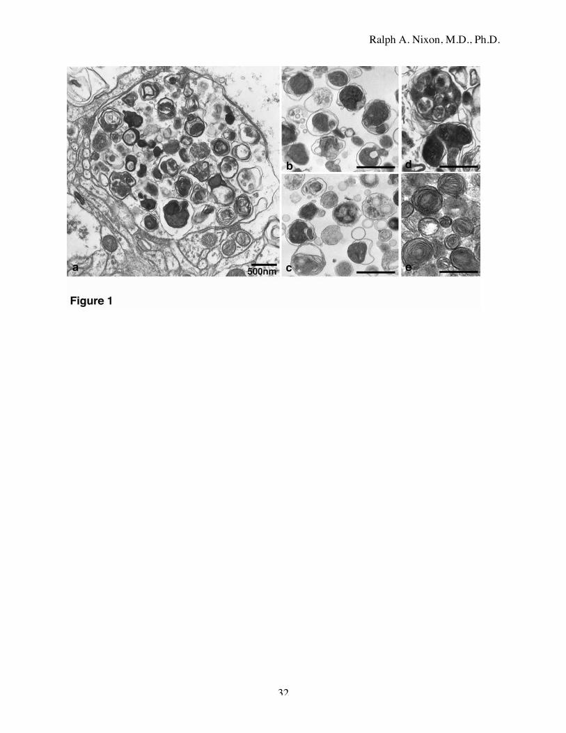
Figure 1: Ultrastructural appearance of autophagic vacuoles in AD brain and highly purified
subcellular fractions from mouse liver.
a: Dystrophic neurites contain abundant vesicles with a range of distinct morphologies similar to
those of AVs highly purified from mouse liver by a well-established subcellular fractionation
techniques (b,c). AV morphologies in brain include large double-membrane-limited vesicles
containing multiple smaller double-membrane vesicles exhibiting heterogeneous intralumenal
contents (d). Multilamellar bodies, another variant of AV [65], are also common in dystrophic
neurites (e).
Figure 2: Different dystrophic neurites contain distinct AV populations.
Abnormal neurites (long arrows) interspersed among normal-appearing neurites (arrowheads)
often differ in the predominating AV subtype present and contain AVs of a specific morphologic
type.
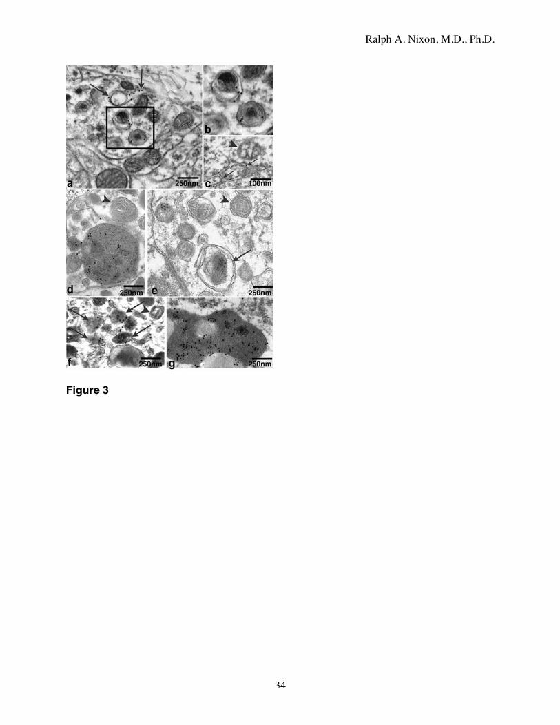
Figure 3: Immunogold electron microscopy of brain AVs.
Highly selective immunogold labeling by anti-calnexin antibodies of the double-limiting
membranes of AVs (a, boxed area; b) in abnormal neurites. A rare ER membrane profile in the
process of autophagosome formation can also be seen (arrows). c: Expected selective labeling
by anti-calnexin immunogold of ER membranes (arrows) in the cytoplasm of a neuronal
perikaryon. Mitochondria (arrowhead) and other cytoplasmic structures are unlabeled. The cat
D antibodies mainly label electron dense intralumenal contents of structures with single-limiting

Ralph A. Nixon, M.D., Ph.D.
30
membranes (d, arrow); micrographs in d-f are lightened to visualize gold more easily. Cat D
rarely labeled multilamellar bodies (d, e arrowheads) or double membrane-limited AVs lacking a
dense core or containing clear membranous elements (e, arrow). The contents of small single-
membrane dense bodies, corresponding to late AVs or lysosomes (f, arrow), are labeled (arrows)
but not most double membrane-limited vesicles (arrowhead). The proteinaceous components of
lipofuscin (g) are strongly immunolabeled by anti-cat D antibodies.
Figure 4: AVs are normally rare in brain but common in neuronal processes and synaptic
terminals in AD.
In non-AD control brains, AVs are infrequently seen in neurites (a) and rarely seen in perikarya.
In AD brain, AVs are common at relatively early stages of neuritic dystrophy in dendrites (b,
arrows) and in synaptic terminals (c, arrows) identified based on the presence of collections of
synaptic vesicles (c, arrowheads).
Figure 5: High incidence of AVs in non-dystrophic terminal dendritic branches.
Fine neuritic processes, which are apposed to a normal appearing neuronal perikarya (a), are
intact and relatively normal in appearance but often contain AVs (b-d, arrows). Higher
magnification images of the boxed areas in a are shown in b-d.
Figure 6: Marked AV accumulation in dystrophic neurites adjacent to amyloid deposits (A).
Increased frequency and severity of neuritic dystrophy (arrowheads) within the vicinity of senile
plaques, where at low magnification (inset) by light microscopic examination cat D
immunoreactivity is particularly strong.

Ralph A. Nixon, M.D., Ph.D.
31
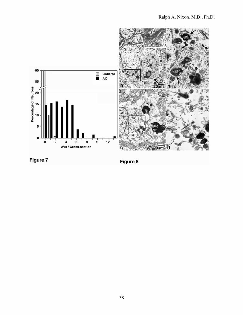
Figure 7: Autophagic vacuole frequency in neuronal perikarya in AD and control neocortex.
AVs counted by visual inspection of electron micrographs (see Materials and Methods) in 130
AD neurons from 7 different AD brains and 137 control neurons from 3 control brains free of
AD pathology. The percentages of neurons exhibiting a particular number of AVs in each group
of cells are plotted. Note that no AVs were detected in 90% of control neurons.
Figure 8: Autophagic vacuole frequency and depletion of other organelles in the perikarya of
degenerating neurons.
a: A tangle-bearing neuron exhibiting scattered bundles of paired helical filaments (arrow and
inset) and a peripherally displaced but otherwise normal nucleus. b: The boxed area of a is
shown at higher magnification. The perikaryon contains numerous AVs that include double-
membrane dense structures and multilamellar bodies (arrows) as well as many small dense
bodies or lysosomes (arrowheads). Autophagosome morphologies are rare in neuronal perikarya
despite their abundance in neurites. Mitochondria, Golgi, ER are relatively depleted in neurons
from AD brains (boxed area). These areas seen at higher magnification in d, also display
abnormal numbers of AVs and dense bodies compared to those seen in an intact neuron in Fig. 5.
Ralph A. Nixon, M.D., Ph.D.
32

Ralph A. Nixon, M.D., Ph.D.
33
Ralph A. Nixon, M.D., Ph.D.
34
Figure 3

Ralph A. Nixon, M.D., Ph.D.
35

Ralph A. Nixon, M.D., Ph.D.
36

Ralph A. Nixon, M.D., Ph.D.
37
Ralph A. Nixon, M.D., Ph.D.
38