extraaxial ependymoma of the posterior fossa - … ependymoma of the posterior fossa ... some...
TRANSCRIPT
Extraaxial Ependymoma of the Posterior Fossa
Melanie B. Fukui, Jeffery P. Hogg, and A. Julio Martinez
Summary: We report an unusual case of an extraaxial ependy-moma of the posterior fossa in an adult. MR imaging showed aheterogeneously enhancing extraaxial mass with a cystic com-ponent. Ependymoma should be included in the differential diag-nosis of uncommon extraaxial masses of the posterior fossa.
Index terms: Ependymoma; Posterior fossa, neoplasms
Two to six percent of all primary brain tumorsare ependymomas (1, 2). These tumors usuallyarise from the fourth ventricle (2, 3), and all arein, or are in proximity to, the fourth ventricle (4,5). In one large series (3), the mean age atpresentation for ependymomas of the posteriorfossa was 19 years. We report an unusual caseof an extraaxial ependymoma of the posteriorfossa in an adult.
Case ReportA 66-year-old man had left-sided facial pain in the
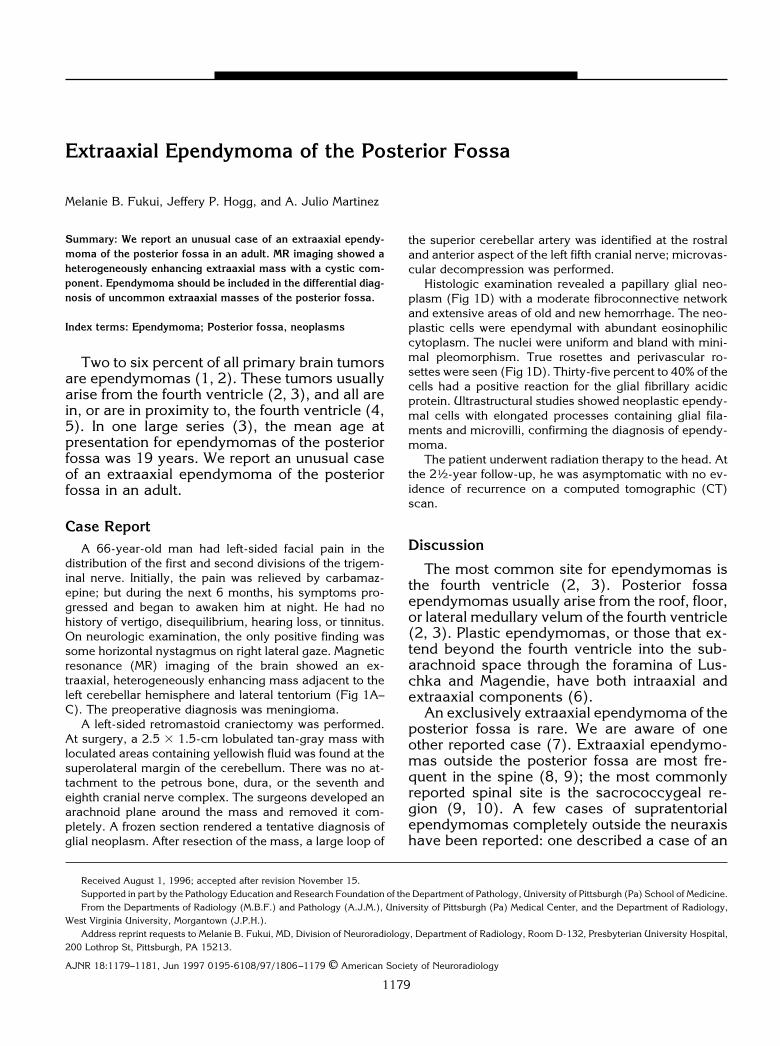
distribution of the first and second divisions of the trigem-inal nerve. Initially, the pain was relieved by carbamaz-epine; but during the next 6 months, his symptoms pro-gressed and began to awaken him at night. He had nohistory of vertigo, disequilibrium, hearing loss, or tinnitus.On neurologic examination, the only positive finding wassome horizontal nystagmus on right lateral gaze. Magneticresonance (MR) imaging of the brain showed an ex-traaxial, heterogeneously enhancing mass adjacent to theleft cerebellar hemisphere and lateral tentorium (Fig 1A–C). The preoperative diagnosis was meningioma.
A left-sided retromastoid craniectomy was performed.At surgery, a 2.5 3 1.5-cm lobulated tan-gray mass withloculated areas containing yellowish fluid was found at thesuperolateral margin of the cerebellum. There was no at-tachment to the petrous bone, dura, or the seventh andeighth cranial nerve complex. The surgeons developed anarachnoid plane around the mass and removed it com-pletely. A frozen section rendered a tentative diagnosis ofglial neoplasm. After resection of the mass, a large loop of
the superior cerebellar artery was identified at the rostraland anterior aspect of the left fifth cranial nerve; microvas-cular decompression was performed.
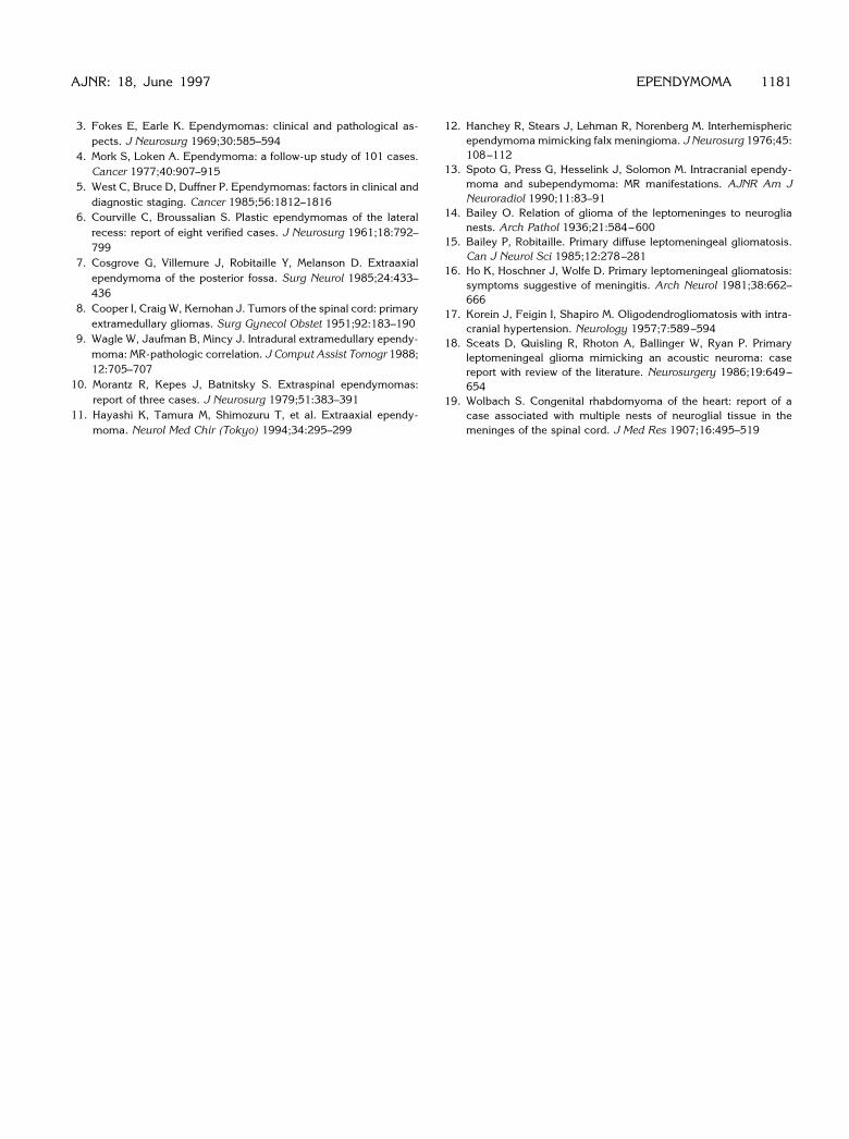
Histologic examination revealed a papillary glial neo-plasm (Fig 1D) with a moderate fibroconnective networkand extensive areas of old and new hemorrhage. The neo-plastic cells were ependymal with abundant eosinophiliccytoplasm. The nuclei were uniform and bland with mini-mal pleomorphism. True rosettes and perivascular ro-settes were seen (Fig 1D). Thirty-five percent to 40% of thecells had a positive reaction for the glial fibrillary acidicprotein. Ultrastructural studies showed neoplastic ependy-mal cells with elongated processes containing glial fila-ments and microvilli, confirming the diagnosis of ependy-moma.
The patient underwent radiation therapy to the head. Atthe 21⁄2-year follow-up, he was asymptomatic with no ev-idence of recurrence on a computed tomographic (CT)scan.
Discussion
The most common site for ependymomas isthe fourth ventricle (2, 3). Posterior fossaependymomas usually arise from the roof, floor,or lateral medullary velum of the fourth ventricle(2, 3). Plastic ependymomas, or those that ex-tend beyond the fourth ventricle into the sub-arachnoid space through the foramina of Lus-chka and Magendie, have both intraaxial andextraaxial components (6).
An exclusively extraaxial ependymoma of theposterior fossa is rare. We are aware of oneother reported case (7). Extraaxial ependymo-mas outside the posterior fossa are most fre-quent in the spine (8, 9); the most commonlyreported spinal site is the sacrococcygeal re-gion (9, 10). A few cases of supratentorialependymomas completely outside the neuraxishave been reported: one described a case of an
Received August 1, 1996; accepted after revision November 15.Supported in part by the Pathology Education and Research Foundation of the Department of Pathology, University of Pittsburgh (Pa) School of Medicine.From the Departments of Radiology (M.B.F.) and Pathology (A.J.M.), University of Pittsburgh (Pa) Medical Center, and the Department of Radiology,
West Virginia University, Morgantown (J.P.H.).Address reprint requests to Melanie B. Fukui, MD, Division of Neuroradiology, Department of Radiology, Room D-132, Presbyterian University Hospital,
200 Lothrop St, Pittsburgh, PA 15213.
AJNR 18:1179–1181, Jun 1997 0195-6108/97/1806–1179 © American Society of Neuroradiology
1179
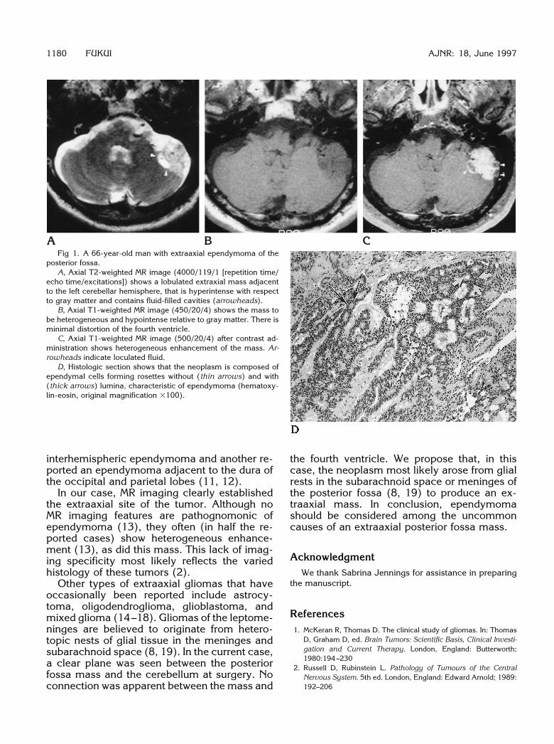
Fig 1. A 66-year-old man with extraaxial ependymoma of theposterior fossa.
A, Axial T2-weighted MR image (4000/119/1 [repetition time/echo time/excitations]) shows a lobulated extraxial mass adjacentto the left cerebellar hemisphere, that is hyperintense with respectto gray matter and contains fluid-filled cavities (arrowheads).
B, Axial T1-weighted MR image (450/20/4) shows the mass tobe heterogeneous and hypointense relative to gray matter. There isminimal distortion of the fourth ventricle.
C, Axial T1-weighted MR image (500/20/4) after contrast ad-ministration shows heterogeneous enhancement of the mass. Ar-rowheads indicate loculated fluid.
D, Histologic section shows that the neoplasm is composed ofependymal cells forming rosettes without (thin arrows) and with(thick arrows) lumina, characteristic of ependymoma (hematoxy-lin-eosin, original magnification 3100).
1180 FUKUI AJNR: 18, June 1997
interhemispheric ependymoma and another re-ported an ependymoma adjacent to the dura ofthe occipital and parietal lobes (11, 12).
In our case, MR imaging clearly establishedthe extraaxial site of the tumor. Although noMR imaging features are pathognomonic ofependymoma (13), they often (in half the re-ported cases) show heterogeneous enhance-ment (13), as did this mass. This lack of imag-ing specificity most likely reflects the variedhistology of these tumors (2).
Other types of extraaxial gliomas that haveoccasionally been reported include astrocy-toma, oligodendroglioma, glioblastoma, andmixed glioma (14–18). Gliomas of the leptome-ninges are believed to originate from hetero-topic nests of glial tissue in the meninges andsubarachnoid space (8, 19). In the current case,a clear plane was seen between the posteriorfossa mass and the cerebellum at surgery. Noconnection was apparent between the mass and
the fourth ventricle. We propose that, in thiscase, the neoplasm most likely arose from glialrests in the subarachnoid space or meninges ofthe posterior fossa (8, 19) to produce an ex-traaxial mass. In conclusion, ependymomashould be considered among the uncommoncauses of an extraaxial posterior fossa mass.
AcknowledgmentWe thank Sabrina Jennings for assistance in preparing
the manuscript.
References1. McKeran R, Thomas D. The clinical study of gliomas. In: Thomas
D, Graham D, ed. Brain Tumors: Scientific Basis, Clinical Investi-gation and Current Therapy. London, England: Butterworth;1980:194–230
2. Russell D, Rubinstein L. Pathology of Tumours of the CentralNervous System. 5th ed. London, England: Edward Arnold; 1989:192–206
3. Fokes E, Earle K. Ependymomas: clinical and pathological as-pects. J Neurosurg 1969;30:585–594
4. Mork S, Loken A. Ependymoma: a follow-up study of 101 cases.Cancer 1977;40:907–915
5. West C, Bruce D, Duffner P. Ependymomas: factors in clinical anddiagnostic staging. Cancer 1985;56:1812–1816
6. Courville C, Broussalian S. Plastic ependymomas of the lateralrecess: report of eight verified cases. J Neurosurg 1961;18:792–799
7. Cosgrove G, Villemure J, Robitaille Y, Melanson D. Extraaxialependymoma of the posterior fossa. Surg Neurol 1985;24:433–436
8. Cooper I, Craig W, Kernohan J. Tumors of the spinal cord: primaryextramedullary gliomas. Surg Gynecol Obstet 1951;92:183–190
9. Wagle W, Jaufman B, Mincy J. Intradural extramedullary ependy-moma: MR-pathologic correlation. J Comput Assist Tomogr 1988;12:705–707
10. Morantz R, Kepes J, Batnitsky S. Extraspinal ependymomas:report of three cases. J Neurosurg 1979;51:383–391
11. Hayashi K, Tamura M, Shimozuru T, et al. Extraaxial ependy-moma. Neurol Med Chir (Tokyo) 1994;34:295–299
AJNR: 18, June 1997
12. Hanchey R, Stears J, Lehman R, Norenberg M. Interhemisphericependymoma mimicking falx meningioma. J Neurosurg 1976;45:108–112
13. Spoto G, Press G, Hesselink J, Solomon M. Intracranial ependy-moma and subependymoma: MR manifestations. AJNR Am JNeuroradiol 1990;11:83–91
14. Bailey O. Relation of glioma of the leptomeninges to neuroglianests. Arch Pathol 1936;21:584–600
15. Bailey P, Robitaille. Primary diffuse leptomeningeal gliomatosis.Can J Neurol Sci 1985;12:278–281
16. Ho K, Hoschner J, Wolfe D. Primary leptomeningeal gliomatosis:symptoms suggestive of meningitis. Arch Neurol 1981;38:662–666
17. Korein J, Feigin I, Shapiro M. Oligodendrogliomatosis with intra-cranial hypertension. Neurology 1957;7:589–594
18. Sceats D, Quisling R, Rhoton A, Ballinger W, Ryan P. Primaryleptomeningeal glioma mimicking an acoustic neuroma: casereport with review of the literature. Neurosurgery 1986;19:649–654
19. Wolbach S. Congenital rhabdomyoma of the heart: report of acase associated with multiple nests of neuroglial tissue in themeninges of the spinal cord. J Med Res 1907;16:495–519
EPENDYMOMA 1181