extravascular albumin concentration of the...
TRANSCRIPT

Investigative Ophthalmology & Visual Science, Vol. 31, No. 1, January 1990Copyright © Association for Research in Vision and Ophthalmology
Exfravascular Albumin Concentration of the UveoCarol D. Toris, Jonathan E. Pederson, Shunji Tsuboi, Dale S. Gregerson, and Thomas J. Rice
The hypothesis that uveal vessels absorb fluid was tested by measuring the albumin in extravascularuveal tissues and in plasma. From these results the effective albumin concentration was calculated inboth rabbits and monkeys. Three separate methods were used to measure uveal albumin, and theresults of these were compared. In method 1, the intravenous fluorescein isothiocyanate (FITC)-albu-min concentration found in the uvea 5 min after injection (intravascular tracer) was subtracted fromthat found 2 hr after injection (intravascular plus extravascular tracer) to determine the extravascularalbumin concentration. In method 2, intravenous FITC-albumin was followed by vascular washoutafter a 2-hr equilibration period to determine extravascular uveal albumin. In method 3, the endoge-nous extravascular albumin concentration of uveal tissues was measured with an enzyme-linked im-munosorbent assay (ELISA) after vascular washout. The effective albumin concentration was deter-mined by dividing the data in methods 1, 2, and 3 by the extravascular albumin space volume. Theeffective albumin concentration in monkey (as percentage of plasma) was, for methods 1, 2, and 3: iris2, 3, and 4%; pars plicata 14, 12, and 7%; pars plana 2, 10, and 12%; and choroid 2, 12, and 10%,respectively. In rabbit, the extravascular albumin concentrations were: iris 10, 21, and 7%; pars plicata69, 26, and 39%; pars plana 41,46, and 10%; and choroid 88,30, and 26%, respectively. These findingsare lower than previously reported in rabbits, yet are consistent with previous estimates in monkeys.These results support the hypothesis that uveal vessels are capable of fluid absorption, since a largecolloid osmotic gradient exists across the vessel wall. Invest Ophthalmol Vis Sci 31:43-53, 1990
Knowledge of the Starling forces acting across theuveal blood vessels is important for understandingthe homeostatic mechanisms that keep the supra-choroidal space dry, prevent uveal edema, and pro-mote aqueous formation. The hydrostatic force act-ing across the vessel wall depends on the intravascularand intraocular pressures. The osmotic force dependsmainly on the colloid osmotic pressures of the intra-vascular and extravascular spaces; these pressures areexerted primarily by albumin concentration differ-ences across these spaces.
Previous measurements of the extravascular albu-min concentration of the uveal tract have been per-formed in rabbits,1'23 and the extravascular albuminconcentration was found to be about 70% of theplasma concentration. In contrast, in monkeys, fluo-rescence microscopy after intravenous Evans blue-albumin, followed by vascular perfusion (washout),
From the Department of Ophthalmology, University of Minne-sota, Minneapolis, Minnesota.
Supported by NIH grant EY-03277 and the James S. AdamsResearch Scholar Award from Research to Prevent Blindness (Dr.Pederson).
Submitted for publication: December 27, 1988; accepted June 6,1989.
Reprint requests: Carol B. Toris, The Department of Physiology,6-255 Millard Hall, University of Minnesota, Minneapolis, MN55455.
showed little fluorescence in the extravascular spaceof the uvea.4 This finding suggests that the extravas-cular albumin concentration of the uvea in monkeysis very low. Low concentrations have been reportedalso in humans (1-35% of serum).5 The current studywas undertaken to determine the extravascular uvealalbumin concentration in monkeys and to verify theextravascular albumin concentration previously re-ported in rabbits.12'3
Three separate methods were used to determine theextravascular albumin concentration in both rabbitsand monkeys. In method 1, FITC-albumin concen-tration in the nonperfused uvea was determined 5min after intravenous injection of the tracer. Thisdetermination was considered intravascular albumin(assuming that tracer did not leak from the vessels inthis time period) and was subtracted from FITC-al-bumin concentrations measured 2 hr after intrave-nous injection. In method 2, FITC-albumin was ad-ministered intravenously 2 hr before vascular wash-out, after which tracer in uveal tissues wasquantitated. In method 3, an enzyme-linked immu-nosorbent assay (ELISA) was used to measure endog-enous extravascular albumin directly.
Materials and Methods
All experiments conformed to the ARVO Resolu-tion on the Use of Animals in Research.
43
Downloaded From: http://iovs.arvojournals.org/pdfaccess.ashx?url=/data/journals/iovs/933150/ on 06/14/2018

44 INVESTIGATIVE OPHTHALMOLOGY 6 VISUAL SCIENCE / January 1990 Vol. 31
Tracer Studies
Method 1: Subtraction of Intravascular Tracer
Monkeys: Cynomolgus monkeys of either sex,weighing 2.5-4 kg, were anesthetized with intramus-cular ketamine hydrochloride (20 mg/kg) and givenan intravenous injection of 2 ml/kg of 10~3 M FITC-albumin (Sigma, St. Louis, MO) in HEPES buffer6
(300 mOsm/kg H2O). In four monkeys, one eye wasenucleated 5 min after FITC-albumin injection andthe fellow eye was enucleated either at 2 hr (2 mon-keys) or at 6 hr (2 monkeys). In one monkey, botheyes were enucleated 14 hr after FITC-albumin injec-tion, and in one monkey a second injection of FITC-albumin (1 ml/kg of 10~3 M) was given at 12 hr andthe eyes were enucleated at 24 hr. In all monkeys,blood samples were collected at 5 min, 2,6, 12 and 24hr where appropriate. Each eye was dissected into iris,pars plicata, pars plana, and choroid. The tissues wereweighed, homogenized in 1 ml phosphate buffer, andcentrifuged for 20 min. The fluorescence of theplasma and tissue extracts were measured with a Far-rand Spectrofluorometer System 3. Exciting thefluorochrome at 302 nm and recording the emissionat 523 nm dramatically reduced the autofluorescenceof the tissue. Intravascular tracer concentration de-termined at 5 min was subtracted from results at allother circulation times.
Rabbits: New Zealand White rabbits of either sex,weighing 4-5 kg, were used. To decrease the observedanaphylatic effects of the FITC-albumin injection,rabbits in this group were given 0.2-0.5 cc of epi-nephrine (1:10,000) subcutaneously 5 min prior tothe administration of 2 ml/kg 10~3 M FITC-albuminin the ear vein. An additional epinephrine injectionwas given if the animal showed signs of distress afterthe tracer injection. Eyes were enucleated at 5 min (n= 7), 2 hr (n = 10), 6 hr (n = 2), and 14 hr (n = 2)after the tracer injection. Blood samples were drawnat 5 min and just prior to sacrifice. Plasma and tissueextracts from the dissected eyes were measured forfluorescence in the same manner as in the monkeystudies.
Method 2: Washout of Intravascular Tracer
Monkeys: Under intramuscular ketamine hydro-chloride anesthesia, 2 ml/kg of 10~3 M FITC-albu-min was administered intravenously to four mon-keys. Two hr later a plasma sample was collected.Each animal was anesthetized deeply with sodiumpentobarbital; the thoracic cavity was opened; andthe heart exposed. A glass cannula with a 1-cm diam-eter opening was inserted into an incision made in theleft ventricle. The cannula was attached by rubbertubing to a reservoir of balanced salt solution (osmo-
lality = 300 mosmol/kg H2O, 37°C). The perfusatewas perfused rapidly at a pressure of 180 cm H2O intothe left ventricle and out of an incision in the rightatrium. Five to seven min later, when the outflowfrom the atrium was clear, seven eyes from the fourmonkeys were enucleated and dissected, and thetracer concentration in each tissue was determined asdescribed above for method 1. The results includeextravascular tracer only.
Rabbits: Four rabbits were anesthetized with 50mg/kg ketamine hydrochloride and 10 mg/kg xyla-zine intramuscularly, after which intravenous ure-thane (25% in 0.9% saline) was administered asneeded to maintain very deep anesthesia. The chestwas opened and a 10-gauge needle attached to poly-ethylene tubing and a reservoir of balanced salt solu-tion or Ringers was perfused at 120 cm H2O into theleft ventricle. An incision was made in the rightatrium for drainage and the animal was perfused for5-6 min. The iris and retinal vessels were observedwith an indirect ophthalmoscope to monitor washoutof blood. The remaining steps were the same as thosefor the monkey studies.
Methods 1 and 2
Extravascular Albumin Space Volume
Seven eyes from four monkeys and nine eyes fromfive rabbits were enucleated immediately after so-dium pentobarbital overdose. Each eye was dissectedinto iris, pars plicata, pars plana, and choroid. Eachtissue was weighed and incubated separately in a so-lution of 10~4 M FITC-albumin for 2 hr. Tissues werewashed briefly (1-2 sec), gently blotted, and weighedagain to determine the amount of tissue swelling dur-ing incubation. The samples were then homogenized,centrifuged for 20 min and the fluorescence of thetissue extract was measured as described above formethod 1.
Tissue Autofluorescence
Without tracer injection, the uvea from five mon-key eyes (four with vascular washout and one with-out) and five rabbit eyes (four with vascular washoutand one. without) were dissected and processed as de-scribed above for method 1. The autofluorescence ofthe tissue extracts were measured. Results with andwithout perfusion were similar and were averaged.
Calculations
Tissue autofluorescence was subtracted as back-ground from all other corresponding tissue fluores-cence measurements. Serial dilutions of FITC-albu-min in HEPES buffer were read on the spectrofluor-ometer, and a linear regression of the concentration
Downloaded From: http://iovs.arvojournals.org/pdfaccess.ashx?url=/data/journals/iovs/933150/ on 06/14/2018

No. 1 UVEAL EXTRA VASCULAR ALBUMIN / Toris er ol 45
of the FITC-albumin to its corresponding voltagereading was established. This standard curve wasused to determine the concentration of FITC-albu-min in each tissue sample, in the incubating solution,and in plasma samples.
The percent extravascular albumin is:
F2CS/F,CP X 100% (1)
where
F! = FITC-albumin concentration in the incu-bated tissues;
Cs = FITC-albumin concentration in the incubat-ing solution (1 X 10"4M);
F2 = FITC-albumin concentration in extravascu-lar uvea from either method 1 or method 2;and
Cp = FITC-albumin concentration in the plasmaat time of sacrifice.
Method 3: ELISA
Endogenous Extravascular Albumin Concentration
Monkeys: Four monkeys were perfused with bal-anced salt solution, and tissue samples from eighteyes were dissected, homogenized, and centrifuged,and the tissue extract collected as in methods 1 and 2.All tissue extracts then were assayed using a modifica-tion of a competition ELISA previously described.7
Initially, the specificity of the assay was tested byspiking chicken serum with known concentrations ofmonkey serum albumin (MSA); the resulting errorwas less than 5%. Then, flat-bottom polystyrene mi-croplates containing 96 wells/plate (Dynatech, Alex-andria, VA) were coated with 50 ng/well of cyno-molgus MSA (Organon Teknika, Malvern, PA) dis-solved in carbonate-bicarbonate buffer (pH 9.6) andincubated overnight (5°C).
Simultaneously, in separate microplates, rabbitanti-MSA (Organon Teknika) diluted 1:16,000 inPBS-Tween was incubated overnight (5°C) withMSA diluted serially from 1 ̂ g-1 ng for a standardcurve and with serial dilutions of tissue samples. Allcoated plates were washed with PBS-Tween andblocked with 2% fish gelatin for 2 hr at room temper-ature. The gelatin was removed and replaced with thepreincubation mixture of anti-MSA and MSA or withtissue extracts for 1 hr at room temperature. Thewells were washed again and goat anti-rabbit IgG al-kaline phosphatase conjugates (ICN, Lisle, IL) di-luted 1:2000 were incubated in the wells for 2 hr atroom temperature. The wells were rinsed in 0.05 MTris buffer (pH 7.5) and incubated with 6 mM p-ni-trophenyl phosphate (pH 9.2) as substrate for the al-kaline phosphatase. The absorbance of the yellowcolor was read at 405 nm in a Titertek Multiscan.
Optical density was converted to mg/ml for each un-known. Each sample was assayed three times and thevalues averaged.
Rabbits: Four rabbits were perfused with saline be-fore enucleation. Tissue dissection and collection oftissue extracts were the same as in method 2. Thetissue extracts were assayed for albumin using themonkey ELISA protocol (described above) except forthree substitutions: rabbit serum albumin (RSA; Or-ganon Teknika, Malvern, PA) for MSA; goat anti-rabbit albumin (Organon Teknika) diluted 1:32,000in PBS-Tween for rabbit anti-monkey albumin; andrabbit anti-goat IgG alkaline phosphatase (Sigma, StLouis, MO) diluted 1:2000 in PBS-Tween for goatanti-rabbit IgG alkaline phosphatase.
Extravascular Albumin Space Volume
Monkeys: Two monkeys were perfused with bal-anced salt solution, and four eyes were dissected intothe four uveal tissues listed above for methods 1 and2. These tissue samples were then incubated for 2 hrin autologous plasma. Tissue swelling was measuredby weighing the samples before and after incubation.Each tissue was rinsed for 2 sec in phosphate bufferand then homogenized and centrifuged, and the tis-sue extract collected. If necessary, samples werestored at -30°C until assayed.
Rabbits: Two rabbits were perfused with saline,and four eyes were dissected into the four uveal tis-sues and incubated in autologous plasma for 2 hr.The tissues were then rinsed, homogenized, and cen-trifuged, and the tissue extracts collected.
Calculations
The percent extravascular albumin was calculatedusing Equation (1) above, with the terms redefinedslightly, as follows:
Fj = albumin concentration in the tissues incu-bated in plasma;
Cs = albumin concentration in the autologousplasma in which the uveal tissues were incu-bated;
F2 = endogenous albumin concentration in extra-vascular uvea; and
Cp = average albumin concentration in monkey orrabbit plasma samples.
Fluorescence Microscopy
Two hr after anesthesia and intravenous injectionof FITC-albumin, one eye from one monkey wasenucleated and quickly frozen in isopentane cooledin liquid nitrogen. The monkey's vasculature wasthen perfused with warmed saline (as described
Downloaded From: http://iovs.arvojournals.org/pdfaccess.ashx?url=/data/journals/iovs/933150/ on 06/14/2018
INVESTIGATIVE OPHTHALMOLOGY & VISUAL SCIENCE / January 1990 Vol. 31
Table 1. Comparison of experimental procedures
Procedure
TracerTracer circulation times (hr)
monkeyrabbit
Vascular washoutIncubation solutionIncubation time (hr)
Method 1(subtraction of intravascular tracer)
FITC-albumin
0.08, 2, 6, 14, 240.08,2,* 6, 14noFITC-albumin2
Method 2(washout of intravascular tracer)
FITC-albumin
2*2*yesFITC-albumin*2
Method 3(ELISA)
none
nonenoneyesplasma2
' Fluorescence microscopic examination of tissue.
above) and the fellow eye was removed and frozen inthe same manner.
One eye from one rabbit was enucleated 2 hr afterintravenous FITC-albumin injection. One eye fromanother rabbit was enucleated 2 hr after intravenousFITC-albumin injection and vascular washout. Oneeye from a third rabbit was enucleated and then in-cubated in FITC-albumin for 2 hr. All tissues werefrozen in isopentane cooled in liquid nitrogen.
All frozen specimens were freeze-dried using amethod described previously8'9 and examined with afluorescence microscope.
A comparison of all experimental procedures isgiven in Table 1.
Results
Fluorescence Microscopy
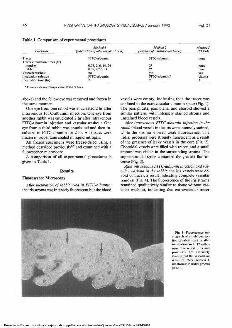
After incubation of rabbit uvea in FITC-albumin:the iris stroma was intensely fluorescent but the blood
vessels were empty, indicating that the tracer wasconfined to the extravascular albumin space (Fig. 1).The pars plicata, pars plana, and choroid showed asimilar pattern, with intensely stained stroma andunstained blood vessels.
After intravenous FITC-albumin injection in therabbit: blood vessels in the iris were intensely stained,while the stroma showed weak fluorescence. Theiridial processes were strongly fluorescent as a resultof the presence of leaky vessels in the core (Fig. 2).Choroidal vessels were filled with tracer, and a smallamount was visible in the surrounding stroma. Thesuprachoroidal space contained the greatest fluores-cence (Fig. 3).
After intravenous FITC-albumin injection and vas-cular washout in the rabbit: the iris vessels were de-void of tracer, a result indicating complete vascularremoval (Fig. 4). The fluorescence of the iris stromaremained qualitatively similar to tissue without vas-cular washout, indicating that extravascular tracer
ttr*
Fig. 1. Fluorescence mi-crograph of an oblique sec-tion of rabbit iris 2 hr afterincubation in FITC-albu-min. The iris stroma andprocesses are intenselystained, but the vasculatureis free of tracer (arrows). I,iris stroma; P, iridial process(XI20).
Downloaded From: http://iovs.arvojournals.org/pdfaccess.ashx?url=/data/journals/iovs/933150/ on 06/14/2018

No. 1 UVEAL EXTRAVA5CULAR ALBUMIN / Toris er ol 47
Fig. 2. Fluorescence mi-crograph of rabbit iris 2 hrafter intravenous injectionof FITC-albumin and novascular washout. The irisvasculature is intensely flu-orescent (arrow), while theiris stroma is only slightlystained. One process is in-tensely fluorescent; itsstroma and vasculature areindistinguishable. I, irisstroma; P, iridial process;AC, anterior chamber; PC,posterior chamber (X120).
was not removed. After vascular washout, the irisprocesses were less fluorescent as a result of washoutof the highly vascular core. The choroidal vesselswere devoid of tracer, but the stroma surroundingsome vessels were ringed by a small amount. Thefluorescence of the suprachoroidal space remainedquite prominent (Fig. 5).
After intravenous FITC-albumin in the monkey:tracer was confined to retinal vessels, but some leak-
age from choroidal vessels into the surroundingstroma was noted (Fig. 6). The iris contained intra-vascular tracer only. Ciliary processes were highly flu-orescent, and the fluorescence of the ciliary musclewas confined mainly to the vasculature.
After intravenous FITC-albumin and vascularwashout in the monkey: the iris and retina containedno visible tracer. The adventitia surrounding choroi-dal vessels were fluorescent, but the lumen of each
Fig. 3. Fluorescence mi-crograph of rabbit retinaand choroid 2 hr after intra-venous injection of FITC-albumin. The avascular ret-ina contains no tracer, whilethe choroidal vessels are in-tensely fluorescent (largearrows). Intense fluores-cence can also be seen in thesuprachoroidal space be-tween the choroid andsclera (small arrow). R, ret-ina; C, choroid; S, sclera(XI20).
Downloaded From: http://iovs.arvojournals.org/pdfaccess.ashx?url=/data/journals/iovs/933150/ on 06/14/2018

INVESTIGATIVE OPHTHALMOLOGY 6 VISUAL SCIENCE / Januory 1990 Vol. 31
Fig. 4. Fluorescence mi-crograph of rabbit iris 2 hrafter intravenous injectionof FITC-albumin and vas-cular washout. The irisstroma and one process arestained with tracer, while ir-idial vessels are devoid oftracer (arrows). I, irisstroma; P? iridial process;AC, anterior chamber; PC,posterior chamber (X120).
vessel was empty. Some tracer was seen in the choroi-dal stroma (Fig. 7).
Quantitation of Uveal Albumin: Methods 1 and 2
Extravascular Albumin Space Volume
The amount of FITC-albumin recovered fromuveal tissues incubated in tracer is summarized in
Table 2 under the columns labeled "Incubate". In themonkey, the pars plana and choroid contained aboutthree times more tracer than did the iris and parsplicata (averaging 18 X 10~7 M and 6 X 10~7 M,respectively). In the rabbit, on the other hand, thetracer is more evenly distributed throughout theuveal tissues, with concentrations averaging 13X 1CT7 M.
Fig. 5. Fluorescence mi-crograph of rabbit retinaand choroid 2 hr after intra-venous injection of FITC-albumin and vascular wash-out. The avascular retina isdevoid of tracer. The cho-roidal vessels are also de-void of fluorescence (largearrows), but the choroidalstroma surrounding vesselsdoes contain tracer. The su-prachoroidal space betweenthe choroid and sclera is in-tensely stained (smallarrow). R, retina; C, cho-roid; S, sclera (XI20).
Downloaded From: http://iovs.arvojournals.org/pdfaccess.ashx?url=/data/journals/iovs/933150/ on 06/14/2018

No. 1 UVEAL EXTRAVASCULAR ALBUMIN / Toris er ol 49
Fig. 6. Fluorescence mi-crograph of monkey retinaand choroid 2 hr after intra-venous injection of FITC-albumin. The retinal andchoroidal vessels and thechoriocapillaris are in-tensely stained with tracer(white arrows). The fluores-cence in the retina is con-fined to retinal vessels butthe fluorescence from thechoroidal vessels can beseen in the choroidal stroma(black arrow). R, retina; C,choroid; S, sclera (X120).
Method 1: Subtraction oflntravenous Tracer
In monkeys, the tracer concentration remained themost stable in the uveal tissues between 2 and 6 hrafter tracer injection. After 6 hr, the amount of recov-ered tracer rose, indicating increased permeability ofuveal vessels. In the rabbit, the tracer concentrationpeaked at 2 hr in the iris, pars plicata, and pars plana,and at 6 hr in the choroid. On the basis of these
results, the optimal circulation time required to equil-ibrate intravenous tracer with uveal tissue in botlimonkey and rabbit was 2 hr.
The amount of tracer recovered in uveal tissues 5min after tracer injection (intravascular albumin) wassubtracted from the amount recovered at 2 hr (totalalbumin) to determine the extravascular albuminconcentration. These results are summarized inTable 2 under the columns labeled "Method 1". The
Fig. 7. Fluorescence mi-crograph of the monkey ret-ina and choroid 2 hr afterintravenous injection ofFITC-albumin and vascularwashout. The entire retinaincluding vasculature is de-void of tracer. The fluores-cent spots are artifact. Cho-roidal vessels are also de-void of tracer (verticalarrows) but the adventitiaand choroidal stroma sur-rounding the vessels are flu-orescent (horizontal arrow).R, retina; C, choroid; S,sclera; N, nerve (X120).
Downloaded From: http://iovs.arvojournals.org/pdfaccess.ashx?url=/data/journals/iovs/933150/ on 06/14/2018

50 INVESTIGATIVE OPHTHALMOLOGY b VISUAL SCIENCE / January 1990 Vol. 31
Table 2.
Tissue
IrisP plicataP planaChoroid
FITC-albumin
Incubate]
5.6 ±0.76.5 ±0.6
17.9 ±0.718.6 ±1.6
recovered in uveal
Monkey
Method I
0.1 ±0.20.9 ± 0.40.4 ± 0.30.3 ± 0.2
tissues*
Method 2
0.2 ±0.10.8 ± 0.041.8 ± 1.072.3 ±0.16
Incubate]
12.9 ±0.716.9 ±0.511.6±1.39.2 ±1.0
Rabbit
Method I
1.4 ±0.511.6 ± 1.24.7 ± 1.28.1 ± 1.0
Method 2
2.7 ± 0.34.4 ± 0.45.3 ± 0.42.8 ± 0.5
* Values are X10"7 M/tissue, mean ± SE.f Incubate, tracer recovered from tissue incubated in tracer.
Methods 1 and 2: experimental procedures used FITC-albumin (see text).P = pars.
amount of tracer recovered in rabbit uvea rangedfrom 1 X 10~7-12 X 10~7 M/tissue, whereas, in themonkey, substantially less tracer was recovered (<1X 10~7 M/tissue). Therefore, an average of at least 10times more tracer was recovered in each rabbit uvealtissue than in each monkey uveal tissue.
Method 2: Washout of Intravenous Tracer
The amount of tracer recovered in uveal tissuesafter vascular washout is summarized in Table 2,under the column labeled "Method 2". In both mon-key and rabbit, the iris contained the least amount oftracer in the uveal tissues, and yet the rabbit iris hadten times more tracer than did the monkey iris. Thepars plicata and pars plana also held substantiallymore tracer in rabbit than monkey. Only the choroidcontained a similar amount of tracer in both species.
For comparison, six rabbit eyes without vascularwashout were processed, and predictably, the albu-min concentration was substantially higher than ineyes with vascular washout. For iris, pars plicata, parsplana, and choroid, 3.5 ± 0.8, 13.0 ± 1.3, 5.0 ± 0.9,and 8.9 ± 1.2 X 10~7 M/tissue, respectively, wererecovered. The highly vascular pars plicata and cho-roid contained the highest albumin concentrations.
Quantitation of Uveal Albumin: Method 3: ELISA
The albumin concentration in the monkey andrabbit uvea as measured by the ELISA method issummarized in Table 3. The columns labeled "Incu-bate" summarize the albumin concentration in uvealtissues incubated 2 hr in autologous plasma. Themonkey ciliary body and choroid contained much(2-6 times) more albumin than did the rabbit. Theextravascular albumin in the rabbit uvea was evenlydivided among the four uveal tissues, whereas in themonkey, the posterior uvea contained nearly threetimes more than did the anterior uvea.
The columns labeled "Method 3" in Table 3 listthe endogenous extravascular albumin measured inuveal tissues. The pars plana and choroid of themonkey contained substantially (2-7 times) more al-
bumin than did the rabbit, while the monkey iriscontained substantially less than did the rabbit.
The plasma albumin concentrations were 38.8mg/ml for monkey and 24.5 mg/ml for rabbit.
Effective Albumin Concentration
Table 4 compares the effective albumin concentra-tion in tissues of monkey and rabbit uvea calculatedfrom the three separate experiments. The effectivealbumin concentration in monkey uvea is small; notissue contained more than 14% of plasma albumin.In the rabbit, on the other hand, the effective albuminconcentration was as high as 88% of plasma. In addi-tion, the results with each method were more variedin the rabbit than in the monkey. For example, de-pending on method used, the extravascular albuminconcentration in the rabbit pars plicata was 26-69%of plasma, whereas that in the monkey pars plicatawas 11-14%.
In the rabbit without vascular washout, the effec-tive albumin concentration was, for iris, pars plicata,pars plana and choroid, 27, 77, 40, and 97%, respec-tively.
Discussion
Two decades ago, the effective albumin concentra-tion of the rabbit uvea was reported to be 68% ofplasma in the choroid and 74% of plasma in ciliaryprocesses.2 Using a technique similar to that reported
Table 3.
IrisP plicataP planaChoroid
Extravascular albumin
Monkey
Incubate] Method 3
0.25 ±0.06 0.007 ±0.0010.85 ±0.06 0.11 ±0.011.76 ±0.55 0.22 ±0.031.38 ±0.29 0.16 ±0.03
ELISA*
Rabbit
Incubate]
0.38 ± 0.070.33 ±0.140.29 ± 0.030.29 ± 0.05
Method 3
0.030.140.030.08
± 0.004±0.02± 0.004±0.02
* Values are mg/ml, mean ± SE.t Incubate, albumin recovered from tissue incubated in autologous
plasma; method 3, endogenous albumin measured in uveal tissues.P = pars.
Downloaded From: http://iovs.arvojournals.org/pdfaccess.ashx?url=/data/journals/iovs/933150/ on 06/14/2018

No. 1 UVEAL EXTRA VASCULAR ALBUMIN / Toris er ol 51
Table 4.
IrisP plicataP planaChoroid
Effective albumin
Method I
1.8 ±4.013.5 ±7.82.2 ± 1.71.8 ± 1.1
concentration*Monkey
Method 2
3.6 ±0.512.3 ± 1.310.1 ±0.612.4 ± 1.4
Method 3
2.4 ± 0.610.9 ± 1.110.5 ± 3.09.7 ±2.3
Method 1
10.8 ±68.6 ±40.5 ±88.0 ±
3.78.0
11.015.2
Rabbit
Method 2
20.9 ± 2.626.0 ± 2.545.7 ± 6.230.4 ± 6.4
Method 3
7.3 ± 1.739.2 ± 17.59.6 ± 1.6
25.5 ± 7.7
* Values are percent of plasma.Values are calculated from equation (1) and are reported as mean ± SE.SE was calculated with the delta method for the Taylor series approximation.P = pars.
previously,2'3 the values in the current study (method1) are 88% in the choroid and 69% in the pars plicata(including ciliary processes). These data indicate thatthere is a small colloid osmotic pressure gradientacross the capillary walls. However, such a smallpressure differential would be insufficient to counter-balance the net outward hydrostatic pressure foundacross the capillary walls (since capillary hydrostaticpressure is greater than intraocular pressure1011); theresult would be in fluid accumulation in the stroma.Since this does not happen in the normal eye, theextravascular albumin concentrations of rabbit uvealtissues measured in method 1 must be greatly overes-timated. Under the same experimental conditions,the effective albumin concentrations of the monkeyuvea is quite low, between 2-14% of plasma. Speciesdifferences may account for some of the dissimilar-ities.
Using two other measuring techniques (methods 2and 3), the extravascular albumin concentration wasas small as 26-30% of plasma in the choroid and26-39% of plasma in the pars plicata of the rabbit.The colloid osmotic pressure gradient in these tissuesis therefore sufficient to cause absorption of fluid intothe capillary. Using methods 2 and 3 in the monkey,the effective albumin concentration in the uvea is lessthan 14% of plasma.
Factors Governing Uveal Albumin
The extravascular uveal albumin concentration isgoverned by two factors: 1) movement of intravascu-lar albumin through the capillary wall (capillary per-meability), and 2) removal of extravascular albuminfrom the uvea and suprachoroidal space.
Capillary permeability is determined by the char-acteristics of the junctional connections between en-dothelial cells. The endothelium of iris blood vesselscontains tight junctions,12 and the capillary perme-ability for blood-borne tracers like horseradish per-oxidase (HRP; MW = 40,000 d) is low in the mon-key12 and rabbit.13 In the current study, the iridialcapillary permeability for blood-borne albumin (MW
= 67,000 d) is also low, and as a result, the albuminconcentration of the iris extravascular space in bothmonkeys and rabbits is small (less than 21% ofplasma).
In contrast to the tight junctions of the iris endo-thelium, the capillaries of the ciliary body (pars pli-cata and pars plana) are studded with fenestrationswhich allow free movement of HRP into the extra-vascular space.1214 In addition, the most extensiveextravascular accumulation of the intra vascular fluo-rescent tracers Evans blue-albumin4 and albumin-bound lisamine rhodamine15 is found in the ciliarybody and especially in the ciliary processes of the parsplicata. In agreement with this, the current studyfound the pars plicata to contain the greatest amountof extravascular albumin in both monkeys and rab-bits (method 3). However, these values are still quitesmall, averaging less than 11% of plasma in monkeyand 40% of plasma in rabbit.
The choroid, like the ciliary body, is highly vascu-larized with fenestrated vessels,12 but intravenousHRP is confined to the choriocapillaris,16 and Evansblue-albumin does not accumulate in choroidal tis-sues of monkey.4 In the current study, even though asmall amount of tracer was visible in the extravascu-lar choroid of monkey (Fig. 7), the effective albuminconcentration was not more than 13% of plasma(Table 4). In the stroma of the rabbit choroid, verylittle tracer was visible, but in the suprachoroidalspace the fluorescence was substantial (Fig. 5) and theeffective albumin concentration was three times thatof monkey. Accumulation of tracer in the supracho-roidal space may have been an inflammatory re-sponse to FITC-albumin. An overestimate of effec-tive albumin concentration in choroidal stroma ofrabbit would result.
The only egress for extravasated uveal albumin isby uveoscleral outflow of aqueous through the supra-ciliary and suprachoroidal spaces and sclera.3 Uveo-scleral outflow is driven by a hydrostatic pressure dif-ferential in these spaces between the anteriorchamber and the posterior choroid; this differential isgenerated mainly by the colloid osmotic absorption
Downloaded From: http://iovs.arvojournals.org/pdfaccess.ashx?url=/data/journals/iovs/933150/ on 06/14/2018

52 INVESTIGATIVE OPHTHALMOLOGY & VISUAL SCIENCE / January 1990 Vol. 31
of choroidal vessels.17 In the rabbit, in which the ef-fective albumin concentration in the choroid wasthree times higher than in the monkey (currentstudy), the colloid osmotic pressure across choroidalvessels would be smaller, and the driving force foruveoscleral outflow would be less than in the mon-key. In agreement with this, uveoscleral outflow inthe rabbit is only 3% of aqueous drainage18 while inthe monkey it is as high as 50%.8
Since extravascular albumin in the uvea leaves theeye mainly by uveoscleral outflow, the rate of wash-out of extra vasated albumin in each tissue of the uveais a function of the proximity of the tissue to theuveoscleral outflow drainage route. For example, theremoval of albumin from the extravascular choroidwhich lies adjacent to the sclera should be quickerthan the albumin removal from the ciliary processeswhich are separated from the sclera by the ciliarymuscle.
Taking into consideration both the rate of albuminleakage into extravascular tissue and the rate of exitby uveoscleral washout, the theoretic albumin con-centration in the uvea is: iris < pars plana < choroid< pars plicata. The iris contains the least amount ofextravascular albumin because of its tight blood ves-sels. The pars plana is not highly vascular, and extra-vasated albumin is readily washed away by theuveoscleral drainage route, making the extravascularalbumin concentration relatively low. The choroidcontains many vessels slightly leaky to albumin,which is readily removed by the uveoscleral drainageroute. Thus, an intermediate amount of extravascularalbumin should be found in the choroid. The parsplicata contains many very leaky vessels, and albu-min is not readily washed away, so this tissue shouldcontain the greatest amount of extravascular albu-min. Results from method 3 in the rabbit conform tothis prediction.
Technical Problems
Three significant technical problems were notedduring the course of this investigation: tissue swelling,difficulties with vascular washout, and tracer toxicity.
Incubating uveal tissues in FITC-albumin for 2 hrcaused tissue to swell to varying degrees. The incuba-tion of rabbit uvea in FITC-albumin resulted in anincrease in tissue weight of the pars plicata (15%) andpars plana (6%) but not of the choroid or iris. Monkeyuvea was more susceptible to swelling, with the irisdoubling in weight (95%) and the remaining uveaswelling 18-19% after incubation in FITC-albumin.Tissue swelling caused an overestimate of F] (Equa-tion (1)) and a proportional underestimate of the ef-
fective albumin concentration in methods 1 and 2.This error was insignificant in method 3 since tissueswelling was not detected after incubation in plasma.
The vasculature washout procedure in methods 2and 3 raised two concerns: either the washout of in-travascular albumin was incomplete, or the removalof some extravascular albumin had occurred. The al-bino rabbit was monitored with an ophthalmoscopeduring perfusion; this monitoring showed that com-plete washout of blood was difficult to achieve. Sinceblood contains a high concentration of albumin, in-complete washout would lead to an overestimate ofF2 and a proportional overestimate of the effectivealbumin concentration. When rabbit uvea withoutvascular washout was assayed, the effective albuminconcentration was overestimated by at least 2-fold. Inthe monkey, vascular washout could not be visuallymonitored, but the washout procedure was accom-plished with less technical difficulty than in the rab-bit, and the extravascular albumin concentration wasquite low. The concern that the vascular washoutprocedure might remove some extravascular albuminhas been discussed previously4 and could lead to anunderestimate of F2 and of effective albumin concen-tration. However, since vascular washout was limitedto 7 min, the extravascular albumin removal shouldbe minimal.
The major technical difficulty encountered was thetoxic effects of FITC-albumin. In the rabbit, thetracer injection caused an anaphylacticlike reactionand a significant mortality rate if not treated withepinephrine. The vasodilation of the anaphylaticlikereaction or the vasoconstriction of the epinephrinewould either increase or decrease F2, respectively.Ideally, these vasoactive effects would cancel eachother out and so cause little error in the calculatedeffective albumin concentrations. The fluorescencein the suprachoroidal space seen in Figs. 3 and 5 maybe an indication of choroidal effusion caused by aninflammatory reaction to the tracer. This effusionwould result in an overestimate of F2 and of the cal-culated effective albumin concentration. In the mon-key, the permeability of uveal vessels to the tracerincreased dramatically 14 hr after intravenous tracerinjection, which suggests that the vessels were af-fected more subtly prior to this time. However, in-flammatory responses were not noted, and accumu-lation of tracer in the suprachoroidal space was notpresent (Figs. 6, 7). In contrast to rabbits, monkeysremained healthy throughout the study and did notsuffer any anaphylactic effects from the tracer.
Of the three measurements made, method 3 is themost meaningful. Tissue incubation in plasma didnot cause swelling, and since no tracers were injected,
Downloaded From: http://iovs.arvojournals.org/pdfaccess.ashx?url=/data/journals/iovs/933150/ on 06/14/2018

No. 1 UVEAL EXTRAVASCULAPv ALBUMIN / Toris er ol 53
toxicity problems did not apply. In addition, theassay was very specific to monkey or rabbit albuminand was sensitive to 1 ng/m\.
In summary: after consideration of the technicaldifficulties, the extravascular albumin concentrationin monkey was very low, (around 10% of plasma). Inrabbit, the values were somewhat higher (around30%, methods 2 and 3), and yet were lower than in aprevious report.2 A small effective albumin concen-tration of the uvea would favor absorption of fluidinto the blood vessels, maintaining relative dehydra-tion of the extravascular space and suprachoroidalspace. The best method to measure extravascular al-bumin concentration is with the ELISA.
Key words: uvea, albumin, monkey, rabbit, FITC-albumin
Acknowledgments
Invaluable technical assistance was provided by KarlanHunt and Volker Knospe.
References
1. Bill A: The albumin exchange in the rabbit eye. Acta PhysiolScand 60:18, 1964.
2. Bill A: A method to determine osmotically effective albuminand gammaglobulin concentrations in tissue fluids, its appli-cation to the uvea and a note on the effects of capillary "leaks"on tissue fluid dynamics. Acta Physiol Scand 73:511, 1968.
3. Bill A: Capillary permeability to and extravascular dynamics ofmyoglobulin, albumin and gammaglobulin in the uvea. ActaPhysiol Scand 73:204, 1968.
4. Radius RL and Anderson DR: Distribution of albumin in thenormal monkey eye as revealed by Evans blue fluorescencemicroscopy. Invest Ophthalmol Vis Sci 19:238, 1980.
5. Allansmith MR, Whitney CR, McClellan BH, and NewmanLP: Immunoglobins in the human eye: Location, type andamount. Arch Ophthalmol 89:36, 1973.
6. Tsuboi S, Fujimoto T, Uchihori Y, Emi K, Iizuka S, KishidaK, and Manabe R: Measurement of retinal permeability tosodium fluorescein in vitro. Invest Ophthalmol Vis Sci25:1146, 1984.
7. Stjernschantz J, Gregerson D, Bausher L, and Sears M: En-zyme-linked immunosorbent assay of Substance P: A study inthe eye. J Neurochem 38:1323, 1982.
8. Toris CB and Pederson JE: Aqueous humor dynamics in ex-perimental iridocyclitis. Invest Ophthalmol Vis Sci 28:477,1987.
9. Grayson M and Laties A: Ocular localization of sodium fluo-rescein: Effect of administration in rabbit and monkey. ArchOphthalmol 4:600, 1971.
10. Bill A: Blood circulation and fluid dynamics in the eye. PhysiolRes 55:383, 1975.
11. Cole DF: Secretion of the aqueous humor. Exp Eye Res25(Suppl):161, 1977.
12. Raviola G: The structural basis of the blood-ocular barriers.Exp Eye Res.25(Suppl):27, 1977.
13. Pedersen OO: A light and electron microscopic study of thepermeability of the rabbit iris vessels to horseradish peroxidasein experimental uveitis. Acta Ophthalmol 52:678, 1974.
14. Smith RS: Ultrastural studies of the blood-aqueous barrier: I.Transport of an eleciron-dense tracer in the iris and ciliarybody of the mouse. Am J Ophthalmol 71:1066, 1971.
15. Machemer R: Angiographic-histologic correlation of eye ves-sel permeability with protein-bound fluorescent dye. Am JOphthalmol 69:27, 1970.
16. Toris CB and Pederson JE: Experimental retinal detachment:VII. Intravenous horseradish peroxidase diffusion across theblood-retinal barrier. Arch Ophthalmol 102:752, 1984.
17. Emi K, Pederson JE, Toris CB: Hydrostatic pressure of thesuprachoroidal space. Invest Ophthalmol Vis Sci 30:233, 1989.
18. Bill A: The routes for bulk drainage of aqueous humour inrabbits with and without cyclodialysis. Doc Ophthalmol20:157, 1966.
Downloaded From: http://iovs.arvojournals.org/pdfaccess.ashx?url=/data/journals/iovs/933150/ on 06/14/2018