eyelid reconstruction - university of texas medical … lower limb (upper limb) angle skin incision...
TRANSCRIPT

Eyelid Reconstruction
Michael Underbrink, M.D.
Faculty Advisor: Karen Calhoun, M.D.
The University of Texas Medical Branch
Department of Otolaryngology
Grand Rounds Presentation
December 18, 2002

Introduction
Goal: restore normal anatomy and function
Various reconstructive techniques
Complex anatomy

Anatomy
Eyelid functions
– Protect eye (light, injury, desiccation)
– Tear production and distribution
Anterior/posterior lamella
Extremely thin skin (upper > lower)
Skin
– Little subcutaneous fat
– Adherent over the tarsus (levator aponeurosis)
Anatomy
Horizontal length – 30 mm
Palpebral fissure – 10 mm
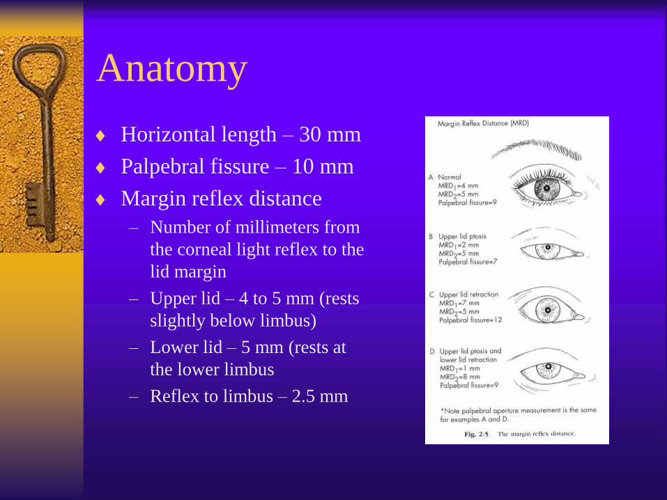
Margin reflex distance
– Number of millimeters from
the corneal light reflex to the
lid margin
– Upper lid – 4 to 5 mm (rests
slightly below limbus)
– Lower lid – 5 mm (rests at
the lower limbus
– Reflex to limbus – 2.5 mm

Anatomy
Tarsus
– Dense, fibrous tissue
– Contour and skeleton
– Contain meibomian
glands
– Length – 25 mm
– Thickness – 1 mm
– Height
• Upper plate – 10 mm
• Lower plate – 4 mm

Anatomy – Muscles
Protractor
– Orbicularis
Retractors
– Levator
– Müller’s

Orbicularis Oculi Muscle

Levator palpebral superioris
and Müller’s muscle

Lower Lid Anatomy

Anatomy
Orbital Septum
– Fascial barrier
– Underlies posterior
orbicularis fascia
– Defines anterior extent
of orbit and posterior
extent of eyelid

Anatomy
Canthal tendons
– Extensions of preseptal & pretarsal orbicularis
– Lateral slightly above medial
– Lateral tendon attaches to Whitnall’s tubercle
1.5 cm posterior to orbital rim
– Medial tendon complex, important for lacrimal
pump function

Canthal Tendons

Lacrimal System

Lacrimal Excretory Pump
Anatomy – Blood Supply
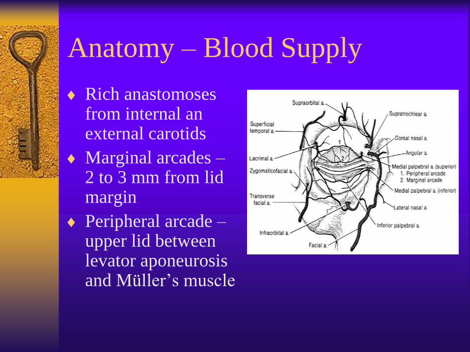
Rich anastomoses from internal an external carotids
Marginal arcades – 2 to 3 mm from lid margin
Peripheral arcade – upper lid between levator aponeurosis and Müller’s muscle

Related Vocabulary
Ptosis – upper eyelid margin abnormally inferiorly
displaced
Entropion – inward rotation of eyelid margin
Ectropion – eversion of eyelid margin
Trichiasis – misdirected eyelashes
Distichiasis – aberrant eyelashes from metaplastic
meibomian glands
Epiblepharon – normal eyelashes pushed toward
the eye by redundant folds of skin
Epicanthal folds – vertical folds of skin over the
medial canthus

Lower Eyelid Reconstruction
Direct Closure
Lateral Cantholysis
Tenzel Rotational Flap
Free Tarsal Grafts
Hughes Procedure
Mustarde (rotational cheek) Flap

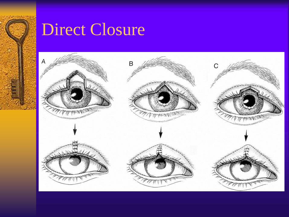
Direct Closure
30% defects in young patients
Up to 45% in older patients with more eyelid
laxity
Lateral cantholysis provides additional 5 mm
Tarsal defect should be squared
Temporal slant to musculocutaneous layer

Direct Closure

Lid Margin Repair
Lateral Cantholysis
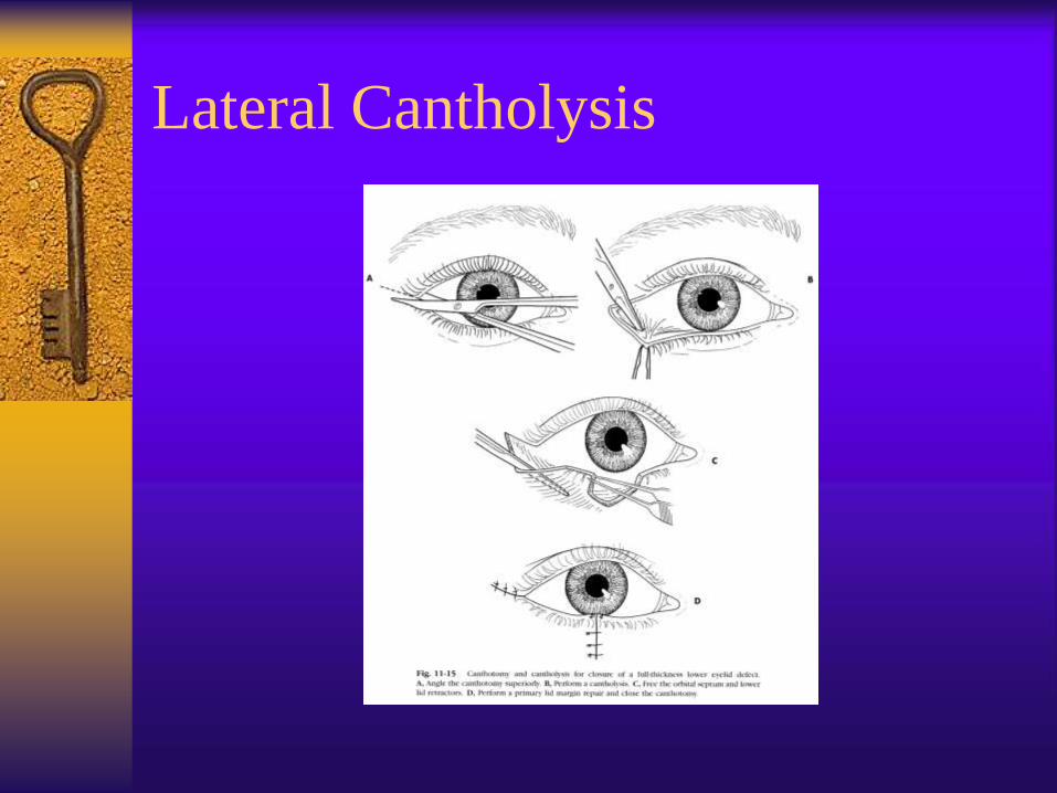
Additional 5 mm of advancement
Split upper and lower canthal tendons
Detach lower limb (upper limb)
Angle skin incision superiorly
Anchor muscle layer to periosteum after
closure of defect
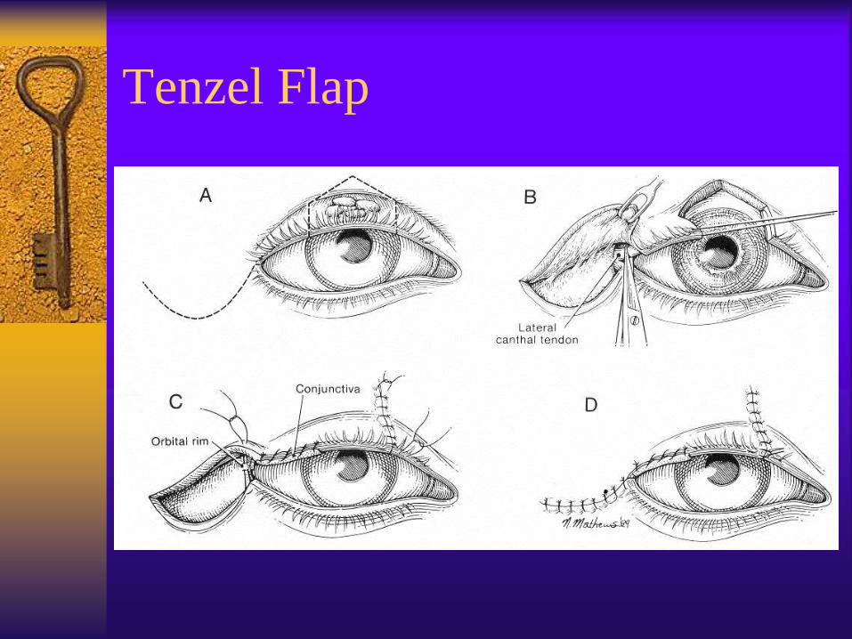
Lateral Cantholysis

Tenzel Rotational Flap
Semicircular musculocutaneous flap
Defects up to 60%
Flap must arch upward
Fixation of muscle to periosteum superior to
canthal attachment avoids droop of lid
Additional support of lateral lid can be
achieved with periosteal strip from lateral
orbital rim
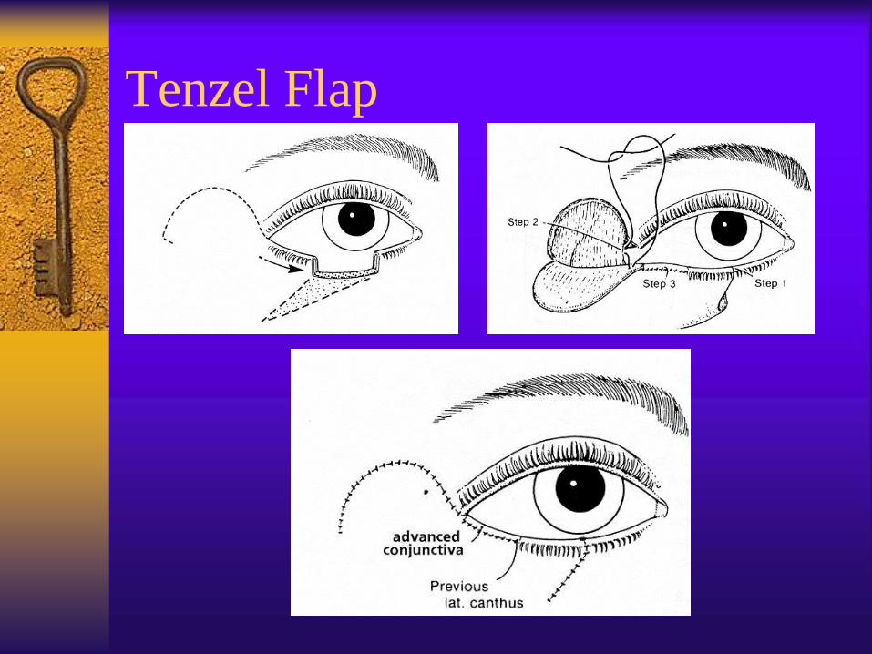
Tenzel Flap

Free Tarsal Graft
Free tarsocunjunctival flap
Harvested from ipsilateral/contralateral lid
Posterior lamellar replacement
Cover with myocutaneous advancement

Free Tarsal Graft

Hughes Procedure
Tarsoconjunctival Flap for posterior lamella
Defects greater than 50%
Vertical upper lid to lower lid sharing
Anterior lamella reconstruction
– Advancement musculocutaneous flap
– Free skin graft
Requires 2nd stage procedure

Hughes Procedure

Hughes Procedure (continued)

Mustarde Rotational Cheek Flap
Good for very large defects
Advantage – single stage procedure
Preferable for patients with:
– Monocular vision
– Children with amblyopia
– Active corneal disease
– Glaucoma
Disadvantages – lacks orbicularis, sagging

Mustarde Technique

Mustarde Technique

Upper Eyelid Reconstruction
Direct Closure +/- lateral cantholysis
Tenzel Flap
Sliding Tarsoconjunctival Flap
Posterior Lamellar Graft with local
myocutaneous flap
Cutler-Beard (Bridge) Flap
Direct Closure
Tenzel Flap

Sliding Tarsoconjunctival Flap
Isolated medial or lateral lid defects
Borrows a sliding portion of remaining lid
segment for posterior lamella
Anterior lamella repaired with skin graft or
local myocutaneous advancement flap

Sliding Tarsoconjunctival Flap

Posterior Lamellar Graft with
Local Myocutaneous Flap Good for patients with skin laxity or
redundancy
Posterior lamella defect
– Conjunctival advancement (upper fornix, lower
lid)
– Supplement with ear cartilage
Anterior lamella
– Myocutaneous flap for blood supply
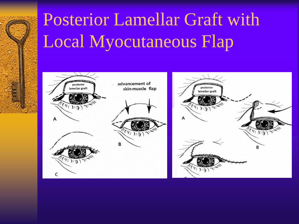
Posterior Lamellar Graft with
Local Myocutaneous Flap

Cutler-Beard (Bridge) Flap
Used for 60% to entire lid defects
Borrows skin, muscle and conjunctiva from
lower eyelid
Autogeneous cartilage to provide support
Requires 2nd stage procedure
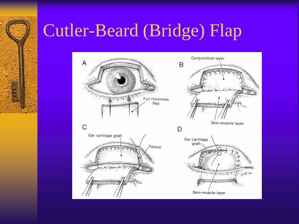
Cutler-Beard (Bridge) Flap

Cutler-Beard (Bridge) Flap – 2nd
Stage Procedure
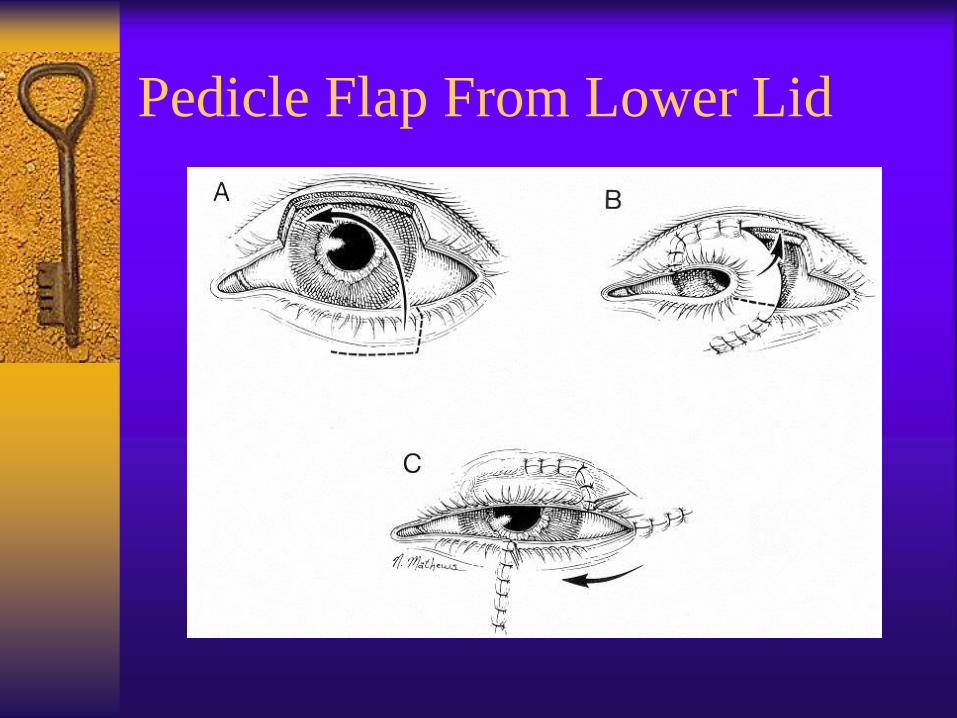
Pedicle Flap From Lower Lid

Lateral Canthal Reconstruction

Lateral Canthal Reconstruction

Medial Canthal Reconstruction
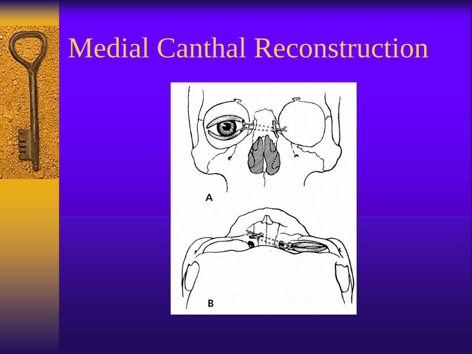
Medial Canthal Reconstruction

Medial Canthal Reconstruction

Decision Making
Conclusion
Thorough understanding of eyelid anatomy
Understand basic techniques of repair
Challenging problem do to complex nature
of eyelid anatomy
Careful attention to detail with delicate
surgical technique required

References
Nerad, J.A. Oculoplastic Surgery: The Requisites in Ophthalmology. Mosby Inc. St. Louis, MO. 2001.
Chen, W.P. Oculoplastic Surgery: The Essentials. Thieme New York. New York, NY. 2001.
McCord, C.D., Codner, M.A. Eyelid Surgery: Principles and Techniques. Lippincott-Raven. Philadelphia, PA. 1995.
Hornblass, A. Oculoplastic, Orbital and Reconstructive Surgery. Vol. 1. Williams and Wilkins. Baltimore, MD. 1988
Rathbun, J.E. Eyelid Surgery. Little, Brown and Company. Boston, MD.
Fischer T, Noever G., Langer M., Kammer E. Experience in upper eyelid reconstruction with the Cutler-Beard technique. Annals of plastic Surgery. 47(3): 338-42, 2001 Sep.
Maloof A., Ng S., Leatherbarrow B. The maximal Hughes procedure. Ophthalmic Plastic & Recon Sur. 17(2): 96-102, 2001 Mar.
Rohrich RJ. Zbar RI. The evolution of the Hughes tarsoconjunctival flap for the lower eyelid reconstruction. Plastic & Recon Sur. 104(2): 518-22, 1999 Aug.
Patrinely JR, O’Neal KD. Kersten RC, Soparkar CN. Total upper eyelid reconstruction with mucosalized tarsal graft and overlying bipedicle flap. Arch of Ophthal. 117(12): 1655-61, 1999 Dec.
Matsumoto K. Nakanishi H. Urano Y. Kubo Y. Nagae H. Lower eyelid reconstruction with a cheek flap supported by fascia lata. Plastic & Recon Sur. 103(6):1650-4, 1999 May
Perry MJ. Langry J. Martin IC. Lower eyelid reconstruction using pedicled skin flap and palatal mucoperiosteum. Dermatologic Sur. 23(5): 395-7, 1997 May.
Werner MS. Olson JJ. Putterman AM. Composite grafting for eyelid reconstruction. Amer J of Ophth. 116(1):11-16, 1993 Jul.
Cohen MS. Shorr N. Eyelid reconstruction with hard palate mucosa grafts. Ophth Plastic & Recon Sur. 8(3):183-95, 1992.