f-18 labelling synthesis, radioanalysis and - helda -
TRANSCRIPT

Report Series in Radiochemistry 27/2007
F-18 LABELLING SYNTHESIS, RADIOANALYSIS AND EVALUATION OF
A DOPAMINE TRANSPORTER AND A HYPOXIA TRACER
Eeva-Liisa Kämäräinen
University of Helsinki
Helsinki 2007

University of Helsinki
Department of Chemistry
Laboratory of Radiochemistry
Faculty of Science
Helsinki, Finland
and
Department of Clinical Physiology and Nuclear Medicine
Helsinki University Central Hospital
Helsinki, Finland
F-18 LABELLING SYNTHESIS, RADIOANALYSIS AND EVALUATION OF
A DOPAMINE TRANSPORTER AND A HYPOXIA TRACER
Eeva-Liisa Kämäräinen
Academic Dissertation
To be presented with the permission of the Faculty of Science of the University of Helsinki, for public criticism in the main lecture hall A 110 of the Department of Chemistry on January 5, 2007, at 12 o’clock noon.
Helsinki 2007

Supervised by:
Professor Olof Solin, PhD
Turku PET Centre and Department of Chemistry
University of Turku
Turku, Finland
Reviewed by:
Professor Raisa Krasikova, PhD
Institute of Human Brain
Russian Academy of Sciences
St. Petersburg, Russia
and
Professor Aapo Ahonen, MD, PhD
Division of Nuclear Medicine
Department of Clinical Physiology and Nuclear Medicine
Helsinki University Hospital
Helsinki, Finland
Dissertation opponent:
Jörgen Bergman, PhD
Turku PET Centre, Radiopharmaceutical Chemistry Laboratory
University of Turku
Turku, Finland
ISSN 0358-7746 ISBN 978-952-10-3604-0 (nid.) ISBN 978-952-10-3605-7 (pdf) http://ethesis.helsinki.fi Helsinki 2007 Yliopistopaino

“Potius sero quam numquam” (T.Livius)

i
ABSTRACT
Positron emission tomography (PET) is an imaging technique in which radioactive positron-emitting tracers are used to study biochemical and physiological functions in humans and in animal experiments. The use of PET imaging has increased rapidly in recent years, as have special requirements in the fields of neurology and oncology for the development of syntheses for new, more specific and selective radiotracers. Synthesis development and automation are necessary when high amounts of radioactivity are needed for multiple PET studies. In addition, preclinical studies using experimental animal models are necessary for evaluating the suitability of new PET tracers for humans. For purification and analysing the labelled end-product, an effective radioanalytical method combined with an optimal radioactivity detection technique is of great importance.
In this study, a fluorine-18 labelling synthesis method for two tracers was developed and optimized, and the usefulness of these tracers for possible prospective human studies was evaluated. N-(3-[18F]fluoropropyl)-2β-carbomethoxy-3β-(4-fluorophenyl)nortropane ([18F]β-CFT-FP) is a candidate PET tracer for the dopamine transporter (DAT), and 1H-1-(3-[18F]fluoro-2-hydroxypropyl)-2-nitroimidazole ([18F]FMISO) is a well-known hypoxia marker for hypoxic but viable cells in tumours. The methodological aim of this thesis was to evaluate the status of thin-layer chromatography (TLC) combined with proper radioactivity detection measurement systems as a radioanalytical method. Three different detection methods of radioactivity were compared: radioactivity scanning, film autoradiography, and digital photostimulated luminescence (PSL) autoradiography.
The fluorine-18 labelling synthesis for [18F]β-CFT-FP was developed and carbon-11 labelled [11C]β-CFT-FP was used to study the specificity of β-CFT-FP for the DAT sites in human post-mortem brain slices. These in vitro studies showed that β-CFT-FP binds to the caudate-putamen, an area rich of DAT. The synthesis of fluorine-18 labelled [18F]FMISO was optimized, and the tracer was prepared using an automated system with good and reproducible yields. In preclinical studies, the action of the radiation sensitizer estramustine phosphate on the radiation treatment and uptake of [18F]FMISO was evaluated, with results of great importance for later human studies. The methodological part of this thesis showed that radioTLC is the method of choice when combined with an appropriate radioactivity detection technique. Digital PSL autoradiography proved to be the most appropriate when compared to the radioactivity scanning and film autoradiography methods. The very high sensitivity, good resolution, and wide dynamic range of digital PSL autoradiography are its advantages in detection of β-emitting radiolabelled substances.

ii
PREFACE The main work of this thesis was carried out at the Laboratory of Radiochemistry, University of Helsinki. Other parts of the work were performed at the Department of Clinical Neuroscience, Karolinska Institutet, Stockholm, Sweden, at the Division of Clinical Physiology and Nuclear Medicine, Helsinki University Central Hospital, and at the Turku PET Centre.
I thank Professor Emeritus Timo Jaakkola for giving me the opportunity to start my work with fluorine-18 labelling and for encouraging me to enter the interesting field of radiopharmaceutical chemistry. Professor Jukka Lehto, present head of the Laboratory of Radiochemistry, and Professor Anssi Sovijärvi, Department of Clinical Physiology and Nuclear Medicine, are gratefully acknowledged for giving me opportunity to complete this work.
I want to express my sincerest thanks to Professor Olof Solin. Without his enthusiasm, endurance and never-failing faith in my ability to accomplish the task this dissertation would never have been completed. I am also very grateful to Professor Solin for acting as my supervisor. His vast professional knowledge, intuition in the field of positron emitters and constructive feedback for my work has been keystones in this thesis.
I thank Professors Aapo Ahonen and Raisa Krasikova, the official reviewers of this thesis, for their constructive and valuable comments that opened my eyes to see many things in a different way. Professor Ahonen is also acknowledged for support and encouragement during the finalisation process of my thesis.
I thank my main co-authors Teija Koivula (née Kyllönen), Merja Haaparanta-Solin, Marja Siitari-Kauppi, Kaarlo Ståhlberg, Outi Nihtilä (née Perhola) and Tiina Lipponen for their valuable advice and contributions to my work.
In particular, I offer my warmest thanks to Merja Haaparanta-Solin for her continuing support, fruitful discussions about the world of preclinical studies and valuable help during the writing my thesis, and to my good friend and former colleague Marja Siitari-Kauppi for frequent supportive conversations and patience when designing the layout of the thesis. Your positive attitudes have helped me greatly during the final course of this work.
I thank Professor Christer Halldin for giving me the opportunity to work in his group at the Karolinska Institutet. I also thank Anu Airaksinen, Camilla Lundkvist, Meixiang Ju, Kjell Någren, Johan Sandell, Oliver Langer, Professor Jouko Vepsäläinen, Jukka Hiltunen, Kim Bergström, Simo Lötjönen, Kalevi Kairemo, Professor Ismo Virtanen, Jan Keyriläinen and Kimmo Taari for their co-operation.
I thank Heikki Björk in VERIFIN for performing the LC-MS analyses.
I express also my special thanks to all my colleagues at the Laboratory of Radiochemistry, especially to Pirkko Hölttä, Tuija Suoranta, Kerttuli Helariutta, Risto Harjula, Risto Koivula, Airi Paajanen, Kaisa Vaaramaa, Martti Hakanen, Esa Puukko and former teachers and colleagues Timo Autio, Jorma Aaltonen, Sirkka-Liisa Karonen, Esko Karttunen, Sinikka Pinnioja and Michael Tillander. In addition, thank you to the radiochemists of a younger generation and many others whom I have not mentioned

iii
individually for their help and support during the many years I have spent working on my research.
Thank you also to all the personnel in the Division of Nuclear Medicine, for their patience and understanding when I was slightly in another world during the last course of my thesis: Marjo Airut, Toni Ihalainen, Kaija Jansson, Ulla Järvinen, Anja Karttunen, Hannele Kivikoski, Tuula Kivimäki, Tuula Kokkola, Tapani Korppi-Tommola, Anu Koskela, Tytti Laakso, Anne Nenonen, Päivi Nikkinen, Varpu Paloheimo, Leena Pekkanen, Jaakko Rintamäki, Leena Salminen, Jukka Schildt, Reijo Takalo, Ritva Teivaala and Taina Väkiparta.
Many thanks to Stewart Makkonen-Craig for revising the English language of the last version of this thesis at only short notice.
To all my good friends and especially to my long-standing friends already since childhood – Hellu, Liisa, Mikkara ja Mari and their families and later friends Piksa, Maija, Seija, Siku, Kaarina, Hannele, Anni, Maija and Ulla, without forgetting my friends Aila, Werner, Mizzi and Ueli in Switzerland – your nice company and get-together have been a great counterbalance to the pressures of this work.
I express my sincere gratitude to my mother Leila, my sisters Ulla and Anu, my brothers Pekka and Martti and all their families for their love, support and for special moments we have shared together over a long period of time.
I address my dearest thanks to my husband Veikko for his support and patience. He first introduced me to the fascinating world of radioactivity and helped me in the very beginning of my radiochemistry studies. Without him I never could have completed this work.
Many thanks also all the colleagues, friends, godchildren and relatives whom I have not mentioned individually.
Financial support from the Finnish Society of Nuclear Medicine is gratefully acknowledged.
Helsinki, December 2006

iv
ABBREVIATIONS β-CFT 2β-carbomethoxy-3β-(4-fluorophenyl)tropane nor-β-CFT 2β-carbomethoxy-3β-(4-fluorophenyl)nortropane β-CFT-FP N-(3-fluoropropyl)-2β-carbomethoxy-3β-(4-
fluorophenyl)nortropane β-CIT 2β-carbomethoxy-3β-(4-iodophenyl)tropane β-CIT-FE N-(3-fluoroethyl)-2β-carbomethoxy-3β-(4-
iodophenyl)nortropane β-CIT-FP N-(3-fluoropropyl)-2β-carbomethoxy-3β-(4-
iodophenyl)nortropane BNCT Boron neutron capture therapy CA Carrier added CNS Central nervous system [14C]PMMA 14C-labelled methylmethacrylate CT Computed tomography DAT Dopamine transporter DMF N,N-Dimethylformamide DMSO Dimethylsulfoxide EC Electron capture EMP Estramustine phosphate EOB End of bombardment EOS End of synthesis FMISO Fluoromisonidazole [18F]β-CFT-FP N-(3-[18F]fluoropropyl)-2β-carbomethoxy-3β-(4-
fluorophenyl)nortropane [18F]EPI-F [18F]Epifluorohydrin [18F]FDG 2-[18F]Fluoro-2-deoxy-D-glucose [18F]FMISO 1H-1-(3-[18F]fluoro-2-hydroxypropyl)-2-nitroimidazole [18F]MPPF 4-[18F]fluoro-N-[2-[1-(2-methoxyphenyl)-1-piperazinyl]ethyl-
N-2-pyridinyl-benzamide GBR 12909 1-(2(bis-(4-fluorophenyl)methoxy)ethyl)-4-(3-
phenylpropyl)piperazine dihydrochloride GM Geiger-Müller GMP Good manufacturing practise GLP Good laboratory practise GRP Good radiopharmacy practise HPLC High performance liquid chromatography HPTLC High performance thin-layer chromatography Kryptofix 222 4,7,13,16,21,24-hexaoxa-1,10-diazabicyclo(8,8,8) hexacosane LC/MS Liquid chromatography/Mass spectrometry MISO Misonidazole NCA No carrier added NITTP 1-(2′-nitro-1′-imidazolyl)-2-O-tetrahydropyranyl-3-O-
toluenesulphonyl-propanediol NIM Nitroimidazole PC Personal computer PET Positron emission tomography PSL Photostimulated luminescence

v
SA Specific radioactivity SERT Serotonin transporter SD Standard deviation SEM Standard error of mean TLC Thin-layer chromatography UV Ultraviolet
vi
LIST OF ORIGINAL PUBLICATIONS
This thesis is based on the following original publications, which are referred to in the text by their Roman numerals.
I. Kämäräinen E-L, Kyllönen T, Airaksinen A, Lundkvist C, Meixiang Yu, Någren K, Sandell J, Langer O, Vepsäläinen J, Hiltunen J, Bergström K, Lötjönen S, Jaakkola T, Halldin C. (2000) Preparation of [18F]β-CFT-FP and [11C]β-CFT-FP, selective radioligands for visualisation of the dopamine transporter using positron emission tomography (PET). J Label Compd Radiopharm 43: 1235-1244.
II. Kämäräinen E-L, Kyllönen T, Nihtilä O, Björk H, Solin O. (2004) Preparation of fluorine-18-labelled fluoromisonidazole using two different synthesis methods. J Label Compd Radiopharm 47: 37–45.
III. Ståhlberg K, Kämäräinen E-L, Keyriläinen J, Virtanen I, Taari K, Kairemo K. (2006) Hypoxia in DU-145 prostate cancer xenografts after estramustine phosphate and radiotherapy. Submitted
IV. Kämäräinen E-L, Haaparanta M, Siitari-Kauppi M, Koivula T, Lipponen T and Solin O. (2006) Analysis of 18F-labelled synthesis products on TLC plates: comparison of radioactivity scanning, film autoradiography and phosphoimaging technique. Appl Radiat Isot 64: 1043-1047
Articles I, II and IV are reprinted with the permission of the copyright holders.
In addition, data published as a scientific abstract are included (Kämäräinen et al. 2000).

vii
CONTENTS ABSTRACT i PREFACE ii ABBREVIATIONS iv LIST OF ORIGINAL PUBLICATIONS vi CONTENTS vii
1 Introduction ............................................................................................ 1
2 Review of literature ................................................................................ 4 2.1 Positron emitters and PET ........................................................................................ 4 2.2 Labelling of radiotracers with short-lived positron emitters .................................... 6 2.3 Labelling of radiotracers with fluorine-18 ............................................................... 9
2.3.1 Nucleophilic radiofluorination ................................................................... 10 2.3.2 Electrophilic radiofluorination ................................................................... 12
2.4 PET tracers for dopamine transporter..................................................................... 12 2.5 PET tracers for hypoxia.......................................................................................... 13 2.6 Radioanalytical techniques..................................................................................... 14 2.7 Preclinical methods for PET tracer evaluation....................................................... 15
2.7.1 Preclinical evaluation of a new dopamine transporter tracer ..................... 15 2.7.2 Preclinical use of a hypoxia tracer ............................................................. 16
3 Aims of the study ................................................................................. 18
4 Materials and methods.......................................................................... 19 4.1 Chemicals and reagents .......................................................................................... 19 4.2 Production of [18F]F- and [11C]CO2........................................................................ 19 4.3 Synthesis and automation of PET tracer production .............................................. 19
4.3.1 Synthesis of [18F]β-CFT-FP and [11C]β-CFT-FP (Paper I)........................ 19 4.3.2 [18F]FMISO synthesis and synthesis automation (Paper II)....................... 21
4.4 Separation and analysis of radiotracers .................................................................. 23 4.4.1 Analysis of radiotracers.............................................................................. 23 4.4.2 Purification of compounds using RadioHPLC........................................... 23 4.4.3 Detection of radioactivity on the TLC plate (Paper IV)............................. 24
4.5 Preclinical studies................................................................................................... 25 4.5.1 Post-mortem human brain autoradiography with [11C]β-CFT-FP
(Kämäräinen et al. 2000)............................................................................ 25 4.5.2 Biodistribution of [18F]FMISO in nude mice with DU-145 human prostate
cancer cell tumours (Paper III)................................................................... 25
5 Results .................................................................................................. 27 5.1 Synthesis of [18F]β-CFT-FP and [11C]β-CFT-FP (Paper I) .................................... 27 5.2 [18F]FMISO synthesis and synthesis automation (Paper II)................................... 28 5.3 Analysis of radioactive compounds with TLC (Paper IV)..................................... 29
5.3.1 Linearity of the autoradiography methods ................................................. 29 5.3.2 Radiochemical yield and purity determination with TLC.......................... 29

viii
5.4 In vitro human post-mortem receptor autoradiography of [11C]β-CFT-FP
(Kämäräinen et al. 2000) ........................................................................................ 30 5.5 Uptake of [18F]FMISO in human prostate tumour cell-bearing mice (Paper III)... 32
5.5.1 Biodistribution of [18F]FMISO in mice with DU 145 human prostate cancer cell tumours ................................................................................................ 32
5.5.2 Histology and immunohistochemistry ....................................................... 33
6 Discussion ............................................................................................ 34 6.1 [18F]β-CFT-FP (Paper I) ......................................................................................... 34
6.1.1 Evaluation of the synthesis of [18F]β-CFT-FP ........................................... 34 6.1.2 In vitro autoradiograhy of [11C]β-FT-FP in human post-mortem brain
(Kämäräinen et al. 2000)............................................................................ 34 6.2 [18F]FMISO (Paper II and III) ................................................................................ 35
6.2.1 Evaluation of the [18F]FMISO synthesis.................................................... 35 6.2.2 Automation in [18F]FMISO synthesis ........................................................ 36 6.2.3 Preclinical studies of [18F]FMISO in tumour mice .................................... 36
6.3 Radiochemical purity analysis of 18F-compounds by TLC (Paper IV) .................. 37 6.3.1 Comparison of radioactivity scanning, film autoradiography, and
phosphoimaging technique......................................................................... 37 6.3.2 Importance of an optimal detection method choice in analysing labelled
compounds on TLC plates ......................................................................... 38 6.4 Future aspects on[18F]β-CFT-FP and [18F]FMISO labelling syntheses and their use
in PET studies......................................................................................................... 39 6.5 Radioanalytical methods and radioactivity measuremets in the development of new
radiotracers ............................................................................................................. 39
7 Conclusions .......................................................................................... 40
8 References ............................................................................................ 41

1
1 Introduction
Fluorine compounds have been known since the Middle Ages, originally in France, where they were used in the form of calcium fluoride (CaF2) for etching glass. The name fluorine arises from the Latin word “fluere”, meaning “to flow”, because fluorine is very reactive and readily forms compounds with other elements. The element fluorine was isolated finally in 1886 by Henri Moissan, for which, among other achievements, he was awarded the Nobel Prize for chemistry in 1906. Fluorine substitution markedly affects the properties of organic compounds, and the very high electronegativity of fluorine can modify electron distribution in a molecule, affecting its absorption, distribution, and metabolism. Fluorine is of considerable importance in the drug industry, and fluorine-containing drugs are used in medicine as anaesthetics, anti-cancer and anti-inflammatory agents, psychopharmaceuticals, and in many other applications (Park and Kitteringham 1994; Park et al. 2001; Isanbor and O’Hagan 2006). Fluorine is isosteric with hydrogen (van der Waals radius 1.2 Å for hydrogen and 1.35 Å for fluorine respectively), and it is usually substituted into a drug molecule in place of a hydrogen or hydroxyl group. Fluorine substitution in a drug molecule can influence not only its pharmacokinetics but also its pharmacodynamics and toxicology. The introduction of fluorine, e.g. to psychopharmaceuticals, improves penetration of the drug across the blood–brain barrier; thus, it affects the lipophilicity of the molecule (Park and Kitteringham 1994; Park et al. 2001). Then the fluorophenyl and trifluoromethyl groups are used, which both are generally resistant to metabolic attack.
Positron emission tomography (PET) is an imaging technique in which radioactive positron-emitting tracers are used to study biochemical and physiological functions in humans and in animal experiments in the fields of cardiology, neurology, and oncology. Fluorine-18, a radioactive isotope of fluorine, is a versatile and remarkable positron-emitting radionuclide for imaging physiological functions in living organisms. Fluorine-18 was discovered as early as 1936 (Snell 1937), but synthetic applications of fluorine-18 lagged behind the use of another positron-emitting radionuclide, carbon-11, in radiochemical applications, largely because of difficulties in the fluorination of organic molecules. Thus the majority of the fluorine-labelling methods have been developed in the last two decades (Snyder and Kilbourn 2003). Fluorine-18 has proved, however, to be an ideal tracer for PET because of its convenient half-life of 109.8 min and low β-energy (max. 0.635 MeV). Due to low energy of β-particles the highest spatial resolution in PET imaging is obtained using 18F-labelled radiotracers. Although the field of fluorine-18 chemistry has expanded in the last decade, fluorine-18-labelled 2-deoxy-D-glucose ([18F]FDG) still remains the most used positron-emitter–labelled radiopharmaceutical in PET studies and it provides some of the highest signal-to-noise ratios to have been observed in the nuclear medicine (Ell 2006).
For the past five years, combined PET and computed tomography (CT), or PET/CT, has spread worldwide especially in oncology. PET itself provides information that is very different from that obtainable from other imaging modalities. The addition of CT to PET has improved not only specificity but also sensitivity in tumour imaging. The combination of [18F]FDG with PET/CT studies is highly synergistic and has proved to be a relevant technique in staging and therapy monitoring of many tumours. Published results in oncology are still limited, but several well designed studies have demonstrated

2
the benefits of PET/CT in staging of non-small cell lung cancer, recurrent colorectal cancer and malignant lymphoma (Ell 2006; von Schultness et al. 2006). However, [18F]FDG in PET/CT has been found to be less sensitive and specific for assessment of some types of cancer, motivating efforts to develop other oncologic tracers (Ell 2006). Among tumour seeking agents the most widely used besides [18F]FDG is O-(2-[18F]fluoroethyl)-L-tyrosine ([18F]FET) for studying amino acid transport (Wester et al. 1999), and in addition 3’-[18F]fluoro-3’-deoxy-thymidine ( [18F]FLT) for assessing tumour proliferation (Shields et al. 1998), [18F]fluorocholine for prostate carcinoma (DeGrado et al. 2000b) and [18F]FMISO (1H-1-(3-[18F]fluoro-2-hydroxypropyl)-2-nitroimidazole) and other nitroimidazole derivatives for assessing hypoxia (Rajendran et al. 2006). In neurology there is also wide interest in developing ligands such as those more specific and selective for dopamine transporter (DAT) because DAT plays an important role in many brain disorders like Parkinson’s disease and schizophrenia. DAT is also an important target for a variety of clinically effective therapeutic drugs, neurotoxic agents and stimulant drugs of abuse such as cocaine and amphetamine (Bannon et al. 1995).
The design and development of automated radiotracer synthesis systems is also an important focus in synthesis development when high amounts of radioactivity are required and the availability of [18F]FDG or other clinically-useful radiotracers for multiple PET studies must be guaranteed. Furthermore, in the development of syntheses, preclinical studies in experimental animal models are needed to evaluate the suitability of the compounds as PET tracers in humans. For purification and analysing the labelled end-product, an effective radioanalytical method combined with an optimal radioactivity detection technique is of great importance. In the radioanalytical part of the synthesis, the status of the almost-neglected method of thin-layer chromatography (TLC), combined with an optimal radioactivity detection technique in analysis and purity determinations of the labelled compounds, needs to be considered.
Several compounds have been labelled and are used in PET studies to evaluate the physiology and pharmacology of DAT in vitro and in vivo. However, poor selectivity or/and unfavourable kinetics of most of the compounds limit their use in quantitative PET studies. The cocaine analogue, 2 β-carbomethoxy-3β-(4-iodophenyl)tropane (β-CIT) has been suggested as a lead structure in the development of radiotracers for DAT. However, radiolabelled β-CIT itself has proven unsuitable for PET because it does not reach peak equilibrium within the time course of the PET experiment (Farde et al. 1994). The estimated order of cocaine analogue selectivity for DAT is β-CIT-FP > β-CIT-FE > β-CIT. A fluorine analogue, N-(3-fluoropropyl)-2β-carbomethoxy-3β-(4-fluorophenyl)nortropane (β-CFT-FP), labelled with fluorine-18 has in preliminary experiments indicated accumulation in the rat striatum (Firnau et al. 1995); thus, it is suggested that β-CFT-FP should have higher selectivity for DAT and should reach peak equilibrium faster than the cocaine analogues mentioned above. The purpose of this study was to prepare radiolabelled β-CFT-FP using two different [18F]fluoroalkylating agents, or to label β-CFT-FP with carbon-11 via [11C]methyltriflate to examine its distribution in the post-mortem human brain using whole hemisphere autoradiography. If these radiotracers are proven valid, these could be used later in human PET studies.
Misonidazole (MISO; 1H-imidazole-1-ethanol, alpha-(methoxymethyl)-2-nitroimidazole) and its nitroimidazole derivatives have been shown to accumulate in hypoxic but viable cells. Consequently, their radiolabelled analogues are used as markers of hypoxic tissues. They bind covalently to cellular molecules at rates that are

3
inversely proportional to intracellular oxygen concentration, and their uptake in hypoxic cells depends on the reduction of the nitro group on the imidazole ring. According to published results (Yang et al. 1995; Rasey et al. 1999; Grönroos et al. 2001; Kumar et al. 2002; Dolbier et al. 2001; Coutier et al. 2004; Grönroos et al. 2004; Grönroos et al. 2005) a number of alternative 2-nitroimidazole derivatives have been evaluated. However, [18F]FMISO has remained the most commonly used agent for hypoxia PET-imaging (Grierson et al. 1989, Rasey et al. 1996, Chapman et al. 1998; Rajendran et al. 2004), so consequently it was chosen for this study. PET and [18F]FMISO can help to estimate the oxygenation status of tumours in any part of the body; for example, they can be used to study the hypoxic volume of tumours during the course of radiation treatment. The first aim in this study was to synthesize [18F]FMISO by comparing two FMISO synthesis methods and to adapt the most valid method to an automated synthesis module. The goal was then to evaluate hypoxia by [18F]FMISO uptake in an experimental prostate tumour model in mice. Estramustine phosphate (EMP), originally an anti-mitotic drug used against advanced, hormone-independent prostate cancer, was applied as a radiosensitizer to sensitize cancer cells to radiotherapy; i.e., to enhance the effect of radiation treatment (Widmark et al. 1994). The effect of EMP on [18F]FMISO uptake in the tumours before and after radiotherapy was evaluated.
In the development of radiotracers chemical, radiochemical and radiopharmaceutical aspects have to be taken into consideration. The radiotracer must be chemically and radiochemically pure and it must be sterile and pyrogen free if it is to be used in PET studies (Vera-Ruiz et al. 1990; Meyer et al. 1992; Långström and Dannals 1995). Chromatography of radiolabelled compounds is an important stage in synthesis both for the isolation and purification of the labelled compounds for eliminating radiochemical and chemical by-products of the synthesis. Furthermore, it is needed for analysis and determination of the radiochemical and chemical purity of the synthesis end-product. At present, for purification of labelled, often complicated radiotracers, high performance liquid chromatography (HPLC) systems are necessary; however, for analysis and radiochemical quality control of labelled compounds, TLC combined with proper radioactivity detection measurement systems is in many cases a good choice. Thus, the chemical resolution of planar chromatographic methods such as TLC and high performance thin-layer chromatography (HPTLC) can be regarded as comparable to that of HPLC (Wilson 1996). Several techniques are available to detect radioactivity of the short-lived positron emitters on the TLC plates. In this study, the aim was to compare and evaluate three different methods: radioactivity scanning, film autoradiography, and digital photostimulated luminescence (PSL) autoradiography (phosphoimaging technique) in analysing radiochemical composition and radiotracer purity on TLC plates by using fluorine-18-labelled synthesis products as an example.
4
2 Review of literature
2.1 Positron emitters and PET
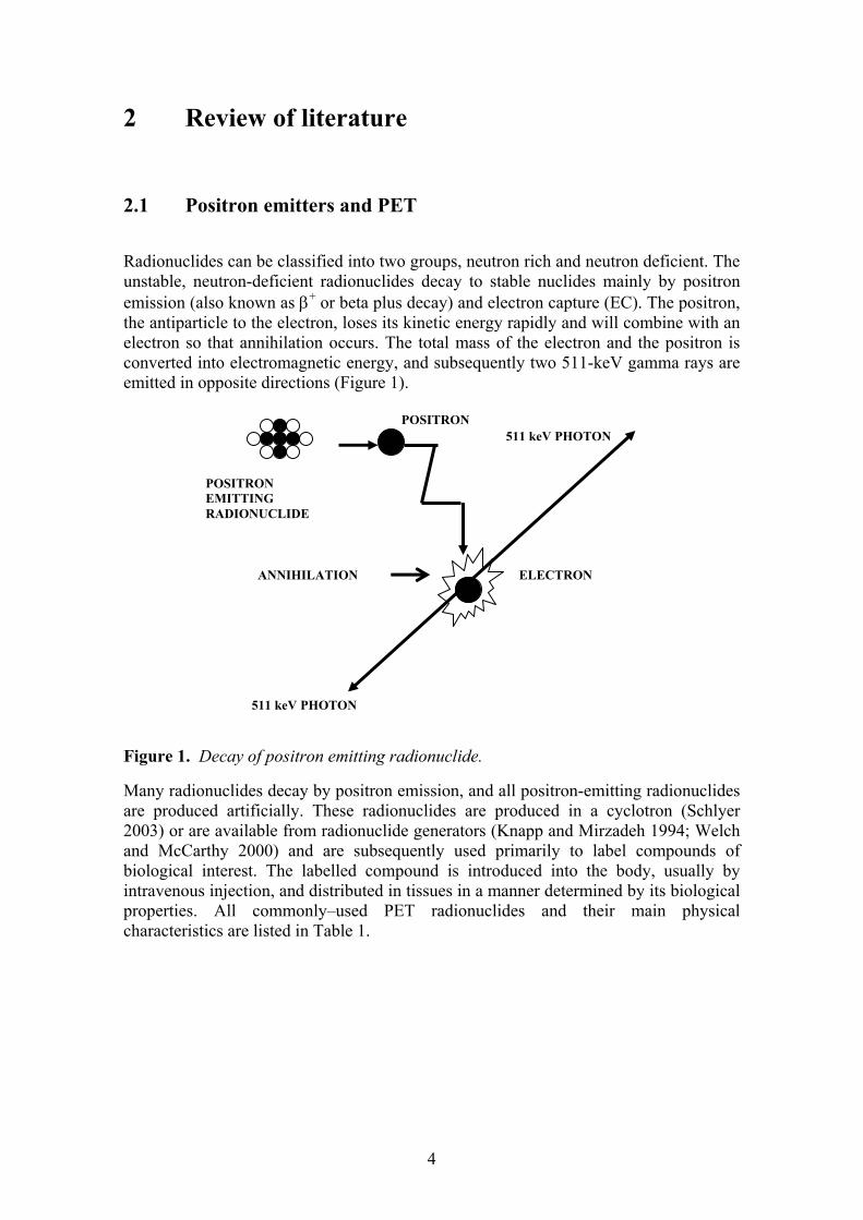
Radionuclides can be classified into two groups, neutron rich and neutron deficient. The unstable, neutron-deficient radionuclides decay to stable nuclides mainly by positron emission (also known as β+ or beta plus decay) and electron capture (EC). The positron, the antiparticle to the electron, loses its kinetic energy rapidly and will combine with an electron so that annihilation occurs. The total mass of the electron and the positron is converted into electromagnetic energy, and subsequently two 511-keV gamma rays are emitted in opposite directions (Figure 1).
Figure 1. Decay of positron emitting radionuclide.
Many radionuclides decay by positron emission, and all positron-emitting radionuclides are produced artificially. These radionuclides are produced in a cyclotron (Schlyer 2003) or are available from radionuclide generators (Knapp and Mirzadeh 1994; Welch and McCarthy 2000) and are subsequently used primarily to label compounds of biological interest. The labelled compound is introduced into the body, usually by intravenous injection, and distributed in tissues in a manner determined by its biological properties. All commonly–used PET radionuclides and their main physical characteristics are listed in Table 1.
511 keV PHOTON
POSITRON EMITTING RADIONUCLIDE
POSITRON
ANNIHILATION
511 keV PHOTON
ELECTRON

5
Table 1. Physical properties of the four most conventional PET radionuclides.
Radio-nuclide
T1/2 (min)
Decay mode
Production reaction
Max. β-
energy (MeV)
Max. range (mm)
Max.theor. specific activity (GBq/μmol)
Measured specific activity (GBq/μmol)
15O 2.04 β+ (99.9%) e-capture (0.1%)
14N(d,n)15O 1.72 8.2 3.39x10⁶ Not applicable
13N 9.96 β+ (99.8%) e-capture (0.2%)
16O(p,α)13N 1.19 5.4 6.99x10⁵ Not applicable
11C 20.4 β+ (99.8%) e-capture (0.2%)
14N(p, α)11C 0.96 4.1 3.41x10⁵ 37-185
18F 109.7 β+(96.9%) e-capture (3.1%)
18O(p,n)18F 20Ne(d, α)18F
0.63 2.4 6.3x10⁴ 37-7401 max. 0.742 303 (Bergman and Solin 1997)
1 Water target, 2 In target production of [18F]F2, 3 Post target production of [18F]F2
Oxygen, nitrogen, and carbon are the main elemental constituents in living organisms. Therefore, it seems natural to label molecules for in vivo investigations with bio-radionuclides. The most commonly used positron emitter, however, is 18F, which is not a constituent in living organisms. However fluorine-18 can be incorporated into organic molecules as a substituent for hydrogen, hydroxyl or some other functional group. These labelled compounds often have biological properties which resemble the parent structures. An advantageous feature of 18F-radiotracers is the applicability of the concept of blocked metabolic pathway, allowing trapping in tissues. For that particular concept [18F]FDG is the classical example (Gallagher et al. 1978) as it is taken up by the cells and converted to [18F]FDG-6-phosphate by the action of hexokinase in a similar manner to the first stage of glucose metabolism. Thereafter, [18F]FDG undergoes further metabolic steps very slowly and remains trapped in the tissue. [18F]FDG is aptly referred to as the “molecule of the millennium” due to its versatility of application in neurology, cardiology and above all in oncology. 18F has some unique advantages over the other conventional PET radionuclides. They include:
-a relatively long half-life allowing longer synthesis time, longer transport from the synthesis laboratory to the patient, and longer PET studies; in addition, if metabolite analyses are needed, the longer half-life enables a wider choice of radioanalytical methods;

6
-it allows the sharpest imaging with PET as a consequence of its low positron energy; and
-incorporating 18F into biologically active molecules can be easily achieved via electrophilic and nucleophilic reactions.
Depending on the labelling position, the introduction of a fluorine atom can affect the metabolic properties of the molecule. For example, a fluorine on an aryl group usually has a stabilising effect as well as increasing the lipophilicity of the molecule (Pike 1988; Park and Kitteringham 1994).
PET is a nuclear imaging technique that uses the unique decay characteristics of radionuclides that decay by positron emission. The annihilation process has a number of very important properties that are advantageous for imaging, and it forms the basis of PET imaging. A PET scanner is designed to detect and localize the simultaneous back-to-back annihilation photons that are emitted following decay of a radionuclide by positron emission. When a radioactive atom on a particular molecule decays, a positron is ejected from the nucleus, and annihilation occurs leading to the emission of high-energy photons that have a good probability of escaping from the body. A PET scanner consists of a set of detectors that surround the object to be imaged and are designed to detect coincident events from annihilation photons and to convert these high-energy photons into an electrical signal that can be fed to subsequent electronics. In a typical PET scan, 106 to 109 events (decays) will be detected. These events are corrected for a number of factors and then reconstructed into a tomographic image via computerised analysis of the acquired emission data (Cherry and Dahlbom 2003). Modern PET cameras, which are increasingly directly associated with CT, can rapidly construct scans from several adjacent planes and so provide kinetic and functional as well as structural information from the human body in the same image.
2.2 Labelling of radiotracers with short-lived positron emitters Chemistry with short-lived positron emitting radionuclides, principally 11C, 15O, 13N, and 18F, has increased over the last decades as a consequence of the breakthrough of PET as a powerful, non-invasive technique for investigating pathophysiology in living people. Because of the short half-lives of these radionuclides, they must be produced immediately prior to use; thus, the need for a cyclotron on the site is a necessity. Time is the most important factor when selecting the synthetic strategy, and consequently, the significance of time has to be considered as a reaction parameter of equal importance to that of chemical yield in the planning of a labelling synthesis. For 11C-labelling, for example, this typically amounts to approximately 10 to 40 minutes for radionuclide production (cyclotron bombardment), 40 minutes for radiotracer synthesis, and up to about 90 minutes for PET imaging. Furthermore, in the synthetic strategy, parameters like the position of specific labelling and specific radioactivity (SA) (especially in receptor studies) are important aspects that require consideration. The label should be ideally in a metabolically stable position (Fowler and Ding 2002; Snyder 2003; Kilbourn 1990; Antoni and Långström 2003; Mason and Mathis 2003; Lasne et al. 2002). In addition, large amounts of radioactivity are needed to compensate for radioactive decay and for the sometimes low synthetic yields; consequently, shielding, remote operations, and automation are of great importance and are thus often integrated

7
into the experimental design when planning labelling syntheses with short-lived positron emitters (Alexoff 2003).
To set up a safe and routine production of a desired radiotracer labelled with a short-lived positron-emitting radionuclide, the several steps depicted in figure 2 have to be completed. Because radiotracers are typically administered intravenously, procedures must be developed following GRP (Good Radiopharmacy Practise) guidelines to yield radiopharmaceuticals that are not only chemically and radiochemical pure but also sterile and free from pyrogens. Special guidelines for the safety, quality assurance, and quality control of short-lived radiotracers have been formulated by several groups of experts (Vera-Ruiz et al. 1990; Meyer 1992; Halldin and Nilsson 1992), and requirements have been increased considerably lasting recent years (EC 2003; EANM 2005; PIC/S 2006). From a biological perspective, the tracer signal in the target should increase to a level significantly above that of the non-specific binding during the time interval of the PET investigation.
Figure 2. Major steps in routine preparation of radiopharmaceuticals. The development of a rapid labelling synthesis is often highly dependent on the availability of suitable labelled precursors. For 11C-chemistry, the most-used primary
Production of radionuclides by cyclotron
Primary precursor or online-produced radiopharmaceutical
Preparation of secondary precursor
Synthesis of radiopharmaceutical
Purification (HPLC)
Formulation of the pharmaceutical for the PET (sterile 0.22 μm membrane filtration)
Quality control before injection (HPLC,TLC)
Ready for use in PET imaging

8
precursors are [11C]CO2 and [11C]CH4; regarding secondary precursors, [11C]-methyl iodide and [11C]methyl triflate (Jewett 1992; Någren et al. 1995) are the most widely used as alkylation agents for introducing 11C into organic molecules. Figure 3 shows the routes for preparing [11C]methyl triflate. In [11C]carboxylation the reaction of [11C]CO2 with a Grignard reagent yields the corresponding carboxylic acid. [11C]Acetate has been prepared by direct labelling using methylmagnesium bromide (Pike et al. 1982), whereas the preparation of [carbonyl-11C]desmethyl-WAY 100635 (Pike et al. 1997) utilised cyclohexylmagnesium chloride as the Grignard reagent for generation of a labelling agent preceding acylation of the precursor.
Figure 3. Production of [11C]methyl triflate from [11C]CO2 or [11C]CH4.
The 10 min half-life of 13N limits still more the reaction time available and creates an unusual challenge for the development of synthesis methods and strategy for its incorporation into suitable PET tracers. Both synthetic and enzymatic approaches have been applied to the preparation of 13N-labelled radiotracers. The most used is the myocardial perfusion tracer [13N]NH3, which can be produced by reduction of 13N-nitrate and nitrite in appropriate conditions (Vaalburg et al. 1975). Nowadays [13N]NH3 is produced directly by on-line processing of the irradiated water (Wieland et al. 1991; Berridge and Landmeier 1993; Krasikova et al. 1999). Preparation of oxygen-15, which has a very short half-life of only 2 min, provides the ultimate challenge in organic synthesis. Despite the very short half-life, tracers such as [15O]O2 and [15O]CO2 for determination of cerebral blood flow, oxygen extraction fraction and oxygen metabolism are produced (Clark and Buckingham 1975) and administered for patients by inhalation. [15O]H2O (Clark and Buckingham 1975) and [15O]butanol (Kabalka et al. 1985) are used for cerebral blood flow measurement, and [15O]butanol has been shown to be a more accurate tracer than labelled water (Berridge et al. 1990).
Labelling using fluorine-18 is presented more specifically in chapter 2.3.
[11C]CH3OTf
[11C]CH4
14N(p,α)11C [11C]CH3I
[11C]CO2 [11C]CH3OH
5-10 % H2 (N2)
0.01 % O2 (N2) HI
I2
[11C]CH3OTf
[11C]CH4
14N(p,α)11C [11C]CH3I14N(p,α)11C [11C]CH3I14N(p,α)11C [11C]CH3I
[11C]CO2 [11C]CH3OH[11C]CO2 [11C]CH3OH
5-10 % H2 (N2)
0.01 % O2 (N2) HI
I2

9
2.3 Labelling of radiotracers with fluorine-18 Although fluorine is not an element in living organisms, fluorine-18 has proved to be an ideal tracer for PET because of its intermediate half-life and low β-energy (Table 1). Its relatively long half-life compared to, for example, that of carbon-11 or oxygen-15, allows for a more complex synthesis of radiotracers to be carried out within the decay time of the radionuclide; moreover, the low energy of the positron gives the highest potential resolution for PET scans. Fluorine-18 is generally produced in a cyclotron via the reaction 20Ne(d,α)18F or 18O(p,n)18F nuclear reactions (Schlyer 2003). The availability of either labelled molecule [18F]F2 or [18F]F- allows flexibility in the development of synthetic routes to organic compounds (Figure 4). However, [18F]F-, which is available in a no-carrier-added (NCA) form and thus with high specific radioactivity (SA), is preferred for most tracer applications. SA is defined as the amount of radioactivity per unit mass of the labelled compound, and it is a major factor in PET studies. It is important to keep the amount of carrier material as low as possible to achieve a high SA in the final preparation. SA of 37-740 GBq/μmol for nucleophilic fluoride (Solin et al. 1988) and a maximum SA of 0.74 GBq/μmol for electrophilic fluorine (Chiracal et al. 1995) have been measured. However, using the production method of Bergman and Solin (1997), a SA for electrophilic fluorine as high as 30 GBq/μmol can be achieved (Table 1). Nevertheless, the theoretical SA of fluorine-18 is still approximately one decade lower than that of carbon-11. In practise, however, the measured specific activity of 37-185 GBq/μmol (Långström and Dannals 1995) for carbon-11 is of about the same magnitude as achieved using 18O-water targets for producing fluorine-18 (Table 1).
Figure 4. Summary of synthesis routes for fluorine-18 labelled radiotracers.
Accelerator productionof 18F
In target gas phaseproduction [18F]F2
Production ofaqueous [18F]F-
Nucleophilic labelling Electrophilic labelling
Post target productionof [18F]F2
Various labellingsynthons
Various labellingsynthons
Accelerator productionof 18F
In target gas phaseproduction [18F]F2
Production ofaqueous [18F]F-
Nucleophilic labelling Electrophilic labelling
Post target productionof [18F]F2
Various labellingsynthons
Various labellingsynthons
10
Radiotracers with very high SA are required for receptor- and gene expression-related studies, and for pharmacologic studies of fluorine-containing drugs that can be rather toxic (Stöcklin 1995). Furthermore, other essential demands are imposed on a radiotracer candidate in receptor studies. For example, when labelling receptors in the brain the compound must cross the blood brain barrier with rapid clearance from the blood. The tracer must have a high affinity constant (Kd) and a high specificity for the site of the interest. Its metabolism should be slow and the binding kinetics should be such that the clearance from the binding site of the interest is slower than that from non-specific binding sites (Långström and Dannals 1995, Barrio 2004).
2.3.1 Nucleophilic radiofluorination In nucleophilic radiofluorination, a precursor molecule reacts with 18F- to produce a radiolabelled compound. The 18F-fluoride ion is first received in an aqueous solution from the 18O-water target after irradiation. However, fluoride in its aqueous form is quite unreactive and requires some simple but important manipulations. [18F]F-(aq) must be dissolved in an organic solvent and transferred into a chemically more reactive form to achieve more reasonable reaction times and better synthesis yields. Usually potassium is used as a counter-ion, and aminopolyethers, e.g. 4,7,13,16,21,24-hexaoxa-1,10-diazabicyclo(8,8,8)-hexacosane (Kryptofix 222) (Hamacher et al. 1986a) or tetra-alkylammonium salts are used as complexing agents for dissolving 18F-. The labelling reactions are performed in dipolar aprotic solvents such as DMSO, acetonitrile, or DMF. Nucleophilic radiofluorinations are used in aliphatic and aromatic substitution reactions.
Pertinent aliphatic reactions are mainly SN2-displacements. The leaving groups in the molecules can be, for example, methanesulfonate (mesyl-) (Berridge and Tewson 1986), p-toluenesulfonate (tosyl-) (Block et al. 1987) or trifluoromethanesulfonate (triflyl-) (Hamacher et al. 1986a) and halogens (Berridge and Tewson 1986; Johnström and Stone-Elander 1995). Amongst the halogens bromine has shown to be the most reactive in, for example, the preparation of ω-[18F]fatty acids or their analogues (DeGrado et al 2000a; Takahashi et al. 1991). The incorporation rate of 18F-fluoride is not the only important factor when choosing the appropriate leaving group. The choice also depends on the stability of the precursors, the ease of the subsequent purification process, or the formation of side products.
Nucleophilic aromatic substitution where 18F is substituted for a proper leaving group has become a method used widely in 18F-chemistry (Ding et al. 1990). Aromatic rings themselves are not suitable for nucleophilic substitution with fluoride, but if the aromatic ring is activated by the presence of one or more electron-withdrawing groups in ortho- or para- positions to the leaving group a direct nucleophilic exchange is possible (Attina et al. 1983). Substituents such as NO2, CN, CHO, or COCH3 can function as strong electron-withdrawing groups. A variety of leaving groups are used, and nitro- and trimethylammonium groups are the most used and efficient in aromatic substitutions with fluoride-18 (Snyder and Kilbourn 2003). Simple isotopic exchange between 18F-fluoride and 19F-fluoride can be rapid but results in low SA and is therefore not suitable in cases where high SA is needed. A direct one-pot nucleophilic 18F-for-NO2 substitution of the nitro precursor is often well suited for routine preparation of

11
radiotracers such as [18F]aryl fluorides, [18F]altanserin (Lemaire et al. 1991) and [18F]butyrophenone neuroleptics (Hamacher et al. 1986b; Katsifis et al. 1993).
Direct nucleophilic substitution is in many cases difficult and sometimes impossible to carry out in certain complex and multisubstituted molecules that have not been activated. An alternative method then is two-step synthesis. Firstly, [18F]fluorinated synthons are prepared by introducing NCA 18F- through nucleophilic substitution into disubstituted alkanes by exchange with a leaving group, X:
X–(CH2)n–X + 18F– → 18F–(CH2)n–X + X–
where X is Br, OMes or OTos.
The means for an efficient reaction is to render fluoride into a reactive form using potassium carbonate and aminopolyether Kryptofix 222 as a complexing agent in a dipolar aprotic solvent such as DMF or acetonitrile. The substitution yield increases in the sequence of leaving groups Br < OMes < OTos and with increasing alkyl chain length (Block et al. 1987). Fluoroalkylation makes it subsequently possible in the second synthesis step to introduce [18F]fluoroalkyl groups into NH, OH and SH functionalities:
18F–(CH2)n–X + R–YH → 18F–(CH2)n–YR + XH
where Y is a functional group containing N, O or S.
The use of [18F]fluorobromoalkanes (Lundkvist et al. 1997; Firnau et al. 1995) or [18F]fluorotosylalkanes (Kazumata et al. 1998; Goodman et al. 2000; Koivula et al. 2005a) in nucleophilic fluoroalkylations of cocaine analogues have been reported. A new alkylating agent dibrosylate has been used to synthetize DAT-ligand 2β-carbomethoxy-3β-(4-chlorophenyl)-8-(2-fluoroethyl)nortropane (FECNT) using a semi-automated system (Voll et al. 2005).
At present, the reactive [18F]fluoride ion can be in general prepared in an organic solvent suitable for chemical synthesis. Utilisation of good commercial sources of 18O-water and of cyclotron targets made of metals such as niobium, tantalum and silver have removed the problems encountered earlier with the nonreactive fluoride ion (Snyder and Kilbourn 1990). Competing anions in target water seem particularly to diminish the reactivity of redissolved [18F]fluoride (Solin et al. 1988). Even though many of the problems have disappeared and a wide range of different nucleophilic reactions have been studied and used for the synthesis of radiotracers, labelling seems to be highly molecule specific, thus demanding a considerable effort to achieve the desired labelled radiotracer.

12
2.3.2 Electrophilic radiofluorination Molecular fluorine (F2) is very reactive in electrophilic fluorination. Electrophilic fluorine-18 labelled fluorine gas ([18F]F2) can be produced only in carrier-added form, because a small amount of carrier fluorine must be used to recover the gas from the radionuclide production target. Two processes are mainly used for production of electrophilic [18F]F2. One method utilizes the 20Ne(d,α)18F process (Casella et al 1980) and the other the 18O(p,n)18F reaction in oxygen-18 gas. In the latter case a two step procedure is needed: irradiation of oxygen-18 gas to form 18F, which attaches to the inside of target surface, followed by a second irradiation in the presence of small amount of carrier fluorine in an inert gas. By this method [18F]F2 of moderate SA can be obtained that is suitable for electrophilic radiofluorination reactions (Nickles et al. 1984). 18F-fluorine from F2 is so reactive that in many cases it must be moderated using an inert gas or through synthesis of a milder fluorinating agent. Acetylhypofluorite is the most well known of these agents, but others, such as perchlorylfluoride and fluoro-N-alkylsulfonamides, have been used. In electrophilic aliphatic fluorination, F2 is attached to an electron-rich reactant such as an alkene. Electrophilic fluorination is relatively easy and fast compared to nucleophilic fluorination. Regioselective aromatic fluorinations can be achieved using [18F]fluorodemetallation reactions. In these syntheses, fluorine replaces a metal substituent such as trialkyltin or mercuryl halide. The preparation of 6-[18F]fluoro-L-DOPA, one of the earliest 18F-labelled compounds proposed as an imaging agent of the dopamine system in CNS (Firnau et al. 1973), has been labelled by using fluorodemercuration (Adam and Jivan 1988) or by fluorodestannylation (Namavari et al. 1992). The limiting factor for the more widespread use of electrophilic fluorination has been the low SA of the fluorine gas produced by gas phase target materials. Better yields and higher SA are achieved when converting NCA [18F]methylfluoride to [18F]F2 in an exchange reaction with F2 by an electrical discharge (Bergman and Solin 1994; Bergman et al. 1995; Bergman and Solin 1997; Bergman et al. 1997).
2.4 PET tracers for dopamine transporter
The dopamine transporter is a presynaptically located protein responsible for the reuptake and thus the removal of dopamine from the synaptic cleft. DAT can therefore serve as a marker for dopaminergic neurons, a possibility that has resulted in considerable interest in studies of this transporter in the human brain. Several studies have shown that DAT density is reduced in patients with degeneration disorders, such as Parkinson’s disease and Alzheimer’s disease, indicating decreased densities of the dopamine neurons.
β-CFT-FP is the N-fluoropropyl analogue of the cocaine congener β-CFT (2β-carbomethoxy-3β-(4-fluorophenyl)tropane). Several cocaine analogues have been labelled with fluorine-18 and carbon-11 and used in PET studies to evaluate the physiology and pharmacology of the central DAT in vitro and in vivo (Carroll et al. 1995). However, poor selectivity or/and unfavourable kinetics of most of the compounds limit their use in quantitative PET. The cocaine analogue β-CIT has been suggested as a lead structure in the development of radiotracers for DAT. However, radiolabelled β-CIT itself has proven unsuitable for PET because it does not reach peak equilibrium within the time course of the PET experiment (Farde et al. 1994). β-CFT
13
has been labelled with either 11C (Wong et al. 1993) or 18F ( Haaparanta et al. 1996; Rinne et al. 1999). Radiolabelled β-CFT has higher selectivity and faster kinetics than β-CIT, but the time to reach peak equilibrium is still too slow. This is particularly true for the 11C-analogue, [11C]β-CFT (Wong et al. 1993).
Two N-fluoroalkyl analogues of β-CIT, which reach peak equilibrium more rapidly, have also been developed for PET (Chaly et al. 1996; Halldin et al. 1996; Lundkvist et al. 1997). These two analogues, N-(3-fluoroethyl)-2β-carbomethoxy-3β-(4-iodophenyl)nortropane (β-CIT-FE) and N-(3-fluoropropyl)-2β-carbomethoxy-3β-(4-iodophenyl)nortropane (β-CIT-FP), are more selective for DAT than the parent compound β-CIT (Günther et al. 1997) but still have considerable affinity for the serotonin reuptake site. The estimated order of selectivity for DAT is β-CIT-FP > β-CIT-FE > β-CIT. A fluorine analogue, N-(3-fluoropropyl)-2β-carbomethoxy-3β-(4-fluorophenyl)nortropane (β-CFT-FP), labelled with fluorine-18 has in preliminary experiments indicated accumulation in the rat striatum (Firnau et al. 1995). Altogether, it is suggested that β-CFT-FP should have higher selectivity for DAT and should reach peak equilibrium faster than the cocaine analogues mentioned above (Kämäräinen et al. 2000).
2.5 PET tracers for hypoxia
Misonidazole (MISO) and its derivatives have been shown to accumulate in hypoxic but viable cells (Jerabek et al.1986; Hodgkiss 1998). Consequently their radiolabelled analogues are used as markers of hypoxic tissues (Jerabek et al. 1986; Rasey et al. 1987, Koh et al. 1992; Martin et al. 1992; Rasey et al. 1996; Varagnolo et al. 2000; Grönroos et al. 2001; Lehtiö et al. 2003). They bind covalently to cellular molecules at rates that are inversely proportional to intracellular oxygen concentration, and their uptake in hypoxic cells is dependant on the reduction of the nitro group on the imidazole ring. Nitroimidazoles are reduced intracellularly in all viable cells and have an established use in the treatment of anaerobic infections. In aerobic cells, the reduced nitroimidazole is immediately re-oxidised and washed out rapidly. By contrast, in cells with a low oxygen concentration, the re-oxidation is slowed, allowing further reductive reactions to take place. This leads to the formation of reactive products that can covalently bind to cell components or are charged and thus diffuse more slowly out of the tissue. However, despite intensive studies the binding mechanism of nitroimidazole compounds to hypoxic cells is not fully understood; thus, validation of the models determining the hypoxic cell fraction still requires further investigation (Hodgkiss 1998).
PET and [18F]FMISO can help to estimate the oxygenation status of tumours in any part of the body. The tracer has also been used to study the relative hypoxic volume of tumours during the course of radiation treatment (Eary and Krohn 2000). Recently, improvement in response to treatment with new selective experimental chemotherapy agents has been observed by using [18F]FMISO and PET (Eary and Krohn 2000). Despite some disadvantages, [18F]FMISO is now the most used radiotracer of the MISO derivatives in humans (Koh et al. 1992; Valk et al. 1992, Rajendran et al. 2003; Coutier et al. 2004) even though its more hydrophilic derivative fluoroerythronitroimidazole (FETNIM) has been labelled and widely used (Yang et al. 1995; Grönroos et al. 2001). In addition, various fluorinated analogues such as [18F]FETA (Rasey et al. 1999),

14
[18F]EFI (Kachur et al. 1999), [18F]EF5 (Dolbier et al. 2001; Coutier et al. 2004; Grönroos et al. 2004; Grönroos et al. 2005) and [18F]FAZA (Kumar et al. 2002) have been labelled, but there are no data available regarding their use in human studies.
2.6 Radioanalytical techniques Chromatography of radiolabelled compounds is an important stage in the synthesis both for the separation and purification and for analysis of labelled compounds. Furthermore, it is needed for determination of radiochemical purity of the synthesis end-product (Vera-Ruiz et al. 1990; Meyer et al. 1992). Chromatographic separations may be regarded as falling into two classes: static and dynamic. TLC and HPTLC fall into the first class. In these techniques, radioactivity after migration is distributed statically on the plates and is subsequently determined using an appropriate detection method. Gas, liquid and high performance liquid chromatography (GC, LC and HPLC, respectively) are the major techniques in the second class. With these techniques, the sample components are eluted from a column within the mobile phase, and radioactivity is detected during elution in the column; when determining sample components, fractions are collected on the column outlet and radioactivity is subsequently measured.
At present, for purification of labelled, and often complicated, radiotracers HPLC systems are necessary. Besides the above-mentioned methods, for analysis and quality control of labelled compounds TLC combined with a proper radioactivity detection measurement system is a good choice (Vera-Ruiz et al. 1990; Meyer et al. 1992). RadioTLC is in several cases the method of choice because the chemical resolution of planar chromatographic methods such as TLC and HPTLC is comparable to that of HPLC (Wilson 1996). However, limitations in quantitative analysis of radioactivity distributed on the TLC plates can be encountered when (1) both high and low amounts of radioactivity are present on the plate, (2) the resolution of the radioactive spots is poor, and (3) the radionuclide is short-lived, and the sample handling and radioactivity analysis are time consuming.
The type of radioactive decay in a study is a determining factor when choosing the appropriate detection method of radioactivity. If β+- or β--emitter labelled synthesis products in complicated mixtures have to be analyzed by TLC, three common detection methods of radioactivity are available: radioactivity scanning, film autoradiography and digital PSL autoradiography. A radioactivity scanning method is a rapid detection method and widely used in radiopharmaceutical laboratories (Solin 1983; Koivula et al. 2005a). Film autoradiography is an old technique that does not require expensive systems (Kubota et al. 1999). Digital PSL autoradiography is a novel technique, its use has increased from the beginning of the 1990s (Okuyama et al. 1993; Shigematsu et al. 1995; Klebovich et al. 1997; Haaparanta et al. 2006). When using HPLC systems both for purification and analytical aims, certain radioactivity detector types specially designed for positron emitters are used, and peak areas can be automatically analyzed using an integrator.

15
2.7 Preclinical methods for PET tracer evaluation Before PET studies in humans, the compounds labelled with short-lived positron emitters have to be evaluated in various in vivo and in vitro models to determine their suitability as PET tracers. This work is carried out as soon as the tracer is available from radiochemistry studies (Maziere et al. 1992; Lambrecht 1996; Gambhir 2004).
Ligands are studied with regard to their pharmacokinetic properties. The biodistribution into tissues as a function of time is determined. The calculation of percent uptake per gram of tissue, and for the total organ, and the determination of target to non-target ratios are included. Furthermore, selectivity and specificity for the process of interest are examined. The clearance time of the radioactivity substrate and its radioactive metabolites from the blood is also part of the screening studies. Methods include ex vivo organ counting for radioactivity distribution, in vitro digital autoradiography of brain cryomicrotome slices for initial selectivity and specificity determinations, ex vivo digital autoradiography of cryomicrotome slices for absolute radioactivity uptake, and distribution determinations. (Okuyama et al. 1993; Lundkvist et al. 1995; Haaparanta et al. 1996; Lundkvist et al. 1997; Sihver et al. 1999; Haaparanta et al. 2004; Haaparanta et al. 2006). Profiles for radioactive metabolites are studied in small animal models with the goal of developing methods for the analysis of samples from humans (Dupois et al. 2004). For determination of metabolic rate, microdialysis is a good method for continuous in vivo sampling of compounds in biological fluids, tissues, and organs of the body (Ungerstedt 1991; Verbeeck 2000; Haaparanta et al. 2004; Haaparanta et al. 2006; Haaparanta 2006). In conjunction with these studies, various immunohistochemical and histological staining techniques are used to determine for example receptor densities and distribution (Lambrecht 1996).
Preclinical experiments using radiotracers can be divided into three different categories.
In vitro: Post-mortem tissue slices or tissue homogenates are incubated with the labelled compound in well-defined conditions.
Ex vivo: Biological processes are studied with living subjects, but samples are collected post mortem.
In vivo: Biological processes are studied with living subjects, and samples are collected during the study.
2.7.1 Preclinical evaluation of a new dopamine transporter tracer
Changes in DAT densities are known to be involved in many neurological and psychiatric disorders like Alzheimer’s disease, Parkinson’s disease, depression and panic disorder. During the development of new DAT tracers various in vitro, ex vivo and in vivo preclinical studies are performed to study the radiopharmacokinetics of potential radiotracers to the monoamine reuptake sites.
Many cocaine analogues have higher affinity to DAT than cocaine itself. Since it was found that there is marked decrease of [3H]CFT binding sites post-mortem in the striata of brains of patient of Parkinson’s disease, CFT and CFT analogues came into focus to study DAT nerve terminals in man (Kaufman and Madras 1991). CFT labelled with

16
carbon-11 has been synthesized, but because of short half-life of carbon-11 (T1/2 = 20.4 min), no equilibrium is reached during the PET investigation. CFT labelled with fluorine-18, which has longer half-life than 11C in preclinical studies (Haaparanta et al. 1996) and CFT-FP (Firnau et al. 1995) proved to be promising radiotracers for PET studies of DAT systems.
Post-mortem human brain slices can be used in vitro for determination of initial specificity and selectivity of radiotracer for DAT in striatum, where the caudate nucleus and putamen are situated, and in regions known to have high DAT density. The whole-hemisphere autoradiography technique provides images with high resolution and is therefore suitable for detailed information of radiotracer uptake and receptor distribution in human brain. Moreover, regional studies on the binding to the other monoamine transporters can be studied simultaneously.
In regional distribution studies of the radiotracer in the animal brain the radiotracer is injected in animals and at the specified times the animals are killed and the brains dissected, cut with a cryostat microtome held at -16 ºC and prepared for autoradiographic studies. The absolute uptake of radioactivity can also be determined in the selected brain areas (Haaparanta et al. 1996; Firnau et al. 1995). In addition the animals can be pre-treated with a selective DAT compound like GBR 12909 (1-(2[bis-(4-fluorophenyl)methoxy]ethyl]-4-(3-phenylpropyl)piperazine dihydrochloride), which inhibits the binding of the radiotracer to DAT-rich areas, thus indicating whether or not non-specific binding exists. Moreover, in competing studies, the effect of a selective serotonin transporter (SERT) compound like norzimeldine on the specific binding of the radiotracer can be examined.
2.7.2 Preclinical use of a hypoxia tracer
The presence of hypoxic cells in tumours is thought to be a major factor limiting efficacy of radiotherapy. Identification of tumour tissue hypoxia will be of importance for individual treatment planning and monitoring as well as predicting the prognosis of the cancer. For non-invasive detection of hypoxia, radiolabelled 2-nitroimidazoles (2-NIM) have been proposed, and a variety of MISO analogues have been labelled with fluorine-18 and used in preclinical evaluations (Grönroos et al. 2001; Grönroos et al. 2004; Grönroos et al. 2005). PET imaging using one of the most extensively studied hypoxia markers [18F]FMISO allows non-invasive assessment of tumour hypoxia. Because of the high incidence of hypoxia as a unique tumour characteristic, chemotherapy agents that operate as radiation sensitizers are being developed to selectively target hypoxic cells; subsequently, improvement in treatment response using FMISO PET has been observed in clinical trials (Eary and Krohn 2000).
In preclinical studies, animal models are used with the aim of developing methods for analysis of samples from humans (Dupois et al. 2004; Wyss et al. 2006). The cellular complexity of humans requires research and testing on animals that are similar to humans to attain reliable and effective results. In animal models, the normative biology or a spontaneously induced pathological process can be investigated. Estramustine phosphate (EMP) has originally been used as an anti-mitotic drug against advanced, hormone-independent prostate cancer and has in recent years also been adopted for use in other carcinomas and neurological malignancies. In addition, it has proven to
17
sensitize cancer cells to radiotherapy (Widmark et al. 1994). In an experimental small animal model, in human prostate cancer xenografts, the uptake of the hypoxia marker FMISO and a level of tumour hypoxia before and after introduction of the radiosensitizer EMP and radiotherapy can be evaluated. After sacrificing the animals, the biodistribution of FMISO into tissues in different experimental conditions can be determined by measuring the radioactivity of various tissue samples by gamma-counting. The uptake value indicates the phase of the disease or, for example, the effect of the estramustine and radiation treatment. This information will be valuable in planning clinical PET studies.
To obtain information complementary to distribution studies, immunohistochemical analysis is used (Cher et al. 2006). Immunohistochemistry is a technique for identification of cellular or tissue constituents (antigens) by means of antigen–antibody interactions that are visualized by a fluorescence dye or enzyme. Subsequently, the immunostained sections are viewed by means of microscopic techniques. Immunohistochemistry allows examination of the relationship between imaging studies and hypoxic markers at the subcellular level.

18
3 Aims of the study
The general aim of this study was to develop the synthesis, radioanalysis and utilization of two important fluorine-18 labelled radiotracers.
The following objectives were set:
1. To develop an optimized synthesis method for [18F]β-CFT-FP.
2. To evaluate the distribution of human dopamine transporters using whole hemisphere autoradiography and [11C]β-CFT-FP.
3. To optimize the synthesis parameters of [18F]FMISO and automate the synthesis for prospective, multiple, human PET studies.
4. To evaluate uptake of [18F]FMISO in mice bearing prostate cancer with and without treatment with the radiation sensitizer estramustine and radiotherapy.
5. To evaluate and compare three methods for radioactivity detection on TLC plates: radioactivity scanning, film autoradiography, and PSL autoradiography for identification and purity determinations of labelled compounds.

19
4 Materials and methods
4.1 Chemicals and reagents
2β-Carbomethoxy-3β-(4-fluorophenyl)tropane (β-CFT), 2β-Carbomethoxy-3β-(4-fluorophenyl)nortropane (nor-β-CFT), N-(3-Fluoropropyl)-2β-carbomethoxy-3β-(4-fluorophenyl)nortropane (β-CFT-FP) and N-(3-Fluoropropyl)-2β-carboxylic acid-3β-(4-fluorophenyl)nortropane (β-CFT-FP-acid) were prepared at the University of Kuopio, Department of Chemistry (Paper I). The inactive precursor of fluoromisonidazole was a gift from Roche (Nutley, NJ, USA) (Paper II). Test compound p-[18F]MPPF’s (4-[18F]fluoro-N-[2-[1-(2-methoxyphenyl)-1-piperazinyl]ethyl-N-2-pyridinyl-benzamide), inactive precursor 4-nitro-N-[2-[1-(methoxyphenyl)-1-piperazinyl]ethyl]-N-2-pyridinylbenzamide (p-MPPNO2), and reference standard (p-MPPF) were synthesized in the Karolinska Institute (Department of Clinical Neuroscience, Psychiatry Section, Karolinska Hospital) Stockholm, Sweden (Paper IV).
All other chemicals, reagents, and TLC plates were obtained from commercial sources and were of analytical grade. The details of the materials are given in the original papers (I-IV).
4.2 Production of [18F]F- and [11C]CO2
[18F]Fluoride was produced by the nuclear reaction 18O(p,n)18F at Karolinska Hospital with a Scanditronix RNP 16 cyclotron using 16 MeV protons, and with a tandem van de Graaf accelerator (Department of Physics, University of Helsinki) with 9.6 MeV protons. It was also produced at the radiochemistry laboratory of the University of Helsinki with a Cyclone 10/5 cyclotron using 10 MeV protons. [11C]Carbon dioxide was produced at the Karolinska Hospital with the Scanditronix RNP cyclotron using 16 MeV protons by the 14N(p,α)11C nuclear reaction on nitrogen.
4.3 Synthesis and automation of PET tracer production 4.3.1 Synthesis of [18F]β-CFT-FP and [11C]β-CFT-FP (Paper I) Preparation of [18F]β-CFT-FP (18F-III) via [18F]fluoropropyl bromide (V) (Figure 5A). The synthesis of 18F-III was performed by N-fluoroalkylation of nor-β-CFT (II) with [18F]fluoropropyl bromide, according to the synthesis method of Lundkvist et al. (1997). In the first step, 1,3-dibromopropane (20 mg) in acetonitrile (500 μl) was reacted with dried residue of NCA Kryptofix 222/K+[18F]F- complex. In the second step of the synthesis after Sep-Pak purification, the purified alkylating agent [18F]fluoropropyl bromide was collected in DMF and then distilled into another vessel containing the precursor nor-β-CFT in DMF with or without potassium carbonate (2–3

20
mg). After the reaction the crude product was purified with HPLC. The synthesis is described in detail in paper I.
Preparation of [18F]β-CFT-FP (18F-III) via [18F]fluoropropyl tosylate (VI) (Figure 5B). The labelling procedure was only slightly different than described above in the preparation via [18F]fluoropropyl bromide. In the first step of the synthesis, 1,3-propanediol di-p-tosylate (2–3 mg) in acetonitrile (500 μl) was reacted with a dried residue of NCA Kryptofix 222/K+[18F]F- complex. After Sep-Pak purification, the labelling agent [18F]fluoropropyl tosylate was transferred into another reaction vessel containing the precursor nor-β-CFT (2 mg). The crude product was analysed with HPLC. The synthesis is described in detail in paper I.
Figure 5. Radiolabelling of [18F]β-CFT-FP (18F-III) starting from nor-β-CFT (II) and using [18F]fluoropropyl bromide (V) (A) or [18F]fluoropropyl tosylate (VI) as the labelling precursor (B).
Preparation of [11C]β-CFT-FP (11C-III) (Figure 6). [11C]β-CFT-FP (11C-III) was labelled by esterification of the corresponding carboxylic acid (IV) using [11C]methyl triflate (VII) (Jewet 1992; Någren et al. 1995; Lundkvist et al. 1998). [11C]Methyl iodide was prepared from cyclotron-produced [11C]carbon dioxide and passed through a heated soda glass column (oven temperature, 170 °C) containing silver-triflate–impregnated graphitised carbon. Subsequently, [11C]methyl iodide was converted to the [11C]methyl triflate (VII) (Figure 6). [11C]β-CFT-FP was prepared by esterification of
NH
HF
COOCH3
Br(CH2)318FBr(CH2)3BrA.
NH
HF
COOCH3
H
CH3CN, 110 oC, 10 min
18FCH2CH2CH2
Br(CH2)318F
DMF, 130 oC, 25 min
(II)
K18F/ Kryptofix
(18F-III)
(V)
CH3CH3CH3
CH3CN, 110 oC, 10 min
B.
NH
HF
COOCH3
HN
H
HF
COOCH3
18FCH2CH2CH2
[18F](CH2)3SO3SO3-(CH2)3-SO3
DMF, 130 oC, 25 min
CH3[18F](CH2)3SO3
(VI)
(II) (18F-III)
K18F/ Kryptofix

21
β-CFT-FP carboxylic acid (IV) with [11C]methyl triflate (VII). No extra reaction time or heating was necessary. After completion of reagent trapping (3–4 min), the crude product was purified with HPLC. The synthesis is described in detail in paper I.
Figure 6. Preparation of [11C]methyl triflate (VII) from [11C]methyl iodide and its use in labelling of [11C]β-CFT-FP (11C-III).
4.3.2 [18F]FMISO synthesis and synthesis automation (Paper II) Two different approaches were evaluated for the synthesis of [18F]FMISO. Method I comprised a two-step reaction sequence starting from 18F-fluorination of commercially available material, glycidyl tosylate (GOTS), followed by reaction with nitroimidazole. The second approach (method II) was a classical one-step aliphatic radiolabelling using a protected precursor with OTosylate as the leaving group. Method II was also adapted to an automatic [18F]FDG synthesis module.
Method I Synthesis of [18F]FMISO (Figure 7) The synthesis of [18F]FMISO was carried out according to the method described by Grierson et al. (1989) and Grierson (1990) with some modifications of our own (Kämäräinen 1992). The [18F]FMISO was produced by displacement of the tosyl group from GOTS precursor reacting with Kryptofix 222/K+[18F]F- complex in acetonitrile to afford [18F]epifluorohydrin ([18F]EPI-F). The subsequent nucleophilic ring opening of the [18F]EPI-F with 2-NIM then afforded the [18F]FMISO; subsequently, the crude product was purified using column chromatography and HPLC. The radiochemical purity of the [18F]FMISO was confirmed by TLC using both autoradiography and radioactivity scanning, and by radioHPLC. The identity of the product was confirmed by comparing the radiochromatograms of HPLC and of TLC of the [18F]FMISO with those of unlabelled reference material. The synthesis is described in detail in paper II.
[11C]CH3I [11C]CH3OSO2CF3
NH
HF
COOH NH
HF
AgOSO2CF3/Graphpac GC
170 oC
[11C]CH3OSO2CF3
FCH2CH2CH2 FCH2CH2CH2
COO11CH3
(VII)
(IV)(11C-III)
TBAOH, acetone, RT

22
Figure 7. Preparation of [18F]FMISO using method I.
Method II and automation of the synthesis
Synthesis of [18F]FMISO (Figure 8)
In this approach, the [18F]FMISO was prepared in one step, where the 18F is directly incorporated into the product structure. The protected precursor 1-(2’-nitro-1’-imidazolyl)-2-O-tetrahydropyranyl-3-O-toluene-sulfonyl-propanediol (NITTP) is fluorinated by reacting with Kryptofix 222/K+[18F]F- complex in acetonitrile solution and, subsequently the protecting group is removed. No extra HPLC purification was needed. The synthesis was done according to the method of Lim and Berridge (1993), with some modifications of our own.
O
N
NO2N
OO S
O
O
(K-222/K2CO3+)18F-
CH3CNO
N
NO2N
F18
O
H+
N
NNH2
F18
OH
Figure 8. Preparation of [18F]FMISO using method II.
NCH2CHCH2 FN
NO2 OH18
O HF18
N N
NO2
O HO S
O
O
GOTS
[18F]FMISO
[18F]EPI-F
(K-222/K2CO3+)18F-
DMSO
NITTP
THP- protected [18F]FMISO
[18F]FMISO

23
Synthesis of [18F]FMISO with an automated synthesis module
The [18F]FMISO was prepared according to the same procedure as described above in method II. The optimization of the synthesis was done by using an [18F]FDG synthesis module made by IBA (Ion Beam Applications, Belgium). The synthesis is fully automated and PC controlled and uses the same PC system as 18F- production with the Cyclone 10/5 IBA cyclotron. The method was evaluated, and some minor modifications have been made to the procedure. The identities of the [18F]FMISO and the intermediates were confirmed by radioTLC using autoradiography, radioactivity scanning, and HPLC by comparing the chromatograms with the unlabelled reference materials. The radiochemical purity of the product was confirmed by TLC using autoradiography and scanning method in detection of radioactivity, and the product was used in preclinical studies (Paper III).
4.4 Separation and analysis of radiotracers 4.4.1 Analysis of radiotracers The analyses of the final product and the reference compounds were carried out with the various HPLC systems. The system in general consisted of an HPLC pump, a specified mobile phase, an automatic sample injector, a column, and detectors. Both normal-phase columns (Silica) and reversed-phase columns (RP-columns) were used for analytical purposes. The detectors used were a UV-absorption detector and NaI-crystal radioactivity detector (scintillation detector). TLC was used in determining the compounds during various synthesis steps. The compounds on the TLC plates were visualized by using a UV lamp at a wavelength of 254 nm. The radioactivity on the plates was detected by film autoradiography or PSL autoradiography or by a radioactivity scanning system. In the case of [18F]FMISO (Paper III), the quality control was carried out by using LC/MS, a Micromass Quattro II mass spectrometer (Altrincham, UK) connected with a Hewlett-Packard 1100 liquid chromatograph. Systems and conditions in determinations are described in detail in the experimental parts of papers I and II. Furthermore, chapter 4.4.3 describes the use of radioTLC in separation and analysis of radiotracers (Paper IV).
4.4.2 Purification of compounds using RadioHPLC Radiotracers were purified by HPLC on a system comprising the specified mobile phase, pump, column, automatic sample injector, UV absorbance detector, and NaI-crystal and Geiger-Müller (gas-counter) radioactivity detectors. Both normal-phase and reversed-phase preparative or semi-preparative columns were used for separations. In addition, for [18F]FMISO, column chromatography was used before HPLC in method I. Systems and conditions in determinations are described in detail in the experimental parts of papers I, II, and III.

24
4.4.3 Detection of radioactivity on the TLC plate (Paper IV) The test compound in this study was 18F–labelled p-[18F]MPPF (Le Bars et al. 1998; Kyllönen et al. 2003). Samples from the crude synthesis product and from the end-product after HPLC purification were applied on the TLC plates. Four different chromatographic systems were used in this study. Mobile phases were dichloromethane:methanol (93:7; vol:vol) and acetonitrile:H2O (90:10; vol:vol), and TLC plates were Silica gel 60 F254 , HPTLC F254 , and RP-18 F254. Systems and conditions are described in detail in paper IV.
Radioactivity scanning After migration, the plates were dried with a hair-dryer and radioactivity was scanned by a TLC scanner (MiniGita, Raytest, Isotopenmessgeräte, GmbH, Straubenhardt, Germany). The gamma-detector of this TLC scanner is a specially designed scintillation probe with a V-shape BGO (Bi12GeO20) crystal mounted in tungsten shielding. A tungsten collimator for energy range >450 keV, which is especially designed for positron emitters, was used. The scanning speed was 1 cm/min.
Film autoradiography After radioactivity scanning, the plates were exposed with a conventional roentgen film (Kodak X Omat MA film, Kodak-Pathé, Paris, France). Film handling must be done in a darkroom, and the optimal exposure times vary from 2 minutes to 10 minutes, depending on the amount of radioactivity. Qualitative and quantitative interpretation is based on digital image analysis of the films. The autoradiographs were digitized with a table scanner (CanoScan 9900F, Canon, optical resolution 2400 dpi) (Siitari-Kauppi et al. 1998; Siitari-Kauppi 2002). The digitized autoradiographs of each TLC run were analysed by the Matlab 7.0 using Image Processing Toolbox (The MathWorks, Natick, MA, USA). Program Mankeli (Version 2, 2005) measures film intensities from 8-bit images and converts them to optical densities for use in analyses.
Digital PSL autoradiography After film exposure, the plates were placed into an exposure cassette with an imaging plate (Fuji Imaging Plate BAS-TR2025, Fuji Photo Film Co., Ltd., Tokyo, Japan). The exposure time used was 1 to 2 minutes. The plates, especially designed for tritium detection, were used because their sensitivity is high and no problems with radioactivity contamination of the plate are encountered with 18F. The imaging plates were then scanned with a Fuji Analyzer BAS-1800 at 200 μm resolution, and the data were stored as digital files for later analysis with an image analysis program, either Tina 2.1 or Aida 4.0 (Raytest Isotopenmessgeräte GmbH, Straubenhardt, Germany).
Linearity of the autoradiography methods The linearity for film autoradiography and digital PSL autoradiography was compared by analyzing 14C-labelled methylmethacrylate ([14C]PMMA) standards. The standards

25
were dilutions of the labelled monomer from the Russian Scientific Centre for Applied Chemistry (St. Petersburg, Russia). The standard solutions were polymerized by 60Co-source to achieve [14C]polymethylmethacrylate preparates. The radioactivity range of the calibration sources used is three decades. 14C is a β--emitting radionuclide (βmax = 150 keV) widely used in autoradiographic applications, and here the calibration sources were exposed on roentgen film and imaging plates using 7- and 3-day exposure times, respectively.
4.5 Preclinical studies 4.5.1 Post-mortem human brain autoradiography with [11C]β-CFT-FP
(Kämäräinen et al. 2000) The post-mortem human brain receptor autoradiography was performed according to the earlier published methods (Persson et al. 1991; Hall et al. 1994; Günther et al. 1997). The human brains used were obtained from the National Institute of Forensic Medicine, Karolinska Hospital, Stockholm, Sweden. The frozen hemisphere was cryosectioned and stored at –85 ˚C. The cryosections were surrounded with plasticine before the solution (approx. 10 ml) containing the radiotracer [11C]β-CFT-FP was carefully pipetted onto the sections into a specially designed incubation chamber. The non-specific binding of [11C]β-CFT-FP was estimated by competing the tracer binding with 10 μM excess of a potent competitive inhibitor of dopamine uptake GBR 12909 and with 1 μM of a potent selective serotonin reuptake inhibitor, norzimeldine. After the 20-min incubation, the sections were rinsed and then placed on a hot plate (35–40 ˚C) in a warm, dry airflow to facilitate rapid drying. The dry sections were put into x-ray cassettes together with β-radiation–sensitive film (Hyperfilm-βmax, Amersham Int. Amersham, UK) for exposure for at least 3 hours. The films were then developed and fixed using conventional techniques. The autoradiograms were analyzed using computerized densitometry using an MTI CCD72 high-resolution video camera connected to a Macintosh IIcx (Image 1.52, NIH, USA).
4.5.2 Biodistribution of [18F]FMISO in nude mice with DU-145 human prostate cancer cell tumours (Paper III)
Tumour mice and measurement of tumour size In the experiment 25 six- to eight-week-old male Balb/c–nude mice were used. Cultured human prostate cancer cells of the cell line DU-145, derived from a lymph node metastasis, were grown in vitro and synchronised to an exponential growth phase. About 2 million cells in 0.2 ml of saline were then inoculated subcutaneously to each flank of the 25 mice. After four weeks, all but three mice had a tumour in at least one flank. The average diameter of the tumours at this stage was about 4 mm. The study protocol was approved by the institutional animal care and use committee.
The 22 mice with tumours were randomised into four groups: E (estramustine treatment), R (radiotherapy), ER (estramustine and radiation), and O (no treatment). Group O was serving as a control group in this study. Measurements of the length, width, and height of the tumours were made after randomisation at the beginning of the treatment and 1 and 3 weeks thereafter (Figure 9). One mouse was excluded from the study because markedly problems with [18F]FMISO injection.

26
1 2 3 4 5 6 7 8 9 10 11 12 13 14 15 16 17 18 19 20 210
E
R
ER
0
Estramustine
Radiotherapy
Estramustine and radiotherapy
[18F]FMISODays
Measurement of tumour size
1 2 3 4 5 6 7 8 9 10 11 12 13 14 15 16 17 18 19 20 210 1 2 3 4 5 6 7 8 9 10 11 12 13 14 15 16 17 18 19 20 210 1 2 3 4 5 6 7 8 9 10 11 12 13 14 15 16 17 18 19 20 210
E
R
ER
0
E
R
ER
0
Estramustine
Radiotherapy
Estramustine and radiotherapy
[18F]FMISODays
Measurement of tumour size
Figure 9. Study protocol. The time scale indicates the time period from the beginning of the treatment. In the beginning of the treatment (day 0) the mice were randomised into four groups: O: no treatment, E: estramustine phosphate only, R: radiotherapy only, ER: estramustine phosphate and radiotherapy.
Estramustine treatment The mice in groups E and ER received a course of estramustine phosphate (Estracyt, Pharmacia & Upjohn GmbH, Erlangen, Germany), 1 mg/ml in 5% glucose, administered intraperitoneally at a dose of 0.2 mg daily. The treatment was continued for 9 days.
Radiotherapy and radiotherapy + estramustine treatment Radiotherapy was begun on the seventh day from the onset of the estramustine treatment. The mice in groups R and ER received radiotherapy daily on three consecutive days; those in group ER received both estramustine and radiotherapy on those 3 days. Experimental conditions are in detail in paper III.
[18F]FMISO biodistribution Three weeks after the beginning of the treatment [18F]FMISO was injected intraperitoneally into each animal. The injected radioactivity per animal ranged from 5.6–7.4 MBq (6.3 ± 0.6 MBq). The animals were killed by decapitation at 120 min after the injection of [18F]FMISO. Tumours, testes, and hearts were immediately removed and weighed, and the 18F-radioactivity of each sample was measured in a gamma counter (LKB 1282 Compugamma, Wallac Oy, Turku, Finland). The decay-corrected uptake of 18F-radioactivity in tissues was expressed as a percentage of the injected dose per gram of tissue (%ID/g). The uptake ratios for tumour to heart and testis to heart were calculated from the [18F]FMISO uptake values.
Histological and immunohistochemical studies Pieces of tumours were snap-frozen in liquid nitrogen and cut to 5 μm-thick slices for histological analysis. After staining with hematoxylin and eosin, the specimens were evaluated for cellularity and necrosis by an experienced pathologist unaware of the treatment groups of the samples. Monoclonal antibody PP-67 against proliferating cell nuclear protein Ki-67 (Sigma, St Louis, MO) was used to assess the proliferative

27
capacity of the xenografts. For this purpose frozen sections were fixed with acetone at –20°C and then exposed to fluorescein isothiocyanate (FITC)-coupled goat anti-mouse immunoglobulin (Jackson Laboratories, West Grove, PA) for 30 min. After washing the specimens were exposed to DAPI (Riedel-de Haen, Hannover, Germany) to detect nuclei, and after washing they were embedded and examined using a Leica Aristolan microscope equipped with appropriate filters.
Statistics Statistical analyses were performed using analysis of variance (ANOVA) to compare uptake ratios (testis/heart and tumour/heart) between treatment groups (O, E, R, ER). A paired t-test was used to compare these uptake ratios within the groups for possible differences. ANOVA was also used to compare groups for the difference in ratios (difference between ratios testis/heart and tumour/heart). Pairwise comparisons in ANOVA were calculated by comparing other groups to a control group (adjusted using Dunnett's method). A p-value less than 0.05 was considered significant. The statistical analyses were carried out using SAS/STAT® software, Version 9.1.3 SP4 of the SAS System for Windows.
5 Results
5.1 Synthesis of [18F]β-CFT-FP and [11C]β-CFT-FP (Paper I) Fluoroalkylation with [18F]fluoropropyl bromide (V) to yield [18F]β-CFT-FP (18F-III) (Figure 5A) In two-step synthesis via [18F]fluoropropyl bromide (V), the incorporation of V to yield [18F]β-CFT-FP (18F-III) was in general 30%–50%. Using 2 mg (7.5 μmol) or 4 mg (15 μmol) of the nor-β-CFT (II) precursor did not significantly change the labelling yield; instead, in all cases the addition of base (2–3 mg) improved the yield, which was 37%–85% with addition of K2CO3 and 25%–58% without the addition, respectively. The best incorporation yield, 85%, was achieved after 30 min heating time (37% after 10 min) with the addition of 2–3 mg of K2CO3. HPLC purification was performed using acetonitrile/0.01 M phosphoric acid (22:78) as a mobile phase and the product peak was collected at a retention time of 5 to 6 min. The identity of the product was confirmed by comparing the chromatogram of the [18F]β-CFT-FP with the authentic unlabelled reference material. The total radiochemical yield of 18F-III calculated from end of bombardment (EOB) and corrected for decay was 2%–3% with a total synthesis time of about 120 min.
Fluorination with the sulfonate ester [18F]fluoropropyl tosylate (VI) to yield [18F]β-CFT-FP (18F-III) (Figure 5B) The preparation via [18F]fluoropropyl tosylate (VI) was also carried out via a two-step procedure. In all cases, 2 mg of precursor nor-β-CFT was used. The radiochemical incorporation of VI to yield 18F-III after 10 min or 30 min reaction time was 27% and 40%, respectively. The longer reaction time improved the yield to some extent.

28
Preparation of [11C]β-CFT-FP (11C-III) from [11C]methyl triflate (VII) (Figure 6) The labelling reaction of the β-CFT-FP acid precursor acid (11C-III) proceeded instantaneously during trapping of VII. The radiochemical incorporation of VII to yield 11C-III was about 40%–50% measured at the end of VII trapping. The reversed-phase HPLC worked well (radioanalytical purity was >99%), and the product eluted between 5–6 min retention times. The total synthesis time was 25–30 min, and the radiochemical yield calculated from the initial [11C]carbon dioxide was 30%–40% decay corrected.
5.2 [18F]FMISO synthesis and synthesis automation (Paper II) Synthesis of [18F]FMISO via [18F]EPI-F (Figure 7) In the two-step [18F]FMISO preparation (method I) via [18F]EPI-F, the radiochemical yield at the end of the first step of the synthesis for the [18F]EPI-F was over 70% (EOB). The radiochemical yield of the [18F]FMISO at the end of the second step of the synthesis was about 40% (EOB) with a total synthesis time of about 180 min. The [18F]EPI-F could not be detected on the TLC plate because of its volatility. In the second step of the synthesis, the [18F]FMISO yield was increased from 10% to 23% (EOB) by optimising the design of the reaction vial. Using an ultrasonic bath for mixing increased the yield from 23% to 37% (EOB). The radiochemical yield of the [18F]FMISO synthesis was at best about 13% (EOS), and the synthesis time was about 180 min. The radiochemical purity of the [18F]FMISO after the HPLC purification was over 99%. The HPLC UV absorption detection demonstrated the presence of a small amount of an unknown compound, some 2-NIM, and the mass peak of fluoromisonidazole.
Synthesis of [18F]FMISO via protected [18F]NITTP (Figure 8) In the synthesis of [18F]FMISO in one step (method II), the radiochemical yield achieved for the [18F]FMISO was 40% (EOB) on average after a synthesis time of 96 min. The identity of the intermediates and the final product were confirmed by comparing the chromatograms with unlabelled reference materials. The radiochemical purity of the final product was over 97%, confirmed by radioTLC and radioHPLC.
Synthesis of [18F]FMISO via protected [18F]NITTP using an automated synthesis module Using an automatic [18F]FDG synthesis module, the radiochemical yield for the final product of [18F]FMISO in one step synthesis (method II) was on an average 34% (EOB) after a synthesis time of about 50 min. The majority of the radioactivity loss occurred in the silica Sep-Paks (mostly as 18F-), indicating an unsatisfactory labelling reaction, and in the fluorination vial. Similar phenomena were observed during the manual synthesis using the same method. The radioactivity in the fluorination vial was mainly the labelled, non-hydrolysed [18F]FMISO intermediate. The radiochemical purity of the product was over 97%, confirmed by HPLC and TLC. Besides the [18F]FMISO (Rf-value of 0.41 ± 0.03), the only one radioactive peak was observed on the TLC plate. This was a very small amount of free 18F-. The LC/MS analysis showed that the product contained a just-detectable mass of FMISO, and, in addition, three impurities in very low amounts, confirming the high chemical purity of the [18F]FMISO.

29
5.3 Analysis of radioactive compounds with TLC (Paper IV) 5.3.1 Linearity of the autoradiography methods Figure 10 shows curves for calibration sources measured by film autoradiographic and digital PSL autoradiographic techniques. The curves clearly indicate the difference in the range of linearity of these two methods. The linearity of the PSL autoradiography covers four decades; that of film autoradiography covers two decades.
Figure 10. Linearity of film autoradiography (filled squares) and PSL technique (open squares) determined for [14C]PMMA standards.
5.3.2 Radiochemical yield and purity determination with TLC Radiochemical yields of labelled crude synthesis product obtained from four TLC methods using three separate radioactivity detection techniques were lower when measured by digital PSL autoradiography than with other two radioactivity detection techniques. The radioactivity scanning method could not separate peaks that were close to each other, whereas in film autoradiography, results of the product radiochemical yield were overestimated. Figure 11 shows an example image of the digital PSL autoradiographs and the corresponding chromatograms measured with all radioactivity detection methods. The mobile phase was acetonitrile:H20 (90:10; vol:vol.) and TLC plates were silica gel 60 F254 HPTLC.
0 20 40 60 80 1000
5000
10000
15000
20000
25000
30000
35000
40000
Activity (kBq/ml)
PSL
0.0
0.2
0.4
0.6
0.8
1.0
1.2
1.4
1.6
1.8
2.0
Optical density

30
Figure 11. Comparison of radioactivity distribution on the TLC plate. Samples are from the crude p-[18F]MPPF synthesis product. Image of TLC plate by digital photo stimulated luminescence (PSL) autoradiography above. Radioactivity scanning chromatogram, film autoradiography chromatogram, and digital PSL autoradiography chromatogram of the same superimposed on each other below. The chromatograms are normalised to the maximum peak intensity in each chromatogram. The radiochemical purities obtained for the end-product using the three radioactivity detection methods were in good agreement, and the radiochemical purities on percentages were about the same using all three systems.
5.4 In vitro human post-mortem receptor autoradiography of [11C]β-CFT-FP (Kämäräinen et al. 2000)
In the post-mortem human autoradiography, high binding was achieved in the striatum, with much lower binding in other brain regions (Figure 12, upper panel). Pre-treatment with the selective DAT compound GBR 12909 inhibited the binding in the striatum (Figure 12, middle panel). The selective serotonin transporter compound norzimeldine had no significant effect on the binding of [11C]β-CFT-FP (Figure 12, lower panel). Thus, in vitro binding studies showed that β-CFT-FP had a high affinity for the DAT and a relatively low affinity for other monoamine transporters.
0
400
800
1200
1600
2000
0 100 200 300 400 500 600 700
ScannerFilm autoradiographyPSL autoradiography
0 1 2 3 4 5 6 7 Migration [cm]
Rad
ioac
tivity
[arb
. uni
ts]

31
Figure 12. Distribution of human dopamine transporters in vitro using whole hemisphere autoradiography and [11C]β-CFT-FP. Axial slices at the caudate-putamen level are shown in all panels. Total binding (upper panel) and binding after displacement with 10 μM GBR 12909 are shown (middle panel). High binding is observed in caudate and putamen. Note that the binding is unaffected by a high concentration of a SERT compound norzimeldine (lower panel) (Kämäräinen et al. 2000).

32
5.5 Uptake of [18F]FMISO in human prostate tumour cell-bearing mice (Paper III)
Tumour size The tumour size increased constantly in the untreated group and less rapidly in the group treated with estramustine only. The group treated by radiation showed a decrease in the relative tumour size between weeks 1 and 3, with the combined treatment group decreasing more rapidly after an initial increase to a relative size larger than that of all other groups (Figure 13). Most tumours had a necrotic centre despite neovascularisation, which was clearly visible superficially.
Figure 13. The average volume of the tumours in different treatment groups during the experiment. O: no treatment, E: estramustine only, R: radiation only, ER: estramustine and radiation. Estramustine was given from days 1 to 9 and radiotherapy from days 7 to 9.
5.5.1 Biodistribution of [18F]FMISO in mice with DU 145 human prostate
cancer cell tumours The mean uptakes and uptake ratios of 18F-radioactivity in the tumours, testes, and hearts in the groups 0, E, R, and ER are shown in Table 2.
Tumor volume
0
50
100
150
200
250
0 1 3
weeks
mm3
OERER

33
Table 2. Biodistribution of [18F]FMISO in mice in four different test groups (estramustine treatment, radiation treatment, estramustine+radiation treatment, and no treatment). Percentage of injected dose per gram of tissue (%ID/g) after uptake of 120 min was calculated. Uptake ratios were calculated by tumour/heart and testis/heart ratio using %ID values. Data are mean ± SEM.
TUMOUR TESTIS HEART
O %ID/g 1.46 ± 0.50 0.91 ± 0.27 0.83 ± 0.28 n=5 Tissue/heart 1.73 ± 0.18 1.14 ± 0.05 - E %ID/g 1.02 ± 0.34 0.61 ± 0.22 0.51 ± 0.17 n=4 Tissue/heart 1.76 ± 0.38 1.05 ± 0.19 - R %ID/g 0.70 ± 0.35 0.28 ± 0.10 0.23 ± 0.07 n=6 Tissue/heart 2.30± 0.68 1.10 ± 0.15 - ER %ID/g 0.92 ± 0.27 0.48 ± 0.12 0.34 ± 0.08 n=6 Tissue/heart 2.64 ± 0.58 1.33 ± 0.14 - O = no treatment E = estramustine treatment R = radiation treatment ER = estramustine and radiation treatment
[18F]FMISO uptake ratio values of tumours were significantly higher than those of testes, being of statistical significance in all groups (p < 0.001) and the difference was significantly higher in groups R (p = 0.019) and ER (p = 0.012) than in the control group.
5.5.2 Histology and immunohistochemistry
Results indicate that there was more necrosis in the tumours of group ER than in the other groups and almost none in group O. The groups that had received radiotherapy (R and ER) had less mitoses and less proliferation.

34
6 Discussion
6.1 [18F]β-CFT-FP (Paper I) 6.1.1 Evaluation of the synthesis of [18F]β-CFT-FP Tropanes such as nor-ß-CFT are complex multisubstituted compounds that do not lend themselves to direct nucleophilic fluorination. In the present approach fluoroalkylation was a possible way to introduce the [18F]fluoroalkyl group into the NH functionality of the nor-ß-CFT precursor to produce [18F]β-CFT-FP. Results demonstrate that both longer reaction time and the addition of base improved the yield of [18F]β-CFT-FP. Using 2 mg (7.5 μmol) or 4 mg (15 μmol) of the nor-β-CFT precursor did not significantly change the labelling yield, which was contrary to the results obtained in the synthesis of the iodine derivative [18F]β-CIT-FP (Lundkvist et al. 1997). The HPLC system used to separate the product from the precursor and by-products using a reversed-phase column and acetonitrile/H2O/triethylamine (60:40:0.1) as the mobile phase failed, and the product and the precursor eluted at the same time. The same mobile phase but slightly more polar (acetonitrile/0.01M phosphoric acid (22:78)), as in the synthesis of [18F]β-CIT-FP, worked well, and the more polar compound [18F]β-CFT-FP eluted in 5–6 min retention time compared to 10 min for [18F]β-CIT-FP. The synthesis time via [18F]fluoropropyl tosylate was shorter; however, better incorporation yields were obtained using [18F]fluoropropyl bromide as the labelled precursor, as described above. The radiochemical incorporation yield using [18F]fluoropropyl bromide or [18F]fluoropropyl tosylate was on average 40-80% or 30-40%, respectively. The total radiochemical yield of [18F]β-CFT-FP using [18F]fluoropropyl bromide as the fluoroalkylating agent was 2-3%, calculated from EOB and corrected for decay, with a total synthesis time of about 120 min.
6.1.2 In vitro autoradiograhy of [11C]β-FT-FP in human post-mortem brain (Kämäräinen et al. 2000)
There was binding in caudate and putamen, regions known to have a high density of dopaminergic nerve terminals (Figure 12, upper panel). GBR 12909 inhibited the binding of [11C]β-CFT-FP in the striatum (Figure 12, middle panel). Norzimeldine instead had a small effect on the binding (Figure 12, lower panel). In vitro binding studies of the affinity of β-CFT-FP for DAT showed that it was higher than that measured for the parent compound cocaine. In addition, β-CFT-FP was found to be more selective for DAT (Kämäräinen et al. 2000) than cocaine and the tropane analogues β-CIT-FP and β-CIT-FE, which have considerable affinity for the serotonin reuptake site (Günther et al. 1997). These results suggest that β-CFT-FP has the potential to be a selective radiotracer for visualization of DATs in vitro and further in vivo studies and in the order of selectivity β-CFT-FP > β-CIT-FP > β-CIT-FE > β-CIT, as was assumed. The latest findings with [18F]β-CFT-FP prepared using the optimized synthesis method (Koivula et al. 2005a) confirmed in preliminary ex vivo studies in rats

35
that β-CFT-FP has high but fast accumulation in DAT-rich brain regions (Koivula et al. 2005b).
6.2 [18F]FMISO (Paper II and III) 6.2.1 Evaluation of the [18F]FMISO synthesis In the first step of synthesis method I, the yields were very low in the early experiments, mainly because of the volatility of [18F]EPI-F. However, with minor modifications, the [18F]EPI-F yield could be increased from 40% to about 70% (EOB). The use of reaction vials specially made for this purpose and increasing the reaction temperature from 115 °C to 125 °C improved the radiochemical yield remarkably. Furthermore, adding acetonitrile in very small portions several times when distilling the [18F]EPI-F also seemed to increase the yield. The yield was approximately at the same level as that reported by Grierson et al. (1989) but clearly lower than McCarthy et al. (1993)
reported; however, our synthesis time was longer (Table 3):
Table 3. Comparison of synthesis yields and times of different authors using synthesis method I.
YIELD OF [18F]FMISO
(%)
TIME
(min)
REFERENCE
40 140 Grierson et al. 1989
40 70 McCarthy et al. 1993
37 180 Kämäräinen et al. 1992
The most problematic issue in the second part of the synthesis was the amount of non-reacted 2-NIM. In this method, 2-NIM was used in excess to achieve a complete reaction between the 2-NIM and the [18F]EPI-F. Normal-phase HPLC failed to separate the 2-NIM and the other impurities (e.g., the rest of the non-reacted [18F]EPI-F) from the product. However, reversed-phase HPLC worked well when a silica gel flash column was used before the HPLC purification. These results suggested that in spite of many improvements, this synthesis was too laborious and time consuming (180 min) with the many purification processes; consequently, it would have been difficult to automate, which was the main aim in this study.
The most problematic issues in method II were the high losses of radioactivity during the purification processes and on the walls of the reaction vials, which decreased the synthesis yield. Although the [18F]FMISO was prepared in one labelling step, the synthesis was too complicated to be performed routinely. The synthetic route required two separate Sep-Pak purifications and two evaporations. These operations lasted approximately 30 min and were not amenable to manual/remote control work, thus leading to large variations in the [18F]FMISO yields. The synthesis time was in general about 100 min, and at best 86 min, thus being much shorter than using method I;

36
consequently, the yield was also better: 40±12% (EOB; n=5). The other advantage of this process was that no HPLC was needed for purification of the labelled [18F]FMISO; thus, this synthesis method was more suitable for automation than method I.
6.2.2 Automation in [18F]FMISO synthesis Using an automated [18F]FDG synthesis module the synthesis and optimization of method II proceeded more rapidly: the synthesis time reduced from 96 for the manual synthesis to 50 min. However, the average yield did not increase being 34±6% (EOB; n=7). The yields, however, were more reproducible than from manual syntheses, which is an important factor in routine PET synthesis. Automated synthesis was also more reliable, and a reasonably high amount of radioactivity could be used because all manipulations were automated. Thus the radiation dose for the radiochemist remains minimal, which is important when large amounts of [18F]FMISO are needed for multiple PET studies. Because the synthesis time used was longer, the yield of the [18F]FMISO synthesis remained lower than that reported by Lim and Berridge (1993) and Patt et al. (1999).
6.2.3 Preclinical studies of [18F]FMISO in tumour mice Hypoxia is a typical feature of any solid malignant tumour. It is also considered a major obstacle in non-surgical cancer treatment, both in radiotherapy and in chemotherapy (Brown 2000). Hypoxia is present in all malignant tumours larger than 100–150 µm in diameter, which is the approximate distance over which oxygen can diffuse through tissues from a capillary vessel. When a solid tumour exceeds this size, it will either develop a central necrosis with a 100–180 μm rim of viable tumour surrounding it, or it will have to promote angiogenesis (Cher et al. 2006).
In this study, most tumours had a necrotic centre despite neovascularisation, which was present superficially. The heart was assumed to have a normal oxygen tension of about 100 mmHg in all cases. The tissue specimen was taken from the viable layer, and no necrotic core was included. With the well-oxygenated neovascularised superficial layer and the intermediate layer included and the central necrotic region excluded, the samples were believed to represent an average of the viable tumour in respect to tissue oxygenation.
Well-oxygenated cancer cells are in general more sensitive to treatment. When treatment is effective against the well-oxygenated cells and destroys them, proportionately more cells in the tumour will be hypoxic. After a period of time, depending on the circumstances, the hypoxic cells may have a better oxygen supply; as a result, a smaller part of the tumour may be hypoxic than before treatment. The timing and extent of this reoxygenation is variable (Hall 1978).
In all groups the uptake ratios of [18F]FMISO for tumour-to-heart were significantly higher than for testis-to-heart. This shows that tumours were more hypoxic than testes. It was also observed, that the relative size of the tumours increased less rapidly in groups ER and R when compared to other groups. The uptake ratio values for the tumours after radiotherapy treatment (group R) and after EMP+radiotherapy treatment (group ER) were slightly higher than in control group. This was however not

37
significant. In groups R and ER the difference between the tomour-to-heart uptake ratio and testis-to-heart uptake ratio was significantly higher than in the O group. However R and ER treatment groups did not differ significantly from each other. This indicates that these treatments had similar effect.
The preliminary results of this preclinical study indicate that using the hypoxia marker [18F]FMISO allows evaluation of the role of EMP as a radiation sensitizer; nevertheless, additional experimental work is needed while this study was limited because the number of animals was low. This fact has certainly been one reason for the differences in [18F]FMISO uptake ratio values between parallel determinations for all treatment groups. In addition, autoradiographical analysis of tissue slices would have been valuable for determining the distribution of activity more accurately in assessments of the uptake of [18F]FMISO. Previous studies have used heart as the surrogate for normal tissue because the oxygen levels may be representative for [18F]FMISO uptake. The uptake ratio of tumour-to-blood, however, would have been more representative for assessing the uptake of the tracer in tumour tissue. Furthermore, it would have been significant to take the size of the tumour into account when randomising the mice into separate treatment groups because the treatments may affect very differently tumours of different sizes. These preliminary findings, however, can be an important step for prospective [18F]FMISO PET studies in humans to identify hypoxic fractions and to follow treatment response of radiation combined with EMP or some other radiosensitizer.
6.3 Radiochemical purity analysis of 18F-compounds by TLC
(Paper IV) 6.3.1 Comparison of radioactivity scanning, film autoradiography, and
phosphoimaging technique As Figure 11 shows, there are great differences in determinations of radioactivity distribution between these three detection methods. There are two reasons for this difference. First, the radioactivity scanning method could not separate peaks that were close to each other. Second, the poor linearity of film autoradiography results in overestimation of the product radiochemical yield. Poor linearity of film autoradiography causes tedious optimizing work, even though the method is cheap to use (Siitari-Kauppi et al. 1998; Siitari-Kauppi 2002). The resolution of the film, however, is good, but the sensitivity of the technique is significantly poorer than the sensitivity of imaging plates used for digital PSL autoradiography. Overall, the results indicate that digital PSL autoradiography is the most reliable technique in detecting radiochemical yield for the test compound p-[18F]MPPF in a crude reaction mixture where the amount of radioactivity varies between the components and the Rf-values are close to each other. If the radioactivity scanning technique is used, the scanning parameters must be validated against the autoradiographic techniques to ensure detection of separated peaks that are close to each other (Solin 1983). Resolution in film autoradiography is also comparable to that of PSL, but the weakness of film autoradiography is the poor linearity. Radioactivity scanning is appropriate only with good chromatographic resolution and previously validated scanning parameters (Solin 1983).

38
The radiochemical purities obtained for the end-product using the three radioactivity detection methods are in good agreement. Because of the good chromatographic resolution of measured compounds and the equal distribution of the radioactivity among the separate peaks, the results among the different radioactivity detection methods are comparable.
6.3.2 Importance of an optimal detection method choice in analysing labelled compounds on TLC plates
Because of the recent focus on HPLC, TLC is employed and used less often for analysis of labelled compounds. The chemical resolution of planar chromatographic methods such as TLC and HPTLC is, however, comparable to that of HPLC (Wilson 1996); consequently, when combined with an optimal radioactivity detection technique, TLC is the method of choice. Findings of the comparison between the three radioactivity detection methods are summarized in Table 4. Each method had its advantages and disadvantages. If rapidity is needed, radioactivity scanning is a good choice, but validating scanning parameters against those from autoradiographic techniques increases the method’s reliability. Film autoradiography does not require high-priced systems, but its low sensitivity and linearity are weaknesses. Digital PSL autoradiography is especially suitable for β-emitting compounds, and it also has the advantage of a wide application area. The high cost of instrumentation can be considered a disadvantage, although the plates are reusable because they can be erased with white light after use. Major advantages of this method are that it enables analysis of radioactivity on planar samples with high resolution, it exhibits high linearity, and it has excellent sensitivity compared with the other methods studied here.
Table 4. Comparison of three radioactivity detection methods in terms of linearity, resolution, sensitivity, cost of instrumentation, simplicity, and rapidity.
Properties Radioactivity
scanning Film auto-
radiography PSL auto-
radiography
LINEARITY ++ + +++ RESOLUTION + ++ +++ SENSITIVITY + ++ +++ COST OF INSTRUMENTATION ++ +++ + SIMPLICITY +++ + +++ RAPIDITY +++ + ++ + = moderate, ++ = good, +++ = excellent

39
6.4 Future aspects on[18F]β-CFT-FP and [18F]FMISO labelling
syntheses and their use in PET studies PET-imaging has developed rapidly during the last decade. Today effective PET-imaging is performed with CT directly associated to PET-cameras. Simultaneously the need for more target-specific radiotracers to the studies has increased. High SA of the radiotracer, as provided by NCA synthesis approaches used in this work, is essential for the brain studies of monoamine transporters. Furthermore, the potential radiotracer to be considered as a candidate for studing DAT sites in the brain has to meet other important requirements. The compound must have fast blood brain barrier penetration, high specific binding and selectivity for DAT and low specific binding to other receptor cites in the brain. From a kinetic perspective, achieving a peak striatal uptake and equilibrium in 60-75 min is ideal and of great importance in PET-imaging when using fluorine-18 labelled radiotracers. ß-CFT-FP used in this study has promising properties, and the first preclinical ex vivo evaluation of fluorine-18 labelled ß-CFT-FP in rats showed fast kinetics and high uptake of [18F]ß-CFT-FP radioactivity in striatum (Koivula et al. 2005b). Consequently it can be considered a potential DAT radiotracer for human PET-studies.
Oncology is an important field in PET-imaging. Hypoxia is a typical feature of any malignant tumour and [18F]FMISO is, despite some of its disadvantages, still the most used radiotracer in hypoxia studies in humans (Rajendran et al. 2006). Consequently the radiotracer is in high demand in large amounts for PET-studies, often also at long-distances from the production site. The automation of its synthesis is therefore a significant part of synthesis development. The work on development of more specific hypoxia tracers is a lively field of research in PET-radiochemistry. According to the literature (Koh et al. 1992), the tumour/muscle or tumour/blood ratio for 18F-FMISO is relatively low (about 1.4). Because of high uptake in normoxic tissues (non-specific binding) PET images with FMISO have to be obtained after 2-3 hours post-injection. To get better contrast uptake several tracers have been prepared and are now under evaluation: [18F]FETA (Rasey et al 1995), [18F]FETNIM (Yang et al. 1995), [18F]FAZA (Kumar et al. 2002), [18F]EF5 (Dolbier et al. 2005) and some others. 64Cu-ATSM (Fujibayashi et al. 1999) is also considered as a promising hypoxia marker with PET. There is a great interest to this tracer as 64Cu-radiotracers can be transported long distances due to the long half-life of the radionuclide (T1/2 = 12.7 h).
6.5 Radioanalytical methods and radioactivity measuremets in the development of new radiotracers
Requirements from regulating authorities concerning radiotracers, especially for those that are planned for use as radiopharmaceuticals in human studies have tightened during last decades. Increasingly, authorities are demanding that GMP, GRP and GLP have to be followed strictly also in the development of the radiotracer synthesis which results in increased demands to the radioanalytical methods. Therefore, also methods used with short-lived positron emitters should be sensitive, reliable and above all validated in the context they are used. TLC combined with PSL technique has proved to be a powerful tool in this field, having an excellent linearity, sensitivity and resolution.

40
7 Conclusions
• [18F]β-CFT-FP was labelled in moderate yields by fluoroalkylation and by using [18F]fluoropropyl bromide or [18F]fluoropropyl tosylate as the labelling precursor. Of the two 18F-fluoroalkylating agents, [18F]fluoropropyl bromide proved to be superior for fluoroalkylation in terms of radiochemical yield. Despite the relatively low yield (2-3%; EOB) this method yields clinically useful amounts of the radiotracer because a high starting activity of 18F-fluoride can be used.
• In vitro autoradiography using human post-mortem brain sections showed that [11C]β-CFT-FP binds to areas that are rich in dopamine transporters. This suggests that this tracer is a potential choice for visualization of dopamine transporters in human PET studies. β-CFT-FP was found to be more selective for DAT than cocaine and the tropane analogues β-CIT-FP and β-CIT-FE, which have considerable affinity for the serotonin reuptake site. These results suggest that β-CFT-FP has the potential to be a selective radiotracer for visualization of DATs in vitro and further in in vivo studies.
• One-step radiolabelling via nucleophilic displacement of the OTos-group of the NITTP-precursor has proved to be an optimal method for routine synthesis of [18F]FMISO. This synthesis approach was fully automated using a commercial IBA [18F]FDG synthesizer. The overall radiochemical yield (34±6%, EOB, n= 7) was high enough for production of several clinical doses of radiotracer. The yield was reproducible, which is essential for routine production.
• In the preclinical study with DU-145 tumour-bearing mice the main finding was that as compared with the tumours treated with EMP, those in the group treated with EMP and radiation or radiation only showed more [18F]FMISO uptake, indicating hypoxia. The group ER also had more necrosis in the histological samples. This information can help in prospective [18F]FMISO PET studies in humans to identify the hypoxic fraction and to follow up treatment response of radiation combined with EMP or some other radiosensitizers.
• When combined with sensitive radioactivity detection like PSL-autoradiography, TLC offers a method incorporating high resolution and high linearity for analyzing radiotracers in different phases of the synthesis. This method can be recommended for evaluation of reaction efficiency on different stages of the synthesis and also for precise quality control of final formulations.
41
8 References
Adam MJ and Jivan S. (1988) Synthesis and purification of L-6[18F]fluorodopa. Appl Radiat Isot 39: 1203-1206.
Alexoff DL. (2003) Automation for the synthesis and application of PET radiopharmaceuticals. In Handbook of Radiopharmaceuticals, eds. Welch MJ and Redvanly CS. pp. 284-305.
Antoni G and Långström B. (2003) Progress in 11C-radiochemistry, In Positron Emission Tomography: Basic Science and clinical practise, eds. Valk PE, Bailey DL, Towsend DW, Maisey MN. Springer-Verlag New York. pp. 237-249.
Attina M, Cacace F, Wolf AP. (1983) Displacement of a nitro group by [18F]-fluoride: A new route to aryl fluorides of high specific activity. J Chem Soc Chem Comm pp.108-109.
Bannon MJ, Granneman JG, Kapatos G. (1995) The dopamine transporter. Potential involvement in neuropsychiatric disorders. In Psychopharmacology, eds. Bloom FE, Kupfer DJ, Fourth Generation of Progress. New York: Raven Press, Ltd., pp.179-188.
Barrio JR. (2004) The molecular basis of the disease. In PET molecular imaging and its biological applications, ed. Phelps ME. Springer Verlag New York. pp. 270-320.
Bergman J and Solin O. (1994) Automated synthesis of [18F]fluoromethane ([18F]CH3F). J Label Compd Radiopharm 34: 476-477.
Bergman J, Johnström P, Haaparanta M, Solin O, Duelfer T, Stone-Elander S. (1995) Radiolabelling of 2-Oxoquazepam with Electrophilic 18F prepared from [18F] Fluoride. Appl Radiat Isot 10: 1027-1034.
Bergman J and Solin O. (1997) Fluorine-18-labeled Fluorine Gas for Synthesis of Tracer Molecules. Nucl Med Biol 24: 677-683.
Bergman J, Lehikoinen P, Solin O. (1997) Specific radioactivity and radiochemical yield in electrophilic fluorination: Case study with [18F]CFT. J Label Compd Radiopharm 40: 38-39.
Berridge MS and Tewson TJ. (1986) Chemistry of fluorine-18 pharmaceuticals. Appl Radiat Isot 37: 685-693.
Berridge MS and Landmeier BJ. (1993) In-target production of [13N]ammonia: target design, products and operating parameters. Appl Radiat Isot 44: 1433-1441.
Berridge MS, Cassidy EH, Terris AH. (1990) A routine, Automated Synthesis of Oxygen-15-Labeled Butanol for Positron Tomography. J Nucl Med 31: 1727-1731.
Block D, Coenen H, Stöcklin G. (1987) The N.C.A Nucleophilic [18F]Fluorination of 1, N-Disubstituted Alkanes as Fluoroalkylation Agents. J Label Compd Radiopharm 24: 1029-1042.

42
Brown JM. (2000) Exploiting the hypoxic cancer cell: mechanisms and therapeutic strategies. Molecular Medicine Today 6: 157-162.
Carroll FI, Scheffel U, Dannals RF, Boja JW, Kuhar MJ. (1995) Development of imaging agents for the dopamine transporter. Med Res Rev 15: 419-444.
Casella V, Ido T, Wolf AP, Fowler JS, Mac Gregor RR, Ruth TJ. (1980) Anhydrous 18F labeled elemental fluorine for pharmaceutical preparation. J Nucl Med 21: 750-757.
Chaly T, Dhawan V, Kazumata K, Antonini A, Mattacchieri R, Yee A, Dahl JR, Margouleff C, Belakhlef A, Margouleff D, Wang S, Tamagnan G, Neumeyer JL, Eidelberg D. (1996) Radiosynthesis of [18F]N-3-fluoropropyl-2-β-carbomethoxy-3-β-(4-iodophenyl) nortropane and the first human study with Positron Emission Tomography. Nucl Med Biol 23: 999-1004.
Chapman JD, Engelhardt EL, Stobbe CC, Schneider RF, Hanks GE. (1998) Measuring hypoxia and predicting tumor radioresistance with nuclear medicine assays. Radiother Oncol 46: 229-237.
Cher LM, Murone C, Lawrentschuk N, Shanker R, Papenfuss A, Hannah A, O’Keefe GJ, Sachinidis JI, Berlangieri SU, Fabinyi G, Scott AM. (2006) Correlation of Hypoxic cell fraction and angiogenesis with glucose metabolic rate in gliomas using 18F-fluoromisonidazole, 18F-FDG PET, and immunohistochemical studies. J Nucl Med 47: 410-418.
Cherry SR and Dahlbom M. (2004) PET: Physics, instrumentation and scanners, In PET molecular imaging and its biological applications, ed. Phelps ME. Springer Verlag New York. pp. 1-124.
Chirakal R, Adams RM, Firnau G, Schrobilgen GJ, Coates G, Garnett ES. (1995) Electrophilic 18F from Siemens 11 MeV proton-only cyclotron. Nucl Med Biol 22: 111-116.
Clark JC and Buckingham PD. (1975) Short lived radioactive gases for clinical use, Butterworths, London and Boston. pp. 122-136.
Couturier O, Luxen A, Chatal J-C, Vuillez J-P, Rigo P, Hustinx R, (2004) Fluorinated tracers for imaging cancer with positron emission tomography. Eur J Nucl Med Mol Imaging 31: 1182-1206.
DeGrado TR, Wang S, Holden JE, Nickles RJ, Taylor M, Stone CK. (2000a) Synthesis and preliminary evaluation of [18F]-labeled 4-thia palmitate as a PET tracer of myocardial fatty acid oxidation. Nucl Med Biol. 27: 221-231.
DeGrado TR, Coleman RE, Wang S, Baldwin SW, Orr MD, Robertson CN, Polascik TJ, Price DT. (2000b) Synthesis and evaluation of 18F-labeled choline as an oncologic tracer for positron emission tomography: initial findings in prostate cancer. Cancer Res 61: 110-117.
Ding YS, Shiue CY, Fowler JS, Wolf AP, Plenevaux A. (1990) No-carrier-added aryl fluorides via the nucleophilic aromatic substitution of electron-rich aromatic rings. J Fluorine Chem 48: 189-205.

43
Dolbier WR Jr, Li AR, Koch CJ, Shiue CY, Kachur AV. (2001) [18F]EF5, a marker for PET detection of hypoxia: Synthesis of precursor and a new fluorination procedure. Appl Radiat Isot 54: 73-80.
EANM, European Association of Nuclear Medicine, Radiopharmacy Committee (2005), Draft Guidelines on Current Radiopharmacy Practises (cGRPP) in the production of radiopharmaceuticals.
EU, European Commission (2003) Manufacture of Sterile Medicinal Products, Eudralex, Annex 1.
Dubois L, Landuyt W, Haustermans K, Dupont P, Bormans G, Vermaelen P, Flamen P, Verbeken E, Mortelmans L. (2004) Evaluation of hypoxia in an experimental rat tumour model by [18F]fluoromisonidazole. Br J Cancer 91: 1947-1954.
Eary JF and Krohn KA (2000) Positron emission tomography: imaging tumor response. Eur J Nucl Med 27: 1737–1739.
Ell PJ. (2006) The contribution of PET/CT to improved patient management. BJR 79: 32-36.
Farde L, Halldin C, Müller L, Suhara T, Karlsson P, Hall H. (1994) PET study of [11C]β-CIT binding to monoamine transporters in the monkey and human brain. Synapse 16: 93-103.
Firnau G, Nahmias C, Garnett S. (1973) The preparation of [18F]5-fluoro-DOPA with reactor-produced fluorine-18. Int J Appl Radiat Isot 24: 182-184.
Firnau G, Chen JJ, Murthy D. (1995) N-[18F]fluoro-alkyl-CFT; Synthesis and Cerebral Distribution. J Label Compd Radiopharm 37: 55-57.
Fowler JS and Ding YS. (2002) Chemistry In Positron Emission Tomography. Ed. Wahl RL, Lippincott Williams&Wilkins, Wolters Kluwer Company, Philadelphia. pp. 16-47.
Fujibayashi Y, Cutler CS, Anderson CJ, McCarthy DW, Jones LA, Sharp T, Yonekura Y, Welch MJ. (1999) Comparative Studies of Cu-64-ATSM and C-11 acetate in an Acute Myocardial Infarction model: Ex Vivo Imaging of Hypoxia in Rats. Nucl Med Biol 26: 117-121.
Gallagher BM, Fowler JS, Gutterson NI, MacGregor RR, Wan CN, and Wolf AP. (1978) Metabolic trapping as a principle of radiopharmaceutical design: some factors responsible for the biodistribution of [18F] 2-deoxy-2-fluoro-D-glucose. J Nucl Med 19: 1154-1161.
Gambhir SS. (2004) Quantitative Assay Development for PET. Ed. Phelps ME. Springer Verlag New York. pp. 125-216.
Goodman MM, Kilts CD, Keil R, Shi B, Martarello L, Xing D, Votaw J, Ely TD, Lambert P, Owens MJ, Camp VM, Malveaux E, Hoffman JM. (2000) 18F-labeled FECNT: a selective radioligand for PET-imaging of brain dopamine transporters. Nucl Med Biol 27: 1-12.

44
Grierson JR, Link JM, Mathis CA, Rasey JS, Krohn KA. (1989) A radiosynthesis of fluorine-18 fluoromisonidazole. J Nucl Med 30: 343–350.
Grierson J.R. Private communication, 1990.
Grönroos T, Eskola O, Lehtiö K, Minn H, Marjamäki P, Bergman J, Haaparanta M, Forsback S, Solin O. (2001) Pharmacokinetics of [18F]FETNIM: A potential Hypoxia Marker for PET. J Nucl Med 42: 1397–1404.
Grönroos T, Tuomela J, Eskola O, Forsback S, Haaparanta M, Evans SM, Koch CJ, Härkönen P, Solin O. (2004) Biodistribution of the Hypoxia Marker 18F-EF5 in Mice Bearing a Prostata Cancer Tumour Model. Eur J Nucl Med Mol Imaging 31 (suppl. 2): S384.
Grönroos T, Tuomela J, Marjamäki P, Eskola O, Seppänen J, Solin O, Härkönen P. (2005) The role of VEGF and FGF8b on the Uptake of the Hypoxia marker 18F-EF5 in Nude Mice Bearing Prostate Cancer Tumours. Eur J Nucl Med Mol Imaging 32 (suppl. 1): S44.
Günther I, Hall H, Halldin C, Swahn C-G, Farde L, Sedvall G. (1997) [125]β-CIT-FE and [125I]β-CIT-FP is superior to [125I]β-CIT for dopamine transporter visualization: autoradiographic evaluation in human brain. Nucl Med Biol 24: 629-634.
Haaparanta M, Bergman J, Laakso A, Hietala J, Solin O. (1996) [18F]CFT ([18F]WIN-35428). A radioligand to study the dopamine transporter with PET: Biodistribution in rats. Synapse 23: 321-327.
Haaparanta M, Grönroos T, Marjamäki P, Eskola O, Bergman J, Paul R, Solin O. (2004) In vivo sampling for pharmacokinetic studies in small experimental animals: a combination of microdialysis, planar chromatography and digital autoradiography. Mol Imaging Biol 6: 27-33.
Haaparanta M, Grönroos T, Eskola O, Bergman J, Solin O. (2006) Planar chromatographic analysis and quantification of short-lived radioactive metabolites from microdialysis fractions. J Chromatogr A 1108: 136-139.
Haaparanta M. (2006) Microdialysis for pharmacocinetic studies with radiotracers labelled with short-lived radionuclides. Academic Dissertation-thesis, University of Turku, Finland.
Hall EJ. (1978) Radiobiology for the radiologist. Harper & Row Publishers, Maryland, USA.
Hall H, Sedvall G, Magnusson O, Kopp J, Halldin C, Farde L. (1994) Distribution of D1- and D2-receptors, and dopamine and its metabolites in the human brain. Neuropsychopharmacology 11: 245-256.
Halldin C and Nilsson S-O. (1992) Carbon-11 radiopharmaceuticals - radiopharmacy aspects. In Progress in Radiopharmacy, eds. Schubiger A and Westera G. Kluwer Academic Publishers, Dordrecht/Boston/London. pp.115-129.
Halldin C, Farde L, Lundkvist C, Ginovart N, Nakashima Y, Karlsson P, Swahn C-G. (1996) [11C]β-CIT-FE, a radioligand for quantitation of the dopamine transporter in the living brain using positron emission tomography. Synapse 22: 386-390.

45
Hamacher K, Coenen HH, Stöcklin G. (1986a) Efficient stereospecific synthesis of no-carrier-added 2-[18F]-fluoro.2-deoxy-D-glucose using aminopolyether supported nucleophilic substitution. J Nucl Med 27: 235-238.
Hamacher K, Coenen HH, Stöcklin G. (1986b) N.C.A radiofluorination of spiperone and N-methylspiperone via aminopolyether supported direct nucleophilic substitution (Abstract). J Label Compd Radiopharm 23: 1047.
Hodgkiss RJ. (1998) Use of 2-nitroimidazoles as bioreductive markers for tumour hypoxia. Anti-Cancer Drug Design 13: 687–702.
Isanbor C and O’Hagan D. (2006) Fluorine in medicinal chemistry: A review of anti-cancer agents J Fluorine Chem 127: 303-319.
Jerabek PA, Patrick TB, Kilbourn MR, Dischino DD, Welch MJ. (1986) Synthesis and biodistribution of 18F-labeled fluoronitroimidazoles: potential in vivo markers of hypoxic tissue. Appl Radiat Isot 37: 599–605.
Jewett DM. (1992) A simple synthesis of [11C]methyltriflate. Appl Radiat Isot 43: 1383-1385.
Johnström P and Stone-Elander S. (1995) The 18F-labelled alkylation agent 2,2,2-trifluoroethyltriflate: Synthesis and specific activity. J Label Compd Radiopharm 36: 537-547.
Kabalka GW, Lambrecht RM, Sajjad M, Fowler JS, Kunda SA, McCollum GW, MacGregor R. (1985). Synthesis of 15O-labled butanol via organoborane chemistry. Int J Appl Radiat Isot 36: 853-855.
Kachur AV, Dolbier WR Jr, Evans SM, Shiue CY, Shiue GG, Skov KA, Baird IR, James BR, Li AR, Roche A, Koch CJ. (1999) Synthesis of new hypoxia markers EFI and [18F]-EFI. Appl Radiat Isot 51: 643-650.
Katsifis A, Hamacher K, Schnitter J, Stöcklin G. (1993) Optimization studies concerning the direct nucleophilic fluorination of butyrophenone neuroleptics. Appl Radiat Isot 44: 1015-1020.
Kazumata K, Dhawan V, Chaly T, Antonini A, Margouleff C, Belakhlef A, Neumeyer, Eidelberg D. (1998) Dopamine transporter imaging with fluorine-18-FPCIT and PET. J Nucl Med 39: 1521-1530.
Kaufman M.J and Madras B.K. (1991) Severe depletion of cocaine recognition sites associated with the dopamine transporter in Parkinson’s-diseased striatum. Synapse 9: 43-49.
Kilbourn MR. (1990) Fluorine-18 labelling of radiopharmaceuticals. Nuclear Science Series NAS-NS3203, National Academy Press, Washington DC. pp. 1-149.
Klebovich I, Szúnyog J, Hazai I. (1997) TLC-DAR for the analysis of biological samples. A newly developed rapid tool for studying drug metabolism. J Planar Chromatogr 10: 399-405.
Knapp FF Jr., Mirzadeh S. (1994) The continuing important role of radionuclide generator systems for nuclear medicine Eur J Nucl Med 21: 1151-1165.

46
Koh WJ, Rasey JS, Evans ML, Grierson JR, Lewellen TK, Graham MM, Krohn KA, Griffin TW. (1992) Imaging of hypoxia in human tumors with [F-18]fluoromisonidazole. Int J Radiat Oncol Biol Phys 22: 199–212.
Koivula T, Perhola O, Kämäräinen E-L, Lipponen T, Vepsäläinen J, Solin O. (2005a) Simplified synthesis of N-(3-[18F]fluoropropyl)-2β-carbomethoxy-3β-(4-fluorophenyl)nortropane ([18F]β-CFT-FP) using [18F]fluoropropyltosylate as the labelling reagent. J Label Compd Radiopharm 48: 463-471.
Koivula T, Marjamäki P, Fagerholm V, Grönroos T, Perhola O, Lipponen T, Haaparanta M, Solin O. (2005b) Preclinical evaluation of [18F]β-CFT-FP, a Novel PET tracer for Dopamine Transporter. Eur J Nucl Med Mol Imaging (Abstract) 32: S43.
Krasikova RN, Fedorova OS, Korsakov MV, Landmeier Bennington B, Berridge MS. (1999) Improved [13N]ammonia yield from the proton irradiation of water using methane gas. Appl Radiat Isot 51: 395-401.
Kubota K, Tada M, Yamada S, Hori K, Saito S, Iwata R, Sato K, Fukuda H, Ido T. (1999) Comparison of the distribution of fluorine-18 fluoromisonidazole, deoxyclucose and methionine in tumour tissue. Eur J Nucl Med 26: 750-757.
Kumar P, Wiebe LI, Asikoglu M, Tandon M, McEwan AJ. (2002) Microwave-assisted (radio)halogenation of nitroimidazole-based hypoxia markers Appl Radiat Isot 57: 697-703.
Kyllönen T, Kämäräinen EL, Björk H, Bergström K, Tarkiainen J, Halldin C, Solin O. (2003) Study on reaction mechanisms and kinetics of [18F]MPPF. J Label Compd Radiopharm 46 (suppl. 1): 199.
Kämäräinen E-L. (1992) Preliminary Results of The [18F]Fluoromisonidazole Synthesis. In: Voipio-Pulkki L-M, Wegelius U (eds.). Proceedings of The Sixth Symposium on the Medical Applications of Cyclotrons, Turku, Finland. Publications of Turku University, Series D 88, A61.
Kämäräinen E-L, Kyllönen T, Hall H, Lundkvist C, Bergström K, Yu M, Någren K, Sandell J, Airaksinen A, Vepsäläinen J, Tuomisto L, Hiltunen J, Jaakkola T, Halldin C. (2000) Autoradiographic examination of A New Dopamine Transporter Radioligand Carbon-11 β-CFT-FP in the Post Mortem Human Brain. Eur J Nucl Med 27: 1221. (Abstract).
Lambrecht RM. (1996) Biological Models In Radiopharmaceutical Development. In Series: developments in Nuclear Medicine, vol. 27, Springer-Verlag New York, LLC. pp. 1-40.
Lasne MC, Perrio C, Rouden J, Barre L, Roeda D, Dolle F, Crouzel C. (2002) Chemistry of β+-emitting compounds based on fluorine-18. In: Topics in Current Chemistry 222, Springer-Verlag, Berlin Heidelberg.
Le Bars D, Lemaire C, Ginovart N, Plenevaux A, Aerts J, Brihaye C, Hassoun W, Leviel V, Mekhsian P, Weissman D, Pujol PF, Luxen A, Comar D. (1998) High yield radiosynthesis and preliminary in vivo evaluation of p-[18F]MPPF. Nucl Med Biol 25: 343-350.

47
Lehtiö K, Oikonen V, Nyman S, Grönroos T, Roivainen A, Eskola O, Minn H. (2003) Quantifying tumour hypoxia with fluorine-18 fluoroerythronitroimidazole [18F]FETNIM and PET using the tumour to plasma ratio. Eur J Nucl Med Mol Imaging 30: 101-108.
Lemaire C, Cantineau MG, Plenevaux A, Christianens L. (1991) Fluorine-18 altanserin: a radioligand for the study of serotonin receptors with PET: radiolabeling and in vivo biologic behaviour in rats. J Nucl Med 32: 2266-2272.
Lim JL and Berridge MS. (1993) An efficient radiosynthesis of [18F]fluoromisonidazole. Appl Radiat Isot 44: 1085-1091.
Lundkvist C, Halldin C, Ginovart N, Swahn C-G, Farde L. (1995) [O-Methyl- [11C]β-CIT-FP, a Potential Radioligand for Quantitation of the Dopamine Transporter: Preparation, Autoradiography, Metabolite Studies, and Positron Emission Tomography Examinations. Nucl Med Biol 22: 905-913.
Lundkvist C, Halldin C, Ginovart N, Swahn C-G, Hall H, Karlsson P, Nakashima Y, Wang S, Milius RA, Neumeyer JL, Farde L. (1997) [18F]β-CIT-FP is superior to [11C]β-CIT-FP for quantitation of dopamine transporter. Nucl Med Biol 24: 621-627.
Lundkvist C, Sandell J, Någren K, Pike VW, Halldin C. (1998) Improved syntheses of PET radioligands, [11C]FLB 457, [11C]MDL 100907, and [11C]β-CFT-FE, by the use of [11C]methyltriflate. J Label Compd Radiopharm 41: 545-556.
Långström B and Dannals RF. (1995) Radiotracer production. In Principles of Nuclear medicine, eds. Wagner HN Jr, Szabo Z, Buchanan JW. 2nd ed. Philadelphia: WB Saunders Company. pp. 166-194.
Martin GV, Caldwell JH, Graham MM, Grierson JR, Kroll K, Cowan MJ, Lewellen TK, Rasey JS, Casciari JJ, Krohn KK. (1992) Noninvasive detection of hypoxic myocardium using fluorine-18-fluoromisonidazole and positron emission tomography. J Nucl Med 33: 2202–2208.
Mason NS and Mathis CA. Radiohalogens for PET imaging. (2003) In Positron Emission Tomography: Basic Science and clinical practise, eds. Valk PE, Bailey DL, Towsend DW, Maisey MN. Springer-Verlag New York. pp. 217-236.
Maziere B, Loc’h M, Maziere M. (1992) Preclinical data necessary to assess PET-examinations. In: Progress in Radiopharmacy, eds. Schubiger A and Westera G. Kluwer Academic Publishers, Dordrecht/Boston/London.
McCarthy TJ, Dence CS, Welch MJ. (1993) Application of Microwawe Heating to the Synthesis of [18F]fluoromisonidazole. Appl Radiat Isot 44: 1129-1132.
Meyer GJ. (1992) General Guidelines for the Quality Assurance and Quality control of Short lived Radiopharmaceuticals. In: Progress in Radiopharmacy. Eds. Schubiger PA and Westera G. Kluwer Academic Publishers, Dorttrecht/Boston/London.
Namavari M, Bishop A, Satyamurthy N, Bida G, Barrio JE. (1992) Regioselective fluorodestannylation with [18F]F2 and [18F]CH3COOF: a high yield synthesis of 6-[18F]fluoro-L-dopa. Appl Radiat Isot 43: 989-996.

48
Nickles RJ, Daube ME, Ruth TJ. (1984) An 18O2 target for the production of [18F]F2. Int J Appl Radiat Isot 35: 117-122.
Någren K, Müller L, Halldin C, Swahn C-G, Lehikoinen P. (1995) Improved synthesis of some commonly used PET ligands by the use of [11C]methyltriflate. Nucl Med Biol 22: 235-239.
Okuyama M, Hatori Y, Shigematsu A. (1993) Autoradioluminography, A novel Quantitative Method of TLC-autoradiography. Biol Pharm Bull 17: 559-563.
Park BK and Kitteringham NR. (1994) Effects of fluorine substitution on drug metabolism: pharmacological and toxicological implications. Drug Metab Rev 26: 605-643.
Park BK, Kitteringham NR, O’Neill PM. (2001) Metabolism of fluorine containing drugs. Ann Rev Pharmacol Toxicol 41: 443-470.
Patt M, Kuntzsch M, Machulla HJ. (1999) Preparation of [18F]fluoromisonidazole by nucleophilic substitution of THP-protected precursor: Yield dependence on reaction prameters. J Radioanal and Nucl Chem 240: 925–927.
Persson A, d’Argy R, Gillberg P-G, Halldin C, Litton J-E, Swahn C-G, Sedvall G. (1991) Autoradiography with a saturation experiments of 11C-RO- 15-1788 binding to human brain sections. J Neurosci Methods 36: 53-61.
PIC/S, Pharmaceutical Inspection Convention and Pharmaceutical Co-operation Scheme (2006) Guide to Good Practises for Preparation of Medicinal Products in Pharmacies, Annex 3.
Pike VW, Eakins MN, Allan RM, Selwyn AP. (1982) Preparation of (1-11C)acetate: An agent for the study of myocardial metabolism by positron emission tomography. Int J Appl Radiat Isot 33: 505-512.
Pike VW. (1988) Organic synthesis with short-lived positron emitting radioisotopes. In Isotopes: Essential Chemistry & Applications II, ed. Jones JR, pp. 1-39.
Pike VW, Halldin C, McCarron JA, Lundkvist C, Hirani E, Olsson H, Hume SP, Karlsson P, Osman S, Swahn C.-G, Hall H, Wikström H, Mensonidas M, Poole KG, Farde L. (1998) [carbonyl-11C]Desmethyl-WAY-100635 (DWAY) is a potent and selective radioligand for central 5-HT1A receptors in vitro and in vivo. Eur J Nucl Med 25: 338-346.
Rajendran JG, Wilson DC, Conrad EU, Peterson LM, Bruckner JD, Rasey JS, Chin LK, Hofstrand PD, Grierson JR, Eary JF, Krohn KA. (2003) [18F]FMISO and [18F]FDG PET imaging in soft tissue sarcomas: correlation of hypoxia, metabolism and VEGF expression. Eur J Nucl Med Mol Imaging 30: 695-704.
Rajendran JG, Mankoff DA, O’Sullivan F, Petersen LM, Schwartz DL, Conrad EU, Spence AM, Muzi M, Farwell DG, Krohn KA. (2004) Hypoxia and glucose metabolism in malignant tumors: evaluation by [18F]fluoromisonidazole and [18F]fluorodeoxyglucose positron emission tomography imaging. Clin Cancer Res 10: 2245-2252.
49
Rajendran JG, Hendrickson KRG, Spence AM, Muzi M, Krohn KA; Mankoff DA. (2006) Hypoxia imaging-directed radiation treatment planning. Eur J Nucl Med Mol Imaging 33: S44-S53.
Rasey JS, Grunbaum Z, Magee S, Nelson NJ, Olive PL, Durand RE, Krohn KA. (1987) Characterization of radiolabeled fluoromisonidazole as a probe for hypoxic cells. Radiat Res 111: 292–304.
Rasey JS, Koh WJ, Evans ML, Peterson LM, Lewellen TK, Graham MM, Krohn KA. (1996) Quantifying regional hypoxia in human tumors with positron emission tomography of [18F]fluoromisonidazole: a pretherapy study of 37 patients. Int J Radiat Oncol Biol Phys 36: 417–428.
Rasey JS, Hofstrand PD, Chin LK, Tewson TJ. (1999) Characterization of [18F]fluoroetanidazole; A new radiopharmaceutical for detecting tumor hypoxia. J Nucl Med 40: 1072-1079.
Rinne JO, Bergman J, Ruottinen H, Haaparanta M, Eronen E, Oikonen V, Sonninen P, Solin O. (1999) Striatal uptake of a novel PET ligand [18F]CFT. Synapse 31: 119-124.
Schlyer David. (2003) Production of radionuclides in accelerators. In Handbook of Radiopharmaceuticals. Ed. Welch MJ and Redvanly CS. pp. 42-70.
Shields AF, Grierson JR, Dohmen BM, Machulla HJ, Stayanoff JC, Lawhorn-Crews JM, Obradovich JE, Muzik O, Mangner TJ. (1998) Imaging proliferation in vivo with [18F]FLT and positron emission tomography. Nature Med 4: 1334-1336.
Shigematsu A, Motoji N, Hatori A, Satoh T. (1995) Progressive application of autoradiography in pharmacokinetic and metabolic studies for the development of new drugs. Regul Toxicol and Pharmacol 22: 122-142.
Sihver S, Sihver W, Bergström M, Höglund AU, Sjöberg P, Långström B, Watanabe Y. (1999) Quantitative autoradiography with short lived positron emission tomography tracers: A study on muscarinic acetylcholine receptors with N-[11C]methyl-4-piperidylbenzilate. J Pharmacol Exp Ther 2: 917-922.
Siitari-Kauppi M, Pinnioja S, Lindberg A. (1998) An autoradiographic method for studying irradiation-induced luminescence in feldspars, in water-rock interaction. Arehart & Hulston (eds) Balkema, Rotterdam, pp. 859-862.
Siitari-Kauppi M. (2002) Academic Dissertation. Report Series in Radiochemistry, 17/2002, University of Helsinki, Finland.
Snell AH. (1937) A new radioactive isotope of fluorine. Phys Rev 51: 43.
Snyder SE and Kilbourn MR. (2003) Chemistry of fluorine-18 radiopharmaceuticals. In Handbook of Radiopharmaceuticals, eds. Welch MJ and Redvanly CS. pp. 195-227.
Solin O. (1983) Counting of positron–emitting radionuclides on Thin Layer Chromatograms. Int J Appl Radiat Isot 34: 1653-1654.
Solin O, Bergman J, Haaparanta M, Reissell A. (1988) Production of 18F from Water Targets. Specific Radioactivity and Anionic Contaminants. Appl Radiat Isot 10: 1065-1071.

50
Stöcklin G. (1995) Fluorine-18 compounds. In Principles of Nuclear Medicine, eds. Wagner HN Jr, Szabo Z, Buchanan JW. pp. 178-194.
Takahashi T, Ido T, Iwata R. (1991) Study of leaving groups for the preparation of [ω-18F]fluorofatty acids via nucleophilic fluorination and its application to the synthesis of 17-[18F]fluoro-3-methylheptadecanoic acid. Appl Radiat Isot 42: 801-809.
Ungerstedt U. (1991) Microdialysis-principles and applications for studies in animals and man. J Intern Med 230: 365-373.
Vaalburg W, Kamphuis JA, Beerling-van der Molen HD, (1975) An improved method for the cyclotron production of 13N-labelled ammonia.Int J Appl Radiat Isot 26: 316-318.
Valk PE, Mathis CA, Prados MD, Gilbert JC, Budinger TF. (1992) Hypoxia in human gliomas: demonstration by PET with fluorine-18 misonidazole. J Nucl Med 33: 2133-2137.
Varagnolo L, Stokkel MPM, Mazzi U, Pauwels EKJ. (2000) 18F-labeled radiopharmaceuticals for PET in oncology excluding FDG. Nucl Med Biol 27: 103–112.
Verbeeck RK. (2000) Blood microdialysis in pharmacokinetic and drug metabolism studies. Adv Drug Deliv Res 45: 217-228.
Vera-Ruiz H, Marcus CS, Pike VW, Coenen HH, Fowler JS, Meyer GJ, Cox PH, Vaalburg W, Cantineau R, Helius F, Lambrecht RM. (1990) Report of An International Atomic Energy Agency’s Advisory Group Meeting on “Quality control of cyclotron produced Radiopharmaceuticals. Nucl Med Biol 17: 445-456.
Voll J, McConathy J, Waldrep MS, Crowe RJ, Goodman MM. (2005) Semi-automated preparation of the dopamine transporter ligand [18F]FECNT for human PET imaging studies. Appl Radiat Isot 63: 353-361.
von Schulthess GK, Steinert HC, Hany TF. (2006) Integrated PET/CT: Current Applications and Future Directions. Radiology 238: 405-422
Welch MJ and McCarthy TJ. (2000) The Potential Role of Generator-Produced Radiopharmaceuticals in clinical PET. J Nucl Med 41: 315-317.
Wester HJ, Weber W, Heiss P, Senekowitsch-Schmidtke R, Schwaiger M, Stöcklin G. (1999) Synthesis and Radiopharmacology of O-(2-[18F]fluoroethyl)-L-tyrosine for tumor imaging. J Nucl Med 40: 205-212.
Wieland B, Bida G, Padgett H, Hendry G, Zippi E, Kabalka G, Morelle J.-L, Verbruggen R, Ghyoot M. (1992) In-target production of [13N]ammonia via proton irradiation of dilute aqueous ethanol and acetic acid mixtures. Appl Radiat Isot 42: 1095-1098.
Widmark A, Damber J-E, Bergh A, Henriksson R. (1994) Estramustine potentiates the effects of irradiation on the Dunning (R 3327) rat prostatic adenocarcinoma. The Prostate 24: 79-83.
Wilson ID. (1996) Thin-layer chromatography: A neglected technique. Ther Drug Monit 18: 484-492.

51
Wong DF, Yung B, Dannals RF, Shaya EK, Ravert HT, Chen CA, Chan B, Folio T, Scheffel U, Ricaurte GA, Neumeyer JL, Wagner HN JR, Kuhar MJ. (1993) In vivo imaging of baboon and human dopamine transporters by positron emission tomography using [11C]CFT. Synapse 15: 130-142.
Wyss MT, Honer M, Schubiger PA, Ametamey SM. (2006) NanoPET imaging of [18F]fluoromisonidazole uptake in experimental mouse tumours. Eur J Nucl Med 33: 311-318.
Yang DJ, Wallace S, Cherif A, Li C, Gretzer MB, Kim EE, Podoloff DA. (1995) Development of F-18 labeled fluoroerytronitroimidazole as a PET agent for imaging tumor hypoxia. Radiology 194: 795-800.