fabrication of nanostructured medical-grade stainless steel by mechanical alloying and subsequent...
TRANSCRIPT
Fabrication of Nanostructured Medical-Grade Stainless Steelby Mechanical Alloying and Subsequent Liquid-Phase Sintering
ERFAN SALAHINEJAD, MOHAMMAD J. HADIANFARD, MOHAMMADGHAFFARI, SHIRAZEH BAGHERI MASHHADI, and ALI K. OKYAY
This article focuses on the microstructure of medical-grade P558 (ASTM F2581) stainless steelproduced by mechanical alloying and liquid-phase sintering. Rietveld X-ray diffraction andtransmission electron microscopy reflect that the mechanically alloyed stainless steel powder is ananocrystal dispersed amorphous matrix composite. Mn-11.5 wt pct Si eutectic alloy as additiveimproves densification of the synthesized P558 alloy via liquid-phase sintering mechanism.X-ray mapping shows that after sintering at 1323 K (1050 �C) for 1 hour, a uniform distribu-tion of dissolved Mn and Si is achieved. Moreover, the development of a nanostructured, fullyaustenitic stainless steel after sintering at the same temperature is realized by X-ray diffractionand transmission electron microscopy.
DOI: 10.1007/s11661-012-1186-5� The Minerals, Metals & Materials Society and ASM International 2012
I. INTRODUCTION
TO meet the best mechanical and corrosion behav-iors of powder metallurgy parts, it is well establishedthat high densities are imperative. To do so, severalapproaches like applying warm compaction, increasingsintering temperature and time, and using properadditives to activate liquid-phase sintering are consid-ered. In the liquid-phase sintering process, the formationof a liquid phase promotes densification via providing aparticle rearrangement, faster diffusion rate, and poreelimination,[1,2] opening up the further commercialdevelopment of powder metallurgy parts. Concerningpowder metallurgy of stainless steels, because high solid-state sintering temperatures generally more than 1573 K(1300 �C) are required to obtain a high density,[3,4] theirliquid-phase sintering process lowering sintering tem-perature and time is a promising field from scientific andtechnological viewpoints. To activate liquid-phase sin-tering of these alloys, various additives like Cu, Sn, Ni,Pt, Ag, Si, Au, B, and P, as well as their compounds andalloys, have been investigated.[1,2]
Austenitic stainless steels, typically AISI 316L, areconventionally used inmedical applications; nevertheless,
the harmful effects of nickel ions released from this type ofimplants have provided a high level of motivation for thefurther development of nickel-free alloys.[5] In the ASTMstandards, two nickel-free, medical-grade stainless steelshave been imported: ASTMF2229 (nominated as Biodur108) andASTMF2581 (nominated as P558). In the recentyears, in vitro and in vivo studies have been conductedon the latter having the nominal composition ofFe–17Cr–10Mn–3Mo–0.4Si–0.5N–0.2C from the view-pointsofbiocompatibility, osseointegration, andcorrosionbehaviors.[6–11]
It is well established that mechanical alloying (MA)can develop a wide variety of equilibrium and nonequi-librium structures including nanostructured and amor-phous powders.[12] In this work, mechanically alloyedP558 stainless steel powder is liquid-phase sintered witha biocompatible sintering aid (Mn–11.5 wt pct Si) andthen the resultant microstructure is evaluated via X-raydiffraction, scanning electron microscopy, X-ray map-ping, and transmission electron microscopy.
II. EXPERIMENTAL WORK
P558 (ASTM F2581, Fe–17Cr–10Mn–3Mo–0.4Si–0.5N–0.2C in wt pct) and Mn–11.5 wt pct Si alloypowders were separately processed by MA of Fe, Cr,Mn, Mo, Si, and C (Merck, Munchen, Germany) andiron nitride (Alfa Aesar, Ward Hill, MA) powders.Milling was performed in a planetary ball mill with aball-to-powder weight ratio of 20:1 at a rotation speedof 500 rpm for 48 hours under an argon atmosphere.Four bearing steel balls of 20 mm and 12 bearing steelballs of 8 mm diameters with a composition ofFe–1.5Cr–0.9C–0.8Si–0.5Mn were used in this work.The microstructure of the stainless steel powder was
studied by X-ray diffraction (XRD; Shimadzu LabX-6000 with Cu Ka radiation) with a step size of 0.03and a step time of 4 seconds. The XRD data were
ERFAN SALAHINEJAD, Ph.D. Student, MOHAMMAD J.HADIANFARD, Professor, and SHIRAZEH BAGHERIMASHHADI, Researcher, are with the Department of MaterialsScience and Engineering, School of Engineering, Shiraz University,7134851154, Shiraz, Iran. Contact e-mail: [email protected] MOHAMMAD GHAFFARI, Research Assistant, iswith the Department of Electrical and Electronics Engineering,UNAM-National Institute of Materials Science and Nanotechnol-ogy, Bilkent University, Ankara 06800, Turkey, and with the Schoolof Electrical and Electronic Engineering, Micro Electronic Division,Nanyang Technological University, Singapore 639798, Singapore.ALI K. OKYAY, Assistant Professor, is with the Department ofElectrical and Electronics Engineering, UNAM-National Institute ofMaterials Science and Nanotechnology, Bilkent University.
Manuscript submitted January 4, 2012.Article published online May 10, 2012
2994—VOLUME 43A, AUGUST 2012 METALLURGICAL AND MATERIALS TRANSACTIONS A

analyzed by the Materials Analysis Using Diffraction(MAUD, Version 2.26) program[13] employing theRietveld refinement to estimate the phase contents andthe crystallite sizes by the Double-Voigt approach. Inaddition, the synthesized powder particles were dis-persed in ethanol, dropped down to a copper grid, andcharacterized by a transmission electron microscope(TEM; FEI–Tecnai G2F30).
Themilled stainless steel powderwasmixedwith 6 wtpctadditive powder, milled in acetone for 1 hour, dried at343 K (70 �C), encapsulated in evacuated quartz tubes,annealed at 1173 K (900 �C) for 15 minutes, and finallyquenched in water. The obtained powders were pressedusing a single acting press at a pressure of 1 GPa withoutany lubrication. The densification process was followed bysintering at 1273 K, 1323 K, 1373 K, 1423 K, and 1473 K(1000 �C, 1050 �C, 1100 �C, 1150 �C, and 1200 �C) for1 hour in evacuatedquartz tubesand thenwater-quenched.Sintered densities were determined by the Archimedeswater immersion method. The distribution of the additiveelements in the structure was analyzed via X-ray mapping(XMAP) by a scanning electron microscope (SEM; FEI-Quanta 200 FEG).Moreover, the resultant microstructureafter sintering was studied by XRD and TEM. For theTEM sample preparation, a selected sintered specimen wascut into a disc of 3 mm in diameter, ground to approxi-mately 100 lm in thickness, and then electropolished.
III. RESULTS AND DISCUSSION
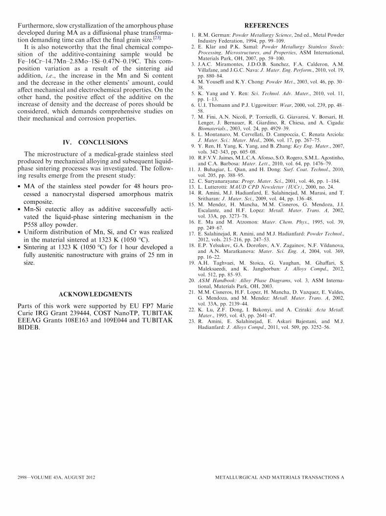
The XRD pattern and TEM micrograph of theas-milled P558 powder are shown in Figure 1. Basedon the Rietveld XRD quantitative analysis (the proce-dure has been detailed in Reference 14), this powderconsists of 10 pct ferrite (a), 48 pct austenite (c), and42 pct amorphous phase, where the crystallite size of theferrite and austenite phases is 11.5 and 12 nm, respec-tively, measured by the Double-Voigt approach. Notethat the relatively low signal-to-noise ratio in the XRDpattern is a result of the presence of the amorphousphase in the material. The selected area diffractionpattern of the TEM micrograph in Figure 1(b) includessome diffraction spots related to the crystalline phasesand a halo pattern related to the amorphous phase,confirming that the material has an amorphous/nano-crystalline structure. It is noteworthy that in bright-fieldTEM images, those crystallites that are close to a zone-axis orientation appear dark; in contrast, all crystallitesthat are far off a zone-axis orientation appear bright likethe amorphous phase. Additionally, Figure 1(c) presentsthe high-resolution TEM micrograph of the samepowder, in which a consideration to the atomic arrange-ment depicts a crystalline region embedded in theamorphous matrix, inferring the heterogeneous nucle-ation of the amorphous phase from grain boundaries of
Fig. 1—XRD pattern (a), TEM micrograph (b), and high-resolution TEM micrograph (c) of the as-milled P558 powder.
METALLURGICAL AND MATERIALS TRANSACTIONS A VOLUME 43A, AUGUST 2012—2995
the crystalline phases as high-energy places that arepreferential for nucleation.[15,16] The significant struc-tural refinement leading to nanocrystallization can beexplained by severe plastic deformation (as a result ofthe actions of the milling media[12]) and the contributionof the interstitially dissolved elements (C and N).Nitrogen[14,17] and carbon[18] atoms are segregated atdislocations and grain boundaries, fixing the disloca-tions and stabilizing the grain boundaries. Conse-quently, the trickling down of mobile dislocations on
the fixed dislocations contributes to the nucleation ofnew boundaries and to a severe grain refinement. Asimilar contribution to structural refinement has beenattributed to the interstitial dissolution of boron inmechanically alloyed Co-based powders.[19] Moreover,amorphization can be explained by severe plastic defor-mation (accordingly extreme structural refinement),large atomic size mismatch, and negative enthalpy ofmixing among the constituent elements.[14,15,17,19]
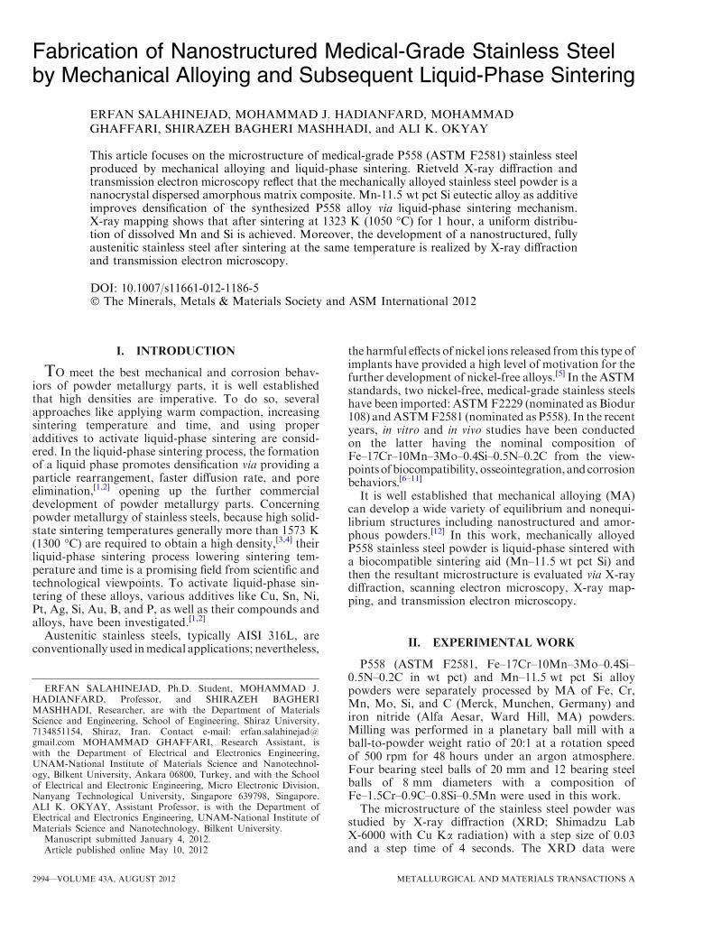
Figure 2 indicates the sintered densities measured bythe Archimedes water immersion method. It can be seenthat by increasing the sintering temperature and addingthe sintering aid, higher densities are achieved. For thesamples containing the sintering aid, a sharp increase indensity occurs when the sintering temperature increasesfrom 1273 K to 1323 K (1000 �C to 1050 �C), followedby a lower rate densification with the further tempera-ture increase. The additive (Mn–11.5 wt pct Si) is aeutectic alloy with a eutectic temperature of 1313 K(1040 �C).[20] The formation of the eutectic liquid phaseat the temperatures higher than 1313 K (1040 �C)activates the liquid phase sintering process. Indeed, theformed liquid at the additive particle sites wets the mainpowder particles, penetrates the particle contacts andpore zones via capillary forces, and provides a path ofhigh diffusivity. With increasing the sintering tempera-ture beyond 1323 K (1050 �C), the decrease in the
Fig. 2—Sintered densities measured by the Archimedes water immer-sion method.
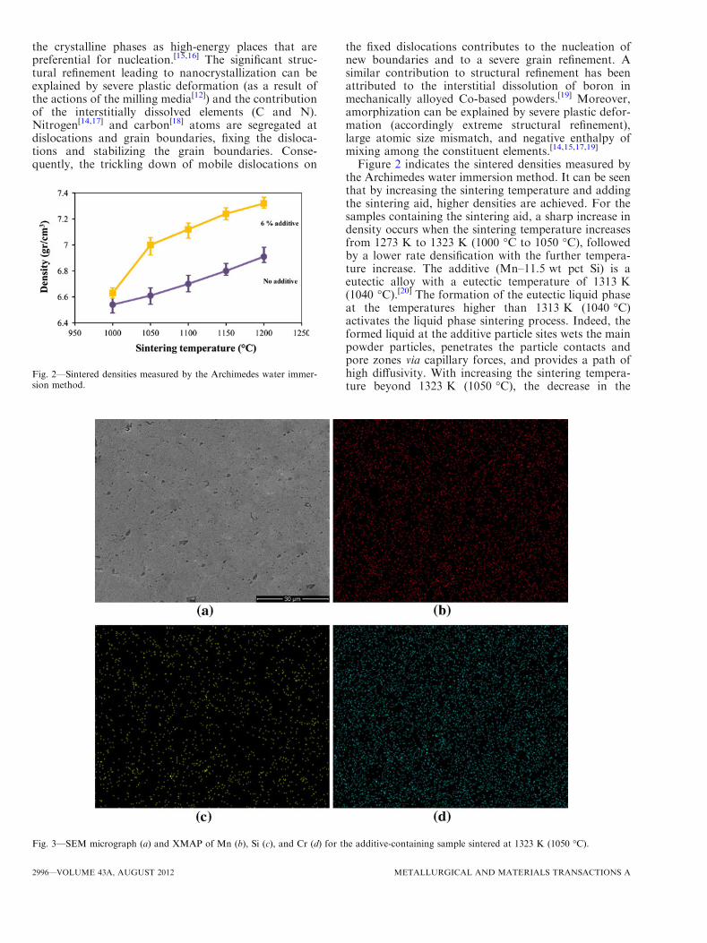
Fig. 3—SEM micrograph (a) and XMAP of Mn (b), Si (c), and Cr (d) for the additive-containing sample sintered at 1323 K (1050 �C).
2996—VOLUME 43A, AUGUST 2012 METALLURGICAL AND MATERIALS TRANSACTIONS A
formed liquid viscosity, the increase in wetting charac-teristics, the increase in diffusivity, and possibly theincrease in the liquid phase amount are responsible fordensification.[1,2]
Figure 3(a) presents the SEMmicrograph of the samplecontaining 6 pct additive sintered at 1323 K (1050 �C) for1 hour. Small, relatively spherical, and isolated poresobserved, despite the practiced sintering temperature thatis low for solid-state sintering, implies activation of theliquid-phase sintering mechanism.Manganese and SiliconXMAP (Figures 3(b) and (c)) confirms the completedissolution, no evidence of precipitation, and the uniformdistribution of the additive elements in the structure in ascale comparable with the stainless steel particle size (SEMobservations of the P558 powder suggested that theparticles are almost 20lm in size). In addition, the uniformdistribution of Cr in the structure (Figure 3(d)), which iscritical for corrosion protection, infers the merit ofprocessing. The achievement of this uniform microstruc-ture is explainedby liquationof theMn–Si eutectic additiveand thereby providing a path of high diffusivity. On theother hand, considering the mean size of the main powderparticles, which is almost 20 lm, the mean diffusion lengthis estimated to be 10 lm. Indeed, this short diffusion lengthand the high-diffusivity liquid path justify the developmentof this homogeneous structure by sintering at 1323 K(1050 �C) for 60 minutes.
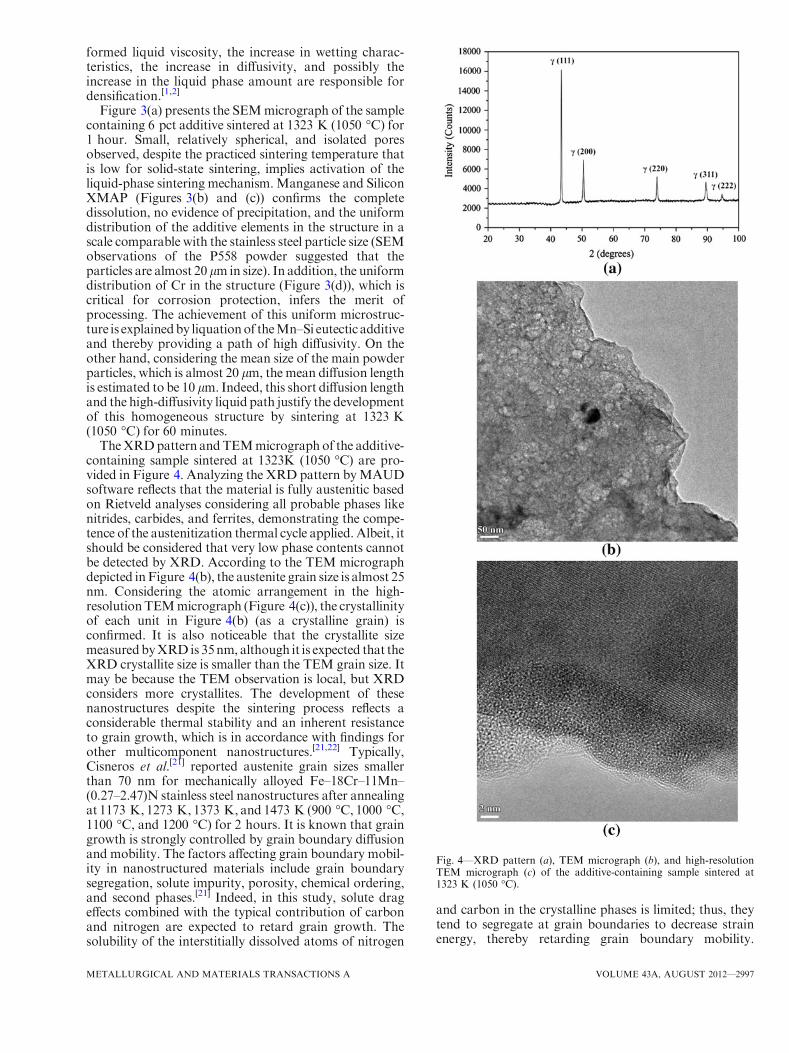
TheXRDpattern andTEMmicrograph of the additive-containing sample sintered at 1323K (1050 �C) are pro-vided in Figure 4. Analyzing the XRD pattern byMAUDsoftware reflects that the material is fully austenitic basedon Rietveld analyses considering all probable phases likenitrides, carbides, and ferrites, demonstrating the compe-tence of the austenitization thermal cycle applied. Albeit, itshould be considered that very low phase contents cannotbe detected by XRD. According to the TEM micrographdepicted inFigure 4(b), the austenite grain size is almost 25nm. Considering the atomic arrangement in the high-resolutionTEMmicrograph (Figure 4(c)), the crystallinityof each unit in Figure 4(b) (as a crystalline grain) isconfirmed. It is also noticeable that the crystallite sizemeasuredbyXRD is 35nm, although it is expected that theXRD crystallite size is smaller than the TEM grain size. Itmay be because the TEM observation is local, but XRDconsiders more crystallites. The development of thesenanostructures despite the sintering process reflects aconsiderable thermal stability and an inherent resistanceto grain growth, which is in accordance with findings forother multicomponent nanostructures.[21,22] Typically,Cisneros et al.[21] reported austenite grain sizes smallerthan 70 nm for mechanically alloyed Fe–18Cr–11Mn–(0.27–2.47)N stainless steel nanostructures after annealingat 1173 K, 1273 K, 1373 K, and 1473 K (900 �C, 1000 �C,1100 �C, and 1200 �C) for 2 hours. It is known that graingrowth is strongly controlled by grain boundary diffusionand mobility. The factors affecting grain boundary mobil-ity in nanostructured materials include grain boundarysegregation, solute impurity, porosity, chemical ordering,and second phases.[21] Indeed, in this study, solute drageffects combined with the typical contribution of carbonand nitrogen are expected to retard grain growth. Thesolubility of the interstitially dissolved atoms of nitrogen
and carbon in the crystalline phases is limited; thus, theytend to segregate at grain boundaries to decrease strainenergy, thereby retarding grain boundary mobility.
Fig. 4—XRD pattern (a), TEM micrograph (b), and high-resolutionTEM micrograph (c) of the additive-containing sample sintered at1323 K (1050 �C).
METALLURGICAL AND MATERIALS TRANSACTIONS A VOLUME 43A, AUGUST 2012—2997
Furthermore, slow crystallization of the amorphous phasedeveloped during MA as a diffusional phase transforma-tion demanding time can affect the final grain size.[23]
It is also noteworthy that the final chemical compo-sition of the additive-containing sample would beFe–16Cr–14.7Mn–2.8Mo–1Si–0.47N–0.19C. This com-position variation as a result of the sintering aidaddition, i.e., the increase in the Mn and Si contentand the decrease in the other elements’ amount, couldaffect mechanical and electrochemical properties. On theother hand, the positive effect of the additive on theincrease of density and the decrease of pores should beconsidered, which demands comprehensive studies ontheir mechanical and corrosion properties.
IV. CONCLUSIONS
The microstructure of a medical-grade stainless steelproduced by mechanical alloying and subsequent liquid-phase sintering processes was investigated. The follow-ing results emerge from the present study:
� MA of the stainless steel powder for 48 hours pro-cessed a nanocrystal dispersed amorphous matrixcomposite.
� Mn-Si eutectic alloy as additive successfully acti-vated the liquid-phase sintering mechanism in theP558 alloy powder.
� Uniform distribution of Mn, Si, and Cr was realizedin the material sintered at 1323 K (1050 �C).
� Sintering at 1323 K (1050 �C) for 1 hour developed afully austenitic nanostructure with grains of 25 nm insize.
ACKNOWLEDGMENTS
Parts of this work were supported by EU FP7 MarieCurie IRG Grant 239444, COST NanoTP, TUBITAKEEEAG Grants 108E163 and 109E044 and TUBITAKBIDEB.
REFERENCES1. R.M. German: Powder Metallurgy Science, 2nd ed., Metal Powder
Industry Federation, 1994, pp. 99–109.2. E. Klar and P.K. Samal: Powder Metallurgy Stainless Steels:
Processing, Microstructures, and Properties, ASM International,Materials Park, OH, 2007, pp. 59–100.
3. J.A.C. Miramontes, J.D.O.B. Sanchez, F.A. Calderon, A.M.Villafane, and J.G.C. Nava: J. Mater. Eng. Perform., 2010, vol. 19,pp. 880–84.
4. M. Youseffi and K.Y. Chong: Powder Met., 2003, vol. 46, pp. 30–38.
5. K. Yang and Y. Ren: Sci. Technol. Adv. Mater., 2010, vol. 11,pp. 1–13.
6. U.I. Thomann and P.J. Uggowitzer: Wear, 2000, vol. 239, pp. 48–58.
7. M. Fini, A.N. Nicoli, P. Torricelli, G. Giavaresi, V. Borsari, H.Lenger, J. Bernauer, R. Giardino, R. Chiesa, and A. Cigada:Biomaterials., 2003, vol. 24, pp. 4929–39.
8. L. Montanaro, M. Cervellati, D. Campoccia, C. Renata Arciola:J. Mater. Sci.: Mater. Med., 2006, vol. 17, pp. 267–75.
9. Y. Ren, H. Yang, K. Yang, and B. Zhang: Key Eng. Mater., 2007,vols. 342–343, pp. 605–08.
10. R.F.V.V. Jaimes, M.L.C.A. Afonso, S.O. Rogero, S.M.L. Agostinho,and C.A. Barbosa: Mater. Lett., 2010, vol. 64, pp. 1476–79.
11. J. Buhagiar, L. Qian, and H. Dong: Surf. Coat. Technol., 2010,vol. 205, pp. 388–95.
12. C. Suryanarayana: Progr. Mater. Sci., 2001, vol. 46, pp. 1–184.13. L. Lutterotti: MAUD CPD Newsletter (IUCr), 2000, no. 24.14. R. Amini, M.J. Hadianfard, E. Salahinejad, M. Marasi, and T.
Sritharan: J. Mater. Sci., 2009, vol. 44, pp. 136–48.15. M. Mendez, H. Mancha, M.M. Cisneros, G. Mendoza, J.I.
Escalante, and H.F. Lopez: Metall. Mater. Trans. A, 2002,vol. 33A, pp. 3273–78.
16. E. Ma and M. Atzomon: Mater. Chem. Phys., 1995, vol. 39,pp. 249–67.
17. E. Salahinejad, R. Amini, and M.J. Hadianfard: Powder Technol.,2012, vols. 215–216, pp. 247–53.
18. E.P. Yelsukov, G.A. Dorofeev, A.V. Zagainov, N.F. Vildanova,and A.N. Maratkanova: Mater. Sci. Eng. A, 2004, vol. 369,pp. 16–22.
19. A.H. Taghvaei, M. Stoica, G. Vaughan, M. Ghaffari, S.Maleksaeedi, and K. Janghorban: J. Alloys Compd., 2012,vol. 512, pp. 85–93.
20. ASM Handbook: Alloy Phase Diagrams, vol. 3, ASM Interna-tional, Materials Park, OH, 2003.
21. M.M. Cisneros, H.F. Lopez, H. Mancha, D. Vazquez, E. Valdes,G. Mendoza, and M. Mendez: Metall. Mater. Trans. A, 2002,vol. 33A, pp. 2139–44.
22. K. Lu, Z.F. Dong, I. Bakonyi, and A. Cziraki: Acta Metall.Mater., 1995, vol. 43, pp. 2641–47.
23. R. Amini, E. Salahinejad, E. Askari Bajestani, and M.J.Hadianfard: J. Alloys Compd., 2011, vol. 509, pp. 3252–56.
2998—VOLUME 43A, AUGUST 2012 METALLURGICAL AND MATERIALS TRANSACTIONS A