fabrication of tissue engineering scaffold from
TRANSCRIPT
Abstract—Scaffolds for bone tissue engineering must meet
the functional requirements, porosity, biocompatibility, and
biodegradability. In this study, hydroxyapatite was prepared by
wet chemical method and incorporated into the alginate gel
solution to improve both the mechanical and cell-attachment
properties of the scaffolds. Next, composite scaffolds were
fabricated from hydroxyapatite/alginate with different weight
ratios by freeze drying method and then some of them coated
with triblock copolymer and compared with others.
Microstructure observation with SEM suggests the formation of
about 50 micrometer size porous structure and interconnected
porosity. Then, human mesenchymal stem cells were cultured
on the composite scaffolds. Cells adhesion to the scaffolds was
observed after three days by DAPI fluorescence microscopy in
which more cells adhesion to the coated scaffolds and cells
diffusion into the pores are visible. Also, cell adhesion within the
structure was observed by SEM in which showed cell
attachment was well in depth which confirms DAPI results.
These results suggest that the triblock-coated HA/Alg porous
scaffolds could provide enhanced cell adhesion and
proliferation which may be a promising approach for bone
tissue-engineering applications.
Index Terms—Alginate, cell adhesion, composite,
freeze-drying, human mesenchymal stem cells, hydroxyapatite,
porous scaffold, tissue engineering.
I. INTRODUCTION
Tissue engineering is the science of design and fabrication
of new tissues for functional restoration of impaired organs
and replacement of lost parts due to cancer, disease and
trauma [1], [2]. The three key ingredients for tissue
engineering and tissue regeneration are signals, stem cells
and Scaffolds [3]. Scaffolds could provide a solid framework
for cell growth and differentiation, allowing cell attachment
and migration [4]-[6]. Several requirements must be
considered in the design of three-dimensional (3D) scaffolds
for tissue engineering [7]-[9]. High porosity is a major factor
that is desired to increase the specific surface area for cell
attachment and tissue in-growth in scaffolds [10]. The pore
size must be large enough to allow accommodation of cells so
that interconnected pores may facilitate uniform distribution
of cells, diffusion of oxygen and nutrient, and waste
exchange by cells deep within the construct [11]-[14].
Polymer/ceramic composites may improve mechanical
properties compared with polymers, and better structural
integrity and flexibility than brittle ceramics. In fact, the
Manuscript received December 9, 2013; revised February 15, 2014.
Masoumeh Haghbin Nazarpak is with New Technologies Research
Center (NTRC), Amirkabir University of Technology, P. O. Box:
1591633311, Tehran, Iran (e-mail: [email protected]).
Farzaneh Pourasgari is with Razi Institute of Serum And Vaccination,
Tehran, Iran (e-mail: [email protected]).
combination of ceramics and polymers could provide
reinforced porous structures with enhanced bioactivity and
controlled resorption rates [15], [16].
Several conventional methods have been used to fabricate
3-D scaffolds [17]. In particular, freeze drying is a technique
including solvent casting method, in which the solvent
removing may be accompanied by freeze drying to a more
porous structure to be obtained. In this procedure, cavities is
produced in the scaffold due to the space occupied by the
solvent after drying, and the smaller pores arising from
sublimation of the solvent serves as interconnection between
the macropores [18].
Natural and synthetic materials can be used for fabrication
of porous scaffolds for bone regeneration. Alginate is a
naturally occurring anionic and hydrophilic polysaccharide.
It is one of the most abundant biosynthesized materials [19],
[20], and is derived primarily from brown seaweed and
bacteria. Unfortunately some drawbacks to alginate include
mechanical weakness and poor cell adhesion. In order to
overcome these limitations, the strength and cell behavior of
alginate have been enhanced by mixtures with other materials,
such as ceramics [20]. In this way, production of composite
scaffolds containing ceramics and synthetic polymers
provided a scaffold with desirable properties. In this way, a
biodegradable Alg/HA composite scaffolds with different
weight ratio for efficient bone tissue engineering were
prepared in the present work. On the next step, we compared
cell adhesion and proliferation on the triblock copolymer
coated scaffolds with uncoated one. The results showed
better cell adhesion on the modified samples.
It is demonstrated in the present work that porous Alg/HA
scaffolds can be successfully prepared by the proposed
freeze-drying method. The prepared scaffolds are highly
porous, and have interconnected pores about 50 µm. Study of
the cell–scaffold interaction also demonstrated the ability of
the scaffold to support hMSC adhesion and proliferation that
suggests its potential application in bone tissue engineering.
II. MATERIALS AND METHODS
A. Synthesis of Hydroxyapatite Powder
Orthophosphoric acid, calcium hydroxide and NH4OH
were obtained from Aldrich Chemical Co. Hydroxyapatite
powders were synthesized by a wet chemical method, based
on the precipitation of HA particles from aqueous solution.
The synthesis procedure involved drop by drop addition of
the H3PO4 solution (0.3 M) into an aqueous suspension of
Ca(OH)2 (0.5 M) while stirring vigorously for about 24 h.
Simultaneously, ammonia hydroxide solution was added to
adjust pH at 10-11. Then, the obtained white precipitate was
aged for 7 days, decanted, rinsed with deionized water, and
Masoumeh Haghbin Nazarpak and Farzaneh Pourasgari
Fabrication of Tissue Engineering Scaffold from
Hydroxyapatite/Alginate Composite
International Journal of Bioscience, Biochemistry and Bioinformatics, Vol. 4, No. 3, May 2014
142DOI: 10.7763/IJBBB.2014.V4.327
filtered. After filtration, the precipitate was dried in an oven
at 70 °C for 24 h. Finally, it was heated at 1000 °C for 1 h in a
conventional furnace under air atmosphere.
B. Scaffold Fabrication
Sodium alginate (29 cP for 1% at 25 °C) was obtained
from Sigma Chemical Co., triblock copolymer were prepared
in our laboratory [21], [22]. To prepare the composite
scaffolds, different weight ratios hydroxyapatite suspension
were prepared using aqua-sonication for 20 min and added
into the prepared alginate solution. Then, the gelation process
was started by spraying 1.0 M CaCl2 on the mixture.
Next, the gels were cast in moulds (12 mm diameter) and
frozen in freezer overnight. Next, the frozen samples were
freeze dried. Some of the samples are used as prepared. For
the coated ones, the triblock copolymer synthesized in
laboratory according to procedure previously reported [21],
[22] dissolved in ethanol was used as coating on some of
samples and finally air dried.
III. RESULTS
A. X-Ray Diffraction
X-ray diffraction was performed to determine phase
structure of the scaffolds using XRD (INEL, EQuinox 3000,
France). The XRD pattern of the sample indicated the
presence of hydroxyapatite phase as shown in Fig. 1. In
addition, there was no evidence of formation of other
unwanted phases, which confirms the purity of the powder is
appropriate for biomedical applications. Also the sharpness
of the peaks revealed its high crystallinity.
Fig. 1. XRD pattern of hydroxyapatite powder prepared in wet chemical
method compared with the reference hydroxyapatite pattern.
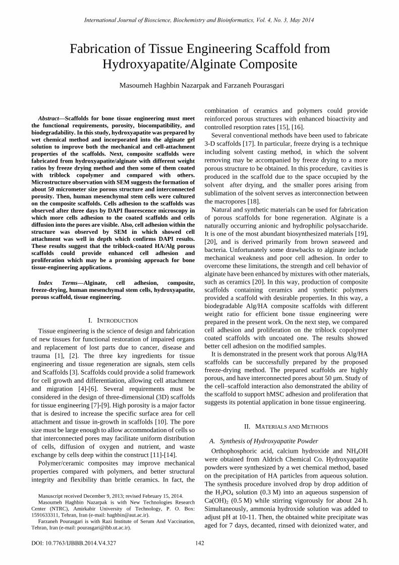
B. FTIR Analysis
FTIR analysis (BRUKER VECTOR 33, Germany) was
performed using KBr. The result is shown in Fig. 2. As it is
seen the 635 and 3570 cm–1 bands correspond to OH– group,
to strongly adsorbed and/or bound H2O. H2O band was also
observed at 1640 cm–1. A strong band of PO43 – group was
also seen at 1046, 962, 602 and 571 cm–1. The bands obtained
for respective phosphate and hydroxyl groups of pure HA,
were in agreement with other published data.
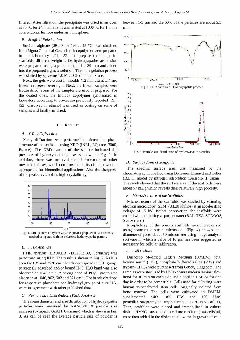
C. Particle size Distribution (PSD) Analysis
The mean diameter and size distribution of hydroxyapatite
particles were measured by NANOPHOX particle size
analyser (Sympatec GmbH, Germany) which is shown in Fig.
3. As can be seen the average particle size of powder is
between 1-5 µm and the 50% of the particles are about 2.5
µm.
Fig. 2. FTIR patterns of hydroxyapatite powder.
Fig. 3. Particle size distribution of hydroxyapatite particles.
D. Surface Area of Scaffolds
The specific surface area was measured by the
chromatographic method using Brunauer, Emmett and Teller
(B.E.T) model by nitrogen adsorbtion (Bellsorp II, Japan).
The result showed that the surface area of the scaffolds were
about 57 m2/g which reveals their relatively high porosity.
E. Microstructure of the Scaffolds
Microstructure of the scaffolds was studied by scanning
electron microscopy (SEM) (XL30 Philips) at an accelerating
voltage of 15 kV. Before observation, the scaffolds were
coated with gold using a sputter coater (BAL-TEC, SCDOOS,
Switzerland).
Morphology of the porous scaffolds was characterized
using scanning electron microscope (Fig. 4) showed the
diameter of pores about 50 micrometer using image analysis
software in which a value of 10 µm has been suggested as
necessary for cellular infiltration.
F. Cell Culture
Dulbecco Modified Eagle’s Medium (DMEM), fetal
bovine serum (FBS), phosphate buffered saline (PBS) and
trypsin–EDTA were purchased from Gibco, Singapore. The
samples were sterilized by UV exposure under a laminar flow
hood for 10 min on each side and placed in DMEM for one
day in order to be compatible. Cells used for culturing were
human mesenchymal stem cells, originally isolated from
bone marrow. The cells were cultivated in DMEM,
supplemented with 10% FBS and 100 U/ml
penicillin–streptomycin–amphotercin, at 37 °C in 5% of CO2.
Then, scaffolds were placed and immobilized in culture
dishes. HMSCs suspended in culture medium (104 cells/ml)
were then added in the dishes to allow the in growth of cells
International Journal of Bioscience, Biochemistry and Bioinformatics, Vol. 4, No. 3, May 2014
143

to the scaffolds. The culture medium was changed every two
days. After incubation in various periods, cells attached on
the scaffolds were harvested for analysis.
Fig. 4. Microstructure of the porous scaffolds.
Fig. 5. SEM photographs of cells morphology on the scaffolds.
G. Morphology of Cells
Cell morphology on the scaffold was also investigated by
scanning electron microscopy that is shown in Fig. 5. The
cell-loaded scaffolds were rinsed with PBS after 3 day of cell
seeding and fixed in glutaraldehyde 2.5% for 1 h. For
dehydrating the scaffolds were placed in a series of gradient
of alcohol concentration and then dried.
The results indicated that the mesenchymal stem cells
cultured in scaffolds can be seen not only in the surface of the
scaffold, but also inside the pores. The images showed the
perfect adhesion of cells to scaffold surface outside and
inside the pores. Adhesion structures resembling tight
junctions were present. The cells elongations and their
interconnection forming a cell net were observed clearly.
Cells that attach themselves to the scaffold, but spread little
might show lower proliferative rates than those with greater
spreading. These scaffolds allowed flattening and spreading
of the cells, showing adequate cell shape for proliferation and
secretion functions.
H. DAPI Fluorescent Staining
The cells on the scaffolds were fixed with 4%
paraformaldehyde. Samples were then washed twice with
PBS, incubated with 4, 6-diamidino-2-phenylindole
(DAPI;Sigma Chemical Co.) for 30 seconds to label nuclei of
the cells and again were rinsed twice with PBS. The
immunofluorescence images were obtained by using a
fluorescence microscope (Nikon, Eclipse).
DAPI fluorescent staining was carried out after 3 days
cultivation which is shown in Fig. 6. In DAPI staining, bright
fluorescence revealed the presence of nuclei. As can be seen,
the samples with more hydroxyapatite percent and/ or coated
with triblock copolymer had better cell adhesion.
International Journal of Bioscience, Biochemistry and Bioinformatics, Vol. 4, No. 3, May 2014
144

IV. CONCLUSION
In the present work, it is demonstrated that porous Alg/HA
scaffolds was successfully prepared by the proposed
freeze-drying method. The prepared scaffolds were
characterized, which the results showed their high porosity,
and interconnected pores about 50 µm. Also, the cell culture
results revealed that the matrix was not cytotoxic and the
cells were strongly adhered to the substrate in the first hours
of cell/ substrate contact. The ability of these scaffolds to
support hMSC adhesion and proliferation suggests its
potential application in tissue engineering.
It is concluded that these scaffolds are promising materials
for tissue engineering, providing a good environment for the
adhesion and proliferation of cells. However, in the next step,
the tests will be focused on the osteogenic differentiation
capability of mesenchymal stem cells seeded on these
scaffolds for bone tissue engineering.
REFERENCES
[1] A. H. Reddi, “Role of morphogenetic proteins in skeletal tissue
engineering and regeneration,” Nat Biotechnol, vol. 16, pp. 247-52,
1998.
[2] A. H. Reddi, “Symbiosis of biotechnology and biomaterials:
applications in tissue engineering of bone and cartilage,” J Cell
Biochem, vol. 56, pp. 192-195, 1994.
[4] G. Chen, T. Ushida, and T. Tateishi, “Development of biodegradable
porous scaffolds for tissue engineering,” Materials Science
Engineering, vol. C17, C17, pp. 63-69, 2001.
[5] D. W. Hutmacher and A. J. Garcia, “Scaffold-based bone engineering
by using genetically modified cells,” Gene, vol. 347, pp. 1-10, 2005.
[6] X. Zhu, W. Cui, X. Li, and Y. Jin, “Electrospun fibrous mats with high
porosity as potential scaffolds for skin tissue engineering,”
Biomacromolecules, vol. 9, pp. 1795-1801, 2008.
[7] L. Budyanto, C. P. Ooi, and Y. Q. Goh, “Fabrication and
characterization of porous poly (L-lactide) PLLA scaffolds using
liquid-liquid phase separation,” IFMBE Proc., vol. 21, pp. 322-325,
2008.
[8] P. A. Gunatillake and R. Adhikari, “Biodegradable synthetic polymers
for tissue engineering,” Eur Cells Materials, vol. 5, pp. 1-16, 2003.
[9] S. Yang, K. F. Leong, Z. Du, and C. K. Chua, “The design of scaffolds
for use in tissue engineering: Part I. Traditional factors,” Tissue
Engineering, vol. 7, pp. 679-689, 2001.
[11] R. A. Horch, N. Shahid, A. S. Mistry, and M. D. Timmer,
“Nanoreinforcement of poly (propylene fumarate)-based networks
with surface modified alumoxane nanoparticles for bone tissue
engineering,” Biomacromolecules, vol. 5, pp. 1990-1998, 2004.
[12] C. T. Buckley and K. U. O’Kelly, “Regular scaffold fabrication
techniques for investigations in tissue engineering,” Topics
Bio-Mechanical Engineering, pp. 147-166, 2004.
[13] F. Lyons, S. Partap, and F. J. O’Brien, “Part 1: Scaffolds and surfaces,”
Technology Health Care, vol. 16, pp. 305-317, 2008.
[14] L. M. Mathieu, P. E. Bourban, and J. E. Ma˚nson, “Processing of
homogeneous ceramic/polymer blends for bioresorbable composites,”
Composites SciTechnol, vol. 66, pp. 1606-1614, 2006.
[15] M. Neumann and M. Epple, “Composites of calcium phosphate and
polymers as bone substitution materials,” Eur Jtrauma, vol. 32, pp.
125-131, 2006.
[16] T. Tyson, A. F. Wistrand, and A. C. Albertsson, “Degradable porous
scaffolds from various L-lactide and trimethylene carbonate
copolymers obtained by a simple and effective method,”
Biomacromolecules, vol. 10, pp. 149-154, 2009.
[17] J. H. de Groot, H. W. Kuijper, and A. J. Pennings, “A novel method for
fabrication of biodegradable scaffolds with high compression moduli,”
J Materials Science: Mater Med., vol. 8, pp. 707-712, 1997.
[18] R. P. Narayanan, G. Melman, N. J. Letourneau, N. L. Mendelson, and
A. Melman, “Photodegradable iron(III) cross-linked alginate gels,”
Biomacromolecules, vol. 13, pp. 2465–2471, 2012.
[19] G. Skjak-Braerk, H. Grasdalen, and O. Smidsrod, “Inhomogeneous
polysaccharide ionic gels,” Carbohydr. Polym, vol. 10, pp. 31–54,
1989.
[20] J. Cuy. (2004). Biomaterials Tutorial: Natural Polymers. University of
Washington Engineered Biomaterials. [Online]. Available: http://www.
uweb.engr.washington.edu/.
[21] F. Najafi and M. N. Sarbolouki, “Synthesis and Characterization of
Block Copolymers from Aromatic Diols, Fumaric Acid,” Sebacic Acid
and PEG, J Appl Polym Sci, 2003, vol. 90, pp. 2358–2363.
[22] F. Najafi and M. N. Sarbolouki, “Biodegradable
micelles/polymersomes from fumaric/sebacic acids and poly(ethylene
glycol),” Biomaterials, vol. 24, pp. 1175–1182, 2003.
Masoumeh Haghbin Nazarpak was born in Iran and
obtained her Ph.D from Amirkabir University of
Technology of Iran in Biomedical Engineering
(Biomaterials). She is now the academic staff of New
Technologies Research Center of Amirkabir
University of Technology and works in the fields of
biomaterials, tissue engineering and drug delivery
systems.
She has published a few papers such as
Nanocrystalline fluorine-substituted hydroxyapatite [Ca5(PO4)3(OH)1-xFx
(0≤x≤1)] for biomedical applications: Preparation and characterization,
Micro & Nano Letters, 7, Issue 2 ( 2012), Injectable and bioresorbable
calcium phosphate delivery system with gentamicin sulphate for treatment of
bone diseases: in vitro study, Advances in Applied Ceramics, Volume 110,
Number 8 (2011), Preparation of a novel porous scaffold from poly
(lacticco-glycolic acid)/hydroxyapatite, Asian Biomedicine, Vol. 5 No. 4
( 2011).
Dr. Masoumeh Haghbin Nazarpak is the member of professional societies
such as Iranian Ceramic Society (ICERS), Iranian Society of Biomedical
Engineering (ISBE), The Australian Research Network for Advanced
Materials (ARNAM), Biomaterials Research Centre, Göteborg.
Farzaneh Pourasgari was born in Iran and obtained
her Ph.D in Biochemistry from University of Tehran,
Institute of Biochemistry and Biophysics. She is now
the academic staff of Razi Vaccine and Serum
Research Institute and one her fields of works is stem
cell culture and differentiation.
She has published a few papers such as
“Preparation of a novel porous scaffold from poly
(lacticco-glycolic acid)/ hydroxyapatite, Asian
Biomedicine, Vol. 5 No. 4 ( 2011)” and “Low cytotoxicity capacity of
Dendrosomes as a carrier for intranasally administered DNA vaccines.
Molecular Biology Reports 2007, 36(1):105-9”.
Dr. Farzaneh Pourasgari is the member of professional societies such as
Biochemistry Society, Iranian Genetic Society and Iranian Stem Cell
Society.
International Journal of Bioscience, Biochemistry and Bioinformatics, Vol. 4, No. 3, May 2014
145
..
[3] A. H. Reddi and C. Huggins, “Biochemical sequences in the
transformation of normal fibroblasts in adolescent rats,” in Proc. Natl
Acad Sci, vol. 69, pp. 1601-1605, USA 1972.
- .
[10] S. Gong, J. Dong, S. T. Xue, and J. Y. Wang, “A novel porous natural
polymer scaffold for tissue engineering,” in Proc. 2005 IEEE
Engineering Medicine Biology 27th Annual Conf, pp. 4884-4887,
Shanghai, Sept. 2005.
Fig. 6. DAPI staining results of coated samples 3% hydroxyapatite
suspension & 2% alginate solution (up) and 6% hydroxyapatite suspension
& 2% alginate solution (down).