factors affecting the outcome of bronchiectasis in
TRANSCRIPT

International Journal of Pediatrics , Vol.2, N.4-3, Serial No.12, December 2014 377
Original Article
http:// ijp.mums.ac.ir
Factors Affecting the Outcome of Bronchiectasis in Pediatric
Patients *Nemat Bilan
1, Mitra Aghakhani
2, Farhad Niafar
3
1Pediatric Pulmonologist. 2 Pediatrician, Tabriz University of Medical Sciences, Tabriz, Iran. 3 Internist, Faculty of Medicine, Tabriz University of Medical Sciences, Tabriz, Iran.
Abstract
Introduction
Bronchiectasis is a common problem in children and early diagnosis can lead to early treatment and
prevent of its complications. This study was aimed to evaluate factors effectiveness on outcome of
bronchiectasis in children.
Methods and Materials
In an analytical cross-sectional study, 347 children with bronchiectasis underwent the study. The patients
were diagnosed based on chronic suppurative cough and Computerized tomography(CT) scan findings.
Results
Disease etiology was asthma in 55.6%, Gastroesophageal reflux disease (GERD) in 7.8%, Cystic
fibrosis (CF) in 4.8%, other causes in 11.2% and idiopathic in 20.6%. All cases complained of
chronic cough. The most common sign was daily sputum production (79.1%) and common symptoms
were ral/crackle in 47.1% and wheezing in 25.4%. Mean treatment period was 32.82±11.56 months. At
the end of follow-up, complete improvement occurred in 35.6%, partial improvement in
40.9% and no improvement in 23.5%.
Conclusion
In children with chronic cough and crackle in physical examination, consideration of
bronchiectasis could be helpful in early diagnosis and complementary evaluations and
treatment initiation. Treatment of the underlying disease could prevent the occurrence and
increase the response to treatment of bronchiectasis.
Keywords: Bronchiectasis, Etiology, Children, Outcome.
*Couresponding Author:
Nemat Bilan, MD, Tabriz Children's Hospital, Sheshgelan St, Tabriz, Iran; Fax: +9841135262280.
E-mail: [email protected]
Received date: Jul 5, 2014 ; Accepted date: Nov 12, 2014

Factors Affecting on Bronchiectasis
International Journal of Pediatrics , Vol.2, N.4-3, Serial No.12, November 2014 378
Introduction
Bronchiectasis remains as an important
health problem in both, developed and
developing countries (1). It results in
impaired quality of life and mortality if
left untreated (2). Bronchiectasis is not a
primary disease, but an anatomical
abnormality which is caused by various
factors. According to the literature, the
primary etiologic factor cannot be found
in 30-74% of the patients (3-5). Early
diagnosis based on detailed medical history
and radiological confirmation is important in
order to establish a treatment focused on the
underlying cause. For this purpose, an
orderly and systematic diagnostic evaluation
is required (1).
The management of bronchiectasis
includes medical interventions as well as
adjunctive therapies and finally surgery.
The therapeutic modalities are provided
with the following goals: aggressive
treatment of infections, treatment of the
underlying disease, promotion of
mucociliary clearance, promotion of
normal growth, avoidance of toxins,
identification and management of
complications, and treatment of the
chronic inflammation to retard disease
progression (2,6). Bronchiectasis is still a
challenge to the pediatric chest physicians in
many developing parts of the world and it
remains a frequent problem being the
final common pathway of several different
lower respiratory tract insults such as
cystic fibrosis, immunodeficiency, ciliary
dyskinesia. There are unanswered
questions about childhood
bronchiectasis, mainly on etiology and
treatment which require more research (7).
It seems that bronchiectasis is a neglected
disorder, with no adequate diagnosis and
treatment
(3). There are few studies published
about bronchiectasis, clinical findings and
response to treatment in children. This
study was aimed to evaluate factors
effective on the outcome of
bronchiectasis in children.
Methods and Materials
This is a cross- sectional and analytic
study performed on 374 pediatric patients
with bronchiectasis presenting to Children
Hospital and Outpatient Pediatric Clinics
of Tabriz University of Medical Sciences
since 2006 till 2010. Inclusion criteria
were having bronchiectasis documented
by clinical manifestations ( including
chronic suppurative cough), and findings on
CT scan, which was reported by one
radiologist and repeated every 6 to 12
months. Exclusion criteria were concurrent
medical disorders, congenital anomalies,
and previous medications. The patients
were treated for 2-3 years, using steroid
inhalers, bronchodilator sprays, and
continuous low- dose oral antibiotic. Then,
according the clinical findings and CT scan
results the patients were classified in
three groups including Recovered,
Partially recovered, and Non- recovered:
• The children in "Recovered group"
were those in whom the clinical findings
including suppurative cough) have been
relieved, and control CT scan showed the
improvement of radiologic findings in
comparison with the basic CT, and the
medication was stopped.
• The children in "Partially recovered"
group continued receiving the decreased
dose of medication because of partial
improvement in suppurative cough.
• The children in "Non- recovered"
group did not have any improvement in
clinical findings and CT scan findings.

Bilan et al.
International Journal of Pediatrics , Vol.2, N.4-3, Serial No.12, December 2014 379
The required data were including
patients' age, sex, symptoms and signs,
imaging results, medical history, family
history, risk factors, medications, duration of
treatment, and response to the therapy. The
data were collected from patients'
medical records using prepared
questionnaire, and then were compared
between the three groups. The study was
confirmed by Regional Ethic Committee,
and the personal information of all
patients was handled as secret.
The collected data were analyzed by SPSS-
16 statistical software by using descriptive
methods (including abundance, percent,
average and standard deviation). The
qualitative variables were compared by
chi- square, and the quantitative variables
were compared by One- way ANOVA test.
Also, independent t- test was applied for
comparison of quantitative variables in
dual groups. The p- values less than 0.05
were considered as statistically significant.
Results
A total of 374 children with
bronchiectasis were studied. The patients
had the age of 2 to 17 years (average:
8.61±3.36; median: 9). Of all the patients
studied, 240 (64.2%) were male and 134
(35.8%) were female. Medical history was
positive in 51 (13.6%) including preterm
birth in 22 (5.9%), recurrent pneumonia in
16 (4.3%), cleft lip in 4 (1%), pulmonary
lobe collapse in 3 (0.8%), asphyxia in 3
(0.8%), congenital metabolic disorders in 2
(0.5%), and pulmonary lobectomy in 1
(0.3%). Familial history of respiratory
disease was positive in 65 (17.4%).
The etiology was idiopathic in 77
(20.6%) and known in 297 (79.4%),
including asthma in 208 (55.6%), GERD in
29 (7.8%), Cystic fibrosis (CF) in 18 (4.8%),
Chest physiotherapy (CP) in 9 (2.4%),
pneumonia in 4 (1.1%), hypotonia and
repeated aspiration in 3 (0.8%), Chronic
liver disease (CLD) in 5 (1.3%), Down
syndrome in 5 (1.3%), esophageal atresia
in 2(0.5%), obesity in 2 (0.5%), and
pulmonary abscess, achalasia, congenital
myopathy, bone deformity and unilateral
paresis of vocal cords, each in one case
(0.3%).
Symptoms were as following: cough in 374
(100%), Chronic suppurative cough (CSC)
in 296 (79.1%), CSC with recurrent
pneumonia in 14 (3.7%), CSC with
dyspnea in 30 (8%), CSC with repeated
aspiration in one (0.3%), CSC with
tachypnea in 3 (0.8%), and with hemoptysis
in 11 (2.9%). Also, there was hemoptysis
with pneumonia in 8 (2.1%), recurrent
pneumonia in 6 (1.6%), dyspnea in 4 (1.1%),
and pneumonia with pulmonary collapse in
one (0.3%).
The clinical findings were as following:
rales (as only sign) in 176 (47.1%),
wheezing in 95 (25.4%), rales and
wheezing in 24 (6.4%), coarse crackle in
75 (20.1%), and clubbing in 4 (1.1%).
CT scan showed the evidences of
bronchiectasis including thickening and
dilatation of bronchi and/or cystic changes
in all patients.
The Medications used in studied were:
antibiotic+ seretide in 216 (57.8%),
antibiotic+ flixotide+ salmeterol in 91
(24.3%), antibiotic+ seretide+ anti-reflux
in 49 (13.1%), and antibiotic+ seretide+
treatment for CF in 18 (4.8%). The duration
of the treatment was 4 to 66 months
(mean=32.82±11.56m, median= 30m).
In follow up, the symptoms and signs were
relived in 227 (60.7%), but did not improve
in 147 (39.3%). Imaging revealed the
relief in 140 patients (37.7%) and the
decrease in disease severity in remaining
234 (62.6%).

Factors Affecting on Bronchiectasis
International Journal of Pediatrics , Vol.2, N.4-3, Serial No.12, November 2014 380
It was reported the need for repeated
presentation to emergency ward and
readmission in 82 cases. The presentation
to emergency ward and readmission was
reported 1 to 7 times ( mean=1.41±0.87,
median= 1). The total of visits of children by
physician was occurred 5 to 60 times
(mean=22.44±10.24, median= 20).
At the end of follow up, the patients'
outcome were as following: complete
recovery and termination of the treatment in
133 (35.6%), partial recovery and continuing
the medications with reduced dose in 153
(40.9%), and not recovery and continuing
the medications with the same dose in 88
(23.5%).
Figure.1 shows the mean age of studied
patients and the disease outcome. As
seen in the figure, the patients without
the response to the therapy are younger
than other groups
(p=0.88). Table.1 shows the relation of
patients' sex and duration of medication with
the disease outcome.
As seen in the table, the male patients
have better response to the therapy than
females (p=0.06). Also, the duration of
medication was not significantly different in
three groups (p=0.16). Also (Table.2) shows
the effect of some variables on response to
the therapy in studied patients.
Table 1: Patients' sex and duration of treatment in three studied groupsp-value No
Recovery
Partial
Recovery
Complete
Recovery
Variables
>0.05 60 (14.97%) 95 (23.80%) 100 (25.40%) Male
>0.05 38 (8.56%) 70 (17.11%) 42 (10.16%) Female
>0.05 32.90±12.8 34.43±10.47 31.35±9.83 Duration of medication (m)

Bilan et al.
International Journal of Pediatrics , Vol.2, N.4-3, Serial No.12, December 2014 381
Table 2: the effect of some variables on response to the therapy in studied patients Variables Complete
Recovery group
Partial
Recovery group
No
Recovery group
P- value
History of previous
disease
19 (14.3%) 19 (12.4%) 13 (14.8%) 0.84
Familial history of
respiratory disease
19 (14.3%) 34 (22.2%) 15 (13.6%) 0.12
Know etiology for
bronchiectasis
110 (37%) 122 (41.1%) 65 (21.9%)
0.27
Unknown etiology
(idiopathic)
23 (29.9%) 31 (40.2%) 23 (29.9%)
With Asthma 82 (39.4%) 98 (47.1%) 28 (13.5%)
<0.001
Without Asthma 51 (30.7%) 55 (33.1%) 60 (68.2%)
With GERD 17 (58.6%) 7 (24.1%) 5 (17.2%)
0.02
Without GERD 116 (33.6%) 146 (42.3%) 83 (24.1%)
With CF 1 (5.6%) 5 (27.8%) 12 (66.7%)
<0.001
Without CF 132 (37.1%) 148 (41.6%) 76 (21.3%)
GERD: Gastroesophageal reflux; CF: Cystic fibrosis
As seen in table 2, the response to
medication was better in patients with
asthma than patients without it (p<0.001).
The presence or absence of known
etiology for bronchiectasis was not
effective on the response to the medication
(p=0.27).
The rate of complete recovery was more in
patients with GERD than patients without
it (p=0.02). The response to medication
was lower in patients with asthma than
patients without it (p<0.001).
The imaging findings were bronchial
thickening and dilatation and other
signs of bronchiectasis, and the response to
the therapy was not significantly different
between various findings in imaging
(p=0.69).
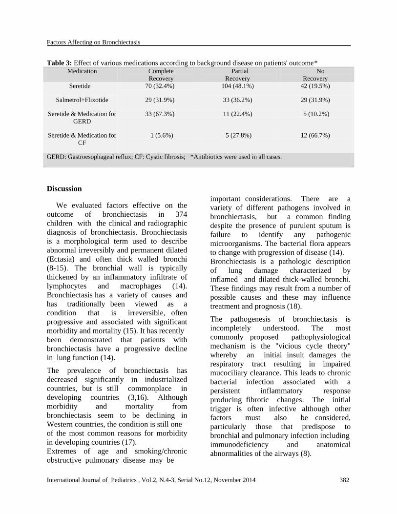
(Table.3) compares the outcome of
various medications scheduled for studied
patients. The complete and partial
recovery was reported at least level in
CF patients with bronchiectasis (cystic
fibrosis bronchiectasis).
Factors Affecting on Bronchiectasis
International Journal of Pediatrics , Vol.2, N.4-3, Serial No.12, November 2014 382
Table 3: Effect of various medications according to background disease on patients' outcome* Medication Complete
Recovery
Partial
Recovery
No
Recovery
Seretide 70 (32.4%) 104 (48.1%) 42 (19.5%)
Salmetrol+Flixotide 29 (31.9%) 33 (36.2%) 29 (31.9%)
Seretide & Medication for
GERD
33 (67.3%) 11 (22.4%) 5 (10.2%)
Seretide & Medication for
CF
1 (5.6%) 5 (27.8%) 12 (66.7%)
GERD: Gastroesophageal reflux; CF: Cystic fibrosis; *Antibiotics were used in all cases.
Discussion
We evaluated factors effective on the
outcome of bronchiectasis in 374
children with the clinical and radiographic
diagnosis of bronchiectasis. Bronchiectasis
is a morphological term used to describe
abnormal irreversibly and permanent dilated
(Ectasia) and often thick walled bronchi
(8-15). The bronchial wall is typically
thickened by an inflammatory infiltrate of
lymphocytes and macrophages (14).
Bronchiectasis has a variety of causes and
has traditionally been viewed as a
condition that is irreversible, often
progressive and associated with significant
morbidity and mortality (15). It has recently
been demonstrated that patients with
bronchiectasis have a progressive decline
in lung function (14).
The prevalence of bronchiectasis has
decreased significantly in industrialized
countries, but is still commonplace in
developing countries (3,16). Although
morbidity and mortality from
bronchiectasis seem to be declining in
Western countries, the condition is still one
of the most common reasons for morbidity
in developing countries (17).
Extremes of age and smoking/chronic
obstructive pulmonary disease may be
important considerations. There are a
variety of different pathogens involved in
bronchiectasis, but a common finding
despite the presence of purulent sputum is
failure to identify any pathogenic
microorganisms. The bacterial flora appears
to change with progression of disease (14).
Bronchiectasis is a pathologic description
of lung damage characterized by
inflamed and dilated thick-walled bronchi.
These findings may result from a number of
possible causes and these may influence
treatment and prognosis (18).
The pathogenesis of bronchiectasis is
incompletely understood. The most
commonly proposed pathophysiological
mechanism is the "vicious cycle theory"
whereby an initial insult damages the
respiratory tract resulting in impaired
mucociliary clearance. This leads to chronic
bacterial infection associated with a
persistent inflammatory response
producing fibrotic changes. The initial
trigger is often infective although other
factors must also be considered,
particularly those that predispose to
bronchial and pulmonary infection including
immunodeficiency and anatomical
abnormalities of the airways (8).

Bilan et al.
International Journal of Pediatrics , Vol.2, N.4-3, Serial No.12, December 2014 383
Identification of predisposing causes may
facilitate prevention of further bronchial
damage (19). Twiss et al. reported
etiology to be unknown (idiopathic) in
54% of cases, post infectious in 22%, as a
result of oncological disease or
chemotherapy in 11%, aspiration 6%, and
primary immunodeficiency 6% (9).
Idiopathic bronchiectasis was found in 13
patients (14%) (20). Nikolaizik and
Warner (19) reviewed 41 cases of
bronchiectasis who presented with chronic
productive cough, and no cause was found
in 37% of their study group. Forty- eight
percent of studied subjects from a New
Zealand cohort, despite extensive
investigations, had no known cause for
their bronchiectasis (21,22). In the
present study, the etiology was unknown
(idiopathic) in 77 (20.6%). This
discrepancy may be due to the
differences in referral patterns and study
populations in the studies.
Unlike the results obtained in this study, the
etiologies of bronchiectasis, reported by a
study in New Zealand were as following:
infections (22%), immunosuppression/
chemotherapy (17%), aspiration (6%), and
idiopathic (54%) (9).
Nikolaizik and Warner suggested that of
4000 children referred for respiratory
disease, 41 (1%) had chronic suppurative
lung disease not due to cystic fibrosis.
Further investigations showed congenital
malformations in six (15%), primary ciliary
dyskinesia syndrome in seven (17%), 11 had
immunological abnormalities (27%), and
two bronchiectasis due to aspiration (5%).
Therefore the underlying cause for the
disease was found in 63% (19).
Bronchiectasis is a relatively common
complication of lower respiratory tract
infections (16,23). It is a major problem
of children in developing countries (24).
In Karadag et al. study, the mean age of
the patients was 7.4 ± 3.7 years at
presentation (17).
The underlying etiology of bronchiectasis
is distinguishable in about 70% and is
considered idiopathic in remaining
(8,9,19). Regarding the initial etiologic
factor and risk factors, the patients with
bronchiectasis have variable outcomes
(15,25). In the present study, the
underlying etiology was determined in
77.4% including asthma (55.6%), GERD
(7.8%), and CF (4.8%). Anomalies,
anatomic disorders, congenital and brain
disorders constituted less than 12% of
etiologies. In the study of Lai et al. the
previous lower respiratory tract infection,
asthma, and primary immunodeficiency
were the most common causes, with
unknown etiology in 31% (26).
In Kim et al. study the underlying
etiologies identified in 85.8% of patients,
included bronchiolitis obliterans (32.6%),
childhood respiratory infection (20.6%),
interstitial lung disease (17.3%),
immunodeficiency (8.6%), and primary
ciliary dyskinesia (4.3%) (20). This
diversity may be due to effects of
geographical and racial differences in the
occurrence of various disease.
The relation between asthma and
bronchiectasis has been controversial
with different prevalence of concurrent
asthma and bronchiectasis (2.7% and
27%) even in a same region (27,28). In
a report from Finland the prevalence of
asthma in patients hospitalized for
bronchiectasis was 19% (29). Possible
mechanisms by which asthma and
bronchiectasis predispose to each other
include asthmatic obstruction
contributing to development of
bronchiectasis, and sensitization of
airways with increased lability due to

Factors Affecting on Bronchiectasis
International Journal of Pediatrics , Vol.2, N.4-3, Serial No.12, November 2014 384
microbial colonization of the ectatic
bronchial tree (28). Misdiagnosis of
asthma is common and the diagnostic
process is further complicated by the fact
that the co-existence of asthma is not
uncommon (30). There is a delay from
onset of bronchiectasis to diagnosis by
HRCT scan.
Usually this may be due to the misdiagnosis
of asthma in children with cough and no
wheeze who had been labeled as having
"cough variant asthma". It has been
reported a potential inaccuracy of a
diagnosis of asthma when based on the
symptom of cough alone (8). In the present
study, the most common known etiology
was asthma in 208 (55.6%).
Cystic fibrosis (CF) is one of the most
important causes, but a great variety of
other causes makes non cystic fibrosis
bronchiectasis a relatively frequent
diagnosis (1). In Western countries, CF is
the most common cause of bronchiectasis
(20,31), and other causes include various
respiratory infections such as pneumonia,
pertussis, measles, and tuberculosis (20).
Bronchiectasis is one of the obvious
manifestations in patients having CF
for years. Evaluation of children with
bronchiectasis has revealed the CF as a
background cause in 3% to 6% (18). CF was
underlying cause of bronchiectasis in 4.8%,
which is compatible with the statistics
indicating its prevalence as 3% to 6% (18).
In the study of Karakoc et al. infection was
the major cause of bronchiectasis. In
8 patients, bronchiectasis developed
after tuberculosis or pneumonia, was
associated with immune deficiency
syndromes in 4 children, and with asthma in
4. Cystic fibrosis was diagnosed in 4 cases
and ciliary dyskinesia in 3. In 10 patients,
only one lobe was involved (16).
Non-CF bronchiectasis in childhood is
still one of the most common causes of
childhood morbidity in developing countries
(17). Various factors have been identified as
predisposing to the development of Non-
CF bronchiectasis. Recurrent respiratory
infections, immune deficiency, a retained
foreign body, asthma, tuberculosis and
primary ciliary dyskinesia are some of the
more common risk factors (17).
Eastham et al. evaluated Non-cystic fibrosis
(CF) bronchiectasis. Associations were
previous pneumonic illness (30%),
immunocompromised (21%), obliterative
bronchiolitis (9%), congenital lung
abnormality (5%), chronic aspiration (3%),
eosinophilic esophagitis (2%), familial
syndrome (2%), primary ciliary
dyskinesia (1%), and right middle lobe
syndrome (1%), while 18% were idiopathic
(8). Banjar evaluated a total of 151 cases
with Non-CF bronchiectasis of which.
There is a period of 5±3.2 years between
the start of symptoms and the diagnosis
of bronchiectasis. More than 2/3 of the
patients had cough, tachypnea, wheezing,
sputum production and failure to thrive.
60% had associated disease: Pulmonary
diseases in 48(32%), immunodeficiency in
27(18%), CNS in 18(12%), cardiac in
12(8%) and asthma in 103(68%) of the
patients. Sixty-eight (67%) were found to
have sinusitis. Forty-nine (32%) developed
GERD (32). In patients with Non-cystic
fibrosis (Non-CF) bronchiectasis,
immunodeficiency, aspiration and primary
ciliary dyskinesia accounted for 67% of the
cases. In 56% of children, the identification
of a cause led to a specific change in
management (21).
Bronchiectasis is currently nearly always
diagnosed early by using High-resolution
computed tomography (HRCT) scanning of

Bilan et al.
International Journal of Pediatrics , Vol.2, N.4-3, Serial No.12, December 2014 385
the chest (8,14,15,31). So, the potential now
exists for the much early detection and
treatment of children with lesser degrees of
bronchial dilation and bronchial wall
thickening than was previously possible
(8,15,31).
HRCT scanning is the most reliable
noninvasive method for assessing the
degree of bronchial wall dilatation, and
thus bronchiectasis can be accurately
diagnosed applying this technique (20,21).
Characteristic findings of bronchiectasis
on CT (33,34) include bronchial wall
thickening with associated bronchial
dilatation. When seen in cross- section,
the internal diameter of the bronchus or
bronchiole becomes larger than that of
its accompanying pulmonary artery (a
broncho arterial ratio greater than 1)
resulting in the "signet ring" sign. The
ectatic airway may be air-filled, fluid-
filled, or have air-fluid levels (14,31,35).
In the present study, CT scan was used
for diagnosis and follow up of
bronchiectasis, and showed one or more
evidences of bronchiectasis in all cases
including bronchial thickening, bronchial
dilatation, or cystic images.
The immune system is less effective in
young children and elderly adults which
results in an increased incidence of
infection in these two groups (36,37).
Bronchiectasis has most commonly been
described as commencing in childhood,
particularly in the first five years of life,
with chronic productive cough and
unresolved infection (12,14,38). In the
present study, the median age at the time
of the diagnosis was 8.61±3.36 years. In
Kim et al. study the median age at the
time of the diagnosis of bronchiectasis was
7.6 years (range, 2 months to 18 years) (20).
In this study of 374 children with
bronchiectasis, 240 (64.2%) were male. This
ratio is compatible with the study of
Eastham et al. in which the male to female
ratio was 2:1 (8). However, in the study of
Banjar et al. in Saudi Arabia, 49.7% were
male (32). Also, in the study of Lai et al. the
male to female ratio was 1:1, and the mean
was 12.1 years (range, 3.1 to 18.1 years)
(21). Karakoc et al. evaluated 23 children
with bronchiectasis (13 boys (57%) and
10 girls (43%), with a mean age of
8.45±4.02 years) (16).
The most common symptom of
bronchiectasis is productive cough with or
without chest pain, hemoptysis, dyspnea,
and failure to thrive (14,15,20). In the study
of Banjar et al., more than two third had
cough, tachypnea, and hemoptysis (32).
These symptoms were common (41.4%)
in another study (26). In our patients, the
most common symptom was cough which
was occurred in all cases and was
productive in 79.1%. Other common
symptoms were recurrent pneumonia,
tachypnea, and dyspnea. Hemoptysis was
seen in 5%. It is a common symptom in
adults while is uncommon in children (23).
The high frequency of hemoptysis in the
study of Lai et al. in comparison with
others may be due to the low number of
patients with hemoptysis (n=31) enrolled by
Lai et al. (26).
The common signs of bronchiectasis are
wheezing (39), crackle, coarse pulmonary
sounds, clubbing, chest deformity, and
cyanosis (14,15). Compatible with these
findings, the most common signs in our
patients were crackle in 47.1% and
wheezing in 25.4%.
Lai et al. reported wheezing and crackle
as the most common findings (26).
Clubbing has reported in 20.7% (26), and
3% to 51% (7,24,39,40). We found clubbing
only in 1.1%. The advancement in the
diagnostic and therapeutic methods has

Factors Affecting on Bronchiectasis
International Journal of Pediatrics , Vol.2, N.4-3, Serial No.12, November 2014 386
led to the early diagnosis and control of
the disease (8,14,15,31). In developed
countries, the incidence of bronchiectasis
has decreased due to improvements in
socioeconomic conditions and effective
treatment of bacterial pneumonia (including
the development of broad spectrum
antibiotics) (20). This has decreased the
prevalence of clubbing as symptom
find in prolonged bronchiectasis
(7,24,39,40). This issue can explain the
lower rate of clubbing in our study series.
Kim et al. investigated the
epidemiological characteristics, clinical
features, underlying etiologic factors, and
distinct change in the management of
patients with bronchiectasis. The median
age at the time of the diagnosis of
bronchiectasis was 7.6 years (range, 2
months to 18 years). Persistent coughing
was the most common symptom. The
underlying etiologies identified in 79
patients (85.8%) included bronchiolitis
obliterans (32.6%), childhood respiratory
infection (20.6%), interstitial lung disease
(17.3%), immunodeficiency (8.6%), and
primary ciliary dyskinesia (4.3%). In 53
children (67%), the identified cause led
to a distinct and individualized change in
management. The distribution of CT
abnormalities had no correlation with the
underlying cause of bronchiectasis (20).
The life expectancy of patients with
bronchiectasis has improved tremendously,
as a result of advances in therapy. Before
the development of surgical resection and
antibiotic treatment, the mortality of
bronchiectasis was as high as 49%, in a
follow up of 3 to 6 years. Even those who
survive, usually have poor quality of life
and incapacitating symptoms (41). The
treatment of childhood bronchiectasis is
medical in initial (10,42). In the study of
Karakoc et al. the initial treatment was
primarily medical, but in 2 patients whose
medical therapy failed, pulmonary resection
was carried out. Three patients died from
severe pulmonary infection and respiratory
failure (16). The goals of treatment are:
a) controlling symptoms; b) improving
quality of life; c) preventing disease
progression. The mainstays of treatment are
antibiotics and physiotherapy. Antibiotics
are used to treat acute exacerbations and as
prophylaxis to reduce the number and
severity of exacerbations (15). It is
aimed at controlling infection and
improving bronchial hygiene with use of
mucolytic agents, inhaled bronchodilators
and corticosteroids, long-acting 2 agonists
and antibiotics (41,43). In the study of
Eastham et al. the total resolution of the
changes in six, improvement in one,
progression in five, and was unchanged
in six at a minimum interval of 18 months
(8).
In our study, 35.6% had complete recovery,
40.9% had relative recovery, and 23.5% had
not recovery. The presence of underlying
disease, asthma, GERD, and CF were
effective on the response to the therapy.
The patients in Kim et al. study were treated
by chest physical therapy and postural
drainage with bronchodilator therapy,
which may assist in mobilizing
endobronchial secretions.
Furthermore, short-term antibiotic therapy
was performed to reduce the volume and
bacterial density of sputum (20).
If an underlying cause can be determined,
this should be treated vigorously.
Immunological problems require the
expert assessment of the pediatric
immunologist. A regular joint respiratory
immunology clinic can be the ideal forum to
manage such complex patients (15).
Conclusions

Bilan et al.
International Journal of Pediatrics , Vol.2, N.4-3, Serial No.12, December 2014 387
Early recognition and institution of
proper treatment and vaccination of
available anti- bacterial and anti-viral
vaccines are encouraged to prevent
progression of the disease. In children
presenting with chronic productive cough
and crackles, considering bronchiectasis
can be helpful in early diagnosis and
providing complementary evaluation and
treatment.
Treatment of the underlying disease can
prevent bronchiectasis and is effective on
its early treatment, and improves
therapeutic response. According to the
study results, it is recommended to
consider additional evaluations in all
patients complaining of chronic cough and
sputum, to exclude bronchiectasis or earlier
onset of treatment to prevent complications.
Conflict of interests: None
Acknowledgment
The authors are grateful to all children and
staffs for their help and cooperation during
the study period.
References
1. Alvarez Caro F, Gómez Farpón A, Ruiz del
Árbol Sánchez P, de Miguel Mallén MÁ,
Alvarez Berciano F. Bronchiectasis in
pediatrics, diagnosis approach and
management. Arch Argent Pediatr.
2012;110(1):52-9.
2. Masekela R, Green RJ. The role of
macrolides in childhood non-cystic
fibrosis-related bronchiectasis. Mediators
Inflamm 2012;2012:134605.
3. Habesoglu MA, Ugurlu AO, Eyuboglu
FO. Clinical, radiologic, and functional
evaluation of 304 patients with
bronchiectasis. Ann Thorac Med
2011;6(3):131-6.
4. King PT, Holdsworth SR, Freezer NJ,
Villanueva E, Holmes PW. Characterisation
of the onset and presenting clinical features
of adult bronchiectasis. Respir Med
2006;100:2183-9.
5. Pasteur MC, Helliwell SM, Houghton SJ,
Webb SC, Foweraker JE, Coulden RA, et
al. An investigation into causative factors in
patients with bronchiectasis. Am J Respir
Crit Care Med 2000;162:1277-84.
6. Feldman C. Bronchiectasis: new
approaches to diagnosis and management. Clinics in Chest Medicine 2011;32(3):535-46.
7. Dagli E. Non cystic fibrosis bronchiectasis.
Paediatr Respir Rev 2000;1(1):64-70.
8. Eastham KM, Fall AJ, Mitchell L,
Spencer DA. The need to redefine non-
cystic fibrosis bronchiectasis in childhood.
Thorax 2004;59(4):324-7.
9. Twiss J, Metcalfe R, Edwards E, Byrnes C.
New Zealand national incidence of
bronchiectasis "too high" for a developed
country. Arch Dis Child 2005;90(7):737-40.
10. Sirmali M, Karasu S, Türüt H, Gezer S,
Kaya S, Taştepe I, Karaoğlanoğlu N.
Surgical management of bronchiectasis in
childhood. Eur J Cardiothorac Surg
2007;31(1):120-3.
11. Barker AF. Bronchiectasis. N Engl J Med
2002;346:1383-93.
12. King PT, Holdsworth SR, Freezer NJ,
Villanueva E, Holmes PW.
Characterisation of the onset and presenting
clinical features of adult bronchiectasis.
Respir Med 2006;100:2183-89.
13. King PT, Holdsworth SR, Freezer NJ,
Villanueva E, Gallagher M, Holmes PW.
Outcome in Adult Bronchiectasis. COPD:
Journal of Chronic Obstructive Pulmonary
Diseases 2005;2:27-34.
14. King PT. The pathophysiology of
bronchiectasis. Int J Chron Obstruct
Pulmon Dis 2009;4:411-9.
15. Fall A, Spencer D. Paediatric bronchiectasis
in Europe: what now and where next?
Paediatr Respir Rev 2006;7(4):268-74.

Factors Affecting on Bronchiectasis
International Journal of Pediatrics , Vol.2, N.4-3, Serial No.12, November 2014 388
16. Karakoc GB, Yilmaz M, Altintas DU,
Kendirli SG. Bronchiectasis: still a problem. Pediatr Pulmonol 2001;32(2):175-8.
17. Karadag B, Karakoc F, Ersu R, Kut A, Bakac
S, Dagli E. Non-cystic-fibrosis bronchiectasis
in children: a persisting problem in
developing countries. Respiration
2005;72(3):233-8.
18. Pasteur MC, Helliwell SM, Houghton SJ,
Webb SC, Foweraker JE, Coulden RA, et
al. An investigation into causative factors in
patients with bronchiectasis. Am J Respir
Crit Care Med 2000;162(4 Pt 1):1277-84.
19. Nikolaizik WH, Warner JO. Aetiology of
chronic suppurative lung disease. Archives
of Disease in Childhood 1994; 70: 141-2.
20. Kim HY, Kwon JW, Seo J, Song YH, Kim
BJ, Yu J, et al. Bronchiectasis in children: 10-
year experience at a single institution. Allergy
Asthma Immunol Res 2011;3(1):39-45.
21. Li AM, Sonnappa S, Lex C, Wong E,
Zacharasiewicz A, Bush A, et al. Non-CF
bronchiectasis: does knowing the aetiology
lead to changes in management? Eur Respir J
2005 ; 26(1):8-14.
22. Edwards EA, Metcalfe R, Milne DG,
Thompson J, Byrnes CA. Retrospective
review of children presenting with non
cystic fibrosis bronchiectasis: HRCT
features and clinical relationships. Pediatr
Pulmonol 2003;36:87-93.
23. Ferkol TW, Davis PB. Bronchiectasis
and bronchiolitis obliterans. In: Taussig
LM, Landau LI, Le Souef PN, Morgan
WJ, Martinez FD, Sly PD editors.
Pediatric respiratory Medicine 1999; St
Louis, Mosby, 784-9.
24. Brown MA, Lemen RJ. Bronchiectasis. In:
Chernick V, Boat TF, Kendig EL, ediors.
Kendig's disorders of respiratory tract in
children. 6 th ed. Philadelphia: Saunders;1998.
538-52.
25. Baker A, Bardana E. Bronchiectasis: update
of on orphan disease. Am Rev Respir Dis
1988; 137: 969-76.
26. Lai SH, Wong KS, Liao SL. Clinical analysis
of bronchiectasis in Taiwanese children.
Chang Gung Med J 2004;27(2):122-8.
27. Pang J, Chan HS, Sung JY. Prevalence of
asthma, atopy, and bronchial hyperreactivity
in bronchiectasis: a controlled study. Thorax
1989; 44, 948-51.
28. Ip MS, So SY, Lam WK, Yam L,
Liong E. High prevalence of asthma in
patients with bronchiectasis in Hong Kong.
Eur Respir J 1992;5(4):418-23.
29. Säynäjäkangas O, Keistinen T, Tuuponen T,
Kivelä SL. Bronchiectasis in Finland: trends
in hospital treatment. Respir Med
1997;91(7):395-8.
30. Chang AB, Redding GJ, Everard ML.
Chronic wet cough: Protracted bronchitis,
chronic suppurative lung disease and
bronchiectasis. Pediatr Pulmonol
2008;43(6):519-31.
31. Kothari NA, Kramer SS. Bronchial diseases
and lung aeration in children. J Thorac
Imaging 2001;16(4):207-23.
32. Banjar HH. Clinical profile of Saudi
children with bronchiectasis. Indian J
Pediatr 2007;74(2):149-52.
33. Coleman LT, Kramer SS, Markowitz RI,
et al. Bronchiectasis in children. J Thorac
Imag 1995; 10: 268-79.
34. Kang E, Miller RR, Muller NL.
Bronchiectasis: comparison of preoperative
thin-section CT and pathologic findings in
resected specimens. Radiology 1995; 195:
649-54.
35. McGuinness G, Naidich DP. CT of
airways disease and bronchiectasis. Radiol
Clin North Am 2002;40:1-19.
36. English BK, Schroeder HW, Wilson CB.
Immaturity of the fetal and neonatal immune
system. In: Rich RR, Fleisher TA, Shearer
WT, Kotzin BL, Schroeder HW, editors.
Clinical Immunology, Principles and
Practice. London, UK: Mosby; 2001. pp.
40.10-40.41.
37. Weksler ME, Szabo P. Aging and the
immune system. In: Rich RR, Fleicher TA,
Shearer WT, Kotzin BL, Schroeder HW,
editors. Clinical Immunology; Principles and
Practice. London, UK: Mosby; 2001. pp.
41.41-48.
38. Pasteur MC, Helliwell SM, Houghton SJ,
et al. An investigation into causative

Bilan et al.
International Journal of Pediatrics , Vol.2, N.4-3, Serial No.12, December 2014 389
factors in patients with bronchiectasis. Am J
Respir Crit Care Med 2000;162:1277-84.
39. Dagli E. Non cystic fibrosis bronchiectasis.
Paediatr Respir Rev 2000; 1: 64-70.
40. Lewinston NJ. Bronchiectasis. In: Hilman
BC, editor. Pediatric respiratory disease:
diagnosis and treatment . Philadelphia:
Saunders:1993.p. 222.
41. Al-Shirawi N, Al-Jahdali HH, Al-
Shimemeri A. Pathogenesis, etiology and
treatment of bronchiectasis. Ann Thorac
Med 2006; 1(1), 41-51.
42. Caylak H, Genc O, Kavakli K, Gurkok S,
Gozubuyuk A, Yucel O, et al. Surgical
management of bronchiectasis: a
collective review of 339 patients with
long-term follow-up. Thorac Cardiovasc
Surg 2011;59(8):479-83.
43. Redding GJ. Update on treatment of
childhood bronchiectasis unrelated to
cystic-fibrosis. Paediatr Respir Rev
2011;12(2):119-23.