facts about myopathies (pdf)
TRANSCRIPT

Facts About
Myopathies
Updated December 2009
2 Myopathies • ©2011 MDA
Dear Friends:
When I was in my early teens, I was having an ice cream at the mall with
some friends, and suddenly I couldn’t move a muscle. The paramedics and the fire department came, and I had to be wheeled out on a stretcher. The doctors, my parents and friends were baffled by what had happened. Many of the doctors doubted there was anything wrong with me. I had similar attacks over the years. Finally, it was found that I had hyperkale-mic periodic paralysis, one of the myopa-thies described in this booklet.
If you’ve recently found out you have an inheritable myopathy, you understand what my family and I went through. Because of the rarity of these diseases, your primary physician may not be aware that many of these myopathies can be managed with medication or changes in diet and exercise. This is why it’s very important that you get all the information you can about your disorder. This booklet will help you get started.
Learning that you or your child has a rare myopathy can be frightening and confus-ing. Some people may think you’re lazy or mentally unbalanced, and that can hurt. One thing you can be sure of is that your disorder wasn’t caused by anything you or your parents did, and you didn’t catch it from anyone. As this pamphlet explains, each inheritable myopathy is caused by a very uncommon genetic defect that people often don’t even know they have. (Two of the myopathies aren’t inheritable; they’re caused by thyroid imbalances that can occur for no known reason.)
I’ve had to make many adjustments to liv-ing with my myopathy. I know what foods and activities can trigger an attack. With a balanced diet keeping my potassium down and my body hydrated, I can exercise and live a full life. I have a career as a legal assistant and a supportive husband who’s
knowledgeable about my hyperKPP, and we have two healthy children. These vic-tories were possible with the assistance of caring doctors I found through the Muscular Dystrophy Association. MDA also has provided endless support and information.
Like my hyperKPP, many myopathies can be controlled so that they cause very little limitation on your life. But, if you have one of the myopathies that has more disabling effects, you can be sure that MDA is your best ally — helping you to find appropri-ate therapists, and to locate and purchase important assistive devices.
And today, people with disabilities have many opportunities to develop and use their abilities. Federal law guarantees us a public education, equal employment opportunity and access to public places. Computers and other technological advances help us to move around, com-municate and work.
“MDA Is Here to Help You,” on page 16, tells of the Association’s services. MDA is also the world leader in research on neuromuscular diseases, and its scien-tists have made many discoveries about myopathies in recent years.
This booklet will give you the basic facts about the inherited and endocrine myopa-thies, and MDA will help you answer all your questions as they arise. As you face the challenges ahead, please be assured that we’re making rapid progress toward better treatments and a cure. And remem-ber, you’re not alone.
Christine (Feigert) Swanson Portland, Oregon
Christine Swanson, with her husband, Scott, and their children, Anna and Nathan, on the Oregon Coast.

3 Myopathies • ©2011 MDA
The word myopathy means “disease of muscle.” More specifically, myopathies
are diseases that cause problems with the tone and contraction of skeletal muscles (muscles that control voluntary move-ments.) These problems range from stiff-ness (called myotonia) to weakness, with different degrees of severity.
Some myopathies, especially when they’re present from birth, have life-threatening complications. But, with time and physical therapy, some people born with myopa-thies can gain muscle strength. Others often can manage their symptoms through medication, lifestyle modifications, or use of orthopedic and respiratory equipment.
This booklet focuses on the six types of inherited myopathy (a myopathy that can be passed from parent to child) in MDA’s program.
• myotonia congenita (Thomsen disease and Becker type)
• paramyotonia congenita (Eulenberg disease)
• periodic paralyses (hyperkalemic, hypo-kalemic, Andersen-Tawil syndrome)
• central core disease/malignant hyper-thermia susceptibility
• nemaline myopathy (rod body disease)
• centronuclear myopathies, including myotubular myopathy
This booklet also contains information on two noninherited myopathies caused by abnormal activity of the thyroid gland — hypothyroid myopathy and hyperthyroid myopathy (see “Endocrine Myopathies,” page 12).
What causes the inherited myopathies?These inherited myopathies are caused by mutations, or changes, in genes — the blueprints for making proteins that are necessary for our bodies to function cor-rectly. Genes are responsible for building our bodies; we inherit them from our parents — along with any mutations or defects they have — and pass them on to our children. (For more information on how myopathies are inherited, see “Does It Run in the Family?” page 14.)
In the inherited myopathies, genetic muta-tions cause defects in various proteins necessary for muscle tone and contrac-tion.
What are muscle tone and contraction, and what controls them?Contraction is the forceful shortening or tightening of muscle, which pulls on the joints to cause movement. In other words, when your brain “tells” a muscle to move, you cause it to contract, and it’s then able to do what you’re asking.
Muscle tone refers to a readiness for contraction that makes resting muscle resistant to stretching. A toned muscle holds its shape and elasticity and is able to respond by contracting when you want it to move. Bodies with poor muscle tone appear “floppy.” Good muscle tone is important for posture and coordination.
A skeletal muscle’s tone and contraction depend on its ability to respond to stimu-lation from nerve cells, which relay signals from the brain, such as the decision to move your hand or leg. A muscle is actu-ally a bundle of individual muscle cells, and a cluster of muscle cells stimulated by a single nerve cell is called a motor unit.
What Are Myopathies?
Myopathies can cause weakness or stiff-ness in all of the body’s voluntary mus-cles. Because muscles support the body’s posture, severe muscle weakness can lead to skeletal deformities.

Myopathies • ©2011 MDA 4
The process of muscle contrac-tion begins when the nerve cells release chemical signals onto the muscle cells. These signals cause the opening of ion chan-nels, pores in each muscle cell’s outer surface that open and close to regulate the movements of charged atoms called ions. Different types of ion channels allow specific ions — sodium, calcium, potassium or chloride — to pass into and out of the muscle cell, creating electrical currents. Opening of sodium and calcium channels causes an electrical excitation that leads to contraction, while opening of potassium and chloride channels keeps the excitation from occur-ring.
The purpose of the electrical excitation is to rapidly spread the signal to contract throughout the entire muscle cell, and to stimu-late the opening of still more channels that release calcium from internal compartments in the muscle cell.
Finally, the freed calcium ions trigger muscle contraction by stimulating the sliding action of filament proteins. These rodlike proteins run lengthwise within the muscle cell and are anchored at opposite ends by scaffolds called Z-discs. When the filament proteins slide past each other — in a ratchet-like mechanism that is fueled by cellular energy sources — they cause shorten-ing of the muscle cell and short-ening (contraction) of the whole muscle.
If this process is disrupted at any stage between the nerve’s signaling the muscle and the
filament proteins’ action, the muscle loses its normal capacity for tone and contraction. At one extreme, the muscle might be limp and weak, and at the other extreme, the muscle may be involuntarily active and unable to relax.
What goes wrong in inherited myopathies?Many of the inherited myopa-thies are caused by mutations that interfere with ion channels, causing either too much or too little current from flowing through the muscle cells. These disorders (myotonia congenita, paramyotonia congenita, peri-odic paralysis and central core disease) are sometimes called channelopathies.
Central core disease seems to damage, and thus weaken, muscles by causing an excess release of calcium from internal storage compartments.
A fifth myopathy, nemaline myopathy, is caused by muta-tions that affect filament pro-teins. When the filament proteins fail to do their jobs, muscles can’t contract properly, causing a loss of tone and strength.
At least one myopathy (a type of myotubular myopathy) is caused by mutations in a muscle pro-tein required for normal muscle development. When this protein is absent or inactive, the mus-cles don’t form properly.
Some of the inherited myopa-thies are congenital, meaning they cause problems from the time of birth. Others have a later onset, with symptoms appearing in childhood or adulthood.
A muscle cell is stimulated to contract by chemical signals sent from an adjoining nerve cell (1). Those signals open ion channels at the muscle cell’s surface, causing an inward/outward flow of ions that acts as an electrical current (2). Inside the muscle cell, the current spreads and causes opening of ion channels that line calcium storage compartments, releasing the calcium ions trapped within (3). The freed calcium ions trigger nearby filament proteins to slide past each other, pulling the Z-discs closer together and shortening the muscle cell (4).
1
2
3
4Z-discFilament proteins
Calcium
NucleusMuscle cell
Ion channels
Nerve cell

5 Myopathies • ©2011 MDA
Myopathies aren’t contagious, and they aren’t caused by overexertion. However, exercise can aggravate some of the myop-athies, because of mutations that change the way muscles respond to activity.
What happens to someone with an inheritable myopathy?Some of the congenital inheritable myopa-thies cause severe, general muscle weak-ness that creates problems with basic activities like swallowing and breathing. These problems can be fatal if not dealt with, but they can be treated with assis-tive medical devices like feeding tubes and mechanical ventilators.
Other inheritable myopathies cause epi-sodes of muscle weakness or stiffness that are milder and more localized and temporary in nature. These episodes often can be managed through medication, or by careful control of exercise and diet.
Unlike muscular dystrophies, myopathies usually don’t cause muscles to die but just keep them from working properly. Also, myopathies are usually nonprogres-sive — that is, a myopathy usually doesn’t grow worse over a person’s lifetime. In fact, some children with myopathies gain strength as they grow older.
Finally, some myopathies can give people a listless facial expression, caused by weakness of muscles in the face. Myop-athies have no effect on intelligence.
Special issues in inheritable myopathies• Anesthesia: People with myopathies can experience a range of adverse reactions to certain anesthetic drugs used during surgery. Although these drugs sometimes just aggravate the myopathy, they also can produce potentially fatal reactions, such as malignant hyperthermia, which refers to a dangerously high increase in body temperature. People with central core disease are especially at risk for malignant hyperthermia because the two conditions
are sometimes caused by the same ion channel defects.
Malignant hyperthermia is triggered by certain inhaled anesthetics (like halothane) and certain muscle relaxants (like succi-nylcholine). These drugs can intensify ion channel defects and boost muscle metab-olism — the set of chemical reactions that provides energy to muscle. The increased metabolism raises body temperature, and causes excessive contraction and rhab-domyolysis — a process of acute muscle breakdown. The resulting leakage of ions and muscle proteins into the circulatory system can cause life-threatening damage to the heart, lungs and kidneys.
People with central core disease aren’t always susceptible to malignant hyper-thermia. Those who are susceptible won’t experience malignant hyperthermia unless they’re exposed to triggering anesthetics.
Before having surgery, people who have a personal or family history of central core disease — or any other myopathy — should consult their doctors about the risks of anesthesia and about the availabil-ity of “nontriggering” anesthetics.
• Respiratory care: Nemaline myopathy and congenital (X-linked) myotubular myopathy may cause weakening of the respiratory muscles (those that control the lungs). Therefore, people with either of these diseases may need to use mechani-cal ventilation to support breathing, and should have their breathing monitored regularly by a specialist. Also, weak lungs are susceptible to infection, so signs of respiratory illness should be taken seri-ously.
• Cardiac care: With the exception of Andersen-Tawil syndrome, the myopathies almost never affect heart muscle directly. However, sometimes they can cause indi-rect damage to the heart.
People with central core disease are at risk for malignant hyperthermia during surgery under general anesthesia. Other adverse reactions to anesthesia also can occur in people with ion channel diseases.

6 Myopathies • ©2011 MDA
In nemaline myopathy and congenital myotubular myopathy, an inadequate oxygen supply to the body during severe bouts of respiratory weakness can lead to heart problems.
In one form of periodic paralysis (the hypokalemic form), attacks of weakness are associated with a decrease in blood potassium level, whereas another type of periodic paralysis (the hyperkalemic form) causes an increase. Either change can indirectly cause an irregularity in with the rhythmic contraction of the heart.
A rare type of periodic paralysis, Andersen-Tawil syndrome, is caused by an inherited defect in a potassium channel that is found in both the heart and skeletal muscle. As a result, these patients may have heart rhythm disturbances, even if their blood potassium level is normal.
People with these diseases should be wary of potential heart problems and have their cardiac function checked by a specialist.
What are the symptoms and treatments for each inheritable myopathy?
Myotonia congenitaCause: This disease is caused by mutations in the gene for a chloride channel that’s neces-sary for shutting off the electrical excita-tion that causes muscle contraction.
Inheritance: autosomal recessive (Becker type), auto-somal dominant (Thomsen)
Onset: early to late childhood
Symptoms: The main problems faced by people with this disease are delayed muscle relaxation and muscle stiffness, typically provoked by sudden movements after rest. The stiff-ness can interfere with simple activities like walking, grasping and chewing, but
is usually manageable by doing warm-up movements. The disease doesn’t cause any muscle wasting; instead, it some-times can cause muscle enlargement and increased muscle strength. Becker-type myotonia is the most common form of myotonia congenita, while Thomsen dis-ease is a very rare, relatively mild form.
Treatment: Someone who has myotonia congenita can lead a long, productive life, and can even excel at sports where strength is more important than agility. Your MDA clinic director can tell you about appropri-ate exercises, and if necessary, appropri-ate medications for dealing with muscle stiffness.
Paramyotonia congenitaAlso called: Eulenberg disease. (Some researchers regard paramyotonia congenita as a form of periodic paralysis.)
Cause: Sodium channels normally open to cause muscle excitation, and then close to end the excitation. In paramyotonia congenita, mutations in the muscle sodium channel gene prolong the channel’s opening, caus-ing higher-than-normal muscle excitation.
Inheritance: autosomal dominant
Onset: congenital (existing at birth)
Symptoms: Paramyotonia congenita causes episodes of muscle stiffness and weakness — mostly in the face, neck, and upper extremities — that can last from minutes to hours. The stiffness is sensitive to exer-cise and cold. During brief exercise, over-excitation of muscles can cause stiffness, and with prolonged exercise, the overex-citation can occasionally lead to a fatigue-like weakness or even complete paralysis.
Be wary of potential heart problems and have your cardiac function checked by a specialist.
7 Myopathies • ©2011 MDA
Cold exposure can have similar effects, but some people experience muscle stiff-ness, weakness or, sometimes, temporary paralysis even when they’re warm.
Treatment: By avoiding strenuous exercise and cold, most people with this condition can largely escape disability. But medications can be beneficial, especially for those who experience symptoms independent of exercise and cold. Your MDA clinic direc-tor can give you more information about these medications.
PERIODIC PARALYSESIn these diseases, faulty ion channels cause “attacks” of temporary muscle weakness that can result in temporary paralysis when severe.
There are different types of periodic paral-ysis, distinguished by what happens to potassium levels in the blood (specifically the serum, or fluid portion of the blood). In the hyperkalemic type (hyperKPP), high serum potassium levels can cause attacks. In the hypokalemic type (hypoKPP), low serum potassium levels can trigger attacks. (Kalemic refers to potassium; hyper means too much and hypo too little.) Total potassium in the body is usu-ally normal. With HyerKPP, the serum potassium is high because it has moved out of muscle into the blood. Conversely, in episodes of hypoKPP, potassium moves from the blood into muscle cells, where it is trapped. Unlike the case for most myop-athies, many people with hypoKPP and some people with hyperKPP experience progressive, permanent muscle damage that occurs independently of the attacks.
Andersen-Tawil syndrome affects ion channels present not only in skeletal muscles but also in the heart. This makes control of episodes even more urgent than in other forms of periodic paralysis.
(Severe bouts of weakness related to a disturbance of thyroid function sometimes
are confused with inherited periodic paral-ysis. Your doctor may order blood tests to check your thyroid function, especially if a family history of this condition is lacking.)
Hyperkalemic periodic paralysisCause: This disease is caused by distinct muta-tions in the muscle sodium channel gene. These mutations may either disrupt the closing of the sodium channel that normally helps end the brief excitation to trigger con-traction (a process called channel inactiva-tion), or cause channels to open too easily (activation). These changes may enhance excitability to produce stiffness (myotonia), or completely overwhelm the muscle fiber, rendering it inexcitable, causing weakness or paralysis. In all of us, higher potassium in the blood will promote muscle excitation. For individuals with hyperKPP, the sodium channel defect amplifies this effect to pro-duce stiffness or weakness.
Inheritance: autosomal dominant
Onset: childhood
Symptoms: Attacks of weakness usually last 15 min-utes to an hour, but can last for a day or more. They can recur daily in severe cases. The attacks commonly occur after vigorous exercise followed by rest, and can be aggravated by stress, pregnancy or foods high in potassium. During attacks not caused by excess potassium intake, a person can become hyperkalemic or remain normokalemic (with no change in serum potassium levels). The frequency of attacks usually declines after middle age.
Treatment: To keep hyperKPP attacks to a minimum, stick to a diet rich in carbohydrates and low in potassium, and avoid strenuous exercise. When you do exercise, be sure to “cool down” with mild activity before resting.
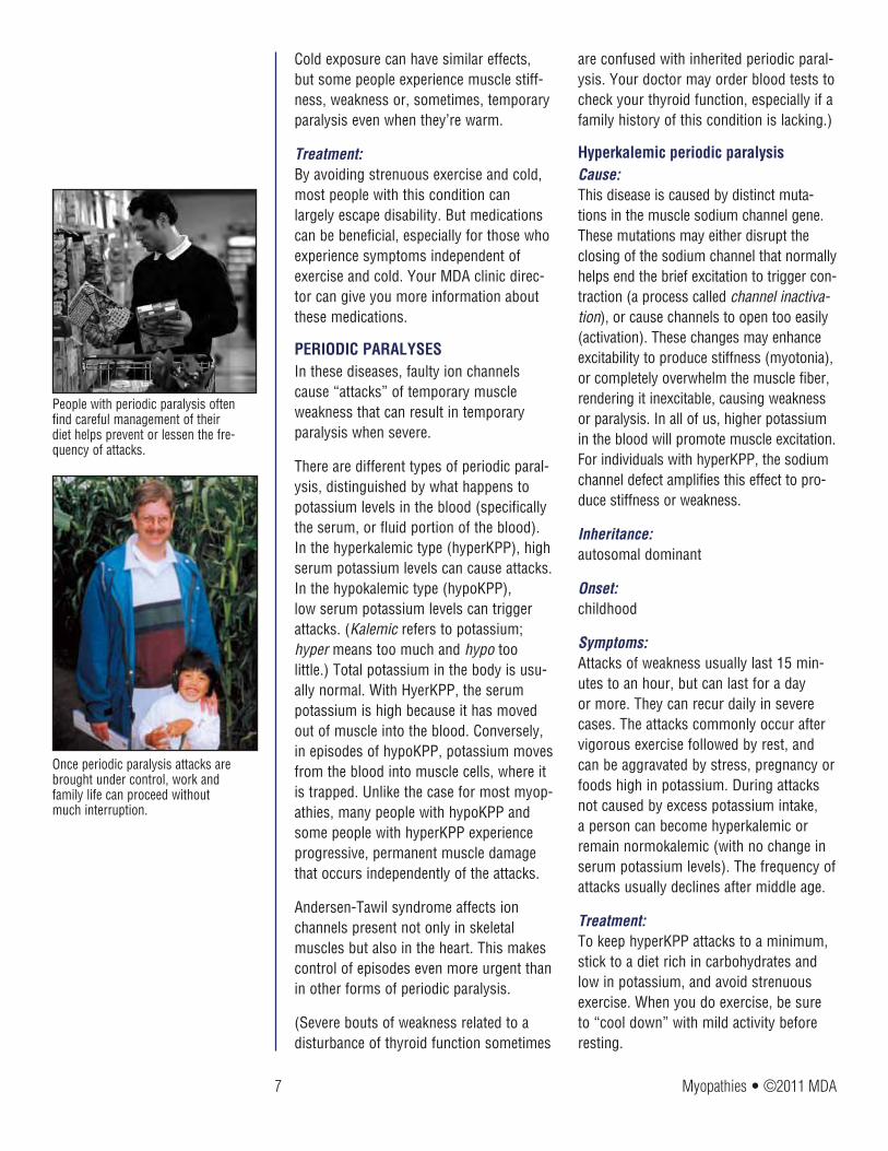
Once periodic paralysis attacks are brought under control, work and family life can proceed without much interruption.
People with periodic paralysis often find careful management of their diet helps prevent or lessen the fre-quency of attacks.

8 Myopathies • ©2011 MDA
During an attack, certain prescription drugs can be used to alleviate symptoms. Your MDA clinic director can give you more specific information on how to man-age hyperKPP through appropriate exer-cise, diet and medication.
Hypokalemic periodic paralysisCauses: As in all forms of periodic paralysis, epi-sodes of weakness in hypoKPP are caused by a temporary loss of muscle excitability. Interestingly, this disease may be caused by genetic defects in either the calcium channel or the sodium channel. The muta-tions cause a loss of muscle excitability when the serum potassium is low.
Inheritance: autosomal dominant
Onset: early childhood to adulthood
Symptoms: Attacks of weakness can occur daily and usually happen in the morning (during waking) or at night. Some people with the disease might experience only a few mild attacks in their lifetime. But the most severe attacks cause nearly full-blown paralysis. Slowly progressive permanent weakness in the legs often develops after age 50.
Treatment: As in hyperKPP, attacks of hypoKPP can be prevented by avoiding strenuous activ-ity and alleviated by medications. The dietary precautions, however, are nearly opposite. High-carbohydrate foods can trigger hypokalemia and contribute to an attack, while potassium intake can restore serum potassium levels and stem an oncoming attack. Ask your MDA clinic director for specific recommendations about diet, exercise and medications.
Andersen-Tawil syndromeCauses: This disease is caused by defects in a potassium channel normally present in
skeletal and cardiac muscles. Mutations in this potassium channel gene interfere with the ability of a muscle to stay poised, ready to contract. As a result, periodic episodes of paralysis may occur, and the heartbeat can become irregular.
Inheritance: autosomal dominant
Onset: childhood to adolescence
Symptoms: Periodic episodes of weakness lasting hours to days occur, as can severe heart-beat irregularities, sometimes accompa-nied by loss of consciousness.
Treatment: Medications called carbonic anhydrase inhibitors and potassium-sparing diuret-ics, as well as potassium supplements, are sometimes prescribed. Cardiac medica-tions also may be part of the treatment plan, and an implantable pacemaker or defibrillator is sometimes recommended.
Central core disease/malignant hyperthermia susceptibilityCauses: This rare disease appears to have multiple origins. But it’s commonly caused by defects in a channel that acts like a gate to internal calcium stores. The defect causes leakage of calcium from the stores, which appears to damage muscle cells.
Inheritance: autosomal dominant, possibly autosomal recessive in rare cases
Onset: congenital
Symptoms: The disease is named for damaged areas within muscle cells (the cores), where the filament proteins are disorganized, and mitochondria (the tiny energy-producing factories that power muscle contraction)
Older children and adults with central core disease typically experience mild disabilities that may worsen slowly over time.

9 Myopathies • ©2011 MDA
are missing. The impact of the cores on disease severity isn’t clear.
This disease causes poor muscle tone (hypotonia) and persistent muscle weak-ness in infants. In rare cases, toddlers with the disease fail to walk at all, but usually they’re just late in reaching motor milestones. Older children and adults typically experience mild disabilities that worsen slowly with time, if at all. Due to chronic muscle weakness, many people develop skeletal deformities, including joint dislocations and scoliosis, or curva-ture of the spine that can compress vital internal organs.
People with this disease should be cau-tious about surgery because they face an especially high risk of malignant hyper-thermia, a potentially fatal reaction to cer-tain anesthetic drugs (see “Anesthesia,” page 5).
Treatment: Someone with a severe form of central core disease might need a walker or other support devices for mobility, but many people require none. Unlike the case for other myopathies, people with this disease can benefit from exercise. Scoliosis and other skeletal problems can usually be corrected by use of orthopedic devices or by surgery. Your doctor or MDA clinic director can tell you more about the risks of surgery, and about anesthetic drugs that are safe.
Nemaline myopathyAlso called: rod body disease
Causes: This disease is caused by a variety of genetic defects, each one affecting one of the filament proteins required for muscle tone and contraction.
Inheritance: autosomal recessive, autosomal dominant
Onset: birth to adulthood
Symptoms: The disease gets its name from the fact that the muscle cells contain abnormal clumps of threadlike material — probably disorganized filament proteins — called nemaline bodies (nema is Greek for “thread”). It causes weakness and poor tone (hypotonia) in the muscles of the face, neck and upper limbs, and often affects the respiratory muscles (those that control breathing).
The infantile-onset cases tend to be the most severe. Usually, infants with the disease lack the muscle strength and tone required for simple postures and movements. They also have serious difficulties with feeding and respiration. Although many infants with the disease die from respiratory failure or lung infections, some survive to adulthood. Affected children usually attain motor mile-stones slowly, and at puberty they might experience further weakening, necessitating use of a wheelchair.
For adults, even noncongenital forms of the disease can cause life-threatening respiratory problems. Adults also might experience swallowing and speech prob-lems, and those with restricted mobility might develop scoliosis. However, even people who have had the disease since birth can lead active lives.
Treatment: An infant with nemaline myopathy usually requires a feeding tube to deliver nutri-tion and mechanical ventilation to support respiration. Children and adults also can benefit from respiratory support, since respiratory failure during sleep can be a persistent danger. Mobility and strength can be improved significantly by physical and orthopedic therapies. If you or your child has nemaline myopathy, your MDA clinic director can provide further informa-tion about treatments.
Infantile-onset nemaline myopathy tends to be the most severe type, but these children sometimes survive to adulthood.

10 Myopathies • ©2011 MDA
CENTRONuCLEAR MYOPATHIESThe centronuclear myopathies are named for the mislocation of cell nuclei in the muscle fibers. Normally, these nuclei are arranged around the periphery of the fiber. In these disorders, many of them are cen-trally located instead.
Myotubular myopathy is a very severe form of centronuclear myopathy. Because of its X-linked inheritance pattern, it affects boys far more commonly than girls. If girls are affected, the disease is usually much less severe than in boys.
The other forms of centronuclear myopa-thies are less severe and are called auto-somal, which is a reference to their inheri-tance pattern. The genetic mutations that underlie these myopathies are not on the X chromosome; they’re on autosomes, the numbered chromosomes.
Myotubular myopathyCauses: The most common and severe form of centronuclear myopathy is myotubular myopathy, which is caused by defects or deficiencies of myotubularin, a protein thought to promote normal muscle devel-opment.
Inheritance: X-linked
Onset: congenital (present at birth)
Symptoms: X-linked myotubular myopathy usually affects only boys, and causes severe muscle weakness and hypotonia notice-able at birth and sometimes before. The weakness and hypotonia interfere with posture and movement, and cause life-threatening difficulties with feeding and respiration. Sometimes, failure or infection of the lungs causes death in early infancy, but others survive into childhood. Usually, these boys require a feeding tube and assisted ventilation.
Contractures (joints frozen in place because of muscle weakness) may develop, particularly in the hips and knees. Spinal curvature (scoliosis) may develop in childhood.
Treatment: Until recently, nearly all infants with X-linked myotubular myopathy died within their first few months of life. But it’s now clear that intensive, continuous support of feeding and ventilation can significantly improve their life expectancy and allow a high quality of life. If spinal curvature develops, surgery to straighten the spine may be necessary.
Autosomal centronuclear myopathiesCauses: As of 2009, two genes have been found that, when flawed, cause an autosomal form of centronuclear myopathy. One gene is for the amphiphysin 2 protein, which normally is involved in maintenance of the membrane surrounding muscle fibers. The other gene is for the dynamin 2 protein, which is part of the transportation system for substances inside cells.
Inheritance: autosomal recessive (amphiphysin 2 type); autosomal dominant (dynamin 2 type)
Onset: childhood, adolescence or adulthood; recessive type tends to have an earlier onset than the dominant type
Symptoms: Autosomal centronuclear myopathies are rarely fatal in childhood and do not seri-ously weaken the respiratory muscles the way the X-linked form does. Weakness is diffuse but generally has a preference for either the proximal (near the center of the body) or distal (away from the center of the body) muscles.
Autosomal dominant centronuclear myop-athy often has a very gradual onset and
X-linked myotubular myopathy affects mostly boys and causes severe weak-ness noticeable at birth.
Autosomal centronuclear myopathies, like the one that affects this young man, are rarely fatal in childhood.
11 Myopathies • ©2011 MDA
may not cause a person to seek medical attention until young adulthood. It’s slowly progressive, and most people are able to walk independently well into adulthood. Drooping of the upper eyelids is common. Contractures may occur.
Autosomal recessive centronuclear myop-athy tends to begin earlier and be more severe. Children typically have some neu-romuscular impairment as they grow and develop. They also may develop weakness of the eye muscles. Some may have an elongated face and a high-arched palate. Contractures may occur.
Treatment: There is no specific treatment. Supportive care, such as physical therapy to minimize contracture development, can be helpful.
How are these six inherited myopathies diagnosed?Usually, diagnosis begins with evaluation of the patient’s personal and family histo-ry, and proceeds with physical and neuro-logical examinations that test reflexes and strength. The exams can detect problems with muscle tone and contraction, and the histories can bring to light patterns of inheritance and conditions that might have aggravated the muscle problems in the past.
Given this information, a doctor can sometimes distinguish an inherited myopathy from other diseases that affect muscle function, such as muscular dystro-phies and neurological disorders. But to accurately identify the myopathy and plan an appropriate course of treatment, the doctor can use several specialized tests:
Genetic (DNA-based) tests, usually per-formed on a blood sample, are available for many of the genetic mutations that underlie various myopathies.
A muscle biopsy, the removal of a small piece of muscle tissue, is sometimes performed. The sample is examined for
physical signs of muscle disease. Under the microscope, muscles affected by various myopathies have fairly distinct appearances.
Also, muscle biopsy can be used to see how isolated muscles respond to different potentially harmful conditions. For exam-ple, to determine a patient’s susceptibility to malignant hyperthermia, a biopsied muscle can be tested for its reaction to potentially dangerous anesthetic drugs.
A muscle’s activity can be measured in the body by electromyography (EMG), which involves observing the electrical signals that a muscle produces during contrac-tion. A needlelike electrode inserted into the muscle “reads” the electrical signals and sends them to a monitor called an oscilloscope. The technique usually causes some discomfort, but is useful for diagnosing channelopathies, which can show telltale abnormal signals on the oscilloscope.
What about myopathies that aren’t inherited?MDA covers noninherited myopathies that are caused by an excess or a deficiency of hormones made by the thyroid gland, which is part of the endocrine system. These myopathies are known as endocrine myopathies. Fortunately, they can almost always be successfully treated by restor-ing normal levels of thyroid hormones with medication or surgery.
Assessing thyroid function is often part of the diagnostic process for a myopathy, especially if there is no family history of the condition.
Doctors frequently measure the level of thyroid-stimulating hormone (TSH) in a blood sample to determine whether the myopathy is endocrine-related or to rule out the endocrine myopathies.
Alan Beggs at Harvard University has had MDA support to study the molecu-lar genetics of inheritable myopathies.

12 Myopathies • ©2011 MDA
Endocrine myopathies can occur when a gland produces too much or too little of a hormone. Hormones travel through the bloodstream and affect metabolism (a set of vital chemical reactions) in a variety of tissues, including muscle. Overproduction of thyroid hormones, known as T3 and T4, by the thyroid gland causes hyperthyroid myopathy, while underproduction causes hypothyroid myopathy.
A common cause of both endocrine myopathies is autoimmunity, a condition in which the immune system turns against part of the body — in this case, the thy-roid.
Both myopathies can be almost complete-ly alleviated by restoring normal thyroid activity, called the euthyroid state.
Hyperthyroid myopathyAlso called: thyrotoxic myopathy
Symptoms: This disease commonly involves weak-ness and wasting of muscles around the shoulders and sometimes the hips. There also can be weakness in muscles of the face and throat, and in the respiratory muscles. Severe cases can cause rhab-domyolysis (acute muscle breakdown). Some people with hyperthyroid myopa-thy develop Grave’s disease, damage to muscles that control movement of the eye and eyelids, which can lead to vision loss. Others develop thyrotoxic periodic paraly-sis, which involves temporary, but severe attacks of muscle weakness in association with low serum potassium. If your doc-tor is concerned about the possibility of hypoKPP, it is important to check the thy-roid function with a blood test to exclude the possibility of an endocrine myopathy (see “Periodic Paralyses,” page 7).
Treatment: Restoring normal levels of the thyroid hor-mones can be achieved with anti-thyroid drugs, but sometimes requires partial or complete surgical removal of the thyroid (thyroidectomy).
Hypothyroid myopathySymptoms: The most common symptoms include weakness around the hips and sometimes the shoulders, and a slowing of reflexes. Some people also experience muscle stiff-ness and painful muscle cramps. Severe cases can cause rhabdomyolysis.
Sometimes, the disease causes muscle enlargement along with muscle weakness.
Treatment: Thyroid hormone levels can be brought up to normal with oral medication.
Too much or too little hormone produc-tion from the thyroid gland can result in a myopathy (muscle disease).
Endocrine Myopathies

Myopathies • ©2011 MDA 13
Acetylcholine leaves the nerve fiber and docks on receptors in the muscle membrane, causing that area of the muscle fiber to become slightly more positive (“depolarized”).
Depolarization of the muscle fiber is sensed by calcium channels and triggers the release of calcium ions from inter-nal storage areas. This flood of released internal calcium is the chemical signal that causes the thick and thin filaments of the muscle fiber to slide past each another (contract).
Sodium channels open in response to this small depolarization, permit-ting a huge flow of posi-tively charged sodium ions to enter the muscle fiber. The depolarization is greatly amplified, and a brief electrical impulse (“action potential”) spreads throughout the fiber.
The sodium channels spontaneously close, potassium channels open, and positively charged potassium ions exit the fiber. Chloride channels also stay open, and negatively charged chloride ions enter the fiber. All these actions cause the inside of the fiber to become more negative (“repolarized”).
acetylcholinenerve fiber muscle fiber
muscle fibermembraneacetylcholine
receptor
thick filament
calciumions
calciumchannel
thin filament
sodium channel
sodium ions
chloride ions
choloride channel
potassium channel
potassium ions
1
2
3
4
How Ion Channels Regulate Muscle Contraction

14 Myopathies • ©2011 MDA
On being told they have a genetic disor-der such as an inheritable myopathy,
patients often ask, “But it doesn’t run in the family, so how could it be genetic?” Inheritable myopathies can run in a fam-ily, even if only one “blood relative” in the family has it.
This is because genetic diseases like inheritable myopathies can be inherited in a variety of ways: X-linked, autosomal dominant and autosomal recessive. Or, a new spontaneous mutation, may occur for the first time in a child.
X-linked means that the genetic mutation (or defect) is located on the X chromo-some. For many X-linked diseases, a nor-mal copy of the gene can compensate for the defective copy. Because males have only one X chromosome while females have two, X-linked diseases almost always affect males.
Autosomal means the mutation occurs on a chromosome other than the X or Y. Therefore, autosomal diseases affect males and females equally.
Autosomal recessive means that two cop-ies of a defective gene are required for the full-blown disease. One copy is inherited from each parent, neither of whom would normally have the disease but would be a “carrier.”
Autosomal dominant means that one copy of a defective gene is enough to cause disease. So, a person who inherits the defective gene from a parent will have the disease, as will the parent.
Inheritable myopathies passed on in an autosomal dominant pattern can be easy to trace through the family tree. By con-trast, X-linked or autosomal recessive disorders might seem to occur “out of the blue.” But in reality, one or both parents might be carriers who silently harbor a
genetic mutation. Many parents have no idea they’re carriers of a disease until they have a child with the disease.
Spontaneous mutations literally come “out of the blue,” when a new mutation occurs during the child’s conception. After they occur, these mutations can be passed on to the next generation.
A good way to find out more about your risk of inheriting or passing on an inherit-able myopathy is to talk to your MDA clinic physician or a genetic counselor. Also, see MDA’s pamphlet “Facts About Genetics and Neuromuscular Diseases.”
Does It Run in the Family?
Inheritable myopathies can run in a family, even if only one “blood relative” in the family has it.

15 Myopathies • ©2011 MDA
The MDA website is constantly updated with the latest information about the
neuromuscular diseases in its program. See the latest research news at www.mda.org.
With MDA’s support, scientists have made significant progress toward understanding and treating the inherited myopathies.
Not long ago, many people with myopa-thies that cause temporary symptoms were told they had psychological problems or were accused of being lazy. MDA-funded scientists helped show that these are treatable, physiologic medical condi-tions.
In the 1990s, researchers discovered that ion channel defects were at the root of several myopathies; that defective filament proteins give rise to nemaline myopathy; and that defects in or loss of a previously unknown protein called myotubularin cause X-linked myotubular myopathy.
The following decade, an MDA research grant to Rabi Tawil at the University of Rochester (N.Y.) helped lead to the iden-tification of potassium channel abnor-malities as the basis of Andersen-Tawil syndrome.
More recently, MDA-supported research-ers have found that centronuclear myopathies encompass a larger group of diseases than just X-linked myotubular myopathy, and that these often have a bet-ter prognosis than the X-linked disease.
Today, MDA continues to support sci-entists in their quest to understand the molecular bases of the myopathies and to find effective treatments. As of 2009, the Association is helping to support a large-scale trial to see whether either of two drugs, acetazolamide or dichlorphena-mide, helps decrease attacks of weakness in hyperkalemic and hypokalemic periodic paralysis.
MDA’s Search for Treatments and Cures

16 Myopathies • ©2011 MDA
The Muscular Dystrophy Association offers a vast array of services to help
you and your family deal with inherited and endocrine myopathies. The staff at your local MDA office is there to assist you in many ways. The Association’s ser-vices include:
• nationwide network of clinics staffed by top neuromuscular disease specialists
• MDA summer camps for kids with neu-romuscular diseases
• help with locating durable medical equipment through its national equip-ment program
• financial assistance with repairs or modifications to all types of durable medical equipment
• annual occupational, physical, respira-tory or speech therapy consultations
• annual flu shots
• support groups for those affected, spouses, parents or other caregivers
• online support services through the e-community myMDA and through myMuscleTeam, a program that helps recruit and coordinate in-home help
MDA’s public health education program helps you stay abreast of research news, medical findings and disability information through magazines, publications, edu-cational speakers, seminars, videos and newsletters.
MDA’s website at www.mda.org contains thousands of pages of valuable informa-tion, including disease specifics, research findings, clinical trials and past magazine articles.
Everyone registered with MDA automati-cally receives Quest, MDA’s award-win-ning quarterly magazine. Quest publishes detailed articles about research findings, medical and day-to-day care, helpful products and devices, social and family issues, and much more. Other MDA pub-lications can be found at www.mda.org/publications; many booklets are available in Spanish. Ask your local office for “MDA Services for the Individual, Family and Community” and for help with obtaining copies of other publications.
If you have any questions about inherited and endocrine myopathies, someone at MDA will help you find the answer. To reach your local MDA office, call (800) 572-1717.
MDA Is Here to Help You
On the cover: Adam, born in 2001, was at first given a diagnosis of X-linked myotubular myopathy, with an uncertain prognosis. His diagnosis has since been changed to centronuclear myopathy.

17 Myopathies • ©2011 MDA
The Muscular Dystrophy Association fights neuromuscular diseases through
an unparalleled worldwide research effort. The following diseases are included in MDA’s program:
Muscular DystrophiesMyotonic dystrophy (Steinert disease) Duchenne muscular dystrophy Becker muscular dystrophy Limb-girdle muscular dystrophy Facioscapulohumeral muscular dystrophy Congenital muscular dystrophy Oculopharyngeal muscular dystrophy Distal muscular dystrophy Emery-Dreifuss muscular dystrophy
Motor Neuron DiseasesAmyotrophic lateral sclerosis (ALS) Infantile progressive spinal muscular atrophy (Type 1, Werdnig-Hoffmann disease) Intermediate spinal muscular atrophy (Type 2) Juvenile spinal muscular atrophy (Type 3, Kugelberg-Welander disease) Adult spinal muscular atrophy (Type 4) Spinal-bulbar muscular atrophy (Kennedy disease)
Inflammatory MyopathiesPolymyositis Dermatomyositis Inclusion-body myositis
Diseases of Neuromuscular JunctionMyasthenia gravis Lambert-Eaton (myasthenic) syndrome Congenital myasthenic syndromes
Diseases of Peripheral NerveCharcot-Marie-Tooth disease Friedreich’s ataxia Dejerine-Sottas disease
Metabolic Diseases of MusclePhosphorylase deficiency (McArdle disease) Acid maltase deficiency (Pompe disease) Phosphofructokinase deficiency (Tarui disease) Debrancher enzyme deficiency (Cori or Forbes disease) Mitochondrial myopathy Carnitine deficiency Carnitine palmityl transferase deficiency Phosphoglycerate kinase deficiency Phosphoglycerate mutase deficiency Lactate dehydrogenase deficiency Myoadenylate deaminase deficiency
Myopathies Due to Endocrine AbnormalitiesHyperthyroid myopathy Hypothyroid myopathy
Other MyopathiesMyotonia congenita Paramyotonia congenita Central core disease Nemaline myopathy Myotubular myopathy Periodic paralysis
MDA’s Purpose and Programs
mda.org • (800) 572-1717
©2009, 2011, Muscular Dystrophy Association Inc.
P-208W 7/11
MDA’s website, mda.org, is constantly updated with the latest research news and information about the diseases in its program. Follow MDA on Facebook, Twitter and YouTube.