faculty of resource science and technology - ir.unimas.my and characterization of carbazole...
TRANSCRIPT

ISOLATION AND CHARACTERIZATION OF CARBAZOLE AND
DIBENZOFURAN DEGRADING BACTERIA FROM SOIL IN MANGROVE
ENVIRONMENT
Fatin Nur Sufinas Bt Shuib
Bachelor of Science with Honours
(Resource Biotechnology)
2013
Faculty of Resource Science and Technology

i
Isolation and Characterization of Carbazole and Dibenzofurans Degrading Bacteria
from Soil in Mangrove Environment
Fatin Nur Sufinas Bt Shuib (26300)
A final project report submitted in partial fulfillment of the Final Year Project
(STF 3015) course
Supervisor: Dr Azham Zulkharnain
Co-supervisor: AP. Dr Awang Ahmad Sallehin Awang Husaini
Resource Biotechnology
Molecular Biology
Faculty of Resource Science and Technology
University Malaysia Sarawak
2013
ii
Acknowledgement
Finally, Alhamdulillah. All praises to Allah for the strengths and His blessing and with
His willing giving me the opportunity to complete this Final Year Project. Foremost, I would
like to express my sincere gratitude to my supervisor, Dr. Azham Bin Zulkharnain for the
continuous support of my final year project, for his patience, motivation, enthusiasm, and
immense knowledge. In order to complete the undergraduate program, his guidance and
encouragement helped me in all the time of the project and writing of this final year project. I
also would like to express my deepest thanks to Assoc. Prof. Dr. Awang Ahmad Sallehin Bin
Awang Husaini, whom given me such valuable information, suggestions and guidance in the
compilation and preparation this final year project report.
Special thanks with lots of appreciation go to all postgraduate students from Molecular
Genetic Laboratory especially to Ms Jane Sebestian Taka and Ms Azizah Ahmad for their
support and help towards my project. They also provided all the required resources, support
and guidance at every step to complete this project and always find time for discussions and
took care the troubleshooting of my project. I would also like to acknowledge with much
appreciation the crucial role of the Laboratory assistants who gave me the permission to use
all required apparatus and the necessary material to complete this final year project.
Sincere thanks to all my fellow lab mates in Final year project laboratory as well as in
Molecular Genetic Laboratory and others that have been contributed by motivating
discussions, for the troubleshooting that we were working together, and for all their kindness
and moral support for carrying out the bench work.
iii
Last but not least, my deepest thanks and appreciation to my beloved parents and
family for their encouragement, constructive suggestion and full of support in term of money
and time in order to complete the project report from the beginning till the end. They always
give their endless love and prayers during the final year project progress till it is fully
completed and supporting me spiritually throughout my life.
I thank you all for your patience and attention.
iv
Declaration
I declared that this project entitled “Isolation and Characterization of Carbazole and
Dibenzofurans Degrading Bacteria from Soil in Mangrove Environment” is the result of my
own research except as cited in the references. This project has not been accepted for any
degree and is not concurrently submitted in candidature of any other degree.
Signature: .......................................................
Name : .......................................................
Date : .......................................................
v
Table of Content
TITLE/COVER PAGE……………………………………………………………………….I
ACKNOWLEDGEMENT...……………………………..…………………….……….........II
DECLARATION……………………………………………………….……….…………..IV
TABLES OF CONTENTS……………………………………………….………………….V
LIST OF ABBREVIATION…………………..………………..………….……..……….VIII
LIST OF TABLES AND FIGURES……………………………………..………………....IX
ABSTRACT .............................................................................................................................. 1
1.0 INTRODUCTION .............................................................................................................. 2
1.0 Background of Study ................................................................................................... 2
1.1 Problem Statement ...................................................................................................... 3
1.2 Objectives .................................................................................................................... 4
2.0 LITERATURE REVIEW .................................................................................................. 5
2.1 Bioremediation ................................................................................................................. 5
2.2 Mangrove environment .................................................................................................... 5
2.3 Dibenzothiophene compound .......................................................................................... 6
2.4 Biodegradation of carbazole and dibenzofurans .............................................................. 7
2.5 Bacterial species identification ...................................................................................... 10
3.0 MATERIALS AND METHODOLOGY ........................................................................ 11
3.1 Samples collection ......................................................................................................... 11
3.2 Bacteria enrichment media preparation ......................................................................... 11
3.2.1 ONR7a agar and suspension preparation ................................................................ 11
3.2.2 First enrichment ...................................................................................................... 13
3.2.3 Second enrichment .................................................................................................. 13

vi
3.3 Screening of heterocyclic hydrocarbon degrading bacteria ........................................... 13
3.4 Physiological testing and biochemical analysis of the isolates ...................................... 14
3.4.1 Motility test ............................................................................................................. 14
3.4.2 PAH growth-substrate experiments ........................................................................ 15
3.4.3 Salt tolerances test ................................................................................................... 15
3.4.4 Hydrogen sulphide test ............................................................................................ 16
3.4.5 Catalase test ............................................................................................................. 16
3.4.6 Oxidase test ............................................................................................................. 17
3.5 Morphological test ......................................................................................................... 17
3.5.1 Gram staining .......................................................................................................... 17
3.6 Total genomic DNA extraction ...................................................................................... 18
3.7 PCR amplification and purifying PCR product ............................................................. 19
3.8 Restriction fragment length polymorphism (RFLP) analysis ........................................ 22
4.0 RESULT ............................................................................................................................ 23
4.1 Bacteria Enrichment ....................................................................................................... 23
4.1.1 First and Second Stage of Bacteria Enrichment ...................................................... 23
4.1.2 Screening of heterocyclic hydrocarbon degrading bacteria .................................... 25
4.2 Physiological testing and biochemical analysis of the isolates ...................................... 26
4.2.1 Motility test ............................................................................................................. 26
4.2.2 PAH growth-substrate experiments ........................................................................ 27
4.2.3 Salt tolerances test ................................................................................................... 28
4.2.4 Hydrogen sulphide test ............................................................................................ 29
4.2.5 Catalase test ............................................................................................................. 30
4.2.6 Oxidase test ............................................................................................................. 31
vii
4.3 Morphological test ......................................................................................................... 32
4.3.1 Gram staining .......................................................................................................... 32
4.4 Total genomic DNA extraction ...................................................................................... 34
4.5 PCR amplification and purifying PCR product ............................................................. 35
4.6 Restriction fragment length polymorphism or RFLP analysis ....................................... 35
5.0 DISCUSSION ................................................................................................................... 37
5.1 Bacteria Enrichment and Isolation ................................................................................. 37
5.1.1 First enrichment and Second enrichment ................................................................ 37
5.1.2 Screening of heterocyclic hydrocarbon degrading bacteria .................................... 37
5.2 Physiological testing and biochemical analysis of the isolates ...................................... 38
5.3 Morphological test ......................................................................................................... 40
5.4 Total genomic DNA extraction ...................................................................................... 42
5.5 PCR amplification and purifying PCR product ............................................................. 44
5.6 Restriction fragment length polymorphism or RFLP analysis ....................................... 46
6.0 CONCLUSION ................................................................................................................. 47
6.1 Conclusion ..................................................................................................................... 47
6.2 Recomendation ............................................................................................................... 48
7.0 REFERENCES ................................................................................................................. 49
viii
List of Abbreviation
% Percentage
⁰C Degree Celsius
μl Micro Liter
AGE Agarose Gel Electrophoresis
bp Basepair
cm Centimeter
CAR Carbazole
DBF Dibenzofurans
DTT Dithiotheitol
DMF Dimethylformamide
dH2O Distilled water
g Gram
kb Kilo base
ml Milliliter
NAH N-heterocyclic aromatic
PAH Polycyclic Aromatic Hydrocarbons
PCR Polymerase Chain Reaction
rDNA Ribosomal Deoxyribunucleic Acid
rpm Rotations per minute
SIM Sulphide Indole Motility
ddH2O Sterile distilled water
UV Ultra-violet
ix
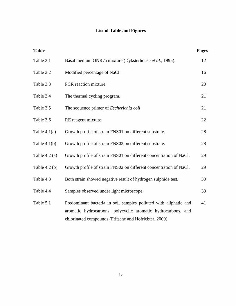
List of Table and Figures
Table Pages
Table 3.1 Basal medium ONR7a mixture (Dyksterhouse et al., 1995). 12
Table 3.2 Modified percentage of NaCl 16
Table 3.3 PCR reaction mixture. 20
Table 3.4 The thermal cycling program. 21
Table 3.5 The sequence primer of Escherichia coli 21
Table 3.6 RE reagent mixture. 22
Table 4.1(a) Growth profile of strain FNS01 on different substrate. 28
Table 4.1(b) Growth profile of strain FNS02 on different substrate. 28
Table 4.2 (a) Growth profile of strain FNS01 on different concentration of NaCl. 29
Table 4.2 (b) Growth profile of strain FNS02 on different concentration of NaCl. 29
Table 4.3 Both strain showed negative result of hydrogen sulphide test. 30
Table 4.4 Samples observed under light microscope. 33
Table 5.1 Predominant bacteria in soil samples polluted with aliphatic and
aromatic hydrocarbons, polycyclic aromatic hydrocarbons, and
chlorinated compounds (Fritsche and Hofrichter, 2000).
41

x
Figure Pages
Figure 2.1 The metabolic pathway for the desulfurization of DBT to HBP and
sulfite.
7
Figure 2.2 Simplified bacterial catabolic pathways of dibenzofuran (X= O) and
carbazole (X = N).
9
Figure 2.3 Dibenzofuran degradation via lateral dioxygenation of Ralstonia sp.
strain SBUG 290.
9
Figure 4.1 First enrichment of ONR7a suspension with two sites of soil samples
supplemented with 1% carbazole (a), dibenzofuran (b) respectively
after 3 weeks of incubation.
23
Figure 4.2 Second enrichment of ONR7a suspension with three sites of soil
samples supplemented with 1% carbazole (a) and dibenzofuran (b)
after 4 weeks of incubation appeared to change color to yellow and
orange/red respectively.
24
Figure 4.3 Control of ONR7a suspension with 1% carbazole (a), dibenzofuran
(b), and after 4 weeks of incubation appeared visibly murky on rotary
shaker.
24
Figure 4.4(a) Bacteria strain FNS01 colonies appeared smooth and milk cream
color.
25
Figure 4.4(b) Bacteria strain FNS02 appeared smooth and yellow color colonies. 25
Figure 4.5 Bacteria colonies from soil sample on ONR7a agar supplemented with
dibenzofurans and carbazole. The colonies appeared as smooth, white,
very small size colonies and grew along streaked line.
26
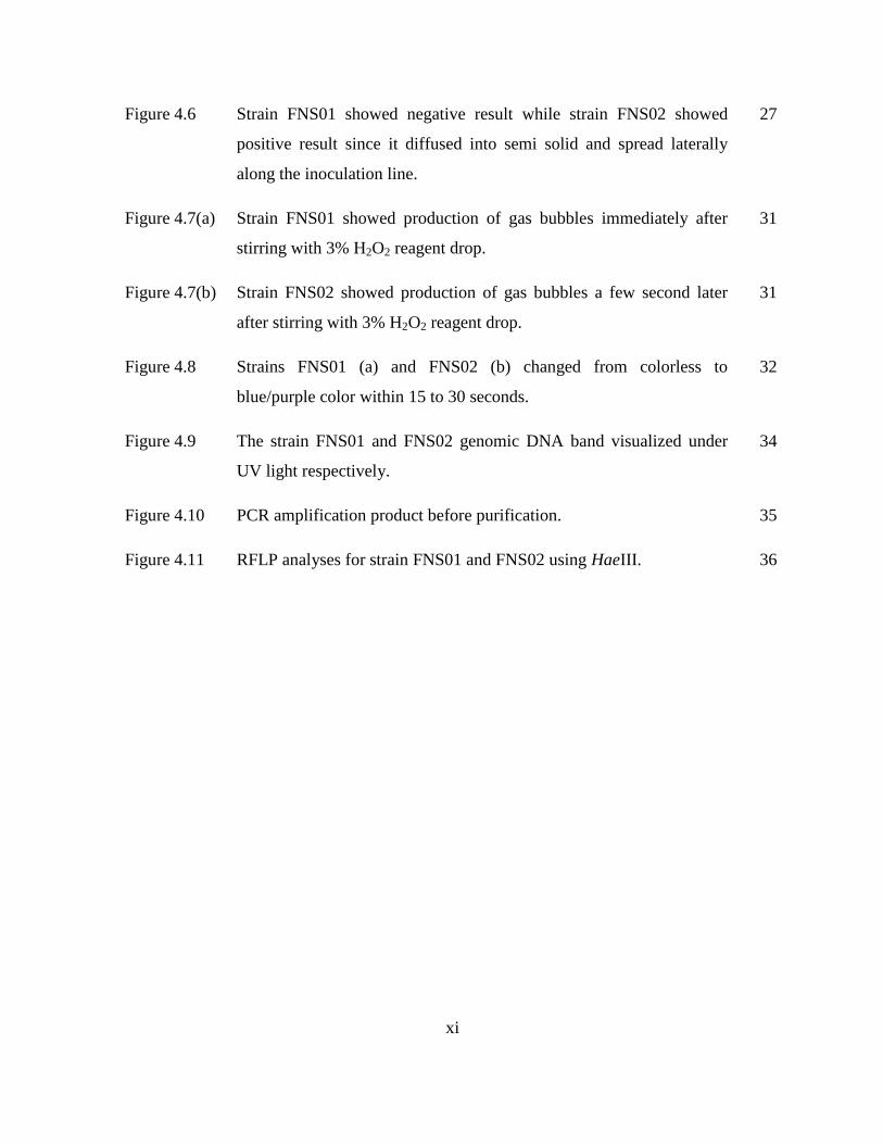
xi
Figure 4.6 Strain FNS01 showed negative result while strain FNS02 showed
positive result since it diffused into semi solid and spread laterally
along the inoculation line.
27
Figure 4.7(a) Strain FNS01 showed production of gas bubbles immediately after
stirring with 3% H2O2 reagent drop.
31
Figure 4.7(b) Strain FNS02 showed production of gas bubbles a few second later
after stirring with 3% H2O2 reagent drop.
31
Figure 4.8 Strains FNS01 (a) and FNS02 (b) changed from colorless to
blue/purple color within 15 to 30 seconds.
32
Figure 4.9 The strain FNS01 and FNS02 genomic DNA band visualized under
UV light respectively.
34
Figure 4.10 PCR amplification product before purification. 35
Figure 4.11 RFLP analyses for strain FNS01 and FNS02 using HaeIII. 36
1
Isolation and Characterization of Carbazole and Dibenzofuran Degrading Bacteria
from Soil in Mangrove Environment
Fatin Nur Sufinas Bt Shuib (26300)
Resource Biotechnology
Molecular Biology
Faculty of Resource Science and Technology
University Malaysia Sarawak
ABSTRACT Heterocyclic hydrocarbons are toxic and mutagenic components of petroleum and creosote contamination that
are frequently found in the groundwater, seawater, sediments, and soil sites. A study was carried out to isolate
and characterize heterocyclic hydrocarbon degrading bacteria from soil samples collected at mangrove
environment at Asajaya, Sarawak. This environment has physicochemical characteristics of high salinity, tidal
range, strong winds, high temperature, crude oil and muddy soil. Crude oil often contains various heterocyclic
compounds that are toxic for most life forms. Despite its toxicity, some bacteria species have the capability to
mineralize these heterocyclic hydrocarbons compound efficiently. Two bacteria strains FNS01 and FNS02 was
isolated and grown with enrichment of carbazole (CAR) and dibenzofuran (DBF) as the sole source of carbon.
Characterization of these two isolated bacteria include motility test with the use of semi-solid medium, catalase
test, salt tolerance test, hydrogen sulfide test, oxidase test, gram staining, and also different polycyclic aromatic
hydrocarbons (PAHs) substrate test such as carbazole (CAR), biphenyl (BPH), fluorene (FLO), dibenzofuran
(DBF), and dibenzothiophene (DBT) were studied. Total DNA was extracted and 16S rRNA amplified by PCR
method and subsequently differentiates the species with restriction fragment length polymorphism (RFLP).
Isolation of these strains might be useful in the bioremediation of environments contaminated by heterocyclic
hydrocarbon.
Key words: heterocyclic hydrocarbon, 16S ribosomal RNA, heterocyclic hydrocarbon degrading bacteria,
bioremediation
ABSTRAK
Hidrokarbon Heterosiklik adalah toksik dan mutagen komponen dalam petrolium dan kontaminasi kreosot yang
kerap ditemui dalam air bawah tanah, air laut, sedimen, dan tapak tanah. Satu kajian telah dijalankan untuk
mengasingkan dan mencirikan bakteria yang menjalankan proses degradasi kepada heterosiklik hidrokarbon
daripada sampel air yang diambil dari persekitaran paya bakau di Asajaya, Sarawak. Persekitaran ini
mempunyai ciri-ciri fizikokimia iaitu kemasinan yang tinggi, pelbagai pasang surut, angin yang kuat, suhu yang
tinggi, minyak mentah dan tanah berlumpur. Minyak mentah sering mengandungi pelbagai sebatian heterosiklik
yang menjadi toksik untuk kebanyakkan kehidupan. Walaupun ia dalam keadaan yang bertoksik, beberapa
spesies bakteria mempunyai keupayaan untuk untuk mengdegradasikan hidrokarbon heterosiklik kompaun
dengan lebih berkesan. Dua bakteria strain iaitu FNS01 dan FNS02 telah diasingkan dan berkembang biak
dengan penambahan karbazol (CAR) dan dibenzofuran (DBF) sebagai satu-satunya sumber karbon. Untuk
pengelasan jenis bacteria ini termasuk ujian motiliti dengan menggunakan agar separuh pejal, ujian katalase,
ujian toleransi garam, ujian hidrogen sulfida, ujian oksidase, pewarnaan Gram, dan juga ujian terhadap
berbeza substrat polisiklik aromatik hidrokarbon (PAH) seperti karbazol (CAR), bifenil (BPH), fluorene (FLO),
dibenzofuran (DBF), dan dibenzothiophene (DBT). Total DNA akan dikeluarkan dan penggandaan 16S rRNA
dengan kaedah PCR dan kemudiannya membezakan spesies dengan sekatan serpihan panjang polimorfisme
(RFLP). Pengasingan jenis-jenis bakteria ini mungkin berguna dalam biopemulihan persekitaran yang tercemar
disebabkan oleh hidrokarbon heterosiklik.
Kata Kunci: hidrokarbon heterosiklik., 16S ribosomal RNA, bakteria yang menjalankan proses degradasi
kepada heterosiklik hidrokarbon, biopemulihan
2
1.0 INTRODUCTION
1.0 Background of Study
Heterocyclic aromatic compounds can be defined as a cyclic compound that has atoms
of at least two different elements as members of its ring. It is also known to possess
toxic and mutagenic activities (Gai et al., 2007). Bacteria that can degrade NHA, such
as carbazole have also been isolated (Shotbolt-Brown et al., 1996). Other examples of
these heterocyclic hydrocarbons are carbazole, biphenyl, fluorene, dibenzofuran, and
dibenzothiophene which their degradation products have been detected in
groundwater, seawater, sediments, and soil sites contaminated with leaks of petroleum
and industrial wastes.
A bioremediation goal is to transform organic pollutants into harmless
metabolites or mineralize the pollutants into carbon dioxide and water using such
microorganisms (Seo et al., 2009). Environments that are contaminated with these
compounds may elicit serious health threats as these heterocyclic compounds are
mutagenic and carcinogenic (Gai et al., 2007). Thus, marine degrading bacteria play a
primary role in the removal of many types of chemical pollutants from the
contaminated water and soil environment. The contribution of these microorganisms
and biodegradation of PAHs can be a useful approach for eliminating these pollutants
(Hop and Omori, n.d). Some example of microorganism such as Ralstonia sp. is the
first reported species that is capable of degrade carbazole (Schneider et al., 2000).
Other is Pseudomonas resinovorans strain CA10 that exhibits the potential to enhance
degradation of carbazole in soil (Widada et al., 2002).

3
Previous study has shown the bacterial degradation pathway of carbazole is
initiated by the angular dioxygenase catalyzed by CAR 1,9a-dioxygenase (CARDO),
which is encoded by the carAa, carAc, and carAd genes. Some example of bacterial
species such as Pseudomonas resinovorans CA10 is shown to degrade CAR to 2'-
aminobiphenyl-2,3-diol via this CARDO. This CARDO is composed of terminal
oxygenase, ferredoxin, and ferredoxin reductase used for bioremediation in marine
environments since it can transforms dioxin compounds and polycyclic aromatic
hydrocarbons (PAHs) (Maeda et al., 2010). Consequently, present study indicates that
the contaminated water and soil samples from mangrove water could contain a diverse
population of N-heterocyclic aromatic (NHA) degrading bacteria since this
environment has various species composition and special characteristic.
1.1 Problem Statement
Currently, an exploration, production, refining, transport and storage of petroleum and
petroleum products, some accidental spill could occur. In addition, industrial effluents
and poor waste management recently has shown contributed to heavy metal
contamination in the sediments. As a result, contamination of certain soil area and
seawater might cause a serious health threats and for our ecosystem. In order to solve
this issue, bioremediation has been focused by researchers since it might aid in
restoration of these contaminated sites. For implementation of effective
bioremediation programs in marine environments, it requires the use of natural
microorganisms like heterocyclic hydrocarbon degrading bacteria. Therefore, this
4
study aims to study degrading bacteria that are useful in the bioremediation that will
lead to a safe environment in an effort to reduce water pollution.
1.2 Objectives
The purposes of this study are to:
a. Isolate heterocyclic hydrocarbon from bacteria strains at mangrove
environment.
b. Study the characterization of heterocyclic hydrocarbon degrading bacteria
isolated from water and soil samples.
c. Assess degradation ability of isolated bacteria using different substrates.

5
2.0 LITERATURE REVIEW
2.1 Bioremediation
Bioremediation is a process that uses microorganisms or their enzymes to return the
environment altered by contaminants to its original condition. Microbial degradation is
natural mechanism to clean up the hydrocarbon pollutants (and crude oil) from the
environment (Cristol, 1983). The recognition of biodegraded derived aromatic
hydrocarbons in marine sediments. Several microorganisms are capable to degrade
crude oil including Arthrobacter, Burkholderia, Mycobacterium, and Pseudomonas
(Hill et al., 1999).
Research has been conducted to understand bioremediation for environmental
pollutants such as N-heterocyclic aromatic (NHA) compounds that are among the
most prevalent and persistent environmental pollutants. Bioremediation can be
successful with the presence of microorganisms with appropriate metabolic
capabilities dependent on nutrients, oxygen, and pH (Im et al., 2004). Biodegradation
of hydrocarbons is a complex process that depends on the nature and on the amount of
the hydrocarbons present.
2.2 Mangrove environment
Mangrove can be defined as a habitat that consist of numerous halophytic (salt-
tolerant) of plant species which there are more than 12 families and 50 species
worldwide (Jennifer, 2012). In nature, mangrove soils normally are acidic which has
high salt concentration and hence, the roots of mangrove plants are adapted to filter

6
salt water, and their leaves can excrete salt for their survival (Kathiresan and Bingham,
2001). Additionally, this environment also has various species that utilize the
mangroves and physicochemical characteristics of high salinity, tidal range, strong
winds, high temperature and muddy soil. For the reason that they are surrounded by
loose sediments, greater diversity of microorganism such as bacteria can be found
since they are able to adapt themselves in such adverse condition. Therefore this study
is carried out to discover which bacteria strains from the mangrove environment have
the ability to degrade the certain heterocyclic hydrocarbon compound.
2.3 Dibenzothiophene compound
One group of compounds that are generally both biohazards and stable are the
polycyclic aromatic hydrocarbons (PAH). Dibenzothiophene compound (DBT) is one
of PAHs compound which can persevere for up to 3 years after an oil spill along with
its derivatives in particular. Meanwhile other more susceptible compounds might have
been biodegraded by heterocyclic hydrocarbon degrading bacteria (Gai et al., 2007).
DBT is a sulfur-containing PAH and numerous reports on biodesulfurization have
been published since DBT is broadly used as a model for biodegradation and
petroleum biodesulfurization (Gai et al., 2007). DBT degradation pathway has been
studied in detail for a few bacterial strains including Pseudomonas, Sphingomonas,
Rhodococcus, Mycobacterium, Terrabacter, Burkholderia, Paenibacillus, Gordonia
and others (Cooper, 2009). DBT degradation pathways as shown in Figure 2.1 when
DszC catalyzes the conversion of dibenzothiophene (DBT) to the sulfoxide
dibenzothiophene 5-oxide (DBTO) and further to the sulfonedibenzothiophene 5,5-

7
dioxide (DBTO2). DszA then further degrades DBTO2 to 2-(2′-hydroxyphenyl)
benzene sulfinate (HBPSi). Finally, DszB catalyzes the conversion of HBPSi to 2-
hydroxybiphenyl (HBP) and sulfite (SO32−
).
Figure 2.1: The metabolic pathway for the desulfurization of DBT to HBP and sulfite (Gai et al., 2007).
2.4 Biodegradation of carbazole and dibenzofurans
Carbazole is nitrogen containing polycyclic aromatic compound and its derivatives
being carcinogenic and mutagenic (Singh et al., 2010). It is an example of heterocyclic
hydrocarbon that derived from creosote, crude oil, and shale oil, which can be
8
mineralized by bacteria strain (Gai et. al., 2007). Ralstonia sp is the first reported of
species that able to degrade carbazole (Schneider et al., 2000). Carbazole degradation
of some previously described Pseudomonas sp. is said to be shared other
characteristics with Ralstonia sp. (Ouchiyama et al., 1993).
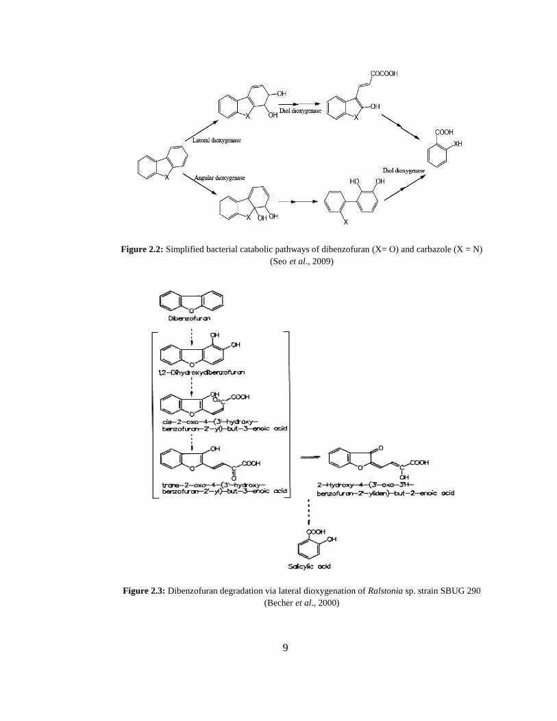
Bacterial catabolism of dibenzofurans starts at insertion of two oxygen atoms
catalyzed by enzyme dioxygenases. The initial reactions of biodegradation pathway
for dibenzofuran and carbazole are classified into angular and lateral dioxygenation
which is then catalyzed by different enzymes (Figure 2.2). These enzymes can be
found in the isolated Gram positive or Gram negative bacteria. For instance, some
bacterial dioxygenases from Pseudomonas sp. CA10 able to catalyze mainly angular
insertion of oxygen meanwhile the commonly known naphthalene dioxygenase from
Pseudomonas sp. that only catalyzes by lateral dioxygenation (Seo et al., 2009).
However some dioxygenase able to catalyze both reactions for instance cloned
dioxygenase of Norcardioides aromaticivorans IC177 from previous studies.
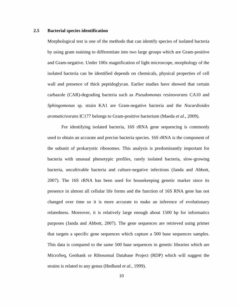
Metabolism of dibenzofuran via lateral dioxygenation has been reported in Ralstonia
sp. strain SBUG 290 (Figure 2.3). It has been shown that during cometabolic
processes, a complete degradation of dibenzofuran via lateral dioxygenation and meta
cleavage of the aromatic structure is possible (Becher et al., 2000).
9
Figure 2.2: Simplified bacterial catabolic pathways of dibenzofuran (X= O) and carbazole (X = N)
(Seo et al., 2009)
Figure 2.3: Dibenzofuran degradation via lateral dioxygenation of Ralstonia sp. strain SBUG 290
(Becher et al., 2000)
10
2.5 Bacterial species identification
Morphological test is one of the methods that can identify species of isolated bacteria
by using gram staining to differentiate into two large groups which are Gram-positive
and Gram-negative. Under 100x magnification of light microscope, morphology of the
isolated bacteria can be identified depends on chemicals, physical properties of cell
wall and presence of thick peptidoglycan. Earlier studies have showed that certain
carbazole (CAR)-degrading bacteria such as Pseudomonas resinovorans CA10 and
Sphingomonas sp. strain KA1 are Gram-negative bacteria and the Nocardioides
aromaticivorans IC177 belongs to Gram-positive bacterium (Maeda et al., 2009).
For identifying isolated bacteria, 16S rRNA gene sequencing is commonly
used to obtain an accurate and precise bacteria species. 16S rRNA is the component of
the subunit of prokaryotic ribosomes. This analysis is predominantly important for
bacteria with unusual phenotypic profiles, rarely isolated bacteria, slow-growing
bacteria, uncultivable bacteria and culture-negative infections (Janda and Abbott,
2007). The 16S rRNA has been used for housekeeping genetic marker since its
presence in almost all cellular life forms and the function of 16S RNA gene has not
changed over time so it is more accurate to make an inference of evolutionary
relatedness. Moreover, it is relatively large enough about 1500 bp for informatics
purposes (Janda and Abbott, 2007). The gene sequences are retrieved using primer
that targets a specific gene sequences which capture a 500 base sequences samples.
This data is compared to the same 500 base sequences in genetic libraries which are
MicroSeq, Genbank or Ribosomal Database Project (RDP) which will suggest the
strains is related to any genus (Hedlund et al., 1999).
11
3.0 MATERIALS AND METHODOLOGY
3.1 Samples collection
This study was conducted throughout mangrove environment at Asajaya, Sarawak.
Soil samples were aseptically collect with a total of soil sample of 20 g. The samples
were collected at three different sites. Then these samples were placed into sterile
polythene bags respectively and stored at 4°C immediately after they were brought to
the laboratory before analyzing it.
3.2 Bacteria enrichment media preparation
3.2.1 ONR7a agar and suspension preparation
The basal medium used was artificial seawater medium ONR7a. A standard
formulation was used for preparing ONR7a medium. Digital balance was used to
weight all the chemical compounds before transferring into 1L conical flask. ONR7a
medium were consisted of as table below:
12
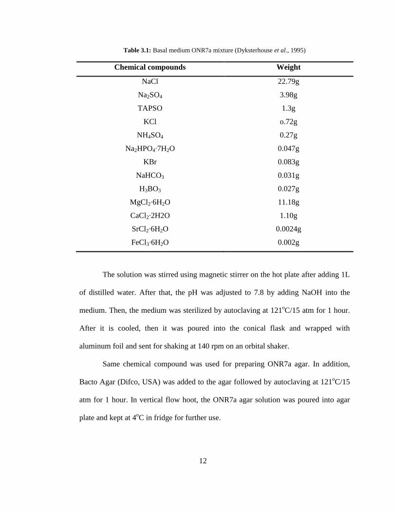
Table 3.1: Basal medium ONR7a mixture (Dyksterhouse et al., 1995)
Chemical compounds Weight
NaCl
Na2SO4
TAPSO
KCl
NH4SO4
Na2HPO4∙7H2O
KBr
NaHCO3
H3BO3
MgCl2∙6H2O
CaCl2∙2H2O
SrCl2∙6H2O
FeCl3∙6H2O
22.79g
3.98g
1.3g
o.72g
0.27g
0.047g
0.083g
0.031g
0.027g
11.18g
1.10g
0.0024g
0.002g
The solution was stirred using magnetic stirrer on the hot plate after adding 1L
of distilled water. After that, the pH was adjusted to 7.8 by adding NaOH into the
medium. Then, the medium was sterilized by autoclaving at 121oC/15 atm for 1 hour.
After it is cooled, then it was poured into the conical flask and wrapped with
aluminum foil and sent for shaking at 140 rpm on an orbital shaker.
Same chemical compound was used for preparing ONR7a agar. In addition,
Bacto Agar (Difco, USA) was added to the agar followed by autoclaving at 121oC/15
atm for 1 hour. In vertical flow hoot, the ONR7a agar solution was poured into agar
plate and kept at 4oC in fridge for further use.