familial primary ovarian insufficiency associated with a ... · 3 31. abstract . 32 objective: to...
TRANSCRIPT
1
Familial primary ovarian insufficiency associated with 1
a SYCE1 point mutation: 2
Defective meiosis elucidated in humanized mice 3
4
Short title: SYCE1 point mutation causing POI 5
6
Diego Hernández-López,a Adriana Geisinger,a,b María Fernanda Trovero,c 7
Federico F. Santiñaque,d Mónica Brauer,e Gustavo A. Folle,c,d Ricardo 8
Benavente,f* and Rosana Rodríguez-Casuriaga.a,b* 9
10
a Department of Molecular Biology, Instituto de Investigaciones Biológicas 11
Clemente Estable, Montevideo, Uruguay. 12
b Biochemistry-Molecular Biology, Facultad de Ciencias, Universidad de la 13
República, Montevideo, Uruguay. 14
c Department of Genetics, Instituto de Investigaciones Biológicas Clemente 15
Estable, Montevideo, Uruguay. 16
d Flow Cytometry and Cell Sorting Core, Instituto de Investigaciones Biológicas 17
Clemente Estable, Montevideo, Uruguay. 18
e Department of Cell Biology, Instituto de Investigaciones Biológicas Clemente 19
Estable, Montevideo, Uruguay. 20
f Department of Cell and Developmental Biology, Biocenter, University of 21
Würzburg, Würzburg, Germany. 22
23
.CC-BY-NC-ND 4.0 International licensepreprint (which was not certified by peer review) is the author/funder. It is made available under aThe copyright holder for thisthis version posted February 7, 2020. . https://doi.org/10.1101/2020.02.07.938639doi: bioRxiv preprint

2
*Corresponding authors: RRC, Department of Molecular Biology, Instituto de 24
Investigaciones Biológicas Clemente Estable, Av. Italia 3318, 11600 25
Montevideo, Uruguay; +59824872605; Email: [email protected] 26
RB, Department of Cell and Developmental Biology, Biocenter, University of 27
Würzburg, D-97074 Würzburg, Germany; Email: [email protected]
wuerzburg.de 29
30
.CC-BY-NC-ND 4.0 International licensepreprint (which was not certified by peer review) is the author/funder. It is made available under aThe copyright holder for thisthis version posted February 7, 2020. . https://doi.org/10.1101/2020.02.07.938639doi: bioRxiv preprint
3
Abstract 31
Objective: To investigate if nonsense mutation SYCE1 c.613C˃T -found in 32
women with familial primary ovarian insufficiency (POI)- is actually responsible 33
for infertility, and to elucidate the involved molecular mechanisms. 34
Design: As most fundamental mammalian oogenesis events occur during the 35
embryonic phase, thus hindering the study of POI’s etiology/pathogeny in 36
infertile women, we have used CRISPR/Cas9 technology to generate a mouse 37
model line with an equivalent genome alteration (humanized mice). 38
Setting: Academic research laboratories. 39
Interventions: We present the characterization of the biallelic mutant mice 40
phenotype, compared to wild type and monoallelic littermates. 41
Animals: Studies were conducted employing the generated humanized mice. 42
All studies were performed for both genders, except otherwise stated. 43
Main outcome measures: reproductive capability by fertility tests; gonadal 44
histological analysis; evaluation of chromosome synapsis and synaptonemal 45
complex (SC) assembly by immunolocalizations; protein studies by Western 46
blotting; transcript quantification by RT-qPCR. 47
Results: The studied mutation proved to be the actual cause of the infertile 48
phenotype, both in female and male mice homozygous for the change, 49
confirming infertility of genetic origin with a recessive mode of inheritance. The 50
mechanisms that lead to infertility are related to chromosome synapsis defects; 51
no putative truncated SYCE1 protein was observed, and Syce1 transcript was 52
hardly detected in biallelic mutants. 53
Conclusions: We present for the first time the generation of humanized mice to 54
study the actual consequences of a SC component mutation found in women 55
.CC-BY-NC-ND 4.0 International licensepreprint (which was not certified by peer review) is the author/funder. It is made available under aThe copyright holder for thisthis version posted February 7, 2020. . https://doi.org/10.1101/2020.02.07.938639doi: bioRxiv preprint
4
with familial POI. By this approach we could confirm the suspected etiology, and 56
shed light on the underlying molecular mechanism. 57
58
Key words 59
Idiopathic infertility; primary ovarian insufficiency; meiosis; synaptonemal 60
complex; humanized mice. 61
62
Capsule 63
Humanized mice were generated to study the effects on fertility of a mutation in 64
a synaptonemal complex-component-coding gene found in women with familial 65
POI, enabling etiology confirmation and mechanism elucidation. 66
67
68
.CC-BY-NC-ND 4.0 International licensepreprint (which was not certified by peer review) is the author/funder. It is made available under aThe copyright holder for thisthis version posted February 7, 2020. . https://doi.org/10.1101/2020.02.07.938639doi: bioRxiv preprint
5
Introduction 69
Primary ovarian insufficiency (POI) is a clinical syndrome characterized 70
by loss of ovarian activity before the age of 40. It is a heterogeneous condition 71
with a broad phenotypic spectrum, sharing the common feature of ovarian 72
follicle dysfunction or follicle depletion. It can have serious noxious effects upon 73
women's psychological and physical health. POI incidence increases with age, 74
affecting one in every 10,000 women at the age of 20, and 1 in 100 at the age 75
of 40 [1]. Most cases (50–90%) have unknown causes and, therefore, are 76
classified as idiopathic [2]. A significant contribution to idiopathic POI resides in 77
the genetic background of the diagnosed females [3], with 10%–15% of them 78
having an affected first-degree relative [4]. Among the already reported causes 79
of POI, there are alterations in chromosome number and structure (e.g. Turner’s 80
syndrome, 45,X), as well as genomic alterations in 46,XX non-syndromic 81
patients [5-7]. During the last two decades, an increasing number of POI-82
associated genes have been identified both on the X chromosome [e.g. 8-11] 83
and in autosomes [e.g. 8, 12-15], as well as in mitochondrial DNA [16], thus 84
confirming the heterogeneous nature of the genetic causal component. 85
Given the requirement of meiotic divisions for normal gamete formation, 86
it is expected that mutations in meiosis-related genes would account for at least 87
part of the idiopathic infertility cases. Specifically, as due to their importance for 88
recombination and proper chromosome segregation synaptonemal complexes 89
(SCs) are essential structures for gametogenesis progression, alterations in SC-90
coding genes are obvious candidates to be at the groundwork of infertility 91
[revised by 17], and particularly of POI. The SC is a meiosis-specific 92
proteinaceous, ladder-like structure that physically binds together homologous 93
.CC-BY-NC-ND 4.0 International licensepreprint (which was not certified by peer review) is the author/funder. It is made available under aThe copyright holder for thisthis version posted February 7, 2020. . https://doi.org/10.1101/2020.02.07.938639doi: bioRxiv preprint
6
chromosomes, and facilitates the resolution of recombination intermediates [18]. 94
SCs are composed of two lateral elements (LEs), a central element (CE), and 95
transverse filaments (TFs) linking both LEs with the CE. The CE together with 96
the TFs constitute the SC central region (CR). So far, eight different SC protein 97
components have been identified, including LE proteins SYCP3 [19,20] and 98
SYCP2 [21-23], TF constituent SYCP1 [24-26], and CE components SYCE1, 99
SYCE2, SYCE3, TEX12, and SIX6OS [27-30]. 100
The involvement of SC components in POI would be supported by loss-101
of-function studies for different SC genes employing KO mice, which have been 102
reported to disrupt SC structure, and lead to infertility [22,25,29-35]. Some 103
human mutations in SC-coding genes have been identified and linked to 104
infertility [revision by 17]. Concerning LE components, various mutations for 105
SYCP3 have been reported, and the first SYCP2 mutations have just been 106
identified [36]. For some of the SYCP3 mutations, a dominant negative effect 107
has been revealed in heterozygous individuals, in which the truncated SYCP3 108
interfered with polymerization of the normal protein [37,38]. 109
Regarding CE components of the SC, in the past years mutations 110
potentially associated to clinical conditions have started to be reported. In 111
particular, deletions in human 10q26.3 encompassing SYCE1 gene were found 112
in POI patients [39-41]. Besides, thus far three reports identifying mutations in 113
SYCE1 gene in infertile patients have been made [42-44]. In one of these 114
reports, a homozygous point mutation was identified in a 13-member-family in 115
which two sisters born to consanguineous parents suffered primary amenorrhea 116
[42]. This mutation, SYCE1 c.613C˃T, would lead to SYCE1 protein truncation. 117
By sequencing studies, the authors determined that of the 11 descendants (five 118
.CC-BY-NC-ND 4.0 International licensepreprint (which was not certified by peer review) is the author/funder. It is made available under aThe copyright holder for thisthis version posted February 7, 2020. . https://doi.org/10.1101/2020.02.07.938639doi: bioRxiv preprint

7
males and six females), only the two affected siblings were biallelic for SYCE1 119
c.613C˃T mutation, suggesting a genetic cause with a recessive mode of 120
inheritance [42]. Although the idea of a possible relation of SYCE1 mutation 121
with pathogenicity would be supported by the phenotype of Syce1 KO mice, 122
which are infertile [35], an unequivocal evaluation linking SYCE1 mutations to 123
the observed medical conditions is lacking so far. 124
As most fundamental mammalian oogenesis events (including SC 125
formation and recombination) occur during the embryonic phase, eventual 126
defects in this process are identified with many years of delay, leaving few 127
possibilities to intervene, and even to study the condition’s etiology and 128
pathogeny. A valid alternative to circumvent this difficulty is the employment of 129
suitable animal models, which has the highest physiological relevance after 130
human studies. However, thus far no transgenic humanized mice mimicking 131
mutations found in humans for any SC component-coding gene have been 132
reported. 133
In order to determine whether mutation SYCE1 c.613C˃T is the actual 134
cause of the observed infertile phenotype, and to study its pathogeny, we have 135
generated a humanized mouse model line containing an equivalent point 136
mutation via CRISPR/Cas9 mutagenesis system. Here, we present the 137
phenotypic characterization of the humanized mutant mice, helping to shed light 138
on the etiology and mechanisms of these infertility cases. We also discuss the 139
potential usefulness of these humanized mouse models as substrates for future 140
development of gene therapy approaches. 141
142
143
.CC-BY-NC-ND 4.0 International licensepreprint (which was not certified by peer review) is the author/funder. It is made available under aThe copyright holder for thisthis version posted February 7, 2020. . https://doi.org/10.1101/2020.02.07.938639doi: bioRxiv preprint

8
Materials and methods 144
145
Ethical approval 146
All animal procedures to generate the mutant line were performed at the SPF 147
animal facility of the Transgenic and Experimental Animal Unit of Institut 148
Pasteur de Montevideo. Experimental protocols were accordingly approved by 149
the institutional Animal Ethics Committee (protocol number 007-18), in 150
accordance with national law 18,611 and international animal care guidelines 151
(Guide for the Care and Use of Laboratory Animals) [45]. All subsequent 152
experimental animal procedures were performed at Instituto de Investigaciones 153
Biológicas Clemente Estable (IIBCE, Montevideo, Uruguay), also in accordance 154
with the national law of animal experimentation 18,611 (Uruguay), and following 155
the recommendations of the Uruguayan National Commission of Animal 156
Experimentation (CNEA, approved experimental protocol 009/11/2016). 157
158
Design of molecules for mutagenesis and generation of humanized mice 159
CRISPR/Cas mutagenesis was employed aiming to obtain the desired 160
humanized mouse cell line [42]. Design and selection of molecules for directed 161
mutagenesis were carried out taking into account on-target ranking, off-target 162
ranking, and distance of single-guide RNA (sgRNA) to target site of 163
mutagenesis (http://www.broadinstitute.org/rnai/public/analysis-tools/sgrna-164
design). The selected sgRNA was acquired as CRISPRevolution Synthetic 165
sgRNA kit (Synthego, USA). The single-stranded oligonucleotide donor 166
(ssODN) employed as template for homology directed repair (HDR) was 167
designed making use of online tools for silent mutation scanning 168
.CC-BY-NC-ND 4.0 International licensepreprint (which was not certified by peer review) is the author/funder. It is made available under aThe copyright holder for thisthis version posted February 7, 2020. . https://doi.org/10.1101/2020.02.07.938639doi: bioRxiv preprint

9
(http://watcut.uwaterloo.ca/template.php) and restriction enzyme analysis 169
(nc2.neb.com/NEBcutter2/), and ordered from IDT as 4 nmole Ultramer DNA 170
Oligo (IDT, USA). Protospacer adjacent motif (PAM) was disrupted in the 171
ssODN in order to avoid repeated nuclease action after the desired edition. 172
Mice and zygote manipulation for genome editing was performed as previouly 173
described [46,47]. For details, see Expanded discussion of the Materials and 174
Methods. 175
176
Genotyping of transgenic mice 177
Offspring genotyping was performed via tail-tips. DNA was extracted by means 178
of GeneJET Genomic DNA Purification Kit (Thermo Fisher Scientific, USA). The 179
genomic region of interest (i.e. where the mutation was directed) was 180
specifically amplified by standard PCR. The primers employed were: Syce1-181
613-FOR: 5’ TCAAGGAAGGTGAGGTCAGG 3’; Syce1-613-REV: 5’ 182
ATGAAGAGACATACCGGCAG 3’. PCR products were run by electrophoresis, 183
recovered by GeneJET Gel Extraction and DNA Cleanup Micro Kit (Thermo 184
Fisher Scientific, USA), and sequenced. 185
186
Fertility tests 187
Fertility was assessed both for females and males by mating 2-month-old 188
mutant mice homozygous for the change with adult WT mice of opposite 189
gender. Heterozygous mutants and WT mice were used as control groups. 190
Assays were performed in triplicate for each gender in breeding pairs or trios 191
(two females and one male). After at least 3 months without offspring, the 192
analyzed individuals were considered infertile. 193
.CC-BY-NC-ND 4.0 International licensepreprint (which was not certified by peer review) is the author/funder. It is made available under aThe copyright holder for thisthis version posted February 7, 2020. . https://doi.org/10.1101/2020.02.07.938639doi: bioRxiv preprint
10
194
Histology 195
Whole adult testes and ovaries were primary fixed in 2.5% glutaraldehyde, 196
postfixed in 1% osmium tetroxide, dehydrated and resin-embedded (Durcupan, 197
Fluka) according to conventional procedures [48]. Thereafter, 250 nm sections 198
were cut using a Power Tome XL ultra-microtome (Boeckeler Instruments, 199
USA), stained with toluidine blue, and examined by bright field microscopy. 200
Photographs were taken by means of an Olympus FV300 microscope equipped 201
with a DP70 camera, and DPController v.1.1.1.65 software. 202
203
Analysis by flow cytometry (FCM) 204
Testicular cell suspensions were prepared using a mechanical method 205
previously described by our group [49,50]. The resulting cell suspensions were 206
stained with Vybrant DyeCycle Green (VDG, Invitrogen Life Technologies, USA) 207
at a final concentration of 10 μM for 1 h at 35 °C in the dark with gentle agitation 208
(80 rpm) as reported earlier [51]. FCM analyses were performed by means of a 209
flow cytometer and cell sorter MoFlo Astrios EQ (Beckman Coulter, USA), using 210
a 488 nm laser, a 100 μm nozzle (25 psi), and Summit software (Beckman 211
Coulter, USA). For details concerning flow cytometer calibration and analyzed 212
parameters, see Expanded discussion of the Materials and Methods. 213
214
Antibodies 215
Primary anti-rabbit antibody against SYCE1 amino-terminal region (i.e. capable 216
of detecting WT SYCE1 and its putative truncated form) was developed at 217
GenScript (GenScript USA Inc.), using peptides ATRPQPLGMEPEGSC and 218
.CC-BY-NC-ND 4.0 International licensepreprint (which was not certified by peer review) is the author/funder. It is made available under aThe copyright holder for thisthis version posted February 7, 2020. . https://doi.org/10.1101/2020.02.07.938639doi: bioRxiv preprint

11
CPEGARGQYGSTQKI from the amino-terminal region of the protein. This 219
affinity-purified antibody was employed both for fluorescence microscopy 220
(1:200) and for Western blots (0.3 µg/mL). 221
The other antibodies employed in this study were either commercial, or 222
described in detail elsewhere [26]. For more information, see Expanded 223
discussion of the Materials and Methods. 224
225
Immunocytochemistry 226
Immunolocalization assays were performed on gonadal spread cells obtained 227
through the dry-down technique [52] with minor modifications (see Expanded 228
discussion of the Materials and Methods). For oocyte spreading, fetal ovaries 229
(E18 embryos) were used, while for spermatocytes spreading we employed 230
mechanically disaggregated adult testes. Slides were afterwards used for 231
incubations with the indicated antibodies for immunofluorescence microscopy. 232
All incubations with primary antibodies were performed overnight at 4°C in the 233
presence of protease inhibitors (P2714, Sigma-Aldrich). Secondary antibody 234
incubations were done at room temperature for 1 h protected from light. 235
236
Microscopy and Imaging 237
All immunofluorescence microscopy acquisitions were performed employing a 238
Zeiss LSM 800 confocal microscope (Carl Zeiss Microscopy, Germany) 239
equipped with Airyscan processing module, a 63X/1.4 N.A. Plan Apochromat oil 240
objective, Axiocam 506 color digital camera, and ZEN Blue 2.3 software (Carl 241
Zeiss Microscopy, Germany). Airyscan image processing was done through the 242
.CC-BY-NC-ND 4.0 International licensepreprint (which was not certified by peer review) is the author/funder. It is made available under aThe copyright holder for thisthis version posted February 7, 2020. . https://doi.org/10.1101/2020.02.07.938639doi: bioRxiv preprint
12
software’s automatic deconvolution step. All images were analyzed by means of 243
FIJI ImageJ software [53]. 244
245
Statistical Analyses 246
Quantitative data of spread nuclei with synapsed chromosomes (zygotene and 247
pachytene stage) from biallelic and monoallelic mutants with WT littermate 248
controls were statistically compared using a chi-square test. Regarding RT-249
qPCR data, statistical significance and p-value were calculated in R 250
bioconductor (http://cran.r-project.org/). 251
252
Western blots 253
Testicular protein lysates corresponding to 7.5 x 10⁵ cells in Laemmli sample 254
buffer were loaded per lane. SDS-polyacrylamide gel electrophoresis was 255
carried out on 12% polyacrylamide gels. Protein gels were transferred to 256
nitrocellulose membranes as instructed [54], and Western blots were performed 257
as previously described [55]. Membranes were incubated for 2 h at room 258
temperature in TBST with primary antibodies (anti-SYCE1-Nt and anti-β-259
tubulin), and for 1 h in blocking solution with anti-rabbit secondary antibody. 260
Bound antibodies were detected by using the Super Signal West Pico substrate 261
(Pierce). All assays were performed more than once, and using biological 262
replicates. 263
264
RT-qPCR assays 265
Total RNA from testicular cell suspensions was extracted with PureLink RNA 266
Mini Kit (Ambion, Life Technologies, Carlsbad, CA), following manufacturer’s 267
.CC-BY-NC-ND 4.0 International licensepreprint (which was not certified by peer review) is the author/funder. It is made available under aThe copyright holder for thisthis version posted February 7, 2020. . https://doi.org/10.1101/2020.02.07.938639doi: bioRxiv preprint
13
recommendations. RNA quantification was done by Nanodrop 1000 268
Spectrophotometer (Thermo Scientific). Retro-transcription and qPCR were 269
performed using Power SYBR Green Cells-to-Ct kit (Ambion), starting from 50 270
ng of RNA, following kit instructions, in a CFX96 Touch Real-Time PCR 271
Detection System 1 (BioRad, Hercules, CA). For qPCR step, 2 µL cDNA in 20 272
µL final volume reaction mix was used. 273
For RT-qPCR on embryonic ovaries, the same kit was directly employed after a 274
lysis reaction with no previous RNA extraction (due to the scarcity of the tissue). 275
The primers used are listed in Supplemental Table 1. We made 3 biological 276
replicas, and chose Ppp1cc (protein phosphatase 1, catalytic subunit, gamma 277
isozyme) as normalizing gene, as it has been previously shown to be a good 278
normalizing gene for testicular RNA [56]. Amplification efficiency of all primers 279
was >93%. The 2-ΔΔCt method and WT mouse RNA as calibrator condition were 280
used [57]. 281
282
.CC-BY-NC-ND 4.0 International licensepreprint (which was not certified by peer review) is the author/funder. It is made available under aThe copyright holder for thisthis version posted February 7, 2020. . https://doi.org/10.1101/2020.02.07.938639doi: bioRxiv preprint
14
Results 283
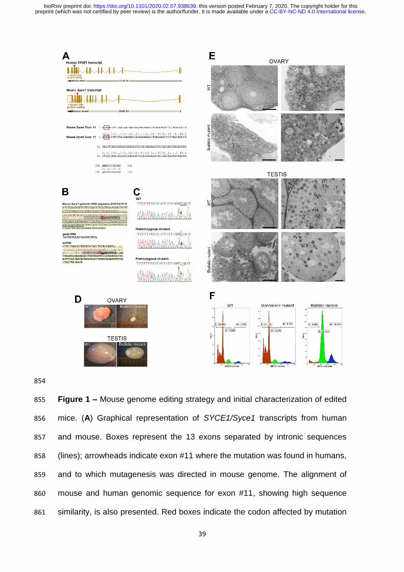
Generation of model mouse line 284
Our first aim was to generate a model mouse line mimicking the SYCE1 285
c.613C>T point mutation observed in humans. To achieve this, we chose the 286
CRISPR/Cas9 technology, and proceeded as described in Materials and 287
Methods. Comparison of SYCE1-coding regions from human and mouse 288
genome evidenced high identity, thus facilitating the choice of the editing target 289
(Fig 1A). SYCE1 c.613C˃T is a nonsense mutation that would lead to a 290
truncated human SYCE1 protein of 240 residues, while its WT counterpart has 291
351 amino acids. In mouse, an equivalent mutation would lead to a truncated 292
protein of 242 amino acids, as compared to the WT 329-residue version. 293
Design of molecules to be used in the directed mutagenesis (sgRNA and 294
ssODN) was optimized to favor the HDR (homology directed repair) pathway 295
(Fig 1B) [58]. Specimens resulting from microinjected zygotes (F0) were 296
genotyped in search of the desired change, and then mated with WT individuals 297
to obtain F1 generation. Afterwards, heterozygous specimens from F1 were 298
intermated to generate F2 offspring. As expected, the latter included WT 299
specimens as well as others heterozygous and homozygous for the desired 300
point mutation, which in mouse corresponds to Syce1 c.727C>T (Fig 1C). 301
302
Syce1 c.727C>T biallelic mutation causes infertility both in female and 303
male mice 304
Fertility was assessed both for females and males by mating mutant 305
mice with WT specimens of opposite gender. Data from three experimental 306
groups was compared in these studies: WT, heterozygous and homozygous 307
.CC-BY-NC-ND 4.0 International licensepreprint (which was not certified by peer review) is the author/funder. It is made available under aThe copyright holder for thisthis version posted February 7, 2020. . https://doi.org/10.1101/2020.02.07.938639doi: bioRxiv preprint
15
mice. No differences were observed between WT and heterozygous mice, 308
which got as easily pregnant, and laid on average 7 pups with an equal ratio of 309
male and female offspring. After 3 months, only individuals homozygous for the 310
Syce1 c.727C>T mutation failed to have offspring. This result was consistently 311
reproduced in triplicates for each gender, and led us to conclude that the sole 312
presence of this mutation in both alleles should be enough to produce the 313
infertile phenotype observed in women [42]. Moreover, the same mutation in 314
men should be able to cause infertility as well. 315
316
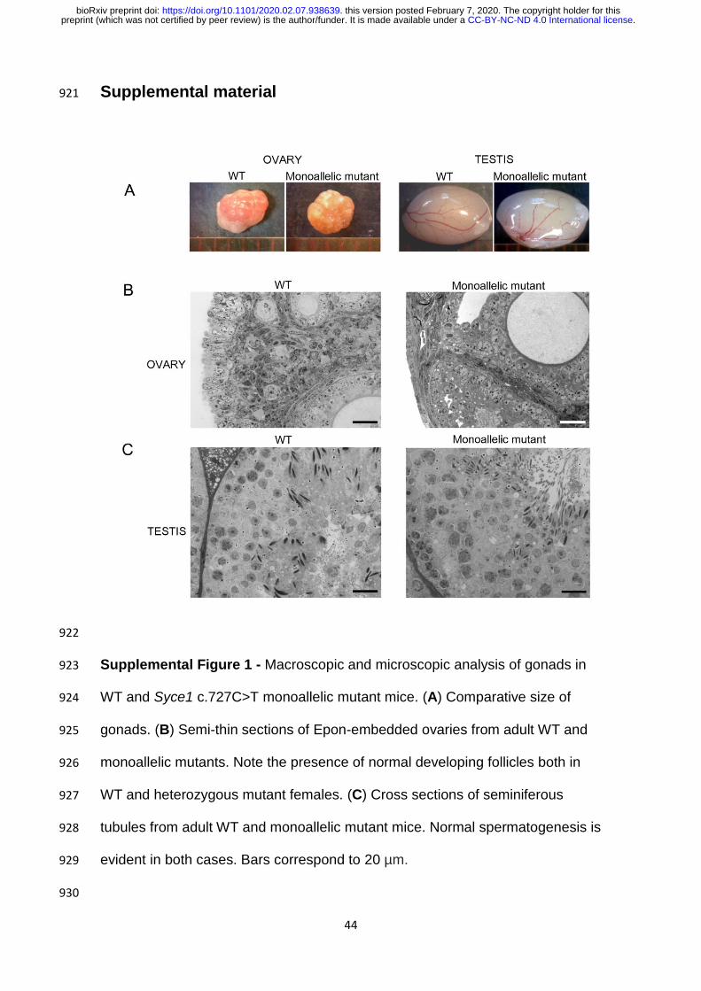
Syce1 c.727C>T biallelic mutation affects gonadal development 317
When gonadal size and aspect were assessed, no evident differences 318
were found between adult WT and heterozygous mutant mice, neither for 319
females nor for males (Supplemental Fig 1A). However, adult homozygous 320
mutants showed striking differences in gonadal size and aspect as compared to 321
their WT littermates, and this proved to be true both for female and male adult 322
individuals (Fig 1D). 323
Concerning microscopic analysis of ovaries, growing oocytes and follicle 324
development were evident in adult WT and heterozygous female animals (Fig 325
1E and Supplemental Fig 1B), while in their biallelic Syce1 c.727C>T littermates 326
no follicles or oocytes were observed (Fig 1E). Regarding testicular 327
development, while both WT and heterozygous adult males showed normal 328
seminiferous tubules with complete spermatogenesis (Fig 1E and Supplemental 329
Fig 1C), the microscopic analysis of gonadal content from adult biallelic male 330
mutant mice evidenced a severely affected spermatogenesis process with 331
complete absence of post-meiotic stages (Fig 1E). The seminiferous tubules of 332
.CC-BY-NC-ND 4.0 International licensepreprint (which was not certified by peer review) is the author/funder. It is made available under aThe copyright holder for thisthis version posted February 7, 2020. . https://doi.org/10.1101/2020.02.07.938639doi: bioRxiv preprint
16
these mutants were also depleted from mid and advanced prophase I stages 333
(i.e. pachytene and diplotene), indicating an arrest in early meiotic prophase I 334
stages. Moreover, the seminiferous tubules were much smaller than those of 335
the WT and monoallelic mutants, and exhibited an unusual aspect (Fig 1E). 336
In order to have stronger quantitative comparative analyses, testicular 337
cell suspensions from adult mice were analyzed by flow cytometry (FCM), 338
mainly based on DNA content. Figure 1F shows representative FCM results. 339
While no significant differences were found between WT and heterozygous 340
mutants, this study confirmed for the biallelic mutant males complete absence 341
of post-meiotic C population (Fig 1F). Regarding the 4C population, the FCM 342
analyses hereby presented were obtained using the DNA-specific fluorochrome 343
VDG that -as we had previously reported- allows the discrimination of two 344
populations of spermatocytes: the early spermatocyte population (leptotene and 345
zygotene stages, L/Z), and the mid/late spermatocyte one (pachytene and 346
diplotene stages, P/D) [51]. Despite these populations are usually visualized in 347
the histograms as a 4C bimodal peak (Fig 1F), this latter could not be observed 348
in the FCM profiles from biallelic mutants (Fig 1F) that resembled those 349
expected for 13-14 dpp WT juvenile mice [59]. 350
351
Evaluation of chromosome synapsis and SC assembly by confocal laser 352
scanning microscopy 353
The dramatic effect of the point mutation on gonadal development, 354
prompted us to study its consequences on SC structure and homologous 355
chromosome synapsis. Immunocytochemical localizations were performed on 356
.CC-BY-NC-ND 4.0 International licensepreprint (which was not certified by peer review) is the author/funder. It is made available under aThe copyright holder for thisthis version posted February 7, 2020. . https://doi.org/10.1101/2020.02.07.938639doi: bioRxiv preprint
17
spread cells from both embryonic ovaries and adult testes, and analyzed by 357
confocal laser scanning microscopy. 358
A first set of studies was centered on SC lateral element component 359
SYCP3, in order to evaluate chromosome synapsis. As the pachytene stage 360
(completely synapsed homologs) is reached by day 13-14 post-partum in male 361
mice and by day 17.5-18 post-coitum in female mice embryos, the ages of the 362
specimens to be analyzed were chosen accordingly. Quantitative data of spread 363
meiocyte nuclei with synapsed chromosomes from Syce1 c.727C>T mutants 364
and WT littermate controls were statistically compared using a chi-square test 365
(X2). Once again, no evident differences were found between monoallelic 366
mutants and WT littermates (X2 [1, N = 84] = 0.26, p = 0.61), with both 367
presenting normally-looking spread chromosomes that had reached the 368
pachytene stage (Fig 2A). However, biallelic mutants consistently showed 369
impaired synapsis, presenting, at most, closely juxtaposed chromosomes that 370
resembled earlier meiotic prophase stages (Fig 2A). These findings proved to 371
be true for both genders (herein shown for females), and indicate that the sole 372
biallelic presence of the point mutation severely affects homologous 373
chromosome synapsis (X2 [1, N = 84] = 134.6, p < 0.00001), and would most 374
probably account for the observed gametogenesis failure. 375
As mentioned above, Syce1 c.727C˃T is a nonsense mutation that 376
would lead to a truncated protein of 242 amino acids, as compared to the WT 377
329-residue version. In order to evaluate the eventual presence of the putative 378
truncated SYCE1 protein in the SC of mutants, we addressed protein 379
immunolocalization employing an antibody specially developed against the 380
SYCE1 amino terminal (N-t) region (see Materials and Methods). We clearly 381
.CC-BY-NC-ND 4.0 International licensepreprint (which was not certified by peer review) is the author/funder. It is made available under aThe copyright holder for thisthis version posted February 7, 2020. . https://doi.org/10.1101/2020.02.07.938639doi: bioRxiv preprint
18
detected SYCE1 in spread meiocytes from WT and monoallelic mutant 382
individuals, but not in those of biallelic mutants. This result was observed for 383
either of both genders (herein shown for females; Fig 2B), and was consistently 384
obtained for all biological and technical replicates. 385
Afterwards, we evaluated the presence of other known protein 386
components of the SC central region (i.e. TFs and CE). Some of the 387
components have been reported to be loaded earlier than SYCE1 onto the SC 388
(i.e. TF SYCP1 and CE SYCE3), while others would be loaded later (e.g. CE 389
TEX12) [22,28-30,60-64]. Representative results are shown in Figure 3. Again, 390
no differences were found between WT and monoallelic mutants for any of the 391
analyzed components either in female (Fig 3A-C) or male meiocytes (e.g. Fig 392
3D). Regarding biallelic mutants, protein components SYCP1 and SYCE3 were 393
detected on spread meiocytes containing SCs in assembling process (Figs 394
3A,B), while TEX12 was not detected at all in the assembling structure (Figs 395
3C,D). 396
For spermatocytes, γH2AX was also immunolabeled along with SC 397
protein components. This histone variant renders a very typical staining on male 398
meiotic chromosomes: dispersed chromosome staining in early stages, then 399
restricted to the XY body in pachytene stage. No difference in this regard was 400
detected between WT and monoallelic male mutants (Fig 3D). However, as 401
expected for a pre-pachytene meiotic arrest, no restricted staining for the sexual 402
chromosome pair was found in biallelic male mutants, which presented a diffuse 403
γH2AX staining pattern, characteristic of earlier meiotic stages (Fig 3D). 404
405
.CC-BY-NC-ND 4.0 International licensepreprint (which was not certified by peer review) is the author/funder. It is made available under aThe copyright holder for thisthis version posted February 7, 2020. . https://doi.org/10.1101/2020.02.07.938639doi: bioRxiv preprint
19
The putative truncated SYCE1 protein is not detected in mutant mice 406
testes 407
Although no SYCE1 protein was detected on assembling SCs of biallelic 408
mutant mice, still, the putative SYCE1 truncated protein could be present in 409
meiocytes, although not incorporated into the SC. In order to shed some light on 410
the molecular mechanism leading to infertility, we assessed the presence of the 411
putative truncated protein in mutant mice through Western blot analysis on 412
testicular material. These protein studies cannot be performed in females due to 413
material requirements unable to be fulfilled with embryonic ovaries (< 0.0001 g). 414
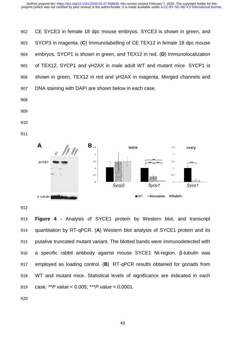
A band with an apparent molecular mass of 38 KDa was detected both 415
for WT mice and monoallelic mutants, in accordance with the predicted 416
molecular weight for WT 329-residue version of murine SYCE1 protein (Fig 4A). 417
No truncated SYCE1 protein (theoretical expected size: 28 KDa) was detected 418
for monoallelic mutants. 419
Concerning biallelic mutants, no protein reactive to SYCE1 antibody was 420
detected at all (see Fig 4A). Protein gels were deliberately overloaded to 421
minimize the effects of detection sensitivity limits, but in all assays no band of 422
28 KDa was observed. 423
424
Syce1 transcript is fairly detected in mutant humanized mice 425
The results from the Western blot assays prompted us to analyze 426
transcript levels. As shown in Figure 4B, Syce1 transcript quantitation rendered 427
big differences between WT and biallelic mutants that exhibited minimum 428
mRNA levels, both for embryonic ovaries (p< 0.0001) and for adult testes (p< 429
0.005). 430
.CC-BY-NC-ND 4.0 International licensepreprint (which was not certified by peer review) is the author/funder. It is made available under aThe copyright holder for thisthis version posted February 7, 2020. . https://doi.org/10.1101/2020.02.07.938639doi: bioRxiv preprint
20
Less pronounced but still significant differences were obtained between 431
WT and monoallelic mutant males that showed intermediate transcript levels 432
(p< 0.01; Fig 4B). On the contrary and as expected, quantitation of Sycp2 433
transcript (coding for SC LE SYCP2) rendered no significant differences 434
between the three conditions. 435
436
.CC-BY-NC-ND 4.0 International licensepreprint (which was not certified by peer review) is the author/funder. It is made available under aThe copyright holder for thisthis version posted February 7, 2020. . https://doi.org/10.1101/2020.02.07.938639doi: bioRxiv preprint
21
Discussion 437
The present work deals with some primary ovarian insufficiency cases 438
presumably related to mutations in SC coding genes, but still classified as 439
idiopathic infertility. We have worked with nonsense mutation SYCE1 440
c.613C˃T, addressing the question on its actual responsibility for the infertility 441
observed in female patients homozygous for this mutation. We have also 442
intended to shed some light on the underlying mechanisms involved. Aiming 443
these, we have applied directed mutagenesis in mouse for genome editing, and 444
successfully generated a humanized mouse line (i.e. the edited murine genome 445
contains a mutation equivalent to the one found in humans). In this regard, the 446
high percentage of identity between human and mouse SYCE1 coding genes 447
allowed us to easily find a SYCE1 c.613C˃T murine equivalent mutation (Syce1 448
c.727C>T). Generation of this animal model has enabled us to study the 449
etiology and pathogeny of these infertility cases. 450
Concerning etiology, it could be established that the biallelic presence of 451
Syce1 c.727C>T mutation is enough to cause infertility in both females and 452
males. On the other hand, mice heterozygous for the mutation were as fertile as 453
WT individuals. Thus, infertility in these cases has now proven genetic origin 454
and recessive mode of inheritance. It is worth reminding that for humans, de 455
Vries and collaborators (i.e. the authors that reported the mutation) found no 456
clinical symptoms for heterozygous individuals of both genders examined in 457
their study [42]. However, the phenotype for males homozygous for the mutated 458
SYCE1 gene could not be known at that time because all the males examined 459
in that study were heterozygous for the mutation [42]. Thus, although the 460
identification of this nonsense mutation was initially connected to cases of 461
.CC-BY-NC-ND 4.0 International licensepreprint (which was not certified by peer review) is the author/funder. It is made available under aThe copyright holder for thisthis version posted February 7, 2020. . https://doi.org/10.1101/2020.02.07.938639doi: bioRxiv preprint
22
women infertility, we can now anticipate that the biallelic mutation would equally 462
cause infertility in men. 463
The fertility results herein reported evidence an absence of sexual 464
dimorphism for this CE-related mutation. This would be in accordance with 465
previous observations for mice with loss-of-function of CR components-coding 466
genes, which were equally infertile in both genders [25,29,30,33-35], as 467
opposed to LE-component mutants that showed sexual dimorphism [22,32]. 468
Regarding gonadal development, no differences were found between 469
monoallelic mutants and unaffected WT individuals of both genders. This is in 470
accordance with the fertile phenotype observed in heterozygous humans, and 471
also with our fertility tests results. However, analysis of gonads from adult 472
biallelic mutant mice did show striking differences both for females and males: 473
minute ovaries with absence of oocytes and follicles (indicative of POI), and 474
testes with incomplete spermatogenic process, and 3-4 times smaller than in 475
unaffected littermates. Female mutant mice bearing POI resemble the clinical 476
description of the human sisters with biallelic Syce1 c.613C˃T, ratifying the 477
validity of the experimental model. 478
In males, the seminiferous epithelium of biallelic mutants showed not 479
only absence of post-meiotic cells, but even of mid/late meiotic prophase 480
stages, thus indicating an early meiotic arrest. We also analyzed testicular cell 481
suspensions by FCM as this methodology represents a widely accepted means 482
to analyze testicular cellular content, bearing very high quantitative analytical 483
power and statistical weight. These analyses let us corroborate a severely 484
affected spermatogenic process in biallelic mutants, with testes completely 485
depleted from postmeiotic haploid cells with C DNA content (i.e. round and 486
.CC-BY-NC-ND 4.0 International licensepreprint (which was not certified by peer review) is the author/funder. It is made available under aThe copyright holder for thisthis version posted February 7, 2020. . https://doi.org/10.1101/2020.02.07.938639doi: bioRxiv preprint
23
elongating spermatids, and spermatozoa). Moreover, concerning the 4C 487
population (mainly composed of primary spermatocytes), FCM profiles obtained 488
for biallelic mutants resemble those of ≈13 dpp mice that have not reached the 489
pachytene stage yet [59]. This result is in agreement with the early meiotic 490
arrest observed by microscopic analysis. 491
Analysis of spread chromosomes immunolabeled against SC protein 492
components, coupled to the use of an Airyscan super-resolution module, 493
enabled a detailed study of chromosome synapsis. This study revealed that the 494
biallelic presence of Syce1 c.727C>T severely affects homologous synapsis. 495
Regarding interactions between SYCE1 and other SC proteins, previous 496
studies based on co-immunoprecipitation and yeast two-hybrid assays have 497
identified interactions with SYCE3 and SIX6OS1 [29,30,62]. Besides, SYCE1 498
recruitment to the SC has been proposed to be mediated by SYCE3 [65]. 499
Neither SYCE1 protein nor SYCE1-downstream-loading SC components (e.g. 500
TEX12) could be detected on meiotic chromosome axes of biallelic mutant 501
humanized mice. As the mutation under study is nonsense, two hypothesis 502
concerning the pathogeny of these infertility cases arose: a) the putative SYCE1 503
truncated protein, which would lack 87 residues from the carboxyl terminus (Ct), 504
would not be recruited and loaded to the SC as its interaction with SYCE3 505
would be impaired, thus affecting normal SC assembly after SYCE3 loading 506
step; b) impaired loading could be due to the absence of the putative truncated 507
SYCE1 protein. 508
In order to shed some light on this matter, SYCE1 protein was assessed 509
by Western blot assays. Even in overloaded protein gels, we have not been 510
able to detect the putative truncated protein in the biallelic mutant mice. 511
.CC-BY-NC-ND 4.0 International licensepreprint (which was not certified by peer review) is the author/funder. It is made available under aThe copyright holder for thisthis version posted February 7, 2020. . https://doi.org/10.1101/2020.02.07.938639doi: bioRxiv preprint

24
Similarly, in monoallelic mutant mice only the WT form of the protein could be 512
detected. Thus, the impaired synapsis phenotype observed in biallelic mutant 513
mice would result from the absence (or at least presence of undetectable levels) 514
of truncated SYCE1, supporting our second hypothesis. Of note, the absence of 515
an interfering shorter version of SYCE1 could explain the unaffected phenotype 516
of monoallelic mutants. This would be quite different from the case of some 517
SYCP3 mutations, where a dominant negative effect has been reported in 518
heterozygous patients [37,38]. The lack of a possibly interfering truncated 519
protein also has important implications concerning the development of eventual 520
therapeutic procedures in biallelic patients, as it would guarantee the 521
occurrence of no relevant interference with an eventually introduced exogenous 522
normal protein, thus facilitating the intervention. 523
As the mutation studied herein is a nonsense one, a possible explanation 524
for the lack of detectable mutant protein could rely on nonsense-mediated 525
mRNA decay (NMD). This regulatory pathway functions to degrade aberrant 526
transcripts containing premature termination codons (PTCs). Since mutations 527
that generate PTCs cause approximately one-third of all known human genetic 528
diseases [66], NMD has been proposed to have a potentially important role in 529
human disease. SYCE1 c.613C>T mutation could be one of these cases. In 530
order to have a primary evaluation of this possibility, we performed Syce1 531
transcript quantitation in humanized mice compared to WT littermates. The 532
results obtained were consistent and pointed to transcript degradation of 533
aberrant transcripts, with hardly detectable levels of transcript in biallelic mutant 534
gonads of both genders (Fig 4B). In addition, monoallelic mutant males showed 535
intermediate Syce1 levels, in accordance with NMD pathway involvement. 536
.CC-BY-NC-ND 4.0 International licensepreprint (which was not certified by peer review) is the author/funder. It is made available under aThe copyright holder for thisthis version posted February 7, 2020. . https://doi.org/10.1101/2020.02.07.938639doi: bioRxiv preprint
25
Thus, the biallelic presence of Syce1 c.727C>T mutation would lead - through a 537
different mechanism - to a similar phenotype to that reported for Syce1 KO 538
mice, in which complete absence of SYCE1 protein causes infertility [35]. 539
The findings herein reported represent a proof of principle, since there 540
are no previous reports on the employment of CRISPR/Cas technology to direct 541
a specific change to a SC component, and generate a humanized mouse model 542
line for its exhaustive study. Besides, the generated mouse model line can be 543
further employed in other studies, including those aiming to develop eventual 544
therapeutic procedures. 545
546
.CC-BY-NC-ND 4.0 International licensepreprint (which was not certified by peer review) is the author/funder. It is made available under aThe copyright holder for thisthis version posted February 7, 2020. . https://doi.org/10.1101/2020.02.07.938639doi: bioRxiv preprint
26
Acknowledgements 547
The authors thank the staff of Transgenic and Experimental Animal Unit core 548
facility from Institut Pasteur de Montevideo, Uruguay, for technical support in 549
the generation of the GM model. 550
551
552
Funding 553
Funding for this research was provided by Agencia Nacional de Investigación e 554
Innovación (ANII, Uruguay; https://www.anii.org.uy/), grant number FCE-3-555
2016-1-126285 awarded to R.R.C., and fellowship POS_NAC_2018_1_151425 556
to D.H.L. Complementary financial support through program aliquots to R.R.C. 557
and D.H.L. was provided by Programa de Desarrollo de las Ciencias Básicas 558
(PEDECIBA, Biología), Universidad de la República (UdelaR). The funders had 559
no role in study design, data collection and analysis, decision to publish, or 560
preparation of the manuscript. 561
562
563
Conflict of interest 564
The authors have declared that no competing interests exist. 565
566
567
568
569
570
571
.CC-BY-NC-ND 4.0 International licensepreprint (which was not certified by peer review) is the author/funder. It is made available under aThe copyright holder for thisthis version posted February 7, 2020. . https://doi.org/10.1101/2020.02.07.938639doi: bioRxiv preprint

27
Reference list 572
1. Goswami D, Conway GS. Premature ovarian failure. Hum Reprod 573
Update 2005;11: 391-410. 574
2. Chapman C, Cree L, Shelling AN. The genetics of premature ovarian 575
failure: current perspectives. Int J Womens Health 2015;7: 799-810. 576
3. Coulam CB, Stringfellow S, Hoefnagel D. Evidence for a genetic factor in 577
the etiology of premature ovarian failure. Fertil Steril 1983; 40: 693-5. 578
4. Van Kasteren YM, Schoemaker J. Premature ovarian failure: a 579
systematic review on therapeutic interventions to restore ovarian function and 580
achieve pregnancy. Hum Reprod Update 1999;5: 483-92. 581
5. Fonseca J, Patino LC, Suarez Y, Rodríguez J, Mateus H, Jimenez K. 582
Next generation sequencing in women affected by nonsyndromic premature 583
ovarian failure displays new potential causative genes and mutations. Fertil 584
Steril 2015;104: 154-62 e2. 585
6. Laissue P. Aetiological coding sequence variants in non-syndromic 586
premature ovarian failure: from genetic linkage analysis to next generation 587
sequencing. Mol Cell Endocrinol 2015;411: 243-57. 588
7. Huhtaniemi I, Hovatta O, La Marca A, Livera G, Monniaux D, Persani L, 589
et al. Advances in the molecular pathophysiology, genetics, and treatment of 590
primary ovarian insufficiency. Trends Endocrinol Metab 2018;29: 400-19. 591
8. Laissue P, Christin-Maitre S, Touraine P, Kuttenn F, Ritvos O, Aittomaki 592
K, et al. Mutations and sequence variants in GDF9 and BMP15 in patients with 593
premature ovarian failure. Eur J Endocrinol 2006;154: 739-44. 594
9. Di Pasquale E, Rossetti R, Marozzi A, Bodega B, Borgato S, Cavallo L, , 595
et al. Identification of new variants of human BMP15 gene in a large cohort of 596
.CC-BY-NC-ND 4.0 International licensepreprint (which was not certified by peer review) is the author/funder. It is made available under aThe copyright holder for thisthis version posted February 7, 2020. . https://doi.org/10.1101/2020.02.07.938639doi: bioRxiv preprint
28
women with premature ovarian failure. J Clin Endocrinol & Metab 2006;91: 597
1976-9. 598
10. Ennis S, Ward D, Murray A. Nonlinear association between CGG repeat 599
number and age of menopause in FMR1 premutation carriers. Eur J Hum Genet 600
2006;14: 253-5. 601
11. Riva P, Magnani I, Fuhrmann Conti AM, Gelli D, Sala C, Toniolo D, et al. 602
FISH characterization of the Xq21 breakpoint in a translocation carrier with 603
premature ovarian failure. Clin Genet 2006;50: 267-9. 604
12. Doherty E, Pakarinen P, Tiitinen A, Kiilavuori A, Huhtaniemi I, Forrest S, 605
et al. A novel mutation in the FSH receptor inhibiting signal transduction and 606
causing primary ovarian failure. J Clin Endocrinol & Metab 2002;87: 1151–5. 607
13. Qin Y, Choi Y, Zhao H, Simpson JL, Chen ZJ, Rajkovic A. NOBOX 608
homeobox mutation causes premature ovarian failure. Am J Hum Genet 609
2007;81: 576-81. 610
14. Watkins WJ, Umbers AJ, Woad KJ, Harris SE, Winship IM, Gersak K, et 611
al. Mutational screening of FOXO3A and FOXO1A in women with premature 612
ovarian failure. Fertil Steril 2006;86: 1518-21. 613
15. Zhao H, Chen ZJ, Qin Y, Shi Y, Wang S, Choi Y, et al. Transcription 614
factor FIGLA is mutated in patients with premature ovarian failure. Am J Hum 615
Genet 2008;82: 1342-8. 616
16. Pagnamenta T, Taanman W, Wilson C, Anderson N, Marotta R, Duncan 617
A, et al. Dominant inheritance of premature ovarian failure associated with 618
mutant mitochondrial DNA polymerase gamma. Hum Reprod 2006;21: 2467-619
73. 620
.CC-BY-NC-ND 4.0 International licensepreprint (which was not certified by peer review) is the author/funder. It is made available under aThe copyright holder for thisthis version posted February 7, 2020. . https://doi.org/10.1101/2020.02.07.938639doi: bioRxiv preprint
29
17. Geisinger A, Benavente R. Mutations in genes coding for synaptonemal 621
complex proteins and their impact on human fertility. Cytogenet Genome Res 622
2016;150: 77-85. 623
18. Zickler D, Kleckner N. Recombination, Pairing, and Synapsis of 624
Homologs during Meiosis. Cold Spring Harb Perspect Biol 2015;7: 625
cshperspect.a016626. 626
19. Lammers JH, Offenberg HH, Van Aalderen M, Vink AC, Dietrich AJ, 627
Heyting C. The gene encoding a major component of the lateral elements of 628
synaptonemal complexes of the rat is related to X-linked lymphocyte-regulated 629
genes. Mol Cell Biol 1994;14: 1137-46. 630
20. Alsheimer M, Baier A, Schramm S, Schutz W, Benavente R. 631
Synaptonemal complex protein SYCP3 exists in two isoforms showing different 632
conservation in mammalian evolution. Cytogenet Genome Res 2010;128: 162-633
8. 634
21. Offenberg HH, Schalk JA, Meuwissen RL, Van Aalderen M, Kester HA, 635
Dietrich AJ, et al. SCP2: a major protein component of the axial elements of 636
synaptonemal complexes of the rat. Nucleic Acids Res 1998;26: 2572-9. 637
22. Yang F, De La Fuente R, Leu NA, Baumann C, McLaughlin KJ, Wang 638
PJ. Mouse SYCP2 is required for synaptonemal complex assembly and 639
chromosomal synapsis during male meiosis. J Cell Biol 2006;173: 497-507. 640
23. Winkel K, Alsheimer M, Ollinger R, Benavente R. Protein SYCP2 641
provides a link between transverse filaments and lateral elements of 642
mammalian synaptonemal complexes. Chromosoma 2009;118: 259-67. 643
.CC-BY-NC-ND 4.0 International licensepreprint (which was not certified by peer review) is the author/funder. It is made available under aThe copyright holder for thisthis version posted February 7, 2020. . https://doi.org/10.1101/2020.02.07.938639doi: bioRxiv preprint
30
24. Meuwissen RL, Offenberg HH, Dietrich AJ, Riesewijk A, Van Iersel M, 644
Heyting C. A coiled-coil related protein specific for synapsed regions of meiotic 645
prophase chromosomes. EMBO J 1992;11: 5091-100. 646
25. De Vries FA, de Boer E, van den Bosch M, Baarends WM, Ooms M, 647
Yuan L, et al. Mouse Sycp1 functions in synaptonemal complex assembly, 648
meiotic recombination, and XY body formation. Genes Dev 2005;19: 1376–89. 649
26. Schücker K, Holm T, Franke C, Sauer M, Benavente R. Elucidation of 650
synaptonemal complex organization by super-resolution imaging with isotropic 651
resolution. Proc Natl Acad Sci U S A 2015;112: 2029-33. 652
27. Costa Y, Speed R, Ollinger R, Alsheimer M, Semple CA, Gautier P, et 653
al. Two novel proteins recruited by synaptonemal complex protein 1 (SYCP1) 654
are at the centre of meiosis. J Cell Science 2005;118: 2755-62. 655
28. Hamer G, Gell K, Kouznetsova A, Novak I, Benavente R, Hoog C. 656
Characterization of a novel meiosis-specific protein within the central element of 657
the synaptonemal complex. J Cell Science 2006;119: 4025-32. 658
29. Schramm S, Fraune J, Naumann R, Hernandez-Hernandez A, Hoog C, 659
Cooke HJ, et al. A novel mouse synaptonemal complex protein is essential for 660
loading of central element proteins, recombination, and fertility. PLoS Genet 661
2011;7: e1002088. 662
30. Gómez-H L, Felipe-Medina N, Sánchez-Martín M, Davies OR, Ramos I, 663
García-Tuñón I, et al. C14ORF39/SIX6OS1 is a constituent of the 664
synaptonemal complex and is essential for mouse fertility. Nat Commun 665
2016;7: 13298. 666
.CC-BY-NC-ND 4.0 International licensepreprint (which was not certified by peer review) is the author/funder. It is made available under aThe copyright holder for thisthis version posted February 7, 2020. . https://doi.org/10.1101/2020.02.07.938639doi: bioRxiv preprint

31
31. Fraune J, Schramm S, Alsheimer M, Benavente R. The mammalian 667
synaptonemal complex: protein components, assembly and role in meiotic 668
recombination. Exp Cell Res 2012;318: 1340-6. 669
32. Yuan L, Liu JG, Hoja MR, Wilbertz J, Nordqvist K, Hoog C. Female germ 670
cell aneuploidy and embryo death in mice lacking the meiosis-specific protein 671
SCP3. Science 2002;296: 1115-8. 672
33. Hamer G, Wang H, Bolcun-Filas E, Cooke H. J, Benavente R, Hoog C. 673
Progression of meiotic recombination requires structural maturation of the 674
central element of the synaptonemal complex. J Cell Science 2008;121: 2445-675
51. 676
34. Bolcun-Filas E, Costa Y, Speed R,Taggart M, Benavente R, De Rooij 677
DG, et al. SYCE2 is required for synaptonemal complex assembly, double 678
strand break repair, and homologous recombination. J Cell Biol 2007;176: 741-679
7. 680
35. Bolcun-Filas E, Speed R, Taggart M, Grey C, de Massy B, Benavente R, 681
et al. Mutation of the Mouse Syce1 Gene Disrupts Synapsis and Suggests a 682
Link between Synaptonemal Complex Structural Components and DNA Repair. 683
PLoS Genet 2009;5: e1000393. 684
36. Schilit SLP, Menon S, Friedrich C, Kammin T, Wilch E, Hanscom C, et al. 685
SYCP2 Translocation-Mediated Dysregulation and Frameshift Variants Cause 686
Human Male Infertility. Am J Hum Genet 2020;106: 41-57. 687
37. Miyamoto T, Hasuike S, Yogev L, Maduro MR, Ishikawa M, Westphal H, 688
et al. Azoospermia in patients heterozygous for a mutation in SYCP3. Lancet 689
2003;362: 1714-49. 690
.CC-BY-NC-ND 4.0 International licensepreprint (which was not certified by peer review) is the author/funder. It is made available under aThe copyright holder for thisthis version posted February 7, 2020. . https://doi.org/10.1101/2020.02.07.938639doi: bioRxiv preprint
32
38. Bolor H, Mori T, Nishiyama S, Ito Y, Hosoba E, Inagaki H, et al. 691
Mutations of the SYCP3 gene in women with recurrent pregnancy loss. Am J 692
Hum Genet 2009;84: 14-20. 693
39. McGuire MM, Bowden W, Engel NJ, Ahn HW, Kovanci E, Rajkovic A. 694
Genomic analysis using high-resolution single-nucleotide polymorphism arrays 695
reveals novel microdeletions associated with premature ovarian failure. Fertil 696
Steril 2011;95: 1595-600. 697
40. Zhen XM, Sun YM, Qiao J, Li R, Wang LN, Liu P. Genome-wide copy 698
number scan in Chinese patients with premature ovarian failure. Beijing Da Xue 699
Xue Bao 2013;45: 841-7. 700
41. Bestetti I, Castronovo C, Sironi A, Caslini C, Sala C, Rossetti R, , et al. 701
High-resolution array-CGH analysis on 46,XX patients affected by early onset 702
primary ovarian insufficiency discloses new genes involved in ovarian function. 703
Hum Reprod 2019;34: 1-10. 704
42. De Vries L, Behar DM, Smirin-Yosef P, Lagovsky I, Tzur S, Basel-705
Vanagaite L. Exome sequencing reveals SYCE1 mutation associated with 706
autosomal recessive primary ovarian insufficiency. J Clin Endocrinol & Metab 707
2014;99: 2129-32. 708
43. Maor-Sagie E, Cinnamon Y, Yaacov B, Shaag A, Goldsmidt H, Zenvirt S, 709
et al. Deleterious mutation in SYCE1 is associated with non-obstructive 710
azoospermia. J Assist Reprod Genet 2015;32: 887-91. 711
44. Pashaei M, Rahimi Bidgoli MM, Zare-Abdollahi D, Najmabadi H, Haji-712
Seyed-Javadi R, Fatehi F, et al. The second mutation of SYCE1 gene 713
associated with autosomal recessive nonobstructive azoospermia. J Assist 714
Reprod Genet 2020; doi: 10.1007/s10815-019-01660-1. 715
.CC-BY-NC-ND 4.0 International licensepreprint (which was not certified by peer review) is the author/funder. It is made available under aThe copyright holder for thisthis version posted February 7, 2020. . https://doi.org/10.1101/2020.02.07.938639doi: bioRxiv preprint
33
45. National Research Council (NRC). Guide for the care and use of 716
laboratory animals. 7th ed. Washington: National Academies Press; 1996. 717
46. Crispo M, Schlapp G, Cárdenas-Rodríguez M, González-Maciel D, 718
Rumbo M. Optimization of transgenesis conditions for the generation of CXCL2-719
luciferase reporter mice line. Electron J Biotechnol 2013;16: 14. 720
47. Schlapp G, Goyeneche L, Fernandez G, Menchaca A, Crispo M. 721
Administration of the nonsteroidal anti-inflammatory drug tolfenamic acid at 722
embryo transfer improves maintenance of pregnancy and embryo survival in 723
recipient mice. J Assisted Reprod Genet 2015;32: 271-5. 724
48. Glauert AM, Lewis PR. Biological Specimen Preparation for 725
Transmission Electron Microscopy. Practical Methods in Electron Microscopy, 726
vol. 17. New Jersey: Princeton Legacy Library; 1998. 727
49. Rodríguez-Casuriaga R, Geisinger A, López-Carro B, Porro V, Wettstein 728
R, Folle GA. Ultra-fast and optimized method for the preparation of rodent 729
testicular cells for flow cytometric analysis. Biol Proced Online 2009;11: 184-5. 730
50. Rodríguez-Casuriaga R, Folle GA, Santiñaque F, López-Carro B, 731
Geisinger A. Simple and Efficient Technique for the Preparation of Testicular 732
Cell Suspensions. J Vis Exp 2013;78: e50102. 733
51. Rodríguez-Casuriaga R, Santiñaque F, Folle GA López-Carro B, 734
Geisinger A. Rapid preparation of rodent testicular cell suspensions and 735
spermatogenic stages purification by flow cytometry using a novel blue-laser-736
excitable vital dye. MethodsX 2014;1: 239-43. 737
52. Peters AH , Plug AW, van Vugt MJ, de Boer P. A drying-down technique 738
for the spreading of mammalian meiocytes from the male and female germline. 739
Chromosome Res 1997;5: 66-8. 740
.CC-BY-NC-ND 4.0 International licensepreprint (which was not certified by peer review) is the author/funder. It is made available under aThe copyright holder for thisthis version posted February 7, 2020. . https://doi.org/10.1101/2020.02.07.938639doi: bioRxiv preprint

34
53. Schindelin J, Arganda-Carreras I, Frise E, Kaynig V, Longair M, Pietzsch 741
T, et al. Fiji: an open-source platform for biological-image analysis. Nat Methods 742
2012;9: 676-82. 743
54. Matsudaira P. Sequence from picomole quantities of proteins electro-744
blotted onto polyvinylidene difluoride membranes. J Biol Chem 1987;262: 745
10035-8. 746
55. Goldman A, Rodríguez-Casuriaga R, González-López E, Capoano A, 747
Santiñaque F, Geisinger A. Mtch2 is differentially expressed in rat testis and 748
mainly related to apoptosis of spermatocytes. Cell Tissue Res 2015 361:869-749
83. 750
56. da Cruz I, Rodríguez-Casuriaga R, Santiñaque FF, Farías J, Curti G, 751
Capoano CA,, et al.Transcriptome analysis of highly purified mouse 752
spermatogenic cell populations: gene expression signatures switch from 753
meiotic-to postmeiotic-related processes at pachytene stage. BMC Genomics 754
2016;17: 294-312. 755
57. Livak KJ, Schmittgen TD. Analysis of relative gene expression data using 756
real-time quantitative PCR and the 2(-Delta Delta C(T)) method. Methods 757
2001;25: 402-8. 758
58. Yang L, Guell M, Byrne S, Yang JL, De Los Angeles A, Mali P, , et al. 759
Optimization of scarless human stem cell genome editing. Nucleic Acids Res 760
2013;41: 9049-61. 761
59. Geisinger A, Rodríguez- Casuriaga R. Flow cytometry for the isolation 762
and characterization of rodent meiocytes. Methods Mol Biol 2017;1471: 217-30. 763
.CC-BY-NC-ND 4.0 International licensepreprint (which was not certified by peer review) is the author/funder. It is made available under aThe copyright holder for thisthis version posted February 7, 2020. . https://doi.org/10.1101/2020.02.07.938639doi: bioRxiv preprint
35
60. Yuan L, Liu JG, Zhao J, Brundell E, Daneholt B, Höög C. The murine 764
SCP3 gene is required for synaptonemal complex assembly, chromosome 765
synapsis, and male fertility. Mol Cell 2000;5: 73-83. 766
61. Pelttari J, Hoja MR, Yuan L, Liu JG, Brundell E, Moens P, , et al. A 767
meiotic chromosomal core consisting of cohesin complex proteins recruits DNA 768
recombination proteins and promotes synapsis in the absence of an axial 769
element in mammalian meiotic cells. Mol Cell Biol 2001;21: 5667-77. 770
62. Lu J, Gu Y, Feng J, Zhou W, Yang X, Shen Y. Structural insight into the 771
central element assembly of the synaptonemal complex. Sci Rep 2014;4: 7059. 772
63. Davies OR, Maman JD, Pellegrini L. Structural analysis of the human 773
SYCE2-TEX12 complex provides molecular insights into synaptonemal complex 774
assembly. Open Biol 2012;2: 120099. 775
64. Dunce JM, Dunne OM, Ratcliff M, Millán C, Madgwick S, Usón I, et al. 776
Structural basis of meiotic chromosome synapsis through SYCP1 self-777
assembly. Nat Struct & Mol Biol 2018; 25: 557-69. 778
65. Hernández-Hernández A, Masich S, Fukuda T, Kouznetsova A, Sandin 779
S, Daneholt B, et al. The central element of the synaptonemal complex in mice 780
is organized as a bilayered junction structure. J Cell Science 2016; 129: 2239-781
49. 782
66. Miller JN and Pearce DA. Nonsense-Mediated Decay in Genetic 783
Disease: Friend or Foe? Mutat Res Rev Mutat Res 2014;762: 52-64. 784
785
786
.CC-BY-NC-ND 4.0 International licensepreprint (which was not certified by peer review) is the author/funder. It is made available under aThe copyright holder for thisthis version posted February 7, 2020. . https://doi.org/10.1101/2020.02.07.938639doi: bioRxiv preprint

36
Expanded discussion of the Materials and Methods 787
Mice manipulation for genome editing 788
Mice were housed in individually ventilated cages (Tecniplast, Milan, Italy), in a 789
controlled environment at 20 ± 1ºC with a relative humidity of 40-60%, in a 790
14/10 h light-dark cycle. Autoclaved food (Labdiet 5K67, PMI Nutrition, IN, USA) 791
and water were administered ad libitum. 792
Cytoplasmic microinjection was performed in C57BL/6J zygotes using a mix of 793
20 ng/µL sgRNA, 30 ng/µL Cas9 mRNA, and 20 ng/µL ssDNA oligo. The same 794
day, surviving zygotes were transferred to B6D2F1 0.5 dpc pseudopregnant 795
females (25 embryos/female in average), following surgery procedures 796
established in the animal facility [46]. Previously, recipient females were 797
anesthetized with a mixture of ketamine (100 mg/kg, Pharmaservice, Ripoll Vet, 798
Montevideo, Uruguay) and xylazine (10 mg/kg, Seton 2%; Calier, Montevideo, 799
Uruguay). Tolfenamic acid was administered subcutaneously (1 mg/kg, 800
Tolfedine, Vetoquinol, Madrid, Spain) to provide analgesic and anti-801
inflammatory effects [47]. Pregnancy diagnosis was determined by visual 802
inspection by an experienced animal caretaker two weeks after embryo transfer, 803
and litter size was recorded on day 21 after birth. 804
805
Flow cytometry analysis 806
Flow cytometer calibration and quality control were carried out using 3.0 µm 807
Ultra Rainbow Fluorescent Particles (Spherotech, USA). Fluorescence emitted 808
from VDG was detected with a 513/26 bandpass filter. The following parameters 809
were analyzed: forward scatter (FSC-Height with P1 Mask), side scatter (SSC-810
Height), 513/26-Area (VDG fluorescence intensity), and 513/26-Width. Doublets 811
.CC-BY-NC-ND 4.0 International licensepreprint (which was not certified by peer review) is the author/funder. It is made available under aThe copyright holder for thisthis version posted February 7, 2020. . https://doi.org/10.1101/2020.02.07.938639doi: bioRxiv preprint

37
were excluded using dot plots of 513/26 pulse-area vs 513/26 pulse-width. FCM 812
data was analyzed with Kaluza software (Beckman Coulter, USA). 813
814
Cell spreading 815
Fetal ovaries were dissected, incubated in hypotonic buffer (30 mM Tris-HCl pH 816
8.2, 17 mM sodium citrate, 5mM EDTA, 50 mM sucrose, 5mM DTT) for 30 817
minutes, mechanically disaggregated on clean slides containing 100 mM 818
sucrose, fixed in 1%-paraformaldehyde/0.15%-TritonX100, and allowed to dry 819
slowly (overnight in closed humidity chamber, then open). Once completely dry, 820
slides were wrapped in aluminum foil, and stored at -80°C until use. For 821
spermatocytes spreading, the same procedure was applied on mechanically 822
disaggregated adult mice testes. 823
824
Antibodies 825
Guinea pig anti-SYCP3 (1:200), guinea pig anti-TEX12 (1:200), rabbit anti-826
SYCP1(1:200) and rabbit anti-SYCE3 (1:200) primary antibodies were used as 827
affinity purified immunoglobulins and described in detail elsewhere [26]. Mouse 828
anti-γH2AX was purchased at Millipore (1:500, 05-636; Millipore, Germany). 829
Primary anti-β-tubulin antibody employed in Western blots as loading control- 830
was acquired from Abcam (ab6046, 1:8,000, Abcam Antibodies), and revealed 831
using an anti-rabbit secondary antibody coupled to horseradish peroxidase 832
(1:30,000, Pierce). 833
Suitable secondary antibodies coupled to AlexaFluor dyes were acquired from 834
Invitrogen Life Technologies, USA: AlexaFluor488 goat anti-rabbit (A11034, 835
.CC-BY-NC-ND 4.0 International licensepreprint (which was not certified by peer review) is the author/funder. It is made available under aThe copyright holder for thisthis version posted February 7, 2020. . https://doi.org/10.1101/2020.02.07.938639doi: bioRxiv preprint

38
1:1,000), AlexaFluor633 goat anti-guinea pig (A21105, 1:1,000), AlexaFluor546 836
goat anti-guinea pig (A11074, 1:1,000). 837
838
839
840
841
842
843
844
845
846
847
848
849
850
851
852
853
.CC-BY-NC-ND 4.0 International licensepreprint (which was not certified by peer review) is the author/funder. It is made available under aThe copyright holder for thisthis version posted February 7, 2020. . https://doi.org/10.1101/2020.02.07.938639doi: bioRxiv preprint
39
854
Figure 1 – Mouse genome editing strategy and initial characterization of edited 855
mice. (A) Graphical representation of SYCE1/Syce1 transcripts from human 856
and mouse. Boxes represent the 13 exons separated by intronic sequences 857
(lines); arrowheads indicate exon #11 where the mutation was found in humans, 858
and to which mutagenesis was directed in mouse genome. The alignment of 859
mouse and human genomic sequence for exon #11, showing high sequence 860
similarity, is also presented. Red boxes indicate the codon affected by mutation 861
.CC-BY-NC-ND 4.0 International licensepreprint (which was not certified by peer review) is the author/funder. It is made available under aThe copyright holder for thisthis version posted February 7, 2020. . https://doi.org/10.1101/2020.02.07.938639doi: bioRxiv preprint
40
c.613C>T in humans that would generate a premature TAG stop codon. (B) 862
Mouse Syce1 genomic sequence corresponding to exon #10 + intron #10 + 863
exon #11. The 20 nt sequence complementary to the sgRNA is shown in grey, 864
protospacer adjacent motif (PAM) in green, and cytosine to be edited in red. 865
Sequences of the sgRNA and single stranded oligonucleotide (ssODN) are 866
indicated. Note in the latter the C>T substitution at the beginning of exon #11, 867
as well as the disrupted PAM sequence. (C) Representative genotyping results 868
obtained through standard sequencing of PCR products amplified from mouse 869
tail-tips. (D) Comparative size of gonads in Syce1 c.727C>T biallelic mutant and 870
control mice. (E) Microscopic analysis of gonads in semi-thin sections of Epon-871
embedded ovaries and testes from adult WT and biallelic mutants. Panoramic 872
view (left) and higher magnification images (right) are shown. Bars correspond 873
to 100 and 20 µm (left and right images, respectively). (F) Flow cytometric 874
analysis of testicular cell suspensions from adult WT and mutant mice. 875
Representative FCM profiles obtained for testis from mutant mice and WT 876
littermates are shown. Relative percentages of C, 2C and 4C cell populations 877
are indicated in each case. 878
879
880
881
882
883
884
885
886
.CC-BY-NC-ND 4.0 International licensepreprint (which was not certified by peer review) is the author/funder. It is made available under aThe copyright holder for thisthis version posted February 7, 2020. . https://doi.org/10.1101/2020.02.07.938639doi: bioRxiv preprint

41
887
Figure 2 – Evaluation of chromosome synapsis and SYCE1 loading to SC in 888
WT and humanized mice. (A) Immunolabelling of LE component SYCP3 on 889
spread meiotic chromosomes from WT (above) and biallelic mutant mice 890
(below). Fluorescence acquisition was performed by means of an Airyscan 891
module that enabled the resolution of LEs, even in completely assembled SCs 892
(see inset above). Closely aligned but unsynapsed LEs are observed for 893
biallelic mice (below). (B) Immunolocalization of SYCE1 protein in female 18 894
dpc WT and mutant mouse embryos. SYCE1 is shown in green, and SYCP3 in 895
magenta. Merged channels and DNA staining with DAPI are also shown 896
below. 897
.CC-BY-NC-ND 4.0 International licensepreprint (which was not certified by peer review) is the author/funder. It is made available under aThe copyright holder for thisthis version posted February 7, 2020. . https://doi.org/10.1101/2020.02.07.938639doi: bioRxiv preprint

42
898
Figure 3 – Immunolocalization of other CR SC components in WT and mutant 899
mice. (A) Immunolabelling of TF SYCP1 in female 18 dpc mouse embryos. 900
SYCP3 is shown in magenta, and SYCP1 in green. (B) Immunolocalization of 901
.CC-BY-NC-ND 4.0 International licensepreprint (which was not certified by peer review) is the author/funder. It is made available under aThe copyright holder for thisthis version posted February 7, 2020. . https://doi.org/10.1101/2020.02.07.938639doi: bioRxiv preprint
43
CE SYCE3 in female 18 dpc mouse embryos. SYCE3 is shown in green, and 902
SYCP3 in magenta. (C) Immunolabelling of CE TEX12 in female 18 dpc mouse 903
embryos. SYCP1 is shown in green, and TEX12 in red. (D) Immunolocalization 904
of TEX12, SYCP1 and γH2AX in male adult WT and mutant mice. SYCP1 is 905
shown in green, TEX12 in red and γH2AX in magenta. Merged channels and 906
DNA staining with DAPI are shown below in each case. 907
908
909
910
911
912
Figure 4 - Analysis of SYCE1 protein by Western blot, and transcript 913
quantitation by RT-qPCR. (A) Western blot analysis of SYCE1 protein and its 914
putative truncated mutant variant. The blotted bands were immunodetected with 915
a specific rabbit antibody against mouse SYCE1 Nt-region. β-tubulin was 916
employed as loading control. (B) RT-qPCR results obtained for gonads from 917
WT and mutant mice. Statistical levels of significance are indicated in each 918
case. **P value < 0.005; ***P value < 0.0001. 919
920
.CC-BY-NC-ND 4.0 International licensepreprint (which was not certified by peer review) is the author/funder. It is made available under aThe copyright holder for thisthis version posted February 7, 2020. . https://doi.org/10.1101/2020.02.07.938639doi: bioRxiv preprint
44
Supplemental material 921
922
Supplemental Figure 1 - Macroscopic and microscopic analysis of gonads in 923
WT and Syce1 c.727C>T monoallelic mutant mice. (A) Comparative size of 924
gonads. (B) Semi-thin sections of Epon-embedded ovaries from adult WT and 925
monoallelic mutants. Note the presence of normal developing follicles both in 926
WT and heterozygous mutant females. (C) Cross sections of seminiferous 927
tubules from adult WT and monoallelic mutant mice. Normal spermatogenesis is 928
evident in both cases. Bars correspond to 20 µm. 929
930
.CC-BY-NC-ND 4.0 International licensepreprint (which was not certified by peer review) is the author/funder. It is made available under aThe copyright holder for thisthis version posted February 7, 2020. . https://doi.org/10.1101/2020.02.07.938639doi: bioRxiv preprint

45
Supplemental Table 1: Primers used for qPCR experiments. 931
Primer name Transcript detected Sequence 5'-3'
Syce1-FOR Syce1 GGGTTCTTCCAGCCTCATTG
Syce1-REV Syce1 CCCATGCTTTTCCAGTTCTTC
Ppp1cc-FOR Ppp1cc CATATCTTGAGTGGTGCTTCA
Ppp1cc-REV Ppp1cc GACAGCATCATCCAACGGCT
Sycp2-FOR Sycp2 TCACTTCCGGCTGACCCATC
Sycp2-REV Sycp2 GAAGACAAACACCCGCAGAC
932
933
.CC-BY-NC-ND 4.0 International licensepreprint (which was not certified by peer review) is the author/funder. It is made available under aThe copyright holder for thisthis version posted February 7, 2020. . https://doi.org/10.1101/2020.02.07.938639doi: bioRxiv preprint