farnesol signalling in candida
TRANSCRIPT

University of Nebraska - LincolnDigitalCommons@University of Nebraska - Lincoln
Dissertations and Theses in Biological Sciences Biological Sciences, School of
4-1-2010
Farnesol Signaling in Candida albicansMelanie L. LangfordUniversity of Nebraska at Lincoln, [email protected]
This Article is brought to you for free and open access by the Biological Sciences, School of at DigitalCommons@University of Nebraska - Lincoln. Ithas been accepted for inclusion in Dissertations and Theses in Biological Sciences by an authorized administrator of DigitalCommons@University ofNebraska - Lincoln. For more information, please contact [email protected].
Langford, Melanie L., "Farnesol Signaling in Candida albicans" (2010). Dissertations and Theses in Biological Sciences. Paper 9.http://digitalcommons.unl.edu/bioscidiss/9

Farnesol Signaling in Candida albicans
By
Melanie L. Langford
A Dissertation
Presented to the Faculty of
The Graduate College at the University of Nebraska
In Partial Fulfillment of Requirements
For the Degree of Doctor of Philosophy
Major: Biological Sciences
Under the Supervision of Professor Audrey L. Atkin
Lincoln, Nebraska
May, 2010

Farnesol Signaling in Candida albicans
Melanie L. Langford, Ph.D.
University of Nebraska, 2010
Advisor: Audrey L. Atkin
Candida albicans is a polymorphic fungus that causes a range of disease in
humans, from mucosal infections to systemic disease. Its ability to cause disease is
linked to conversion between yeast and filamentous forms of growth, and the first
quorum-sensing molecule discovered in an eukaryote, farnesol, blocks this transition. In
C. albicans, farnesol also kills mating-competent opaque cells, inhibits biofilm formation,
protects the cells from oxidative stress, and can be a virulence factor or protective agent
in disseminated and mucosal mouse models of infection, respectively. While much
emphasis has been placed on determining its effect on C. albicans morphology, the
molecular response to farnesol is not completely understood. The overall theme for this
dissertation was to better understand the C. albicans molecular response to farnesol under
quorum sensing conditions. Due to the duplicitous nature of the farnesol response in C.
albicans, i.e., its ability to kill cells or simply alter morphology, we clearly defined the
environmental conditions in which farnesol acts as a quorum sensing molecule or as a
toxic agent towards C. albicans. This clarification enabled a subsequent two-pronged
approach to study the molecular response to farnesol during morphological regulation. A
direct approach was used to investigate the role of a likely candidate, Tup1, a negative
regulator of hyphal development, in farnesol signaling. Secondly, a screening approach
was utilized to identify new farnesol resistant mutants that may participate in the farnesol

response. From the mutants identified, Czf1 (C. albicans zinc finger) was selected for
further characterization and was shown to play a vital role in the morphological response
to farnesol as well as farnesol tolerance. Overall, this study identified two new factors
involved in farnesol signaling, and highlights the power of farnesol as a tool with which
to unravel the complex signaling networks present in C. albicans.

iv
ACKNOWLEDGEMENTS
No one ever gets through their Ph.D. program on their own, and I am certainly no
exception. I would like to begin by thanking my wonderful advisor, Audrey Atkin. What
a happy accident joining your lab turned out to be- I never would have guessed that I
would switch from studying bacteriology to fungal biology! It makes an incredible
difference to work for someone that not only encourages quality research but is so
pleasant on a personal level as well, things that I will miss. During my time spent in your
lab, I have learned nearly everything I know about fungi, and I have had the chance to
learn several new lab techniques as well, such as working with RNA. I am happy you
encouraged me to give several oral and poster presentations, both locally and nationally;
what initially seems like more work always pays off in learning experiences. I also think
it says a lot about the career development information you provide your lab members
when I have heard nearly everything discussed in our Professionalism course this
semester from you already over the course of my graduate program. Lastly I always
appreciated your support whenever a crisis arose (be it professional, experimental, or
personal).
I would also like to extend my thanks to Kenneth Nickerson, my favorite fungal
biology neighbor. I will miss having a chemistry expert right next door and your
enthusiasm for nearly everything and anything in science. I have truly enjoyed having
joint Atkin-Nickerson lab meetings, and I feel they have been critical to the completion
of my degree. To both Audrey and Ken, I am always amazed at how well your
collaboration works (given your entirely different research styles, personalities, etc), but I
am happy that it does (maybe I fall somewhere in the middle of you two?). I think I have
v
learned about balancing careful experimental design with exploratory experiments
moving in new directions, and I have also enjoyed writing papers with both of you and
learning about the process of paper submission.
Next I would like to thank all my committee members: Steve Harris, Jack Morris,
and Luwen Zhang. You always provided interesting ideas and suggestions during my
committee meetings, and I appreciate that you always did so in a very constructive
manner. Thanks to Cheryl Bailey for teaching me real-time PCR from scratch- it is easy
to find people that will let you borrow their machines, but not so easy to find someone to
teach you the method and theory behind it. There are also so many past and current lab
members to thank. First, I would like to thank Bessie Kebaara, a former post-doc in the
lab that I still miss having around. I could always go to you with technical questions,
scientific ideas, or just to complain about the cold weather. Dhammika Navarathna and
Raluca Dumitru, former Ph.D. students in the Nickerson lab, both taught me important
Candida techniques and were great role models for me as a new student. And thanks to
Suman Ghosh, another former Ph.D. student, for teaching me farnesol extraction
techniques and for being a good friend. Of the current students in our research group, I
would first like to extend my gratitude to Sahar Hasim, for being such an excellent
collaborator on the farnesol toxicity project. You are such an amiable and reliable person
to work with, and even though things took longer to finish than we originally thought
(seems they always do), I think we learned some very valuable information over the
course of that project. Thanks also to Swetha Tati (my technical support aide prior to my
defense), Krista Fager, Jeff Bunker, and Narmin Tahirova for all your helpful comments
and suggestions during journal club and lab meetings.

vi
To my family: Mom, Dad, Courtney, Gina, Jim, and all the Langford brothers, I
would like to thank you for all of your support during both my M.S. and Ph.D. degrees.
Believe it or not, attempting to explain to you what I do in the lab has actually improved
my oral presentations to more general scientific audiences! I am very lucky to have such
supportive family members on my side.
And lastly, I would like to thank my husband, Gabe. I am certain that I would not
have been able to get through graduate school without you, much less have the
confidence to even pursue my Ph.D. degree. It has been chaotic but fun to go through the
ups and downs of graduate school with you, as you were always the perfect confidant. I
am so excited to start the next phase of our life together with our new family.

vii
TABLE OF CONTENTS
ACKNOWLEDGEMENTS. ........................................................................................... iv
TABLE OF CONTENTS ............................................................................................... vii
LIST OF FIGURES ...........................................................................................................x
LIST OF TABLES ........................................................................................................... xi
CHAPTER 1: Introduction ..............................................................................................1
References..........................................................................................................................23
Figure Legends...................................................................................................................18
Figures................................................................................................................................19
Tables.................................................................................................................................20
CHAPTER 2: Activity and toxicity of farnesol on Candida albicans is dependent on
growth conditions.............................................................................................................35
Abstract ..............................................................................................................................36
Introduction........................................................................................................................37
Methods..............................................................................................................................39
Results................................................................................................................................40
Discussion..........................................................................................................................42
References..........................................................................................................................44
Figure Legends...................................................................................................................49
Figures................................................................................................................................50

viii
CHAPTER 3: Candida albicans Tup1 is involved in farnesol-mediated inhibition of
filamentous growth induction .........................................................................................53
Abstract ..............................................................................................................................54
Introduction........................................................................................................................55
Methods..............................................................................................................................59
Results................................................................................................................................62
Discussion..........................................................................................................................68
References..........................................................................................................................73
Figure Legends...................................................................................................................80
Figures................................................................................................................................82
Tables.................................................................................................................................87
CHAPTER 4: A novel role for Czf1 in farnesol tolerance and the morphological
response to farnesol in Candida albicans .......................................................................89
Abstract ..............................................................................................................................90
Introduction........................................................................................................................91
Methods..............................................................................................................................94
Results................................................................................................................................97
Discussion........................................................................................................................106
References........................................................................................................................111
Figure Legends.................................................................................................................119
Figures..............................................................................................................................122
Tables...............................................................................................................................130

ix
CHAPTER 5: Summary and Future Directions ........................................................147
References........................................................................................................................157

x
LIST OF FIGURES
Figure 1-1...........................................................................................................................19
Figure 2-1...........................................................................................................................50
Figure 2-2...........................................................................................................................51
Figure 2-3...........................................................................................................................52
Figure 3-1...........................................................................................................................82
Figure 3-2...........................................................................................................................83
Figure 3-3...........................................................................................................................84
Figure 3-4...........................................................................................................................85
Figure 3-5...........................................................................................................................86
Figure 4-1.........................................................................................................................122
Figure 4-2.........................................................................................................................123
Figure 4-3.........................................................................................................................124
Figure 4-4................................................................................................................. 125-126
Figure 4-5.........................................................................................................................127
Figure 4-6.........................................................................................................................128
Figure 4-7.........................................................................................................................129

xi
LIST OF TABLES
Table 1-1. ..................................................................................................................... 20-21
Table 1-2 ............................................................................................................................22
Table 3-1 ............................................................................................................................87
Table 3-2 ............................................................................................................................88
Table 4-1 ..........................................................................................................................130
Table 4-2 ..........................................................................................................................131
Supplementary Table 4-1......................................................................................... 132-146

1
CHAPTER 1
INTRODUCTION
Reference:
Langford, M.L., Atkin, A.L., and Nickerson, K.W. (2009) Cellular Interactions of
Farnesol, a Quorum Sensing Molecule Produced by Candida albicans. Future Microbiol.
4: 1353-62.

2
INTRODUCTION
The discovery of E,E-farnesol as a quorum sensing molecule in Candida albicans
(Hornby et al., 2001) demonstrated the existence of quorum sensing in a eukaryotic
organism, a process once thought to be confined to bacteria. The earlier work on the
discovery and characterization of farnesol was reviewed by Nickerson et al (Nickerson et
al., 2006). C. albicans is a polymorphic fungus that is capable of growing in yeast,
hyphal, pseudohyphal, opaque, and chlamydospore cell morphologies. Farnesol is
capable of blocking the yeast to hyphal/pseudohyphal (filaments) switch (Hornby et al.,
2001), a conversion that is the focus of intense study in C. albicans because of its role in
virulence (Lo et al., 1997; Saville et al., 2003). Another quorum sensing molecule
(QSM) described in C. albicans is farnesoic acid (Oh et al., 2001). Farnesoic acid, like
farnesol, acts to block the yeast to filament transition (Oh et al., 2001), but it is far less
active than farnesol (Shchepin et al., 2003) and is produced by only one known strain of
C. albicans, 10231 (Hornby and Nickerson, 2004; Oh et al., 2001). Additional possible
quorum sensing molecules are reviewed by Kruppa (Kruppa, 2009). Farnesol also blocks
biofilm formation by C. albicans (Alem et al., 2006; Cao et al., 2005; Martins et al.,
2007; Ramage et al., 2002) in a manner consistent with its inhibition of the yeast to
filament conversion. The mating-competent form of C. albicans, opaque cells, can be
adversely affected by farnesol; in aerobic conditions the cells die by necrosis (Dumitru et
al., 2007) but remain unharmed in anaerobic conditions (Dumitru et al., 2004). Lastly,
the production of chlamydospores, a C. albicans cell morphology with unknown
function, is increased in the presence of very high (10 mM) levels of farnesol (Martin et
al., 2005). Given that it may affect all five C. albicans growth morphologies, it has

3
become clear that farnesol plays an influential role in C. albicans physiology, and the
search is underway to elucidate the molecular mechanism(s) of these effects.
The Molecular Response to Farnesol in C. albicans
The first described function of farnesol was to block filamentation induced by a
variety of environmental signals (Hornby et al., 2001; Mosel et al., 2005), and several
factors involved in the filamentation process are known to play a role in the C. albicans
farnesol response. Ras1 of the cyclic AMP pathway, as discussed later, is a candidate for
direct inhibition by farnesol (Davis-Hanna et al., 2008). Repressors of filamentation,
Tup1 and Nrg1, also play critical roles in the farnesol response, as tup1Δ/tup1Δ and
nrg1Δ/nrg1Δ mutants are unable to respond to farnesol (Kebaara et al., 2008). The
presence of farnesol increases TUP1 expression and suppresses the haploinsufficient
phenotype of a TUP1/tup1Δ mutant (Kebaara et al., 2008). A two-component signal
transduction pathway histidine kinase, Chk1 (Kruppa et al., 2004), and mitogen activated
protein (MAP) kinase pathway components, Cph1 and Hst1 (Sato et al., 2004), may also
be involved in the response to farnesol, but Cph1 might play a downstream role since a
cph1Δ/cph1Δ mutant retains the ability to respond to farnesol (Davis-Hanna et al., 2008).
Regulators of filamentation are not the only genes involved in the farnesol response.
Microarray analyses have identified other categories of genes affected by farnesol
treatment such as heat shock genes, drug resistance genes, cyclin and cell proliferation
genes, histone genes, genes expressed at high cell density, phagocytosis response genes,
and adhesion genes (Cao et al., 2005; Cho et al., 2007; Enjalbert and Whiteway, 2005;
Uppuluri et al., 2007). With all of the different pathways and responses induced by
farnesol, it promotes the question: Does farnesol have one or more specific target(s) in

4
the cell, or does it somehow elicit a more general and nonspecific response with
pleomorphic consequences?
Are there farnesol receptors or farnesol binding proteins? The morphological
response to farnesol in C. albicans appears to be very sensitive to minor changes in the
structure of farnesol, providing support for the specific target(s) model, rather than a
scenario involving general membrane disruption. For example, there are four isomers of
farnesol but only E,E-farnesol is capable of blocking filamentation in C. albicans
(Shchepin et al., 2003). This conclusion is based on a comparison between pure (96%)
E,E-farnesol and two commercial mixed isomers containing 56 and 36% E,E-farnesol.
These three farnesol preparations reduced germ tube formation (early stage filaments) to
50% at 1.2, 3.5, and 4.4 µM, respectively (Shchepin et al., 2003). Thus, there was
sufficient E,E-farnesol in the two samples with mixed isomers to account for all of their
QSM activities. Further, the 12-carbon backbone of farnesol appears to be critical for its
ability to block filamentation because altering the chain length generally results in
decreased filament inhibitory activity (Hogan et al., 2004; Kim et al., 2002; Shchepin et
al., 2003; Shchepin et al., 2005). Other farnesol analogs and related molecules from
other organisms, such as 3-oxo-C12-homoserine lactone and dodecanol, are capable of
blocking filamentation in C. albicans, suggesting some limited flexibility for the
inhibitory action of farnesol-related molecules (Davis-Hanna et al., 2008; Hogan et al.,
2004; Kim et al., 2002; Shchepin et al., 2003; Shchepin et al., 2005). One, but not the
sole explanation for these results is the presence of a farnesol receptor(s) or farnesol
binding protein(s) in C. albicans. Fluorescent farnesol (Shchepin et al., 2005) and a

5
farnesol affinity column (Shchepin, 2006) were developed to help isolate farnesol binding
proteins, so far with limited success.
Farnesol and Ras1/cAMP Signaling
Ras1 and the adenylyl cyclase-cAMP-protein kinase A (PKA)-Efg1 pathway (Fig.
1) have been proposed as a potential direct target for farnesol inhibition (Davis-Hanna et
al., 2008). This idea is very reasonable and it is persuasive that farnesol was able to
reverse filamentation in the dominant active (CAI4-RaslG13V) mutant of C. albicans and
farnesol inhibition of filament formation was itself reversed by addition of dibutyryl-
cAMP (Davis-Hanna et al., 2008, discussed in more detail by Paula Sundstrom and
Deborah Hogan in this issue). It may be important that Ras1 is one of the few
farnesylated proteins in C. albicans, and it has been estimated that the farnesylated,
membrane-bound form of Ras1 is ca. 100 X more effective in activating adenylyl cyclase
than is the cytoplasmic form (Kuroda et al., 1993). Thus, there are several ways by
which farnesol could inhibit the Ras1-cAMP-PKA-Efg1 pathway (Fig. 1). A partial list
includes: blocking the farnesylation of Ras1, see review by Hancock et al (Hancock,
2003), releasing farnesylated Ras1 from the membrane (Fig. 1A), binding directly to
Ras1 (Fig. 1B), and binding directly to a downstream protein such as adenylyl cyclase
(Fig. 1C). It is unlikely that farnesylated Ras1 has the farnesyl tail removed because it is
attached by a very stable thioether bond and, where measured, farnesylated Ras1 has a
long half life.
Having Ras1 as a direct target for farnesol can provide some unity for many of the
filamentation genes involved in the farnesol response since it is a common regulator for
the Hst7/Cph1 MAP kinase pathway and the cAMP pathway (Castilla et al., 1998; Feng

6
et al., 1999; Leberer et al., 2001). While it is unknown what factors regulate Tup1/Nrg1
and Chk1, it is possible that they are also under the control of Ras1. A second possibility
for farnesol signaling is the presence of multiple farnesol targets, each affecting different
aspects of filamentation in response to different inducing stimuli, while a third possibility
is that farnesol interacts with the cell membrane in a very specific manner to induce a
pleiotropic signaling response in the rest of the cell. Notice that Figure 1 has been drawn
with both intracellular and extracellular farnesol. This was done to emphasize that
further research is warranted to determine the exact nature of the molecular response to
farnesol during yeast to filament inhibition. It is difficult to say whether farnesol acts
from the outside or the inside because it can cross the cytoplasmic membrane by
diffusion. On the one occasion when cellular localization was attempted (Navarathna et
al., 2005), the values for extracellular, intracellular, and membrane-associated farnesol
were 0.115, 0.009, and 0.106 mg/g dry weight of cells, respectively (Navarathna et al.,
2005). These values correspond to concentrations of ca. 4 µM extracellular (Nickerson et
al., 2006) and 13.5 µM intracellular, with the intracellular calculation based on a yeast
cytoplasmic volume of ca. 3 µl/mg dry weight. Similarly, the membrane-associated
farnesol would constitute 1-2% of the total extractable membrane lipids based on a value
of 8.5 mg extractable lipid/g dry weight (Hitchcock et al., 1986).
Possible Artifacts
We have found several artifacts associated with the cellular response to farnesol.
Caveats which must be considered include: 1) some auxotrophic mutants (his1Δ/his1Δ,
arg4Δ/arg4Δ) produce germ tubes faster than their wild type, prototrophic counterparts
and 2) as a consequence, when examining a mutant for its response to farnesol, it is

7
always safer to run a time course with and without farnesol until 90-100% germ tube
formation has been achieved in the control lacking farnesol (our unpublished data). This
prevents a misinterpretation for lack of farnesol response rather than a faster rate of
filamentation. A third potential artifact is derived from farnesol’s lipophilic nature. We
previously pointed out how ca. 150x more farnesol was needed to block germ tube
formation in the presence of 10% serum than in its absence (Mosel et al., 2005). The
farnesol concentration would be effectively reduced because of the lipid binding capacity
of serum albumins. A similar trap could occur when farnesol’s action in blocking germ
tube formation or inhibiting growth is reversed by added diacylglycerol (DAG) or 1-
oleoyl–2-acetyl-sn-glycerol (OAG). Machida et al (Machida et al., 1999), Voziyan et al
(Voziyan et al., 1995) and Uppuluri et al (Uppuluri et al., 2007) attributed internal modes
of action, i.e. protein kinase C activation (Voziyan et al., 1995) or interference with
phosphatidylinositol signaling (Machida et al., 1999; Uppuluri et al., 2007), to this
DAG/OAG reversal. However, micelle forming detergents or lipids such as DAG and
OAG could incorporate the farnesol and effectively reduce its concentration. Thus, a
simple way of distinguishing between reversal by an external “micelle trap” versus a
more specific cytoplasmic mechanism is to find out whether equivalent amounts of
detergents such as Triton X-100 and NP40 also reverse farnesol’s action. This idea was
recently tested for farnesol’s growth inhibition of Candida parapsilosis. OAG and Triton
X-100 showed equivalent reversal of farnesol’s growth inhibition (T. Rossignol and G.
Butler, personal communication), and in this case, no conclusions could be drawn on
farnesol’s intracellular mode of action.
Farnesol Production by Candida Species

8
Weber et al (Weber et al., 2008) recently compared the farnesol production levels
from 56 strains of eight Candida species. C. albicans had the highest production levels
(35 + 16 µM) compared to the other Candida species tested: C. dubliniensis, C.
tropicalis, C. parapsilosis, C. guilliermondii, C. kefyr, C. krusei, and C. glabrata (Weber
et al., 2008). The closely related species C. dubliniensis produced 8.7 + 3.8 µM farnesol
while the remaining Candida species all produced < 1 µM farnesol. This value for C.
albicans (35 µM) is substantially higher than our best estimates (Nickerson et al., 2006)
of 2 to 4 µM farnesol at a yeast cell density of 108/ml, based on production of ~ 0.13 mg
farnesol per g dry weight of fungus (Hornby and Nickerson, 2004). Production levels for
two laboratory and four clinical isolates were tightly clustered from 0.11 to 0.14 mg per g
dry weight (Hornby and Nickerson, 2004) whereas the seven strains of C. albicans
studied by Weber et al (Weber et al., 2008) varied from 13 to 58 µM farnesol. This wide
variance (Weber et al., 2008) might be resolved by normalizing the data on a per g dry
weight basis. Also, despite the different analytical procedures employed, GC/MS by
Hornby and Nickerson (Hornby and Nickerson, 2004) and derivatization with 9-anthroyl
nitrile followed by HPLC by Weber et al (Weber et al., 2008), the estimates of 35 µM vs.
2-4 µM farnesol may be compatible. If the cell densities achieved following 24 hrs
growth in RPMI at 37°C (Weber et al., 2008) were ca. 109/ml, compared to only 108/ml
in GPP at 30°C (Hornby and Nickerson, 2004), then the two data sets would be in
remarkably close agreement.
Careful consideration must also be given to the growth medium when measuring
farnesol production levels. For example, the addition of 10% fetal calf serum (FCS)
lowered farnesol production ca. 18-fold (Weber et al., 2008) but the mechanism behind

9
this reduction remains unclear. The nonspecific lipid binding capability of serum
albumin was evident in the fact that 150-fold more farnesol was needed to block
filamentation than in its absence (Mosel et al., 2005). Similarly, industrial stratagems to
maximize farnesol production (Muramatsu et al., 2008, 2009) commonly used an external
lipid sink such as 5% soybean oil to shift the equilibrium toward production and
excretion of farnesol. Thus, 10% FCS should have increased farnesol production rather
than decreased it (Weber et al., 2008). Possibly the low levels of farnesol detected with
10% FCS used an extraction protocol which did not fully denature the lipid-binding
albumins. The importance of a nearby lipid sink will be revisited in our discussion on the
role of farnesol in pathogenicity.
Manipulating the sterol biosynthetic pathway in C. albicans and other fungi with
inhibitors such as fluconazole, terbinafine, SQAD, or zaragozic acid can result in
increased farnesol production levels (Hornby et al., 2003; Hornby and Nickerson, 2004),
and three genes have been identified in C. albicans thus far that regulate farnesol
production: DPP3, TUP1, and NRG1 (Kebaara et al., 2008; Navarathna et al., 2007a).
C. albicans Dpp3 is an ortholog of the S. cerevisiae phosphatase Dpp1 (Toke et al.,
1998), and it presumably acts as a phosphatase to convert farnesyl pyrophosphate to
farnesol (Navarathna et al., 2007a). Deletion of the C. albicans DPP3 gene results in
farnesol production levels that are six times lower than wild type and parental levels
(Navarathna et al., 2007a). Conversely, Tup1 and Nrg1 appear to play a negative role in
farnesol production; tup1Δ/tup1Δ and nrg1Δ/nrg1Δ mutant strains produce 17 and 19-
fold higher farnesol, respectively, than their parents, but it is unclear whether this is a
result of direct or indirect sterol biosynthesis regulation (Kebaara et al., 2008). Further

10
research is required to fully understand the regulation of farnesol production in both C.
albicans and other Candida species.
Farnesol and Interspecies Communication
A developing branch of research has focused on the effects of farnesol signaling
on interspecies communication. A brief summary of the effects of farnesol on other
organisms is described in Table 1. Filament and biofilm development are each blocked
by farnesol in the closely related C. dubliniensis, similar to the C. albicans morphological
response to farnesol (Henriques et al., 2007; Jabra-Rizk et al., 2006b; Martins et al.,
2007). In contrast, C. parapsilosis, which normally produces very low levels of farnesol,
appears to respond in a different manner; farnesol reduces C. parapsilosis biofilm
formation independently from filament inhibition (Laffey and Butler, 2005; Rossignol et
al., 2007). The use of physiologically relevant farnesol concentrations to avoid
unintended cytotoxic effects will determine whether farnesol blocks dimorphism and/or
biofilm formation in other Candida species. In other cell types, farnesol can be inhibitory
or toxic, often accompanied by elevated reactive oxygen species (ROS) production.
However, these reports encompass a huge range of farnesol concentrations, many of
which are above the ca. 1 mM solubility limit for farnesol, increasing the likelihood that
the responses observed are both specific and non-specific, or detergent-like.
Given that the production of extracellular signaling molecules is certainly not
unique to C. albicans, there has also been some recent work describing the converse
situation in which the response of C. albicans to bacterial signaling molecules is
examined (Table 2). It is important to distinguish between general growth inhibition (an
antifungal antibiotic) and specific inhibition of one type of growth, e.g. a QSM blocking

11
the yeast to filament transition in C. albicans. For instance (Table 2), while Burkholderia
diffusible signal factor (BDSF) at a 5 µM concentration inhibited 70% of germ tube
formation for C. albicans cells, that same concentration also strongly inhibited growth
(Boon et al., 2008). It is intriguing how a similar molecule produced by Xanthomonas
campestris, diffusible signal factor (DSF), can inhibit filamentation without any growth
inhibition (Boon et al., 2008; Wang et al., 2004), even though the two molecules differ
by only one methyl group. Molecules that are not inhibitory for the general growth of C.
albicans, yet induce a signaling response are legitimate candidates for cross-kingdom
signaling molecules (Table 2).
An interesting consideration to make when evaluating the variety of responses to
QSMs such as farnesol in different organisms is the likelihood of the cells to interact with
one another during the commensal state versus during an infection. Similarly, one must
also consider the location of the interaction within the host, as farnesol production by C.
albicans results in different host responses depending on the site of infection. For
instance, it is likely important for C. albicans commensal life style in the gastrointestinal
tract (Kumamoto and Vinces, 2005b) that farnesol production is turned off during
anaerobic growth (Dumitru et al., 2004). Lastly, farnesol must be used carefully and at
the appropriate physiologically relevant concentrations in order to avoid artifactual
observations, such as unintended cytotoxic effects.
Farnesol-Mediated Cell Death in C. albicans
With regard to the possibility of farnesol-mediated cell death, the literature has
conflicting reports on the appropriate levels of farnesol with which to treat C. albicans.
Several groups have used 150-250 µM farnesol with no apparent cell death (Davis-Hanna

12
et al., 2008; Henriques et al., 2007; Hogan et al., 2004; Kruppa et al., 2004); one study
by Jabra-Rizk et al (Jabra-Rizk et al., 2006b) concluded the minimum inhibitory
concentration (MIC) for C. albicans was >250 µM while another study concluded that 40
µM farnesol induced cell death (Shirtliff et al., 2009). Conflicts have also emerged
regarding the concentration of farnesol needed to block filamentation in C. albicans, with
effective concentration reports ranging from 4 µM (Mosel et al., 2005), up to 250 µM
(Kruppa et al., 2004). Another important finding was that of Davis-Hanna et al (Davis-
Hanna et al., 2008) who noted that higher concentrations of farnesol were required to
block filamentation in plastic microtiter plates compared to borosilicate glass tubes or
flasks, suggesting a possible farnesol adsorption effect by plastic that might lower the
effective concentration of farnesol. Our summary view is that the appropriate level of
farnesol to use for filament inhibition is ≤ 50 µM farnesol when using glassware; this
corresponds to physiologically relevant concentrations of farnesol produced by
stationary-phase C. albicans cultures (Weber et al., 2008). While the plastic versus
glassware variable may explain some discrepancies in the literature, additional variables
need to be considered when evaluating farnesol-mediated cell death in C. albicans.
Farnesol is clearly a bioactive molecule. With the caveat that inappropriately high
concentrations were often used, the recurrent theme in Table 1 is that farnesol is
inhibitory or lethal to a great many other cell types. Thus, a fundamental question is how
C. albicans has evolved to withstand the farnesol which it produces in great abundance.
This question is exactly analogous to an antibiotic producing strain being resistant to that
antibiotic. A corollary to this idea is that reports of farnesol mediated cell death be

13
viewed as circumstances where C. albicans has let down its guard, i.e. the molecular
defense mechanisms are regulable rather than constant or intrinsic.
Farnesol has been reported to cause growth defects (Uppuluri et al., 2007),
necrosis (Dumitru et al., 2007) and apoptosis (Shirtliff et al., 2009) in C. albicans,
although the precise concentrations required are somewhat unclear, because different
conditions were used to grow the cells and different methods were used to assess cell
damage. Work is underway in our laboratories to investigate farnesol tolerance by C.
albicans, taking into account many environmental variables, in an effort to unify and
validate existing data in the literature (Langford et al, data not shown). Our conclusions
so far are that four factors are significant. First, only white cells of C. albicans are
resistant. Dumitru et al (Dumitru et al., 2007) observed that the opaque cells were very
sensitive to farnesol, being lysed rapidly by ≥ 40 µM. Following this line of thinking, in
a saturated aerobically grown culture of predominantly white cells, the farnesol levels
could possibly reach levels high enough to kill opaque cells. Second, farnesol resistance
is somewhat energy dependent. The farnesol-induced death and apoptosis reported by
Shirtliff et al (Shirtliff et al., 2009) was for cells which had been stored for 24h in PBS
buffer with no exogenous energy sources. Third, as was first pointed out by Uppuluri et
al (Uppuluri et al., 2007), the starting growth phase of the inoculum is critical. At 25°C,
C. albicans log-phase cells were considerably more sensitive to growth inhibition by
farnesol than were stationary-phase cells. Stationary-phase inocula maintained similar
growth rates and viability with 0-100 µM farnesol, while log-phase cells grew
considerably slower with 40 µM farnesol and viability was only 18% with 100 µM
farnesol added (Uppuluri et al., 2007). Fourth, ROS are generated during farnesol

14
treatment of both white and opaque cells, and evidence supports mitochondrial
perturbation when cytotoxic levels of farnesol are present (Dumitru et al., 2007; Shirtliff
et al., 2009; Uppuluri et al., 2007). The importance of ROS is also implicit in the
findings of Dumitru et al (Dumitru et al., 2007); under anaerobic conditions C. albicans
tolerated mM levels of farnesol. Careful use of physiologically relevant farnesol
concentrations combined with clear indications of the previously mentioned variables
will hopefully prevent future confusion on this issue.
Is farnesol-mediated death in C. albicans induced in a specific or non-specific
manner? Evidence exists supporting both models. Ras1/cyclic AMP signaling again
becomes relevant as it is known that an activated Ras-cAMP-PKA pathway and cAMP-
stimulatory drugs promoted apoptotic cell death in C. albicans (Phillips et al., 2006).
However, the connection of farnesol to apoptosis (Shirtliff et al., 2009) is less clear.
Based on the findings of Phillips et al (Phillips et al., 2006), one would expect farnesol to
lessen apoptosis rather than to promote it. Additionally, the lysis of opaque cells by
farnesol was not apoptotic (Dumitru et al., 2007). Using the same procedures as had
been used for showing apoptosis in A. nidulans (Semighini et al., 2006), including the
definitive Annexin V test (Semighini et al., 2006), we were unable to demonstrate
apoptosis in farnesol lysed opaque cells (Dumitru and Nickerson, unpublished data).
Since farnesol is lipophilic, it has the ability to disrupt cell membranes, and this supports
the nonspecific-mediated killing model. Understanding how farnesol regulates C.
albicans growth morphologies, growth rates, and even cell death, may be essential
components for understanding the role of farnesol in the host.
The Role of Farnesol In Vivo

15
Our final topic is the role of farnesol during infection. At the time of its discovery
as a QSM, two ideas were put forward regarding its in vivo importance (Hornby et al.,
2001). One was that, due to its ability to block the yeast to filament transition, farnesol
treatment would act as a therapeutic in a mouse model of infection. The other was that
farnesol would act as a virulence factor. Research on the topic has shown that the role of
farnesol in vivo is not so simplistic. Navarathna et al (Navarathna et al., 2005;
Navarathna et al., 2007a) showed that farnesol acts as a virulence factor in the mouse tail
vein injection assay, leading to the question of just how much farnesol is produced by C.
albicans in the mouse. A definitive answer to this question will be difficult. The in vitro
production estimates were for cells grown in glass flasks whereas in vivo production
would be for cells growing in close proximity to mouse membranes of many types which
could easily act as external lipid sinks, thus enhancing farnesol production.
C. albicans is capable of evading part of the host immune system by escaping
from macrophages through the production of filaments (Ghosh et al., 2009; Lorenz et al.,
2004). While external farnesol reduces the viability of macrophages in vitro (Abe et al.,
2009), the interesting question remains whether farnesol levels naturally produced by C.
albicans are high enough to affect macrophage function/viability and thus enable fungal
survival and escape from the macrophage. Does farnesol production in the
phagolysosome contribute to escape from the macrophage? Experiments which add
exogenous farnesol to the macrophage cannot answer this question. Also, an unfortunate
obstacle for answering this question directly with the dpp3Δ/dpp3Δ mutant currently
available is twofold. First, it is knocked out in only one of two genes which convert
farnesyl pyrophosphate to farnesol and thus it still produces 15% as much farnesol as its

16
parent and, second, it is an arginine auxotroph (Navarathna et al., 2007a). Ghosh et al
(Ghosh et al., 2009) demonstrated the requirement of arginine biosynthesis by C.
albicans to effectively penetrate and escape murine macrophages. Briefly (Fig 1), Ghosh
et al (Ghosh et al., 2009) showed that the rapid upregulation of arginine biosynthetic
genes following macrophage ingestion, originally noted by Lorenz et al (Lorenz et al.,
2004), occurred so that the arginine could be degraded to urea and then to CO2, a known
signal for hyphal switching (Bahn and Muhlschlegel, 2006) necessary for C. albicans to
escape. Therefore, the dpp3Δ/dpp3Δ mutant and its parental strain SN152 are unable to
escape from macrophages, regardless of the presence of DPP3. New prototrophic
mutants defective in farnesol production must be constructed before this question can be
answered.
Consistent with early hypotheses, Hisajima et al (Hisajima et al., 2008) recently
published the finding that farnesol has a protective role against C. albicans in an oral
model of murine infection. This presents an intriguing conundrum: how does farnesol
enhance infection for disseminated candidiasis (Navarathna et al., 2007a) while treating
infection for mucosal (oral) candidiasis? A similar conundrum is how farnesol can act
externally to protect yeast cells from oxidative stress (Westwater et al., 2005) but also
inhibit growth by enhancing ROS production by mitochondria (Machida et al., 1998b).
This suggests other factors are at play other than or in addition to the effect of farnesol on
C. albicans, potentially the host’s response to farnesol.
Acknowledgements
We wish to thank Geraldine Butler, Tristan Rossignol, Suman Ghosh, and Raluca
Dumitru for allowing us to cite their unpublished data, as well as Deborah Hogan for

17
helpful reading of the manuscript. This work was supported by a faculty seed grant from
the Constance Miriam and Ethel Corrine Syford Memorial Fund (to A.L.A.), the
Hammond-Maude Fling Fellowship (to M.L.L.), and the Farnesol and Candida albicans
Research Fund, University of Nebraska Foundation (to K.W.N.). These authors have no
other relevant affiliations or financial involvement with any organization or entity with a
financial interest in or financial conflict with the subject matter or materials discussed in
the manuscript.

18
Figure legends
Figure 1-1. Farnesol impacts the cyclic AMP pathway in C. albicans. Three possible
points of inhibition by farnesol.

19
Figure 1-1
Ras1
Cyr1
cAMP
Farnesol B.
Efg1
Hyphal-Specific Genes
Tpk1/2
HCO3-
C.
A.
L-arginine
Urea
Car1
Dur1,2
NH3
L-ornithine
Farnesol

20
Table 1-1. The effects of farnesol treatment in other cell types. Organism
Farnesol Concentration(s) Used
Summary of Farnesol Response
C. dubliniensis 1.5 nM- 300 µM Filament inhibition, no effect on growth, (Henriques et al., 2007; Martins et al., 2007), biofilm inhibition, MIC = 200µM, lowered tolerance of fluconazole (Jabra-Rizk et al., 2006b)
C. parapsilosis 1.5 nM-100 µM Biofilm inhibition (Laffey and Butler, 2005), growth arrest ≥50 µM, lipid metabolism, ribosome biogenesis, and amino acid biosynthesis genes affected, altered lipid polarization (Rossignol et al., 2007)
Acinetobacter baumannii
50 µM- 200 µM Growth inhibition (Peleg et al., 2008)
Aspergillus fumigatus
10 µM - 250 µM Growth inhibition, apoptosis (Semighini et al., 2006)
Aspergillus nidulans
1 µM - 250 µM Apoptosis and ROS production (10 – 250 µM) (Semighini et al., 2006), hyphal inhibition (1-10 µM) (Semighini et al., 2006), autophagy, lipid metabolism, amino acid response, transcription, and translation genes affected (10-100 µM) (Savoldi et al., 2008)
Aspergillus niger
100 µM- 10mM Inhibition of conidiation (Lorek et al., 2008)
Fusarium graminearum
100 µM - 300 µM
Apoptosis, altered spore germination and lysis (Semighini et al., 2008)
Human gingival cells
10 µM- 300 µM Reduced proliferation and adhesion (Saidi et al., 2006)
Human oral carcinoma cells
10 µM - 60 µM Apoptosis (Scheper et al., 2008)
Murine macrophages
56 µM - 112 µM Apoptosis, ROS production, decreased phagocytic activity (Abe et al., 2009)
Paracoccidiodes brasiliensis
5 µM - 300 µM Inhibition of yeast to hyphal and hyphal to yeast growth at low concentrations, growth inhibition at high concentrations (Derengowski et al., 2009)
Pseudomonas aeruginosa
30 µM - 300 µM Inhibition of Pseudomonas quinolone signal (PQS) and pyocyanin production, decreased swarming motility, (Cugini et al., 2007; McAlester et al., 2008)
Saccharomyces cerevisiae
5 µM- 5 mM Growth inhibition by cell cycle arrest, effect on mitochondria and increased reactive oxygen species (ROS) production (Machida et al., 1998a; Machida et al., 1999), cell death (Fairn et al.,

21
2007) Staphylococcus aureus
30 µM - 4.5 mM Cell death, membrane disruption, biofilm inhibition, lipase inhibition, increased sensitivity to antibiotics (Jabra-Rizk et al., 2006a; Kuroda et al., 2007; Togashi et al., 2008)
Staphylococcus epidermidis
100 µM -200 µM Cell death (Gomes et al., 2009)
Streptococcus mutans
250 µM - 5 mM Growth inhibition, biofilm inhibition, prevention of dental caries in rats when in combination with fluoride and apigenin (Koo et al., 2002a; Koo et al., 2002b; Koo et al., 2003; Koo et al., 2005)
Tobacco (Nicotiana tabacum L. cv Bright Yellow-2)
5 µM -150 µM Cell death (Hemmerlin and Bach, 2000; Hemmerlin et al., 2006)

22
Table 1-2. The effects of secreted bacterial molecules on C. albicans. Bacterium Secreted
Molecule(s) Summary of C. albicans Response
Acinetobacter baumannii
Unknown, found in cell-free supernatant
Filament inhibition, biofilm inhibition (Peleg et al., 2008)
Burkholderia cenocepacia
Cis-2-dodecenoic acid (Burkholderia diffusible signal factor, BDSF)
Growth inhibition and filament inhibition (Boon et al., 2008)
P. aeruginosa 3-oxo-C12-homoserine lactone, dodecanol, and C12-acyl homoserine lactone
Filament inhibition and reversion to yeast morphology (Davis-Hanna et al., 2008; Hogan et al., 2004)
P. aeruginosa Phenazines Cell death (Gibson et al., 2009) Salmonella enterica serovar Typhimurium
Unknown, found in cell-free supernatant
Filament inhibition, biofilm inhibition, and reduced viability (Tampakakis et al., 2009)
Streptococcus gordonii
Unknown, found in cell-free supernatant and missing/reduced in ΔluxS mutant
Induction of filamentous growth, suppression of farnesol-mediated filament inhibition (Bamford et al., 2009)
Xanthomonas campestris
Cis-11-methyl-2-dodecenoic acid (diffusible signal factor, DSF)
Filament inhibition (Boon et al., 2008; Wang et al., 2004)
23
References
Abe, S., Tsunashima, R., Iijima, R., Yamada, T., Maruyama, N., Hisajima, T., Abe, Y.,
Oshima, H., and Yamazaki, M. (2009) Suppression of anti-Candida activity of
macrophages by a quorum-sensing molecule, farnesol, through induction of
oxidative stress. Microbiol Immunol 53: 323-330.
Alem, M.A., Oteef, M.D., Flowers, T.H., and Douglas, L.J. (2006) Production of tyrosol
by Candida albicans biofilms and its role in quorum sensing and biofilm
development. Eukaryot Cell 5: 1770-1779.
Bahn, Y.S., and Muhlschlegel, F.A. (2006) CO2 sensing in fungi and beyond. Curr Opin
Microbiol 9: 572-578.
Bamford, C.V., d'Mello, A., Nobbs, A.H., Dutton, L.C., Vickerman, M.M., and
Jenkinson, H.F. (2009) Streptococcus gordonii modulates Candida albicans
biofilm formation through intergeneric communication. Infect Immun.
Boon, C., Deng, Y., Wang, L.H., He, Y., Xu, J.L., Fan, Y., Pan, S.Q., and Zhang, L.H.
(2008) A novel DSF-like signal from Burkholderia cenocepacia interferes with
Candida albicans morphological transition. Isme J 2: 27-36.
Cao, Y.Y., Cao, Y.B., Xu, Z., Ying, K., Li, Y., Xie, Y., Zhu, Z.Y., Chen, W.S., and Jiang,
Y.Y. (2005) cDNA microarray analysis of differential gene expression in Candida
albicans biofilm exposed to farnesol. Antimicrob. Agents Chemother. 49: 584-
589.
Castilla, R., Passeron, S., and Cantore, M.L. (1998) N-acetyl-D-glucosamine induces
germination in Candida albicans through a mechanism sensitive to inhibitors of
cAMP-dependent protein kinase. Cell Signal 10: 713-719.

24
Cho, T., Aoyama, T., Toyoda, M., Nakayama, H., Chibana, H., and Kaminishi, H. (2007)
Transcriptional changes in Candida albicans genes by both farnesol and high cell
density at an early stage of morphogenesis in N-acetyl-D-glucosamine medium.
Nippon Ishinkin Gakkai Zasshi 48: 159-167.
Cugini, C., Calfee, M.W., Farrow, J.M., 3rd, Morales, D.K., Pesci, E.C., and Hogan, D.A.
(2007) Farnesol, a common sesquiterpene, inhibits PQS production in
Pseudomonas aeruginosa. Mol Microbiol 65: 896-906.
Davis-Hanna, A., Piispanen, A.E., Stateva, L.I., and Hogan, D.A. (2008) Farnesol and
dodecanol effects on the Candida albicans Ras1-cAMP signalling pathway and
the regulation of morphogenesis. Mol Microbiol 67: 47-62.
Derengowski, L.S., De-Souza-Silva, C., Braz, S.V., Mello-De-Sousa, T.M., Bao, S.N.,
Kyaw, C.M., and Silva-Pereira, I. (2009) Antimicrobial effect of farnesol, a
Candida albicans quorum sensing molecule, on Paracoccidioides brasiliensis
growth and morphogenesis. Ann Clin Microbiol Antimicrob 8: 13.
Dumitru, R., Hornby, J.M., and Nickerson, K.W. (2004) Defined anaerobic growth
medium for studying Candida albicans basic biology and resistance to eight
antifungal drugs. Antimicrob. Agents Chemother. 48: 2350-2354.
Dumitru, R., Navarathna, D.H., Semighini, C.P., Elowsky, C.G., Dumitru, R.V., Dignard,
D., Whiteway, M., Atkin, A.L., and Nickerson, K.W. (2007) In vivo and in vitro
anaerobic mating in Candida albicans. Eukaryot. Cell 6: 465-472.
Enjalbert, B., and Whiteway, M. (2005) Release from quorum-sensing molecules triggers
hyphal formation during Candida albicans resumption of growth. Eukaryot. Cell
4: 1203-1210.

25
Fairn, G.D., Macdonald, K., and McMaster, C.R. (2007) A chemogenomic screen in
Saccharomyces cerevisiae uncovers a primary role for the mitochondria in
farnesol toxicity and its regulation by the Pkc1 pathway. J Biol Chem 282: 4868-
4874.
Feng, Q., Summers, E., Guo, B., and Fink, G. (1999) Ras signaling is required for serum-
induced hyphal differentiation in Candida albicans. J Bacteriol 181: 6339-6346.
Ghosh, S., Navarathna, D.H., Roberts, D.D., Cooper, J.T., Atkin, A.L., Petro, T.M., and
Nickerson, K.W. (2009) Arginine-induced germ tube formation in Candida
albicans is essential for escape from murine macrophage line RAW 264.7. Infect
Immun 77: 1596-1605.
Gibson, J., Sood, A., and Hogan, D.A. (2009) Pseudomonas aeruginosa-Candida
albicans interactions: localization and fungal toxicity of a phenazine derivative.
Appl Environ Microbiol 75: 504-513.
Gomes, F.I., Teixeira, P., Azeredo, J., and Oliveira, R. (2009) Effect of farnesol on
planktonic and biofilm cells of Staphylococcus epidermidis. Curr Microbiol 59:
118-122.
Hancock, J.F. (2003) Ras proteins: different signals from different locations. Nat Rev Mol
Cell Biol 4: 373-384.
Hemmerlin, A., and Bach, T.J. (2000) Farnesol-induced cell death and stimulation of 3-
hydroxy-3-methylglutaryl-coenzyme A reductase activity in tobacco cv bright
yellow-2 cells. Plant Physiol 123: 1257-1268.

26
Hemmerlin, A., Reents, R., Mutterer, J., Feldtrauer, J.F., Waldmann, H., and Bach, T.J.
(2006) Monitoring farnesol-induced toxicity in tobacco BY-2 cells with a
fluorescent analog. Arch Biochem Biophys 448: 93-103.
Henriques, M., Martins, M., Azeredo, J., and Oliveira, R. (2007) Effect of farnesol on
Candida dubliniensis morphogenesis. Lett Appl Microbiol 44: 199-205.
Hisajima, T., Maruyama, N., Tanabe, Y., Ishibashi, H., Yamada, T., Makimura, K.,
Nishiyama, Y., Funakoshi, K., Oshima, H., and Abe, S. (2008) Protective effects
of farnesol against oral candidiasis in mice. Microbiol Immunol 52: 327-333.
Hitchcock, C.A., Barrett-Bee, K.J., and Russell, N.J. (1986) The lipid composition of
azole-sensitive and azole-resistant strains of Candida albicans. J Gen Microbiol
132: 2421-2431.
Hogan, D.A., Vik, A., and Kolter, R. (2004) A Pseudomonas aeruginosa quorum-sensing
molecule influences Candida albicans morphology. Mol. Microbiol. 54: 1212-
1223.
Hornby, J.M., Jensen, E.C., Lisec, A.D., Tasto, J.J., Jahnke, B., Shoemaker, R., Dussault,
P., and Nickerson, K.W. (2001) Quorum sensing in the dimorphic fungus
Candida albicans is mediated by farnesol. Appl. Environ. Microbiol. 67: 2982-
2992.
Hornby, J.M., Kebaara, B.W., and Nickerson, K.W. (2003) Farnesol biosynthesis in
Candida albicans: cellular response to sterol inhibition by zaragozic acid B.
Antimicrob. Agents Chemother. 47: 2366-2369.
Hornby, J.M., and Nickerson, K.W. (2004) Enhanced production of farnesol by Candida
albicans treated with four azoles. Antimicrob Agents Chemother 48: 2305-2307.
27
Jabra-Rizk, M.A., Meiller, T.F., James, C.E., and Shirtliff, M.E. (2006a) Effect of
farnesol on Staphylococcus aureus biofilm formation and antimicrobial
susceptibility. Antimicrob Agents Chemother 50: 1463-1469.
Jabra-Rizk, M.A., Shirtliff, M., James, C., and Meiller, T. (2006b) Effect of farnesol on
Candida dubliniensis biofilm formation and fluconazole resistance. FEMS Yeast
Res 6: 1063-1073.
Kebaara, B.W., Langford, M.L., Navarathna, D.H., Dumitru, R., Nickerson, K.W., and
Atkin, A.L. (2008) Candida albicans Tup1 is involved in farnesol-mediated
inhibition of filamentous-growth induction. Eukaryot Cell 7: 980-987.
Kim, S., Kim, E., Shin, D.S., Kang, H., and Oh, K.B. (2002) Evaluation of morphogenic
regulatory activity of farnesoic acid and its derivatives against Candida albicans
dimorphism. Bioorg Med Chem Lett 12: 895-898.
Koo, H., Pearson, S.K., Scott-Anne, K., Abranches, J., Cury, J.A., Rosalen, P.L., Park,
Y.K., Marquis, R.E., and Bowen, W.H. (2002a) Effects of apigenin and tt-
farnesol on glucosyltransferase activity, biofilm viability and caries development
in rats. Oral Microbiol Immunol 17: 337-343.
Koo, H., Rosalen, P.L., Cury, J.A., Park, Y.K., and Bowen, W.H. (2002b) Effects of
compounds found in propolis on Streptococcus mutans growth and on
glucosyltransferase activity. Antimicrob Agents Chemother 46: 1302-1309.
Koo, H., Hayacibara, M.F., Schobel, B.D., Cury, J.A., Rosalen, P.L., Park, Y.K., Vacca-
Smith, A.M., and Bowen, W.H. (2003) Inhibition of Streptococcus mutans
biofilm accumulation and polysaccharide production by apigenin and tt-farnesol.
J Antimicrob Chemother 52: 782-789.

28
Koo, H., Schobel, B., Scott-Anne, K., Watson, G., Bowen, W.H., Cury, J.A., Rosalen,
P.L., and Park, Y.K. (2005) Apigenin and tt-farnesol with fluoride effects on
Streptococcus mutans biofilms and dental caries. J Dent Res 84: 1016-1020.
Kruppa, M., Krom, B.P., Chauhan, N., Bambach, A.V., Cihlar, R.L., and Calderone, R.A.
(2004) The two-component signal transduction protein Chk1p regulates quorum
sensing in Candida albicans. Eukaryot. Cell 3: 1062-1065.
Kruppa, M. (2009) Quorum sensing and Candida albicans. Mycoses 52: 1-10.
Kumamoto, C.A., and Vinces, M.D. (2005) Alternative Candida albicans lifestyles:
growth on surfaces. Annu Rev Microbiol 59: 113-133.
Kuroda, M., Nagasaki, S., Ito, R., and Ohta, T. (2007) Sesquiterpene farnesol as a
competitive inhibitor of lipase activity of Staphylococcus aureus. FEMS
Microbiol Lett 273: 28-34.
Kuroda, Y., Suzuki, N., and Kataoka, T. (1993) The effect of posttranslational
modifications on the interaction of Ras2 with adenylyl cyclase. Science 259: 683-
686.
Laffey, S.F., and Butler, G. (2005) Phenotype switching affects biofilm formation by
Candida parapsilosis. Microbiology 151: 1073-1081.
Leberer, E., Harcus, D., Dignard, D., Johnson, L., Ushinsky, S., Thomas, D.Y., and
Schroppel, K. (2001) Ras links cellular morphogenesis to virulence by regulation
of the MAP kinase and cAMP signalling pathways in the pathogenic fungus
Candida albicans. Mol Microbiol 42: 673-687.
Lo, H.J., Kohler, J.R., DiDomenico, B., Loebenberg, D., Cacciapuoti, A., and Fink, G.R.
(1997) Nonfilamentous Candida albicans mutants are avirulent. Cell 90: 939-949.

29
Lorek, J., Poggeler, S., Weide, M.R., Breves, R., and Bockmuhl, D.P. (2008) Influence of
farnesol on the morphogenesis of Aspergillus niger. J Basic Microbiol 48: 99-
103.
Lorenz, M.C., Bender, J.A., and Fink, G.R. (2004) Transcriptional response of Candida
albicans upon internalization by macrophages. Eukaryot Cell 3: 1076-1087.
Machida, K., Tanaka, T., Fujita, K., and Taniguchi, M. (1998a) Farnesol-induced
generation of reactive oxygen species via indirect inhibition of the mitochondrial
electron transport chain in the yeast Saccharomyces cerevisiae. J Bacteriol 180:
4460-4465.
Machida, K., Tanaka, T., Fujita, K., and Taniguchi, M. (1998b) Farnesol-induced
generation of reactive oxygen species via indirect inhibition of the mitochondrial
electron transport chain in the yeast Saccharomyces cerevisiae. 180 17.
Machida, K., Tanaka, T., Yano, Y., Otani, S., and Taniguchi, M. (1999) Farnesol-induced
growth inhibition in Saccharomyces cerevisiae by a cell cycle mechanism.
Microbiology 145: 293-299.
Martin, S.W., Douglas, L.M., and Konopka, J.B. (2005) Cell cycle dynamics and quorum
sensing in Candida albicans chlamydospores are distinct from budding and
hyphal growth. Eukaryot Cell 4: 1191-1202.
Martins, M., Henriques, M., Azeredo, J., Rocha, S.M., Coimbra, M.A., and Oliveira, R.
(2007) Morphogenesis control in Candida albicans and Candida dubliniensis
through signaling molecules produced by planktonic and biofilm cells. Eukaryot
Cell 6: 2429-2436.

30
McAlester, G., O'Gara, F., and Morrissey, J.P. (2008) Signal-mediated interactions
between Pseudomonas aeruginosa and Candida albicans. J Med Microbiol 57:
563-569.
Mosel, D.D., Dumitru, R., Hornby, J.M., Atkin, A.L., and Nickerson, K.W. (2005)
Farnesol concentrations required to block germ tube formation in Candida
albicans in the presence and absence of serum. Appl. Environ. Biol. 71: 4938-
4940.
Muramatsu, M., Ohto, C., Obata, S., Sakuradani, E., and Shimizu, S. (2008) Various oils
and detergents enhance the microbial production of farnesol and related prenyl
alcohols. J Biosci Bioeng 106: 263-267.
Muramatsu, M., Ohto, C., Obata, S., Sakuradani, E., and Shimizu, S. (2009) Alkaline pH
enhances farnesol production by Saccharomyces cerevisiae. J Biosci Bioeng 108:
52-55.
Navarathna, D.H., Hornby, J.M., Hoerrmann, N., Parkhurst, A.M., Duhamel, G.E., and
Nickerson, K.W. (2005) Enhanced pathogenicity of Candida albicans pre-treated
with subinhibitory concentrations of fluconazole in a mouse model of
disseminated candidiasis. J. Antimicrob. Chemother. 56: 1156-1159.
Navarathna, D.H., Hornby, J.M., Krishnan, N., Parkhurst, A., Duhamel, G.E., and
Nickerson, K.W. (2007) Effect of farnesol on a mouse model of systemic
candidiasis, determined by use of a DPP3 knockout mutant of Candida albicans.
Infect Immun 75: 1609-1618.
Nickerson, K.W., Atkin, A.L., and Hornby, J.M. (2006) Quorum sensing in dimorphic
fungi: farnesol and beyond. Appl Environ Microbiol 72: 3805-3813.

31
Oh, K.B., Miyazawa, H., Naito, T., and Matsuoka, H. (2001) Purification and
characterization of an autoregulatory substance capable of regulating the
morphological transition in Candida albicans. Proc Natl Acad Sci U S A 98:
4664-4668.
Peleg, A.Y., Tampakakis, E., Fuchs, B.B., Eliopoulos, G.M., Moellering, R.C., Jr., and
Mylonakis, E. (2008) Prokaryote-eukaryote interactions identified by using
Caenorhabditis elegans. Proc Natl Acad Sci U S A 105: 14585-14590.
Phillips, A.J., Crowe, J.D., and Ramsdale, M. (2006) Ras pathway signaling accelerates
programmed cell death in the pathogenic fungus Candida albicans. Proc. Natl.
Acad. Sci. U S A. 103: 726-731.
Ramage, G., Saville, S.P., Wickes, B.L., and Lopez-Ribot, J.L. (2002) Inhibition of
Candida albicans biofilm formation by farnesol, a quorum-sensing molecule.
Appl Environ Microbiol 68: 5459-5463.
Rossignol, T., Logue, M.E., Reynolds, K., Grenon, M., Lowndes, N.F., and Butler, G.
(2007) Transcriptional response of Candida parapsilosis following exposure to
farnesol. Antimicrob Agents Chemother 51: 2304-2312.
Saidi, S., Luitaud, C., and Rouabhia, M. (2006) In vitro synergistic effect of farnesol and
human gingival cells against Candida albicans. Yeast 23: 673-687.
Sato, T., Watanabe, T., Mikami, T., and Matsumoto, T. (2004) Farnesol, a morphogenetic
autoregulatory substance in the dimorphic fungus Candida albicans, inhibits
hyphae growth through suppression of a mitogen-activated protein kinase
cascade. Biol Pharm Bull 27: 751-752.

32
Saville, S.P., Lazzell, A.L., Monteagudo, C., and Lopez-Ribot, J.L. (2003) Engineered
control of cell morphology in vivo reveals distinct roles for yeast and filamentous
forms of Candida albicans during infection. Eukaryot. Cell 2: 1053-1060.
Savoldi, M., Malavazi, I., Soriani, F.M., Capellaro, J.L., Kitamoto, K., da Silva Ferreira,
M.E., Goldman, M.H., and Goldman, G.H. (2008) Farnesol induces the
transcriptional accumulation of the Aspergillus nidulans Apoptosis-Inducing
Factor (AIF)-like mitochondrial oxidoreductase. Mol Microbiol 70: 44-59.
Scheper, M.A., Shirtliff, M.E., Meiller, T.F., Peters, B.M., and Jabra-Rizk, M.A. (2008)
Farnesol, a fungal quorum-sensing molecule triggers apoptosis in human oral
squamous carcinoma cells. Neoplasia 10: 954-963.
Semighini, C.P., Hornby, J.M., Dumitru, R., Nickerson, K.W., and Harris, S.D. (2006)
Farnesol-induced apoptosis in Aspergillus nidulans reveals a possible mechanism
for antagonistic interactions between fungi. Mol. Microbiol. 59: 753-764.
Semighini, C.P., Murray, N., and Harris, S.D. (2008) Inhibition of Fusarium
graminearum growth and development by farnesol. FEMS Microbiol Lett 279:
259-264.
Shchepin, R., Hornby, J.M., Burger, E., Niessen, T., Dussault, P., and Nickerson, K.W.
(2003) Quorum sensing in Candida albicans: probing farnesol's mode of action
with 40 natural and synthetic farnesol analogs. Chem. Biol. 10: 743-750.
Shchepin, R., Dumitru, R., Nickerson, K.W., Lund, M., and Dussault, P.H. (2005)
Biologically active fluorescent farnesol analogs. Chem. Biol. 12: 639-641.
Shchepin, R.V. (2006) Investigation of quorum sensing in Candida albicans. PhD Thesis
University of Nebraska-Lincoln.

33
Shirtliff, M.E., Krom, B.P., Meijering, R.A., Peters, B.M., Zhu, J., Scheper, M.A., Harris,
M.L., and Jabra-Rizk, M.A. (2009) Farnesol-induced apoptosis in Candida
albicans. Antimicrob Agents Chemother 53: 2392-2401.
Tampakakis, E., Peleg, A.Y., and Mylonakis, E. (2009) Interaction of Candida albicans
with an intestinal pathogen, Salmonella enterica serovar Typhimurium. Eukaryot
Cell 8: 732-737.
Togashi, N., Inoue, Y., Hamashima, H., and Takano, A. (2008) Effects of two terpene
alcohols on the antibacterial activity and the mode of action of farnesol against
Staphylococcus aureus. Molecules 13: 3069-3076.
Toke, D.A., Bennett, W.L., Dillon, D.A., Wu, W.I., Chen, X., Ostrander, D.B., Oshiro, J.,
Cremesti, A., Voelker, D.R., Fischl, A.S., and Carman, G.M. (1998) Isolation and
characterization of the Saccharomyces cerevisiae DPP1 gene encoding
diacylglycerol pyrophosphate phosphatase. J Biol Chem 273: 3278-3284.
Uppuluri, P., Mekala, S., and Chaffin, W.L. (2007) Farnesol-mediated inhibition of
Candida albicans yeast growth and rescue by a diacylglycerol analogue. Yeast 24:
681-693.
Voziyan, P.A., Haug, J.S., and Melnykovych, G. (1995) Mechanism of farnesol
cytotoxicity: further evidence for the role of PKC-dependent signal transduction
in farnesol-induced apoptotic cell death. Biochem Biophys Res Commun 212: 479-
486.
Wang, L.H., He, Y., Gao, Y., Wu, J.E., Dong, Y.H., He, C., Wang, S.X., Weng, L.X.,
Xu, J.L., Tay, L., Fang, R.X., and Zhang, L.H. (2004) A bacterial cell-cell

34
communication signal with cross-kingdom structural analogues. Mol Microbiol
51: 903-912.
Weber, K., Sohr, R., Schulz, B., Fleischhacker, M., and Ruhnke, M. (2008) Secretion of
E,E-farnesol and biofilm formation in eight different Candida species. Antimicrob
Agents Chemother 52: 1859-1861.
Westwater, C., Balish, E., and Schofield, D.A. (2005) Candida albicans-conditioned
medium protects yeast cells from oxidative stress: a possible link between quorum
sensing and oxidative stress resistance. Eukaryot. Cell 4: 1654-1661.

35
CHAPTER 2
Activity and toxicity of farnesol on Candida albicans is dependent on growth
conditions
Reference:
Langford, M.L., Hasim, S., Nickerson, K.W., and Atkin, A.L. (2010) Activity and
toxicity of farnesol on Candida albicans is dependent on growth conditions. Antimicrob.
Agents Chemother. 54(2): 940-2.
36
Abstract
Farnesol interacts with Candida albicans as both a quorum sensing molecule and
toxic agent, but confusion abounds regarding which conditions promote these distinct
responses. Farnesol sensitivity was measured when inoculum cell history and size,
temperature, and media were altered. Parameters for farnesol tolerance/sensitivity are
defined, validating previous studies and identifying new variables, such as energy
sources. This study provides a clear understanding of what farnesol concentrations are
lethal to C. albicans, based on environmental conditions.
37
Introduction
Candida albicans is a dimorphic fungus of great medical importance. It is also
the model system for studying quorum sensing for fungal dimorphism (Hornby et al.,
2001). C. albicans excretes a quorum sensing molecule (QSM) called farnesol and when
extracellular levels exceed a threshold of 1-5 μM (Mosel et al., 2005), farnesol blocks the
yeast-to-filament conversion. One feature of farnesol’s action as a QSM was that
exogenous farnesol up to 200-300 μM does not alter the growth rate; instead, the cells
grow as yeasts rather than as filaments. These results were quickly confirmed by
Ramage et al (Ramage et al., 2002), Hogan et al (Hogan et al., 2004), and Kruppa et al
(Kruppa et al., 2004). Farnesol also impacts other aspects of C. albicans biology: it
blocks biofilm formation (Ramage et al., 2002), acts as a virulence factor during systemic
infection (Navarathna et al., 2007a), and is a protective factor during mucosal infection
(Hisajima et al., 2008). Farnesol production is regulated in that it is turned off in opaque
cells (Dumitru et al., 2007) and during anaerobic growth (Dumitru et al., 2004) but
elevated in some mutants which are altered in morphology (Jensen et al., 2006) or locked
in the filamentous morphology (Kebaara et al., 2008). Farnesol production also
increases in the presence of sublethal levels of sterol biosynthesis inhibitors (Hornby and
Nickerson, 2004; Navarathna et al., 2005).
Farnesol is a bioactive molecule with mild detergent-like properties. At
concentrations in the 20-50 μM range it has been reported to inhibit or induce cell death
in Saccharomyces cerevisiae (Machida et al., 1998a; Machida et al., 1999), Aspergillus
nidulans (Semighini et al., 2006), Fusarium graminearum (Semighini et al., 2008),
Paracoccidioides brasiliensis (Derengowski et al., 2009), and C. albicans opaque cells

38
(Dumitru et al., 2007). Similarly, farnesol has been reported to trigger cell death in
several mammalian cell lines (Abe et al., 2009; Saidi et al., 2006; Scheper et al., 2008)
and bacteria (Gomes et al., 2009; Jabra-Rizk et al., 2006a; Koo et al., 2003; Togashi et
al., 2008), and it induces cytokine production, such as IL-6, by macrophages. Thus, the
view to this point was that C. albicans exhibited exceptional tolerance to farnesol, as a
necessary corollary to its production as an antagonistic molecule. This view was recently
challenged by Shirtliff et al (Shirtliff et al., 2009) who reported that farnesol, at
concentrations as low as 40 μM, killed C. albicans by inducing apoptosis. Thus, in the
spirit of constructive dialogue, we draw attention to the differences between the growth
conditions used in our previous body of work under which farnesol would act as a
signaling molecule, innocuous to growth, and the conditions under which Shirtliff et al
(Shirtliff et al., 2009) observed cell death.
Previous studies examining C. albicans sensitivity to farnesol (Dumitru et al.,
2007; Jabra-Rizk et al., 2006b; Shirtliff et al., 2009; Uppuluri et al., 2007) have used
vastly different assay conditions (varying temperatures, media, inoculum sizes, and
inoculum growth phases), adding to confusion on the matter of farnesol-induced cell
death. Critically, Shirtliff et al (Shirtliff et al., 2009) used cells that were grown
overnight, washed and resuspended in PBS for farnesol sensitivity assays. Since it is well
known that detergent resistance in bacteria is an energy dependent process (Aspedon and
Nickerson, 1993; Rajagopal et al., 2003), we were particularly interested whether
farnesol resistance in C. albicans is similarly energy dependent.
39
Methods
We examined farnesol sensitivity under a series of different growth conditions in
plastic microtiter dishes. Variables included temperature, cell density, growth medium
(rich vs. minimal), and inoculum history. We followed cell growth by means of optical
density and cell death by methylene blue staining (Gibson et al., 2009). C. albicans cells
were grown to stationary phase (unbudding cells; cultures were inoculated to an OD600
=0.1 and grown at 30°C for 16-18h) or mid-log phase (OD600 =0.5), washed three times
in PBS, and inoculated at the indicated levels with variable concentrations of farnesol.
We used 10 mM and 100 mM stocks of E, E-farnesol in methanol so that the final
methanol concentration never exceeded 1%, and this level of methanol had no effect on
cell growth or cell death (data not shown).

40
Results
In rich growth medium (YPD), no cell death was observed at farnesol
concentrations up to 300 μM and growth inhibition was only observed with 300 μM
farnesol (data not shown). When we switched to a defined glucose-proline (GPP pH6.8)
(Kebaara et al., 2008)medium (Fig. 1), very similar growth curves were observed with all
concentrations of farnesol up to 300 μM when starting with stationary phase inocula (Fig.
1A). The cell growth experiments (Fig. 1) were simultaneously examined for cell death
by staining with methylene blue (Fig. 2B, 2D). Minimal cell death occurred in GPP with
a stationary phase inoculum, our standard growth conditions (Hornby et al., 2001; Mosel
et al., 2005), and up to 300 μM farnesol (Fig. 2D) consistent with growth curves seen in
Fig. 1A. However, when we used inocula of exponentially growing cells, 40 μM farnesol
partially inhibited growth and further inhibition correlated with increasing farnesol levels
(Fig. 1B). Log phase cells were killed by 100 and 300 μM farnesol in GPP (Fig. 2B),
consistent with the delayed growth seen in Fig. 1B. These results support the growth
phase-dependent sensitivity described by Uppuluri et al (Uppuluri et al., 2007).
Temperature does not play a prominent role in farnesol growth inhibition because we
obtained similar growth curves at 25ºC, 30°C, and 37°C (data not shown).
To examine the effects of different media on farnesol sensitivity, cells were
compared under both growth (GPP) and storage (PBS) conditions, using both exponential
and stationary phase inocula (Fig. 2). For exponential phase cells inoculated in PBS,
even low levels of farnesol, i.e. 40 μM, caused cell death (Fig. 2A), consistent with the
findings of Shirtliff et al (Shirtliff et al., 2009). The cells in PBS were far more sensitive
to farnesol when they had come from an exponential phase inoculum than a stationary

41
phase inoculum (Fig. 2A, 2C). Interestingly, both exponential and stationary phase cells
showed increased tolerance to farnesol when incubated in growth media (GPP or YPD),
compared to PBS (Fig. 2). As with growth rates, similar results for cell death were
obtained at all 3 temperatures tested, 25°C, 30°C, and 37°C. These observations suggest
a role for energy source(s) in C. albicans farnesol tolerance.
The previous experiments (Figs. 1 and 2) were conducted in 96 well plates with
farnesol added at time zero to washed cells. Because of a possible farnesol adsorption
effect with plastic (Davis-Hanna et al., 2008), we confirmed the farnesol sensitivity of
exponentially growing cells in glass flasks (Fig. 3). We compared the farnesol sensitivity
of C. albicans cells in exponential phase (OD600 = 0.5, Fig. 3A, 3B) and stationary phase
(OD600 = 4.0, Fig. 3C, 3D) by adding farnesol directly to unwashed growing cultures.
The results with glass flasks (Fig. 3) were consistent with those obtained with plastic 96
well plates.

42
Discussion
A distinct benefit of this controversy is to focus attention on the conditions in
which C. albicans tolerates farnesol while so many other cell types are killed by it.
Stationary phase C. albicans cells exhibit extreme farnesol tolerance, even at 100-300
μM farnesol, and these levels are lethal to most other organisms tested. Throughout, cell
death was not accompanied by cell lysis because there was no drop in OD600 and the
methylene blue positive cells remained intact. Similarly, optical densities of cells in PBS
were not significantly affected by the presence of farnesol (data not shown). This lack of
cell lysis with white cells of C. albicans is in marked contrast with opaque cells where ≥
40 µM farnesol caused rapid cell lysis (Dumitru et al., 2007). The differences in
susceptibility between white and opaque cells (Dumitru et al., 2007), aerobic and
anaerobic cells (Dumitru et al., 2004), stationary and exponential cells [(Uppuluri et al.,
2007) and Figs. 1-3], and between cells in buffer alone versus minimal media suggest a
physiological adaptation to farnesol, depending on environmental conditions. Two places
where this selective farnesol tolerance might be localized are the mitochondria, where
farnesol stimulates ROS production in C. albicans and S. cerevisiae (Machida et al.,
1998a; Machida et al., 1999; Shirtliff et al., 2009), and the lipid portion of the
cytoplasmic membrane. Changes in fatty acid composition, sterol-rich domains (Alvarez
et al., 2007), sphingolipids (Brown and London, 2000), and GPI-anchored proteins
preferentially associated with lipid rafts (Brown and London, 2000) could be involved, as
supported by the lipid domain alteration seen in C. parapsilosis after farnesol treatment
(Rossignol et al., 2007). Although the mechanisms(s) involved in farnesol tolerance

43
remain unclear, conditions in which C. albicans is either sensitive or resistant to farnesol
have now been outlined.
In summary, stationary phase cells inoculated into minimal or rich media are ideal
candidates for studying farnesol signaling, as these do not undergo cell death with high
farnesol concentrations. This generalization was true for all three commonly used
temperatures (25°C, 30°C, 37°C) as well as for experiments with starting cell densities of
OD600 = 0.05 or 0.10. Conversely, optimal conditions to examine farnesol-mediated cell
death include the use of log phase cells in PBS, under energy-starved conditions.
Understanding these environmental parameters may unify many previous discrepancies
in the literature and provide unambiguous conditions to induce each of farnesol’s unique
effects on C. albicans.
Acknowledgements
This work was supported by a faculty seed grant from the Constance Miriam and
Ethel Corrine Syford Memorial Fund (to A.L.A.), the Hammond-Maude Fling Fellowship
(to M.L.L.), and the Farnesol and Candida albicans Research Fund, University of
Nebraska Foundation (to K.W.N.).

44
References
Abe, S., Tsunashima, R., Iijima, R., Yamada, T., Maruyama, N., Hisajima, T., Abe, Y.,
Oshima, H., and Yamazaki, M. (2009) Suppression of anti-Candida activity of
macrophages by a quorum-sensing molecule, farnesol, through induction of
oxidative stress. Microbiol Immunol 53: 323-330.
Alvarez, F.J., Douglas, L.M., and Konopka, J.B. (2007) Sterol-rich plasma membrane
domains in fungi. Eukaryot Cell 6: 755-763.
Aspedon, A., and Nickerson, K.W. (1993) A two-part energy burden imposed by growth
of Enterobacter cloacae and Escherichia coli in sodium dodecyl sulfate. Can J
Microbiol 39: 555-561.
Brown, D.A., and London, E. (2000) Structure and function of sphingolipid- and
cholesterol-rich membrane rafts. J Biol Chem 275: 17221-17224.
Davis-Hanna, A., Piispanen, A.E., Stateva, L.I., and Hogan, D.A. (2008) Farnesol and
dodecanol effects on the Candida albicans Ras1-cAMP signalling pathway and
the regulation of morphogenesis. Mol Microbiol 67: 47-62.
Derengowski, L.S., De-Souza-Silva, C., Braz, S.V., Mello-De-Sousa, T.M., Bao, S.N.,
Kyaw, C.M., and Silva-Pereira, I. (2009) Antimicrobial effect of farnesol, a
Candida albicans quorum sensing molecule, on Paracoccidioides brasiliensis
growth and morphogenesis. Ann Clin Microbiol Antimicrob 8: 13.
Dumitru, R., Hornby, J.M., and Nickerson, K.W. (2004) Defined anaerobic growth
medium for studying Candida albicans basic biology and resistance to eight
antifungal drugs. Antimicrob. Agents Chemother. 48: 2350-2354.

45
Dumitru, R., Navarathna, D.H., Semighini, C.P., Elowsky, C.G., Dumitru, R.V., Dignard,
D., Whiteway, M., Atkin, A.L., and Nickerson, K.W. (2007) In vivo and in vitro
anaerobic mating in Candida albicans. Eukaryot. Cell 6: 465-472.
Gibson, J., Sood, A., and Hogan, D.A. (2009) Pseudomonas aeruginosa-Candida
albicans interactions: localization and fungal toxicity of a phenazine derivative.
Appl Environ Microbiol 75: 504-513.
Gomes, F.I., Teixeira, P., Azeredo, J., and Oliveira, R. (2009) Effect of farnesol on
planktonic and biofilm cells of Staphylococcus epidermidis. Curr Microbiol 59:
118-122.
Hisajima, T., Maruyama, N., Tanabe, Y., Ishibashi, H., Yamada, T., Makimura, K.,
Nishiyama, Y., Funakoshi, K., Oshima, H., and Abe, S. (2008) Protective effects
of farnesol against oral candidiasis in mice. Microbiol Immunol 52: 327-333.
Hogan, D.A., Vik, A., and Kolter, R. (2004) A Pseudomonas aeruginosa quorum-sensing
molecule influences Candida albicans morphology. Mol. Microbiol. 54: 1212-
1223.
Hornby, J.M., Jensen, E.C., Lisec, A.D., Tasto, J.J., Jahnke, B., Shoemaker, R., Dussault,
P., and Nickerson, K.W. (2001) Quorum sensing in the dimorphic fungus
Candida albicans is mediated by farnesol. Appl. Environ. Microbiol. 67: 2982-
2992.
Hornby, J.M., and Nickerson, K.W. (2004) Enhanced production of farnesol by Candida
albicans treated with four azoles. Antimicrob Agents Chemother 48: 2305-2307.

46
Jabra-Rizk, M.A., Meiller, T.F., James, C.E., and Shirtliff, M.E. (2006a) Effect of
farnesol on Staphylococcus aureus biofilm formation and antimicrobial
susceptibility. Antimicrob Agents Chemother 50: 1463-1469.
Jabra-Rizk, M.A., Shirtliff, M., James, C., and Meiller, T. (2006b) Effect of farnesol on
Candida dubliniensis biofilm formation and fluconazole resistance. FEMS Yeast
Res 6: 1063-1073.
Jensen, E.C., Hornby, J.M., Pagliaccetti, N.E., Wolter, C.M., Nickerson, K.W., and
Atkin, A.L. (2006) Farnesol restores wild-type colony morphology to 96% of
Candida albicans colony morphology variants recovered following treatment with
mutagens. Genome 49: 346-353.
Kebaara, B.W., Langford, M.L., Navarathna, D.H., Dumitru, R., Nickerson, K.W., and
Atkin, A.L. (2008) Candida albicans Tup1 is involved in farnesol-mediated
inhibition of filamentous-growth induction. Eukaryot Cell 7: 980-987.
Koo, H., Hayacibara, M.F., Schobel, B.D., Cury, J.A., Rosalen, P.L., Park, Y.K., Vacca-
Smith, A.M., and Bowen, W.H. (2003) Inhibition of Streptococcus mutans
biofilm accumulation and polysaccharide production by apigenin and tt-farnesol.
J Antimicrob Chemother 52: 782-789.
Kruppa, M., Krom, B.P., Chauhan, N., Bambach, A.V., Cihlar, R.L., and Calderone, R.A.
(2004) The two-component signal transduction protein Chk1p regulates quorum
sensing in Candida albicans. Eukaryot. Cell 3: 1062-1065.
Machida, K., Tanaka, T., Fujita, K., and Taniguchi, M. (1998) Farnesol-induced
generation of reactive oxygen species via indirect inhibition of the mitochondrial
electron transport chain in the yeast Saccharomyces cerevisiae. 180 17.

47
Machida, K., Tanaka, T., Yano, Y., Otani, S., and Taniguchi, M. (1999) Farnesol-induced
growth inhibition in Saccharomyces cerevisiae by a cell cycle mechanism.
Microbiology 145: 293-299.
Mosel, D.D., Dumitru, R., Hornby, J.M., Atkin, A.L., and Nickerson, K.W. (2005)
Farnesol concentrations required to block germ tube formation in Candida
albicans in the presence and absence of serum. Appl. Environ. Biol. 71: 4938-
4940.
Navarathna, D.H., Hornby, J.M., Hoerrmann, N., Parkhurst, A.M., Duhamel, G.E., and
Nickerson, K.W. (2005) Enhanced pathogenicity of Candida albicans pre-treated
with subinhibitory concentrations of fluconazole in a mouse model of
disseminated candidiasis. J. Antimicrob. Chemother. 56: 1156-1159.
Navarathna, D.H., Hornby, J.M., Krishnan, N., Parkhurst, A., Duhamel, G.E., and
Nickerson, K.W. (2007) Effect of farnesol on a mouse model of systemic
candidiasis, determined by use of a DPP3 knockout mutant of Candida albicans.
Infect Immun 75: 1609-1618.
Rajagopal, S., Eis, N., Bhattacharya, M., and Nickerson, K.W. (2003) Membrane-derived
oligosaccharides (MDOs) are essential for sodium dodecyl sulfate resistance in
Escherichia coli. FEMS Microbiol Lett 223: 25-31.
Ramage, G., Saville, S.P., Wickes, B.L., and Lopez-Ribot, J.L. (2002) Inhibition of
Candida albicans biofilm formation by farnesol, a quorum-sensing molecule.
Appl Environ Microbiol 68: 5459-5463.

48
Rossignol, T., Logue, M.E., Reynolds, K., Grenon, M., Lowndes, N.F., and Butler, G.
(2007) Transcriptional response of Candida parapsilosis following exposure to
farnesol. Antimicrob Agents Chemother 51: 2304-2312.
Saidi, S., Luitaud, C., and Rouabhia, M. (2006) In vitro synergistic effect of farnesol and
human gingival cells against Candida albicans. Yeast 23: 673-687.
Scheper, M.A., Shirtliff, M.E., Meiller, T.F., Peters, B.M., and Jabra-Rizk, M.A. (2008)
Farnesol, a fungal quorum-sensing molecule triggers apoptosis in human oral
squamous carcinoma cells. Neoplasia 10: 954-963.
Semighini, C.P., Hornby, J.M., Dumitru, R., Nickerson, K.W., and Harris, S.D. (2006)
Farnesol-induced apoptosis in Aspergillus nidulans reveals a possible mechanism
for antagonistic interactions between fungi. Mol. Microbiol. 59: 753-764.
Semighini, C.P., Murray, N., and Harris, S.D. (2008) Inhibition of Fusarium
graminearum growth and development by farnesol. FEMS Microbiol Lett 279:
259-264.
Shirtliff, M.E., Krom, B.P., Meijering, R.A., Peters, B.M., Zhu, J., Scheper, M.A., Harris,
M.L., and Jabra-Rizk, M.A. (2009) Farnesol-induced apoptosis in Candida
albicans. Antimicrob Agents Chemother 53: 2392-2401.
Togashi, N., Inoue, Y., Hamashima, H., and Takano, A. (2008) Effects of two terpene
alcohols on the antibacterial activity and the mode of action of farnesol against
Staphylococcus aureus. Molecules 13: 3069-3076.
Uppuluri, P., Mekala, S., and Chaffin, W.L. (2007) Farnesol-mediated inhibition of
Candida albicans yeast growth and rescue by a diacylglycerol analogue. Yeast 24:
681-693.

49
Figure Legends Fig. 2-1. Effect of farnesol on C. albicans cell growth. A) stationary phase inoculum, B)
exponential phase inoculum. Cells were grown in duplicate on at least two separate
occasions in defined GPP medium with indicated levels of farnesol (see legend in B) at
30°C in 96-well plates, and OD600 values were recorded on an automated plate reader
(Molecular Devices, Sunnyvale, CA). Note the different y-axis scales in graphs A and B.
Fig. 2-2. Effect of farnesol on C. albicans cell death. Percent death was determined by
methylene blue staining (Gibson et al., 2009). Cells were incubated in either PBS (A, C)
or GPP (B, D) with the indicated levels of farnesol in 96-well plates at 30°C. Incubations
were initiated with either exponential (A, B) or stationary phase (C, D) inocula.
Fig. 2-3. Toxicity of farnesol to exponential cultures of C. albicans. One culture (A, B)
was subdivided (in six) when it had reached exponential phase (OD600 = 0.5) and the
other (C, D) was subdivided when it had reached stationary phase (OD600 = 4.0).
Cultures were not washed prior to subdivision and farnesol addition. Cultures containing
the indicated levels of farnesol were shaken at 30°C and 250 rpm for 4 hrs and at the
indicated times cell growth (A, C) and % dead cells (B, D) were determined. All cultures
were in glass flasks in GPP both before and after subdivision.

50
Figure 2-1

51
Figure 2-2

52
Figure 2-3

53
CHAPTER 3
Candida albicans Tup1 is involved in farnesol-mediated inhibition of filamentous-
growth induction
Reference:
Kebaara, B.W., Langford, M.L., Navarathna, D.H., Dumitru, R., Nickerson, K.W.,
and Atkin, A.L. (2008) Candida albicans Tup1 is involved in farnesol-mediated
inhibition of filamentous-growth induction. Eukaryot. Cell. 7: 980-7.

54
Abstract
Candida albicans is a dimorphic fungus that can interconvert between yeast and
filamentous forms. Its ability to regulate morphogenesis is strongly correlated with
virulence. Tup1, a transcriptional repressor, and the signaling molecule farnesol are both
capable of negatively regulating the yeast to filamentous conversion. Based on this
overlap in function, we tested the hypothesis that the cellular response to farnesol
involves, in part, the activation of Tup1. Tup1 functions with the DNA binding proteins
Nrg1 and Rfg1 as a transcription regulator to repress expression of hyphal specific genes.
The tup1/tup1 and nrg1/nrg1 mutants, but not the rfg1/rfg1 mutant, failed to respond to
farnesol. Treatment of C. albicans cells with farnesol caused a small but consistent
increase in both TUP1 mRNA and protein levels. Importantly, this increase corresponds
with the commitment point, beyond which added farnesol no longer blocks germ tube
formation, and it correlates with a strong decrease in the expression of two Tup1-
regulated, hyphal-specific genes, HWP1 and RBT1. Tup1 probably plays a direct role in
the response to farnesol because farnesol suppresses the haploinsufficient phenotype of a
TUP1/tup1 heterozygote. Farnesol did not affect EFG1 (a transcription regulator of
filament development), NRG1, or RFG1 mRNA levels, demonstrating specific gene
regulation in response to farnesol. Further, the tup1/tup1 and nrg1/nrg1 mutants produced
17- and 19-fold more farnesol, respectively, than the parental strain. These levels of
excess farnesol are sufficient to block filamentation in a wild-type strain. Our data are
consistent with Tup1 being a crucial component of the response to farnesol in C.
albicans.

55
Introduction
Candida albicans is the most commonly isolated opportunistic fungal pathogen in
humans. C. albicans is part of the normal flora, and it resides in the gastrointestinal and
genitourinary tracts, as well as on the skin. However, C. albicans is capable of causing a
wide range of disease, from mild mucosal infections to life-threatening systemic
infections termed candidemia (Jarvis and Martone, 1992). Vulnerable patients include
AIDS patients, patients undergoing chemotherapy, and organ transplant patients (Jarvis
and Martone, 1992). The annual cost of treating candidiasis in the United States was
estimated to be one billion dollars, and the mortality rates for patients with candidiasis
are 30-50% even with antifungal treatment (Kullberg and Filler, 2002), indicating a need
for new antifungal drugs.
The ability of C. albicans to cause disease has been strongly linked to its
conversion between two distinct morphological forms: yeast and filaments. Recently,
our research has focused on farnesol, the first quorum sensing molecule (QSM)
discovered in a eukaryote (Hornby et al., 2001). Farnesol is a virulence factor
(Navarathna et al., 2007a) that is excreted continuously by C. albicans (Hornby et al.,
2001), and when it accumulates beyond a threshold level it blocks the yeast to filament
conversion (Hornby et al., 2001). Stationary phase cultures of C. albicans have
accumulated 2-4 µM farnesol (Hornby et al., 2001) and the IC50 value for blocking germ
tube formation (GTF) in an N-acetylglucosamine stimulated assay is ca. 1-2 µM (E, E)
farnesol (Hornby et al., 2001; Mosel et al., 2005; Shchepin et al., 2003) and,
consequently, these farnesol production levels are physiologically relevant. Other roles
described for farnesol include biofilm inhibition (Ramage et al., 2002), protection from

56
oxidative stress (Deveau et al., 2010; Westwater et al., 2005), and induction of apoptosis
in another fungus, Aspergillus nidulans (Semighini et al., 2006). While many
phenotypic effects produced by farnesol have been described, little is understood about
farnesol’s mode of action.
In addition to farnesol, C. albicans yeast and filamentous growth is controlled by
an assortment of signaling pathways (Berman and Sudbery, 2002; Dhillon et al., 2003).
The yeast to filamentous conversion is activated by many pathways, including:
Components of the CEK1 MAP kinase pathway, Ras/cAMP-dependent pathway,
Calcium/Calmodulin signaling pathway, Rim101-independent pathway, and Chk1 two-
component signal transduction pathway. Although each has been implicated in
filamentation (Csank et al., 1998; Kruppa et al., 2004; Ramon et al., 1999), these
pathways show some degree of specialization in that they respond to different
environmental inducers. Activation and inhibition of filament development is largely
accomplished through changes in gene expression mediated by transcription activators
and repressors. Efg1 is a major transcription regulator of filamentous growth and is a
central control point for many signaling pathways involved in filamentation (Eckert et al.,
2007). Efg1 also regulates the expression of multiple genes including those involved in
virulence (Eckert et al., 2007; Kumamoto and Vinces, 2005a). Mechanisms have also
been identified that block filament development with transcriptional repression by Tup1
playing a key role (Braun and Johnson, 1997; Braun and Johnson, 2000).
Farnesol is able to block filamentous growth induced by environmental signals for
most, and possibly all of the signaling pathways activating filament development. These
signals include: 10% serum, 10 mM L-proline, 2.5 mM N-acetylglucosamine, or the

57
combination of N-acetylglucosamine and L-proline, all at 37oC (Hornby et al., 2001).
Thus, farnesol may individually block each of the morphogenic signaling pathways
and/or act at a common control point in morphogenesis. Tup1 repression of filament
specific genes is an attractive candidate for a common control point that may be regulated
by farnesol (Kadosh and Johnson, 2005).
The C. albicans Tup1 protein is a transcription regulator that plays two key roles
in the cell: 1) regulation of phase switching, and 2) inhibition of filamentous growth.
Tup1 interacts with either Ssn6 or Tcc1 corepressor proteins. This complex functions
with DNA binding proteins to repress gene expression (Kaneko et al., 2006; Sprague et
al., 2000). At least three DNA-binding proteins have been identified that function with
Tup1: Nrg1 (homologous to Saccharomyces cerevisiae Nrg1p), Rfg1 (homologous to S.
cerevisiae Rox1p), and Mig1 (homologous to S. cerevisiae Mig1p). Homozygous tup1
mutants are unable to grow as yeast and instead remain locked in the filamentous form in
all media tested (Braun and Johnson, 1997). Deletion of TUP1 results in the up-
regulation of approximately one-third of C. albicans genes (Murad et al., 2001a; Murad
et al., 2001b) and these mutants are also avirulent in a murine model of infection.
Activation of Tup1 transcription repressor complexes results in the repression of
filament-specific gene expression (Braun and Johnson, 1997; Braun and Johnson, 2000;
Murad et al., 2001a; Murad et al., 2001b).
Here, we tested the hypothesis that the C. albicans response to farnesol involves
Tup1. The morphological response to farnesol was tested in wild-type, tup1/tup1,
tup1/TUP1, nrg1/nrg1, and rfg1/rfg1 strains to assess the requirement for these genes in
the farnesol response. The gene expression pattern for MIG1 was not determined because

58
Mig1 protein does not play a role in filamentous growth of C. albicans (Murad et al.,
2001a). The gene expression patterns of TUP1, NRG1, RFG1, and EFG1, as well as
genes under their control were examined in the presence or absence of farnesol by
quantitative northern and western analyses. Finally, we compared farnesol production
levels in tup1, nrg1, and rfg1 homozygous mutants relative to wild type cells.

59
Methods
Strains and media
Candida albicans SC5314 is an independent clinical isolate and the reference
strain for the Candida genome sequence. C. albicans strains CAF-2
(ura3:imm434/URA3) and CAI-4 (ura3::imm434/ura3::imm434) are derived from
SC5314 by gene replacement . Strains BCa2-9 (tup1/tup1, in CAI-4; (Braun and
Johnson, 1997), BCa2-10 (tup1/tup1, frameshift disruption fragment in CAI-4; (Braun
and Johnson, 1997), DU152 (nrg1/nrg1, in CAI-4; (Braun and Johnson, 1997), DU129
(rfg1/rfg1, in CAI-4; (Backen et al., 2000; Kadosh and Johnson, 2001), BCa05 that
expresses TUP1 ectopically (tup1/tup1, MAL3:p455, in CAI-4; (Braun and Johnson,
1997) and BCa2-3 (TUP1/tup1, in CAI-4; (Braun and Johnson, 1997) were obtained from
Alexander Johnson, University of California, San Francisco, CA. Strain MEN was
provided by Richard Cannon, University of Otago, Dunedin, New Zealand.
Resting cells were obtained by growing cells in modified glucose salts biotin
media (mGSB) overnight, washing three times with 50 mM phosphate pH 6.5,
resuspended in 10 ml of 50 mM phosphate, and stored at 4°C to be used within a month.
The defined glucose-salts medium GPP pH 4.8 contained (per liter distilled
water): glucose, 20g; L-proline, 1.15g; NaH2PO4, 3.2g; KH2PO4, 4g; MgSO4•7H20, 0.5g;
CuSO4•5H20, 1mg; ZnSO4•7H20, 1mg; MnCl2, 1mg; FeSO4, 1 mg; biotin, 20 μg;
pyridoxine•HCl, 200 μg; thiamine•HCl, 200 μg,. The glucose (20% w/v) and L-proline
(100 mM) were autoclaved separately and added aseptically, as were the filter-sterilized
vitamins (Kulkarni and Nickerson, 1981). Modified GPP (mGPP) also contained 2.5 mM
N-acetylglucosamine (Hornby et al., 2001). GPP pH 6.8 contained 3.2g/L Na2HPO4

60
instead of NaH2PO4. For maltose phosphate proline media (MPP), filter-sterilized
maltose replaced the glucose. Cornmeal agar (Difco, Detroit, MI) was also used. Solid
media included 1.5% (w/v) agar. All media for CAI-4 included uridine at 40 μg/ml
(Backen et al., 2000).
Microscopy
Differential interference contrast (DIC) images were photographed with an
Olympus BX51 microscope and colony morphology images were photographed with an
Olympus SZX12 microscope.
Quantitative Northern blot analysis
To measure mRNA accumulations, SC5314 resting cells were inoculated in
mGPP to an OD600 of 0.5-0.6 and allowed to equilibrate at 37°C for 5 minutes whereupon
20 μM farnesol was added to half of the flasks. Cells were grown at 37°C for 0, 20, 40,
60, and 80 minutes until the cells were harvested and total RNA extracted by the hot
phenol method (Backen et al., 2000; Kebaara et al., 2003). Equal amounts of RNA
(15μg) were resolved on 1.0 % agarose-formaldehyde gels and the RNA was transferred
to GeneScreen Plus (NENTM Life Science Products, Inc., Boston, MA) using the capillary
blot transfer protocol recommended by the manufacturer. The Northern blots were probed
with radiolabeled DNA probes. The probe DNAs used for synthesis were prepared by
PCR using MEN genomic DNA. The probes were labeled with 32P-dCTP (GE Health
Sciences, Piscataway, NJ) using an oligolabeling kit, RadPrime DNA labeling system
following the protocol recommended by the manufacturer (Invitrogen, Carlsbad, CA).
Northern blots were PhosphorImaged using a Storm (Amersham Pharmacia Biotech Inc.,
Piscataway, NJ) and quantified using ImageQuant software (Molecular Dynamics version

61
5.0, Sunnyvale, CA). mRNA abundance measurements were done using a minimum of
three independent Northern blots.
Western blot analysis
Western blots were prepared as previously described (Atkin et al., 1995) and
Tup1 and Act1 proteins detected with the Supersignal® West Pico Chemiluminescent
Substrate using the manufacturers protocol (Pierce, Rockford, IL) with the exception that
blocking was done with 5% nonfat dry milk. Rabbit polyclonal antibodies against Tup1
were previously described (Inglis and Johnson, 2002). Mouse monoclonal Anti-Act1
antibodies and horseradish peroxidase (HRP) labeled ant-rabbit IgG antibodies were from
Amersham Pharmacia Biotech Inc. (Piscataway, NJ). HRP labeled anti-mouse antibody
was from Perkin-Elmer (Boston, MA).
Analysis of farnesol levels
Extracellular farnesol was extracted from cell free-supernatants of cultures grown
in mGPP at 30o and analyzed by GC/MS as described (Hornby et al., 2001).

62
Results
tup1/tup1 and nrg1/nrg1 mutants lack a morphological response to farnesol while
the rfg1/rfg1 mutant responds to farnesol
The juxtaposition of farnesol’s ability to inhibit differentiation, and the role of
Tup1 as a transcription repressor for filamentation genes suggests that farnesol could
function by activating Tup1 and/or one of its co-regulators, Nrg1 and Rfg1.
Consequently, we examined the effect of farnesol on the morphology of null mutants
lacking TUP1, NRG1, and RFG1. As a control, the wild type C. albicans SC5314 in
filament-inducing media grew as yeasts in the presence of 20 μM farnesol and as
filaments in media lacking farnesol, demonstrating a positive response to farnesol (Fig.
1). The rfg1/rfg1 mutant responded to 20 μM farnesol in a similar manner to SC5314
(Fig. 1). Unlike SC5314 and rfg1/rfg1, the tup1/tup1 and nrg1/nrg1 mutants lacked a
detectable response to farnesol and remained filamentous in the presence of 20 μM
farnesol (Fig. 1). The filamentous-only cell morphology is the expected phenotype for
these known mutants (Braun and Johnson, 1997; Braun et al., 2001; Khalaf and Zitomer,
2001; Murad et al., 2001a; Murad et al., 2001b). However, in this regard, tup1/tup1 and
nrg1/nrg1 mutants differ from the great majority of filamentous-only mutants recovered
from a previous study, 96% of which reverted to a smooth colony (yeast) morphology on
YM agar plates with 50 μM farnesol (Jensen et al., 2006). For the tup1/tup1 mutant, the
lack of response to farnesol was specific for loss of Tup1 because we found ectopic
expression of TUP1 (Braun and Johnson, 1997) restores the ability to respond to farnesol
(data not shown).

63
TUP1 mRNA levels increase in the presence of farnesol while RFG1 and NRG1
mRNA levels were not affected by farnesol
We analyzed the effect of farnesol on TUP1 mRNA levels over time in C.
albicans SC5314 cells which had been induced to differentiate from yeasts to filaments
by growth at 37°C in mGPP. We previously showed (Mosel et al., 2005) that, in these
conditions, the first germ tubes appeared at 30 min and the process was complete by 110
min. Further, farnesol no longer blocked germ tube formation when added 60-90 min
after inoculation (Kulkarni and Nickerson, 1981). Here our analysis was designed to
evaluate changes in TUP1 mRNA just before germ tube formation when the cells were
still responsive to farnesol. Filamentation was induced by transferring resting cells into
mGPP (pH 4.8) at 37°C in the presence and absence of 20 μM farnesol and mRNA levels
were determined at 0, 20, 40, 60 and 80 min following induction. In all experiments, the
TUP1 mRNA levels decreased over the first 20 min and then increased (Fig. 2A). This
pattern is consistent with the single time point results of Toyoda et al. (Toyoda et al.,
2004) who showed that TUP1 mRNA levels increased slightly at 180 minutes after
induction of filamentation. In the presence of farnesol, we found that TUP1 mRNA
consistently increased 2.5±0.6 (n=4) fold from 20 to 60 min. Importantly, this is the time
period just prior to that at which the cells become committed and are no longer
responsive to farnesol (Mosel et al., 2005). In contrast, in the absence of farnesol, there
was very little increase (1.4±0.3, n=4) in TUP1 mRNA levels from 20 to 60 min (Fig
2A). Thus, farnesol (20 µM) causes a consistent increase in TUP1 mRNA levels during
the precise time period when it blocks differentiation from yeasts to filaments. This

64
increase of 2.5-fold in TUP1 mRNA corresponded to an increase in SC5314 Tup1 protein
levels at 60 min following induction (Fig.3). Tup1 protein in SC5324 was increased in
all three replicate experiments by an average of 2.5-fold.
Since Tup1 functions with DNA binding proteins such as Rfg1 or Nrg1, and in C.
albicans strain JCM9061 NRG1 mRNA levels decreased during filamentation (Toyoda et
al., 2004), we tested the effect of farnesol on RFG1 and NRG1 mRNA levels during
differentiation from yeasts to filaments. Like TUP1, RFG1 mRNA levels initially
decreased and then increased (data not shown). However, unlike TUP1, the timing and
magnitude of the changes were similar in the presence and absence of farnesol (Fig. 4,
data not shown). Under the same conditions, NRG1 mRNA levels did not change during
development and they too were the same in the presence and absence of farnesol (Fig. 4).
Thus, we conclude that farnesol does not affect RFG1 or NRG1 mRNA levels.
Expression of the Tup1-regulated filamentous genes HWP1 and RBT1 is inhibited
by farnesol
To test whether the increased TUP1 expression in the presence of farnesol was
biologically significant, we examined the expression of two Tup1-regulated genes, HWP1
and RBT1 [Fig. 2B and C, (De Groot et al., 2003)]. In the absence of farnesol, the HWP1
and RBT1 transcripts were undetectable at time 0 but they were strongly expressed from
40 to 80 minutes (Fig. 2B and C). Farnesol delays and dramatically reduces the
magnitude of HWP1 and RBT1 mRNA expression (Fig. 2B and C). At 80 minutes,
HWP1 and RBT1 levels were 30- and 7.6-fold lower, respectively, in farnesol treated than
untreated cells. Similar results were observed by Davis-Hanna et al. (Davis-Hanna et al.,

65
2008) for HWP1 mRNA 2 hours after treatment with 75 μM farnesol. Thus there is a
strong correlation between elevated TUP1 expression in response to farnesol and the
expression of Tup1-regulated genes.
EFG1 mRNA levels remain unaffected by farnesol
Efg1 is a transcription regulator for genes required for filamentation. EFG1
mRNA levels are down regulated at the initiation of filament development and then
increase as filament formation progresses (Tebarth et al., 2003). HWP1 and RBT1 are
activated by Efg1 during filamentation. Therefore, we tested whether farnesol also
affects EFG1 mRNA levels (Fig. 4). The EFG1 mRNA levels were high at time 0,
decreased to a minimum at 20 minutes, and then increased steadily throughout the
remaining time (data not shown). However, farnesol had no influence on EFG1 mRNA
levels since the timing and magnitude of the changes were similar in the presence and
absence of farnesol (Fig.4 and data not shown).
Farnesol suppresses the haploinsufficient phenotype of a TUP1/tup1 heterozygote.
Braun and Johnson (Braun and Johnson, 1997) showed that BCa2-3, a TUP1/tup1
heterozygote, is haploinsufficient in that these cells develop a higher proportion of
filaments compared to wild-type cells on most media (Braun and Johnson, 1997).
Presumably these cells do not make enough Tup1 to compensate for the reduced gene
copy number. We hypothesized that farnesol might suppress this phenotype because it
increases TUP1 expression 2.5-fold in SC5314 and 4.2 fold in TUP1/tup1 (Fig. 3). This
increase should restore Tup1 to roughly wild type levels. To test this hypothesis, we

66
examined the effect of farnesol on C. albicans BCa2-3 on cornmeal agar plus Tween 80
under a coverslip for 25 hours at 25°C. In these conditions, the TUP1/tup1 mutant was
more filamentous than wild-type colonies but less filamentous than the tup1/tup1 mutant
(BCa2-10; 4 and Table 2). As a control, we showed the TUP1/tup1 mutant responds to
farnesol because, although it forms filamentous cells when grown in mGPP, the addition
of 20 μM farnesol results in growth as yeasts (Fig. 1), and Tup1 protein levels were ca.
4.2-fold higher in the TUP1/tup1 mutant treated with farnesol. In contrast to the
haploinsufficient phenotype observed in the absence of farnesol, in the presence of
farnesol the TUP1/tup1 mutant looked identical to wild type C. albicans (Table 2). Thus
farnesol suppresses the haploinsufficiency phenotype of the TUP1/tup1 heterozygote.
tup1/tup1 and nrg1/nrg1 mutants produce excess farnesol.
Jensen et al (Jensen et al., 2006) tested the farnesol production levels for several
filamentous-only mutants. A subset of these mutants produced levels of farnesol
significantly higher than wild type strains. This overproduction suggests the ability to
respond to farnesol may be linked to regulation of farnesol production. Here we tested
farnesol production levels in CAI-4, CAF-2, tup1/tup1, nrg1/nrg1, and rfg1/rfg1 strains.
Farnesol production levels were dramatically increased in the tup1/tup1 and nrg1/nrg1
mutants (Table 1) that were unable to respond to farnesol (Fig. 1). The tup1/tup1 and
nrg1/nrg1 mutants produced ca. 17- and 19-fold more farnesol, respectively, than did
CAF-2 and CAI-4. In contrast, the farnesol responsive rfg1/rfg1 mutant only produced
ca. 2.6-fold more farnesol than the wild-type strains (Table 1). Thus, the two mutants

67
that are unable to respond to farnesol (tup1/tup1 and nrg1/nrg1) produced much higher
levels of farnesol than did strains that do respond to farnesol.
tup1/tup1 overproduction of farnesol inhibits SC5314 filamentation.
We tested the biological significance of farnesol overproduction by sequentially
plating tup1/tup1 and SC5314 next to one another and observing the resultant colony
morphologies. When SC5314 was plated and followed one day later by another streak
with SC5314, a small area of filament inhibition was observed (Fig. 5A). In contrast,
when tup1/tup1 was plated first, followed by SC5314, a much larger area of filament
inhibition was observed (Fig. 5B). These results are consistent with the tup1/tup1
overproduction of farnesol. As controls, whenever tup1/tup1 was plated second, no
filament inhibition was observed (Fig. 5, C and D).

68
Discussion
C. albicans responds to farnesol, in part, by changing gene expression (Cao et al.,
2005; Enjalbert and Whiteway, 2005). We hypothesize that some of these changes are
mediated by changes in the activity of the signaling pathways regulating morphogenesis.
Here we show that the tup1/tup1 and nrg1/nrg1 null mutants are strictly filamentous and
the cells remain filamentous in the presence of added farnesol (Fig. 1). In these cases the
total farnesol levels are actually much higher than the added farnesol because the mutants
themselves produce elevated levels of farnesol (Table 1, see below). Further, Tup1
mRNA and protein levels increased in the presence of farnesol while two Tup1-regulated
genes, HWP1 and RBT1 mRNA levels decrease (Fig. 2, and 3). Importantly, the timing
of this increase (40-60 min, Fig. 2) corresponds with the commitment point, beyond
which added farnesol no longer blocks germ tube formation (Mosel et al., 2005). Finally,
we believe that Tup1 is part of the farnesol response pathway because farnesol suppresses
the haploinsufficient phenotype of a TUP1/tup1 strain (Table 2).
Cell synchrony, farnesol concentration, and timing were all important
considerations for our experimental design. Previous work examining farnesol-
dependent changes in the global transcription profiles of developing biofilms (Cao et al.,
2005; Enjalbert and Whiteway, 2005) and during resumption of growth following
stationary phase (Enjalbert and Whiteway, 2005) were done with mixed cell populations
that differed in their ability to respond to farnesol. Further, the effect of farnesol addition
on the global gene expression during biofilm formation was determined at a single time
point, 24 hours after addition of farnesol (Cao et al., 2005). This point is significant
because such a study could only measure stable long-term farnesol-dependent changes in

69
gene expression. Timing is also important because of the commitment phenomenon.
This is the point at which a switch in the environmental stimulus no longer causes the
expected switch in morphology (Chaffin and Wheeler, 1981; Mitchell and Soll, 1979;
Mosel et al., 2005). It is relevant to farnesol’s mode of action because, while farnesol
blocks the yeast to filament switch, it does not block the elongation of preexisting
filaments (Mosel et al., 2005). Thus for our experiments, we added farnesol at time zero
in order to avoid commitment to filamentous growth, and we harvested cells at 20 minute
increments to observe changes in transcript levels during the early stages of the farnesol
response (Mosel et al., 2005). We also achieved a synchronous cell population by
starting with resting cells and inoculating them in mGPP; in these conditions we routinely
get 95-100% filaments within 3-4 hrs. Exposing a synchronized cell population to
farnesol allowed us to detect subtle and consistent changes in transcript abundance.
Small changes in expression of a transcription regulator can have profound effects
on the genes it regulates. For example, we have shown that nonsense-mediated mRNA
decay (NMD) in S. cerevisiae regulates accumulation of the mRNA for Adr1, a
transcription regulator of the genes responsible for making acetyl CoA and NADH from
nonfermentable substrates. In particular, the respiratory impairment seen in NMD
mutants is due, in part, to overexpression of Adr1 (Taylor et al., 2005). The change in
ADR1 mRNA levels is small (2.6-fold), but sufficient to affect expression of Adr1-
regulated genes. Thus even though the change in Tup1 expression is relatively small it
can have a profound effect on expression of the genes it regulates.
Two Tup1 co-regulators, encoded by NRG1 and RFG1, were unaffected by
farnesol at the mRNA level (Fig. 4). In this regard, it is reasonable that farnesol regulates
70
only one part of the complex, i.e. that farnesol elevates TUP1 mRNA but not NRG1 or
RFG1 mRNA. By analogy, for Ca2+ and calmodulin where only the Ca2+-calmodulin
complex is active (Soto et al., 2004a), fungi have the calmodulin in excess and regulate
the activity of the complex by regulating the availability of cytoplasmic Ca2+
(Muthukumar et al., 1987).
Because the tup1/tup1 and nrg1/nrg1 mutants did not respond to farnesol, it
suggests that farnesol acts through a pathway requiring Tup1 and Nrg1. The rfg1/rfg1
mutant responded to farnesol, indicating that the genes regulated by Rfg1 are not required
for the response to farnesol. Furthermore, the tup1/tup1 and nrg1/nrg1 mutants
overproduced farnesol while the rfg1/rfg1 mutant produced only slightly elevated
farnesol (Table 1). This tup1/tup1 mutant overproduction is biologically significant
because the excess farnesol produced by the tup1/tup1 mutant inhibits filamentation of
wild-type C. albicans grown on the same plate (Fig. 5). The juxtaposition between
farnesol non-responsive mutants and the overproduction of farnesol implies that a
farnesol-Tup1 feedback loop may exist, and that Nrg1 may work in concert with Tup1 to
negatively regulate farnesol synthesis. This regulation may be direct or indirect. The
enzyme responsible for ca. 90% of farnesol synthesis is Dpp3 (Navarathna et al., 2007a).
DPP3 mRNA levels were not significantly elevated in the whole genome profiles of
tup1/tup1 or nrg1/nrg1 mutants (Kadosh and Johnson, 2005); however, DPP3 does have
a putative Nrg1 binding site in its promoter region.
The increased TUP1 expression we observed for farnesol blockage of filament
development (Fig. 2A) is smaller than that reported for farnesol blockage of biofilm
development, ca 6.6-fold as determined with DNA arrays (Cao et al., 2005). The

71
difference in the response intensity may reflect filamentous vs. biofilm growth conditions
as well as the fact that Cao et al. (Cao et al., 2005) used one time point 24 hours after
farnesol addition.
Efg1 is a transcriptional factor that activates hyphal gene expression including
HWP1 and RBT1. EFG1 mRNA levels are regulated during filamentation, but they were
not affected by farnesol since the timing and magnitude of the changes were similar in
the presence and absence of farnesol (Fig. 4). These results are consistent with those of
Soto et al (Soto et al., 2004b) who also found no change in EFG1 mRNA levels at a
single time point with added farnesol (Soto et al., 2004b). Together with our results, this
suggests that farnesol does not regulate EFG1 mRNA levels but, at this time we can not
exclude the possibility that post translational regulation of Efg1 is affected by farnesol.
Two other farnesol-related findings regarding filamentous growth can be
accommodated in a Tup1-dependent model because they are downstream from Tup1.
Soto et al (Soto et al., 2004b) suggested that farnesol acts by causing decreased CPH1
and HST7 mRNA levels. CPH1 is a transcription factor that regulates filamentous growth
and HST7 is a MAP kinase kinase involved in filamentous growth. Both are down-
regulated by Tup1 and thus their downregulation by farnesol (Soto et al., 2004b) is
consistent with a secondary effect of farnesol on Tup1. Additionally, Chk1, a histidine
kinase shown to be required for the farnesol response (Kruppa et al., 2004), is also a
Tup1 repressed gene; CHK1 was elevated 6.5-fold in the tup1/tup1 mutant (Kadosh and
Johnson, 2005). Taken together, these findings indicate that Tup1 is involved in
mediating the C. albicans response to farnesol.

72
Acknowledgements
We thank Alexander Johnson and Richard Cannon for providing us with C.
albicans strains and the Tup1 antibody. We also thank Jessica A. Wiles for assisting with
the germ tube assays and RNA work.
This work was supported by grants from the National Science Foundation (MCB-
0110999), the University of Nebraska Tobacco Settlement Biomedical Research
Enhancement Fund, and the Farnesol and Candida albicans Research Fund, University of
Nebraska Foundation. Any opinions, findings, conclusions, or recommendations
expressed in this report are ours, and do not necessarily reflect the views of the National
Science Foundation.

73
References
Atkin, A.L., Altamura, N., Leeds, P., and Culbertson, M.R. (1995) The majority of yeast
UPF1 co-localizes with polyribosomes in the cytoplasm. Mol. Biol. Cell 6: 611-
625.
Backen, A.C., Broadbent, I.D., Fetherston, R.W., Rosamond, J.D., Schnell, N.F., and
Stark, M.J. (2000) Evaluation of the CaMAL2 promoter for regulated expression
of genes in Candida albicans. Yeast 16: 1121-1129.
Berman, J., and Sudbery, P.E. (2002) Candida albicans: a molecular revolution built on
lessons from budding yeast. Nat. Rev. Genet. 3: 918-930.
Braun, B.R., and Johnson, A.D. (1997) Control of filament formation in Candida
albicans by the transcriptional repressor TUP1. Science 277: 105-109.
Braun, B.R., and Johnson, A.D. (2000) TUP1, CPH1 and EFG1 make independent
contributions to filamentation in Candida albicans. Genetics 155: 57-67.
Braun, B.R., Kadosh, D., and Johnson, A.D. (2001) NRG1, a repressor of filamentous
growth in C.albicans, is down-regulated during filament induction. Embo J 20:
4753-4761.
Cao, Y.Y., Cao, Y.B., Xu, Z., Ying, K., Li, Y., Xie, Y., Zhu, Z.Y., Chen, W.S., and Jiang,
Y.Y. (2005) cDNA microarray analysis of differential gene expression in Candida
albicans biofilm exposed to farnesol. Antimicrob. Agents Chemother. 49: 584-
589.
Chaffin, W.L., and Wheeler, D.E. (1981) Morphological commitment in Candida
albicans. Can J Microbiol 27: 131-137.

74
Csank, C., Schroppel, K., Leberer, E., Harcus, D., Mohamed, O., Meloche, S., Thomas,
D.Y., and Whiteway, M. (1998) Roles of the Candida albicans mitogen-activated
protein kinase homolog, Cek1p, in hyphal development and systemic candidiasis.
Infect Immun 66: 2713-2721.
Davis-Hanna, A., Piispanen, A.E., Stateva, L.I., and Hogan, D.A. (2008) Farnesol and
dodecanol effects on the Candida albicans Ras1-cAMP signalling pathway and
the regulation of morphogenesis. Mol Microbiol 67: 47-62.
De Groot, P.W., Hellingwerf, K.J., and Klis, F.M. (2003) Genome-wide identification of
fungal GPI proteins. Yeast 20: 781-796.
Deveau, A., Piispanen, A.E., Jackson, A.A., and Hogan, D.A. (2010) Farnesol induces
hydrogen peroxide resistance in Candida albicans yeast by inhibiting the Ras-
cAMP signaling pathway. Eukaryot Cell Epub.
Dhillon, N.K., Sharma, S., and Khuller, G.K. (2003) Signaling through protein kinases
and transcriptional regulators in Candida albicans. Crit. Rev. Microbiol. 29: 259-
275.
Eckert, S.E., Sheth, C.C., and Muhlschlegel, F.A. (2007) Candida:Comparative and
Functional Genomics. Norfolk, UK: Caister Academic Press.
Enjalbert, B., and Whiteway, M. (2005) Release from quorum-sensing molecules triggers
hyphal formation during Candida albicans resumption of growth. Eukaryot. Cell
4: 1203-1210.
Hornby, J.M., Jensen, E.C., Lisec, A.D., Tasto, J.J., Jahnke, B., Shoemaker, R., Dussault,
P., and Nickerson, K.W. (2001) Quorum sensing in the dimorphic fungus

75
Candida albicans is mediated by farnesol. Appl. Environ. Microbiol. 67: 2982-
2992.
Inglis, D.O., and Johnson, A.D. (2002) Ash1 protein, an asymmetrically localized
transcriptional regulator, controls filamentous growth and virulence of Candida
albicans. Mol Cell Biol 22: 8669-8680.
Jarvis, W.R., and Martone, W.J. (1992) Predominant pathogens in hospital infections. J
Antimicrob Chemother 29 Suppl A: 19-24.
Jensen, E.C., Hornby, J.M., Pagliaccetti, N.E., Wolter, C.M., Nickerson, K.W., and
Atkin, A.L. (2006) Farnesol restores wild-type colony morphology to 96% of
Candida albicans colony morphology variants recovered following treatment with
mutagens. Genome 49: 346-353.
Kadosh, D., and Johnson, A.D. (2001) Rfg1, a protein related to the Saccharomyces
cerevisiae hypoxic regulator Rox1, controls filamentous growth and virulence in
Candida albicans. Mol Cell Biol 21: 2496-2505.
Kadosh, D., and Johnson, A.D. (2005) Induction of the Candida albicans Filamentous
Growth Program by Relief of Transcriptional Repression: A Genome-wide
Analysis. Mol. Biol. Cell 16: 2903-2912.
Kaneko, A., Umeyama, T., Utena-Abe, Y., Yamagoe, S., Niimi, M., and Uehara, Y.
(2006) Tcc1p, a novel protein containing the tetratricopeptide repeat motif,
interacts with Tup1p to regulate morphological transition and virulence in
Candida albicans. Eukaryot Cell 5: 1894-1905.

76
Kebaara, B., Nazarenus, T., Taylor, R., Forch, A., and Atkin, A.L. (2003) The Upf-
dependent decay of wild-type PPR1 mRNA depends on its 5'-UTR and first 92
ORF nucleotides. Nucleic Acids Res. 13: 3157-3165.
Khalaf, R.A., and Zitomer, R.S. (2001) The DNA binding protein Rfg1 is a repressor of
filamentation in Candida albicans. Genetics 157: 1503-1512.
Kruppa, M., Krom, B.P., Chauhan, N., Bambach, A.V., Cihlar, R.L., and Calderone, R.A.
(2004) The two-component signal transduction protein Chk1p regulates quorum
sensing in Candida albicans. Eukaryot. Cell 3: 1062-1065.
Kulkarni, R.K., and Nickerson, K.W. (1981) Nutritional control of dimorphism in
Ceratocystis ulmi. Exp. Mycol. 5: 148-154.
Kullberg, B.J., and Filler, S.G. (2002) In Candida and Candidiasis. Washington: ASM
Press.
Kumamoto, C.A., and Vinces, M.D. (2005) Contributions of hyphae and hypha-co-
regulated genes to Candida albicans virulence. Cell Microbiol 7: 1546-1554.
Mitchell, L.H., and Soll, D.R. (1979) Commitment to germ tube or bud formation during
release from stationary phase in Candida albicans. Exp Cell Res 120: 167-179.
Mosel, D.D., Dumitru, R., Hornby, J.M., Atkin, A.L., and Nickerson, K.W. (2005)
Farnesol concentrations required to block germ tube formation in Candida
albicans in the presence and absence of serum. Appl. Environ. Biol. 71: 4938-
4940.
Murad, A.M., d'Enfert, C., Gaillardin, C., Tournu, H., Tekaia, F., Talibi, D., Marechal,
D., Marchais, V., Cottin, J., and Brown, A.J. (2001a) Transcript profiling in

77
Candida albicans reveals new cellular functions for the transcriptional repressors
CaTup1, CaMig1 and CaNrg1. Mol. Microbiol. 42: 981-993.
Murad, A.M., Leng, P., Straffon, M., Wishart, J., Macaskill, S., MacCallum, D., Schnell,
N., Talibi, D., Marechal, D., Tekaia, F., d'Enfert, C., Gaillardin, C., Odds, F.C.,
and Brown, A.J. (2001b) NRG1 represses yeast-hypha morphogenesis and hypha-
specific gene expression in Candida albicans. EMBO J. 20: 4742-4752.
Muthukumar, G., Nickerson, A.W., and Nickerson, K.W. (1987) Calmodulin Levels in
Yeasts and Filamentous Fungi. FEMS Microbiol. Lett. 41: 253-255.
Navarathna, D.H., Hornby, J.M., Krishnan, N., Parkhurst, A., Duhamel, G.E., and
Nickerson, K.W. (2007) Effect of farnesol on a mouse model of systemic
candidiasis, determined by use of a DPP3 knockout mutant of Candida albicans.
Infect Immun 75: 1609-1618.
Ramage, G., Saville, S.P., Wickes, B.L., and Lopez-Ribot, J.L. (2002) Inhibition of
Candida albicans biofilm formation by farnesol, a quorum-sensing molecule.
Appl Environ Microbiol 68: 5459-5463.
Ramon, A.M., Porta, A., and Fonzi, W.A. (1999) Effect of environmental pH on
morphological development of Candida albicans is mediated via the PacC-related
transcription factor encoded by PRR2. J Bacteriol 181: 7524-7530.
Semighini, C.P., Hornby, J.M., Dumitru, R., Nickerson, K.W., and Harris, S.D. (2006)
Farnesol-induced apoptosis in Aspergillus nidulans reveals a possible mechanism
for antagonistic interactions between fungi. Mol. Microbiol. 59: 753-764.

78
Shchepin, R., Hornby, J.M., Burger, E., Niessen, T., Dussault, P., and Nickerson, K.W.
(2003) Quorum sensing in Candida albicans: probing farnesol's mode of action
with 40 natural and synthetic farnesol analogs. Chem. Biol. 10: 743-750.
Soto, T., Ueno, Y., Watanabe, T., Mikami, T., and Matsumoto, T. (2004a) Role of
Ca2+/calmodulin signaling pathway on morphological development of Candida
albicans. Biol. Pharm. Bull 27: 1281-1284.
Soto, T., Watanabe, T., Mikami, T., and Matsumoto, T. (2004b) Farnesol, a
morphogenetic autoregulatory substance in the dimorphic fungus Candida
albicans, inhibits hyphae growth through suppression of a mitogen-activated
protein kinase cascade. Biol Pharm Bull 27: 751-752.
Sprague, E.R., Redd, M.J., Johnson, A.D., and Wolberger, C. (2000) Structure of the C-
terminal domain of Tup1, a corepressor of transcription in yeast. EMBO J. 19:
3016-3027.
Taylor, R., Kebaara, B.W., Nazarenus, T., Jones, A., Yamanaka, R., Uhrenholdt, R.,
Wendler, J.P., and Atkin, A.L. (2005) Gene set coregulated by the Saccharomyces
cerevisiae nonsense-mediated mRNA decay pathway. Eukaryot Cell 4: 2066-
2077.
Tebarth, B., Doedt, T., Krishnamurthy, S., Weide, M., Monterola, F., Dominguez, A., and
Ernst, J.F. (2003) Adaptation of the Efg1p morphogenetic pathway in Candida
albicans by negative autoregulation and PKA-dependent repression of the EFG1
gene. J Mol Biol 329: 949-962.

79
Toyoda, M., Cho, T., Kaminishi, H., Sudoh, M., and Chibana, H. (2004) Transcriptional
profiling of the early stages of germination in Candida albicans by real-time RT-
PCR. FEMS Yeast Res 5: 287-296.
Westwater, C., Balish, E., and Schofield, D.A. (2005) Candida albicans-conditioned
medium protects yeast cells from oxidative stress: a possible link between quorum
sensing and oxidative stress resistance. Eukaryot. Cell 4: 1654-1661.

80
Figure Legends
Figure 3-1. Response to farnesol by C. albicans in conditions that promote germ tube
formation and hyphal growth. SC5314, CAI4, CAF2, rfg1/rfg (DU129), nrg1/nrg1
(DU152), tup1/tup1(BCa2-9), tup1/tup1 (BCa2-10) and TUP1/tup1 (BCa2-3) resting
cells were inoculated to mGPP (pH 4.8) medium at 37°C in the presence or absence of 20
µM farnesol, and their cell morphology examined at 4 hours. Scale bar = 10µm
Figure 3-2. TUP1 mRNA levels increased, while two Tup1-regulated genes, HWP1 and
RBT1 were downregulated in the presence of farnesol. C. albicans SC5314 resting cells
were inoculated into mGPP (pH 4.8) in the presence or absence of 20 μM farnesol and
incubated at 37°C. Cells were then harvested at 0, 20, 40, 60 and 80 minutes post
inoculation. Northern blots were prepared with total RNA from cells incubated both in
the presence or absence of farnesol. Shown is a PhosphorImage of a representative
Northern blot probed with radiolabeled TUP1 DNA (A), HWP1 DNA (B) and RBT1
DNA (C), and a plot of average mRNA levels from a minimum of three independent
experiments. ACT1 mRNA levels were used as a loading control.
Figure 3-3. Tup1 protein levels are higher in the presence of farnesol. Total protein
extracts were prepared from SC5314 and TUP1/tup1 (BCa2-3) resting cells inoculated to
mGPP (pH 4.8) medium at 37°C in the presence or absence of 20 µM farnesol and
incubated at 37°C for 60 minutes. The average fold Tup1 protein accumulation for
farnesol treated cells relative to untreated cells is shown. Act1 levels were used as a
loading control.

81
Figure 3-4. Farnesol does not affect expression of RFG1 or NRG1, which encode DNA
binding proteins that function with Tup1, or EFG1, which encodes a transcription
activator of hypal-specific genes. Quantitative Northern blot analysis was used to
measure TUP1, NRG1, RFG1, and EFG1 mRNA levels in SC5314 at 60 min after
inoculation of resting cells into conditions that promote germ tube formation and hyphal
growth in the presence and absence of 20μM farnesol. The results are an average of three
independent experiments.
Figure 3-5. Overproduction of farnesol by the tup1/tup1 mutant inhibits SC5314
filamentation. Resting cells were grown at 37 °C for 24 hours on YPD agar plates to
allow for farnesol accumulation in the agar (horizontal streak, C.albicans strain SC5314
(B, C), or tup1/tup1 (BCa2-10, A, D)). Subsequently, either SC5314 (A, B) or
tup1/tup1(C, D) resting cells was plated (vertical streak) and incubated at 37 °C for an
additional 24 hours. Area above the two arrows (left panels A, B) are zones of filament
inhibition (as evident by smooth morphology) resulting from the farnesol produced by the
horizontally streaked strains. Filamentation gives the wrinkled colony morphology seen
below the arrows. The pictures in the two white boxes have been magnified 2.5X so that
the colony morphology can be seen more clearly (center panels A, B). Micrographs of
individual cells from the two bracketed regions are shown in the right panels (A, B; scale
bar = 10μm). The cells from the smooth regions are mainly yeasts, while there is a much
larger proportion of filaments in the wrinkled region.

82
Figure 3-1

83
Figure 3-2

84
Figure 3-3

85
Figure 3-4
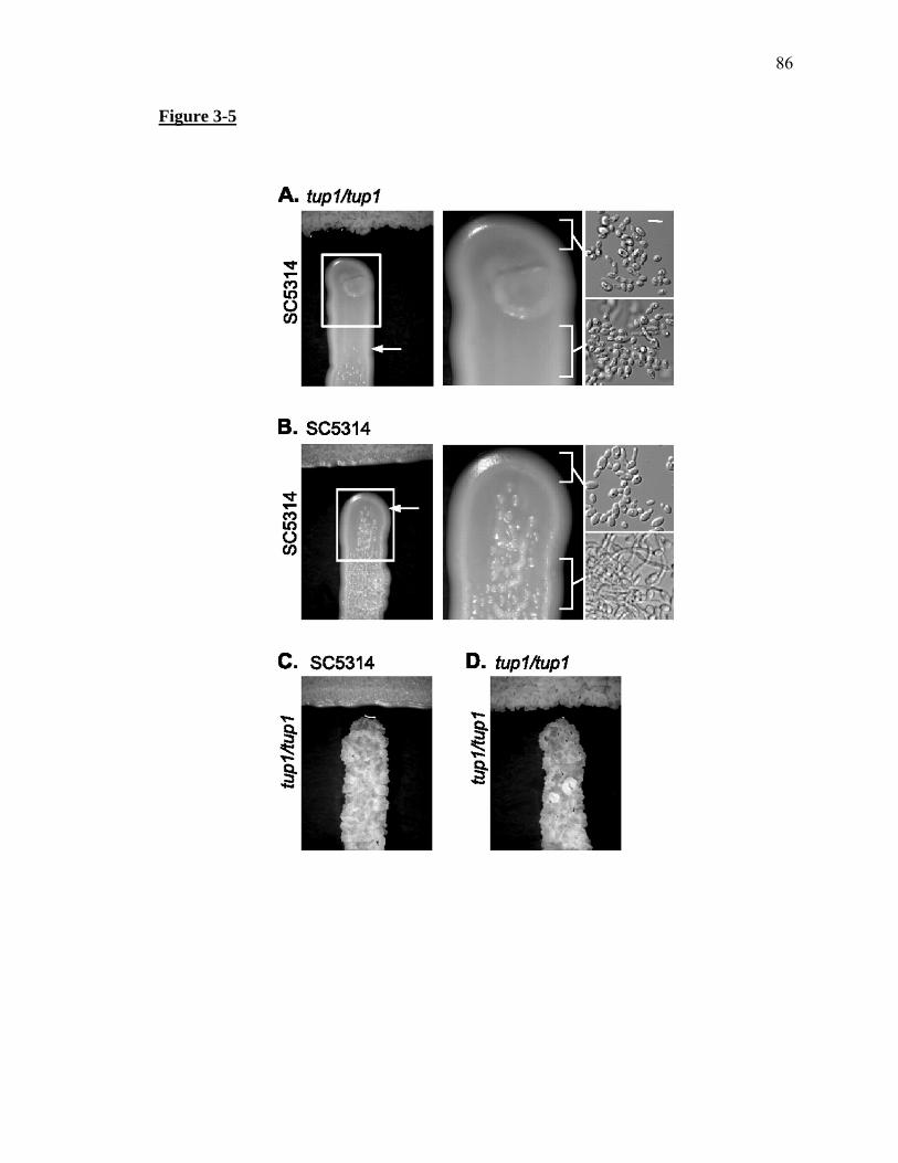
86
Figure 3-5

87
Table 3-1. tup1/tup1 and nrg1/nrg1 null mutants do not respond to farnesol but overproduce
farnesol.
C. albicans strain Farnesol responsea/ Farnesol productionb/ Fold increase in farnesolc/
(CAI-4) Positive 1.6 ± 0.36
(CAF-2) Positive 2.0 ± 1.30
tup1/tup1(BCa2-10) Negative 30.6 ± 6.40 17
nrg1/nrg1 (DU152) Negative 34.5 ± 12.2 19
rfg1/rfg1 (DU129) Positive 4.8 ± 2.0 2.6
a/ Farnesol responses on GPP agar with and without 20μM farnesol incubated at 37oC for
48 hours. A positive response to farnesol indicates smooth colony morphology (yeast
cells) in the presence of farnesol and rough colony morphology (filamentous cells)
without added farnesol. A negative response to farnesol indicates rough colony
morphology in the presence and absence of farnesol.
b/ Farnesol production (μg/g dry weight of cells) was the average of three measurements
c/ Values based on fold increase over 1.8, the average value for CAI-4 and CAF-2 strains.

88
Table 3-2. Farnesol suppresses the haploinsufficient phenotype of a TUP1/tup1
heterozygote. Cells were plated on a cornmeal agar plus Tween 80 plate under a coverslip
and grown at 25°C for 25 hours.
Cell morphology at the colony periphery C. albicans Strain
No Farnesola/ 20 μM Farnesol
Wild type (SC5314) Yeasts + few filaments Yeast + few filaments
TUP1/tup1 (BCa2-3) Yeasts + filaments Yeast + few filaments
tup1/tup1 (BCa10) filaments filaments
a/The phenotype for these strains grown on a cornmeal agar plus Tween 80 plate under a
coverslip without farnesol was also previously reported (Braun and Johnson, 1997).

89
CHAPTER 4
A novel role for Czf1 in farnesol tolerance and the morphological response to
farnesol in Candida albicans

90
Abstract
Candida albicans is the primary cause of hospital acquired fungal infections in
the United States. The quorum sensing phenomenon in Candida albicans is a process
that inhibits the transition to filamentous growth and may be directly related to its ability
to cause both mucosal and systemic disease. The Ras1-cyclic AMP signal transduction
pathway is a proposed target for farnesol inhibition, resulting in filamentous growth
obstruction, but a clear understanding of the downstream effectors and signaling
networks vital for the morphological farnesol response have yet to be unraveled. To
address this issue, we screened a mutant library for farnesol resistant mutants; among the
resistant mutants was a czf1Δ/czf1Δ mutant. Here, we demonstrate for the first time the
capability of C. albicans to respond to farnesol in embedded conditions, and in these and
liquid assays, Czf1 was required for a wild-type farnesol response. The CZF1 mRNA
transcript levels in response to farnesol, farnesol toxicity, and farnesol production levels
were also examined. Finally, CZF1 ectopic expression restored farnesol response in
strains lacking Czf1, highlighting a new role for Czf1 as a critical downstream effector of
the morphological response to farnesol in C. albicans.

91
Introduction
Candida albicans is an opportunistic pathogen that is part of the normal flora but
can cause mild to severe opportunistic infections in immunocompromised patients. A
polymorphic fungus, C. albicans can switch between yeast and filamentous forms of
growth, and this transition is necessary for causing disease in a mouse model of infection
(Lo et al., 1997; Saville et al., 2003). Two additional growth morphologies in C.
albicans include mating-competent opaque cells and structures with unknown function
called chlamydospores. All of these cell types are affected by the quorum sensing
molecule, farnesol, highlighting its influential role in C. albicans morphology.
Farnesol was the first eukaryotic quorum sensing molecule identified and was
initially shown to block the transition from yeast to filaments (Hornby et al., 2001).
Subsequent studies identified farnesol as having an inhibitory role in biofilm formation
(Ramage et al., 2002) and a protective role against oxidative stress (Deveau et al., 2010;
Westwater et al., 2005). In addition, very high levels of farnesol can increase
chlamydospore formation (Martin et al., 2005), and low levels of farnesol induces
necrosis in opaque cells when oxygen is present, thereby regulating opaque cell stability
(Dumitru et al., 2007). White cells can also be killed by farnesol in the right
environmental conditions; log phase cells that are energy-deprived are particularly
sensitive, while stationary phase cells in growth medium are quite farnesol tolerant
(Langford et al., 2010; Shirtliff et al., 2009; Uppuluri et al., 2007). Given its important
role in physiology, it comes as no surprise that in vivo studies provide physiological
relevance for farnesol signaling during infection and suggest farnesol plays distinct roles
at different sites of infection. For example, farnesol is a virulence factor in a
92
disseminated mouse model of infection (Navarathna et al., 2007a), yet it protects mice
from oral candidiasis (Hisajima et al., 2008). These results highlight the need for a
complete understanding of the signaling response induced by farnesol in C. albicans.
Factors identified that play a role in the C. albicans farnesol response include
Tup1/Nrg1 (Kebaara et al., 2008), Chk1 (Kruppa et al., 2004), the Hog1 MAP kinase
pathway (Smith et al., 2004), the Cek1 MAP kinase pathway (Roman et al., 2009), and
the Ras1-cyclic AMP (cAMP) signaling pathway (Davis-Hanna et al., 2008). In this
paper, we have identified Czf1 (C. albicans zinc finger) as an important factor in the
response to farnesol by screening a mutant library for mutants with a defective farnesol
response. Known roles of Czf1 include induction of contact-induced filamentous growth
(Brown et al., 1999), biofilm formation (Stichternoth and Ernst, 2009), and white to
opaque cell switching (Vinces and Kumamoto, 2007). It contains an unusually large 5’
untranslated region (UTR) of approximately 2 kb (Vinces et al., 2006), and can
negatively regulate its own mRNA expression (Vinces et al., 2006). Czf1 also has ties to
the cAMP pathway, as a transcription factor downstream of cAMP signaling, Efg1, can
bind the promoter of CZF1 and tightly regulate its expression, since no CZF1 mRNA was
detected in an efg1Δ/ efg1Δ background (Vinces et al., 2006). This positive regulation by
Efg1 is intriguing because Czf1 and Efg1 appear to play opposing roles in the cell with
respect to morphology (Giusani et al., 2002; Noffz et al., 2008). Of note, regulation
between Efg1 and Czf1 may also occur at the protein level as well since these two
proteins interact during a yeast two hybrid assay (Giusani et al., 2002; Noffz et al., 2008).
Here we have defined a new role for Czf1 in the quorum sensing response of C.
albicans. We show that Czf1 is not only necessary for a wild type morphological

93
response to farnesol in a variety of conditions, but in combination with Efg1, is required
for farnesol tolerance as well. Our data suggest a vital downstream role for Czf1 in the
signaling cascade elicited by farnesol in C. albicans.

94
Methods
Strains and media
Candida albicans strains and plasmids are listed in Table 1. The defined medium,
modified glucose phosphate proline (mGPP) pH 6.8 was prepared as described in
Kebaara et al (Kebaara et al., 2008) with 2.5 mM N-acetylglucosamine (GlcNAc) and
uridine (40 µg/ml). Yeast peptone dextrose (YPD) medium contained 1% yeast extract,
0.5% peptone, and 2% dextrose, and solid medium included 2% agar. Resting cells were
prepared as in Kebaara et al (Kebaara et al., 2008) with modifications: single colonies
were grown in 25 mL YPD broth at 30°C (unless otherwise noted) for 22-24h to reach
stationary phase, washed three times with 50 mM potassium phosphate buffer (pH6.8),
resuspended in potassium phosphate buffer, and stored at 4°C overnight before use. For
liquid farnesol response assays, resting cells were inoculated at 106 cells/mL in mGPP or
mSPP (2% sucrose replacing glucose) broth with indicated farnesol concentrations. Cells
were then grown at 37ºC, shaking in glass flasks for the indicated times. Trans-trans
farnesol (Sigma, St. Louis, MO) was stored under nitrogen and freshly prepared as a 100
mM stock solution in methanol for each experiment.
Mutant library screen
Mutants were obtained in 96-well plates and plated on mGPP (with 40 µg/ml uridine,
arginine, and histidine added) agar plates containing 0, 10, or 50 µM farnesol. Plates
were incubated at 37°C for 2 days before colony morphology was assessed and compared
to the parental strain, BWP17 for farnesol response. Wrinkled/hairy colony
morphologies were considered to be composed primarily of filaments and smooth colony
morphologies were considered to be mostly yeast cells. A positive response to farnesol

95
was viewed as filamentous colonies without farnesol and smooth colonies in the presence
of farnesol. Farnesol resistant mutants maintained a filamentous morphology, even in the
presence of 50 µM farnesol.
Embedded cell growth
Embedded media were prepared by mixing 104 cells/mL in 30 ml GPP (no GlcNAc
added) or SPP molten agar (cooled to 50°C) with appropriate concentrations of farnesol
and plated. Embedded plates were incubated at 37°C for 12 or 17 hours as indicated.
Only colonies beneath the agar surface were examined.
Microscopy and cell death determination
Cellular morphology during the germ tube assays was determined using a Zeiss Stemi
2000-C light microscope. Embedded micrographs were taken using a custom MVI
TDM400 tetrad dissecting microscope and Sony Cybershot camera. DIC micrographs
were taken using an Olympus BX51 microscope and a Photometrics CoolSnap HQ CCD
camera. Cell death was determined by methylene blue staining as described by Gibson et
al (Gibson et al., 2009).
DNA analysis and transformation
To create strain AAC2, CKY283 was plated on 5-fluoroorotic acid (5-FOA) containing
media to select for ura- mutants. AAC2 was subsequently transformed with BsgI-
digested pDB212 to create strain AAC6. Transformations were performed by the lithium
acetate method and transformants were selected on media lacking uridine (Gietz and
Woods, 2002). Newly created strains were confirmed by PCR and Southern blot analysis
(data not shown); restriction digestion and Southern blotting were performed as described

96
in the GeneScreen Plus Hybridization Transfer and Detection Protocols (DuPont NEN
Research Products, Boston, MA).
Quantitative real- time PCR
Resting cells were inoculated into 75 mL mGPP broth at 5 x 10⁶ cells/mL, and 0, 50, or
100 µM farnesol was added to each flask. Cells were incubated at 37°C and harvested at
40, 60, or 80 minutes. For harvesting cells, cultures were passed through glass fiber
filters and cells were scraped off the filters to reduce the loss of filamentous cells during
centrifugation. Data shown are from a representative independent experiment performed
in triplicate. Extraction of mRNA was performed with the RiboPure Yeast Kit (Applied
Biosystems, Foster City, CA), and mRNA integrity was assessed by measuring OD260/280
ratios and by examining the appearance on agarose gels. Reverse transcription and PCR
amplification were performed according to manufacturer’s specifications using
MultiScribe Reverse Transcriptase and Power SYBR Green PCR Master Mix (Applied
Biosystems). Real-time PCR reactions were carried out in an ABI Prism 7500 Real-Time
PCR machine (Applied Biosystems). ACT1 mRNA levels were used as controls.
Primers used to amplify CZF1 and ACT1 and PCR efficiencies were as previously
described (Bassilana et al., 2005).
Farnesol Production Measurements
Extracellular farnesol was extracted from cell-free supernatants of stationary phase
cultures grown in GPP at 30ºC and analyzed by gas chromatography-mass spectrometry,
as described by Hornby et al (Hornby et al., 2001).

97
Results
Identification of new genes required for the response to farnesol
To identify important regulators of the farnesol response in C. albicans, a
transposon insertion mutant library of approximately 560 mutants (Supp. Table 1) was
screened for mutants defective in the morphological response to farnesol. The majority
of mutants in this collection are Tn7-UAU1 plasmid insertion mutants (Nobile et al.,
2006); therefore it is desirable to obtain knockout mutants for further characterization due
to the possibility of partial loss-of-function mutations. Mutants that were identified as
farnesol resistant in the original screen were re-streaked on individual plates to rule out
interference from neighboring mutant colonies and to confirm the farnesol resistant
phenotype. Eight mutants were confirmed as farnesol resistant, and they are summarized
in Table 1. Mutants that were unable to interconvert between yeast and filaments (at 30°
and 37°, respectively) were not examined further in this study to rule out mutants that are
merely defective in morphological switching and not the specific response to farnesol.
No mutants were identified that were hypersensitive to farnesol, nor were any detected
that demonstrated growth inhibition in the presence of farnesol.
CZF1 is required for a wild type morphological response to farnesol in both liquid
and embedded conditions
The czf1/czf1 mutant was identified as farnesol-resistant in the mutant library
screen (Table 1) and selected for further characterization based on its known roles in
morphogenesis, white/opaque cell switching, and its connection to the cAMP pathway.
To test the role of Czf1 in the morphological response to farnesol, a double null

98
czf1Δ/czf1Δ mutant was compared with its parental strain (CKY101), a wild type clinical
isolate (SC5314), the heterozygous mutant, and the double null mutant ectopically
expressing CZF1 under the control of the MAL2 promoter in a liquid farnesol response
assay (Fig. 1). In both glucose and sucrose-containing media, SC5314 and CKY101
displayed the expected reduction of early stage filaments (germ tubes) when in the
presence of farnesol. However, the czf1Δ/czf1Δ mutant showed only a minimal reduction
of germ tubes, even in the presence of 100 μM farnesol, and a haploinsufficient farnesol
response phenotype was observed for the heterozygous mutant. Results were similar
whether glucose or sucrose was used as a carbon source. Ectopic complementation of the
czf1Δ/czf1Δ mutant was able to restore the farnesol response similar to that of the
heterozygous mutant in pMAL2-inducing conditions (Fig. 1B; sucrose-containing media),
while there was only a minimal farnesol response in non-inducing conditions, similar to
that of the czf1Δ/czf1Δ mutant (Fig. 1A; glucose-containing media). These results show
that the presence of Czf1 is critical to the ability of C. albicans to respond to farnesol in
liquid media.
Since the first described role for Czf1 was to promote filamentation in embedded
conditions (Brown et al., 1999), we sought to determine whether: 1) C. albicans could
respond to farnesol under such conditions and 2) Czf1 was needed for farnesol response
when embedded in a matrix. For consistency with our prior work studying farnesol
signaling and physiological significance (plates were incubated at 37°C), we utilized
defined GPP or SPP agar plates (lacking GlcNAc, see experimental procedures) in our
embedded assays. Although the growth media and incubation time and temperature used
in this study are different from those initially used by Brown et al (Brown et al., 1999),

99
we still observed a strong filamentation response from C. albicans SC5314 and CKY101
cells in response to growth embedded in a matrix (Fig. 2). When SC5314 and CKY101
cells were grown in embedded media containing 50 μM farnesol, filamentous colonies
were not observed. As with the liquid germ tube formation assays, using glucose versus
sucrose as a carbon source made no difference in filamentation or farnesol response (Fig.
2). This demonstrates the capability of C. albicans to respond to farnesol in an embedded
assay.
As expected, the czf1Δ/czf1Δ mutant exhibited a defective growth pattern in
embedded conditions; filamentation was still observed, but far fewer colonies were
present compared with the wild type and parental strains and colony morphology
appeared different as well (Fig. 2). When the czf1Δ/czf1Δ mutant was grown in agar
containing farnesol, only a moderate farnesol response was observed in that the hyphae
appeared shorter than in untreated samples; however, colonies were still filamentous. As
with the liquid farnesol response assays, the czf1Δ/CZF1 heterozygote maintained a
haploinsufficient phenotype in both farnesol treated and untreated samples: in untreated
samples, the overall level of growth was more similar to that of wild type strains, and in
farnesol-treated samples, colonies were reduced in hyphal formation but still more
filamentous than wild type colonies treated with farnesol. Ectopic CZF1
complementation of the czf1Δ/czf1Δ mutant produced positive farnesol responses similar
to that of the heterozygote in both GPP and SPP, conditions which should turn off and on,
respectively, the MAL2 promoter (Fig. 2). This suggests unintended leaky promoter
activation in GPP media, possibly resulting in the expression of CZF1 and
complementation of the farnesol response in both GPP and SPP embedded media. These

100
results indicate that Czf1 is required for farnesol response in at least two distinct
environmental conditions: liquid, aerobic growth media and embedded in a semi-solid
matrix, new conditions in which to test C. albicans for a morphological farnesol
response.
CZF1 mRNA levels are decreased in the presence of farnesol
Since CZF1 is regulated at the mRNA level, we asked whether CZF1 mRNA
levels are affected by the presence of farnesol. A time-course experiment was performed
to measure CZF1 expression levels in the presence of 0, 50 or 100 μM farnesol, and 40,
60, and 80 minute time points were selected based on previous studies showing the
importance of this time frame to the farnesol response (Kebaara et al., 2008). As with
TUP1 mRNA levels (Kebaara et al., 2008; Mosel et al., 2005), CZF1 levels were most
dramatically affected 60 minutes after treatment with farnesol, showing a greater than 5-
fold decrease in expression relative to a sample lacking farnesol (Fig. 3B). At all 3 time
points tested, both 50 and 100 μM farnesol reduced CZF1 levels, relative to samples
containing no farnesol (Fig. 3). These data indicate that CZF1 mRNA levels are
decreased in the presence of farnesol, during time points crucial to the farnesol response.
An efg1Δ/ efg1Δ czf1Δ/ czf1Δ double mutant exhibits decreased farnesol tolerance
that is partially temperature dependent
Upon growth of the efg1Δ/efg1Δ czf1Δ/czf1Δ mutant in liquid media for
preparation of resting cells, an unusual temperature-regulated phenotype was observed.
Mutant cells grown in YPD broth at 30°C appeared small but elongated, reminiscent of
101
opaque cells (Fig. 4A), and when the efg1Δ/efg1Δ czf1Δ/czf1Δ double mutant was grown
in YPD broth at 37°C, cells appeared larger and more rounded (Fig. 4B). When these
efg1Δ/efg1Δ czf1Δ/czf1Δ double mutant cells (pre-grown at 30°C) were tested for
farnesol response in the previously described mGPP/mSPP liquid germ tube formation
assays (experimental procedures), we observed what appeared to be cell death. Given the
importance of environmental conditions in the ability of C. albicans to survive and
tolerate farnesol, methylene blue staining was performed during liquid germ tube assays
to more accurately assess the level of cell death occurring in these conditions (Fig. 4C-F).
For the cell death assays, resting cells were prepared as described in experimental
procedures, except that cells were grown at either 30°C or 37°C in YPD broth until
stationary phase was reached. The parental strain CAI4 was used as a control in the 30°C
resting cell group, and as observed for SC5314 in Langford et al (Langford et al., 2010),
no significant cell death was observed in mGPP or in mSPP, even in the presence of 100
μM farnesol. The czf1Δ/czf1Δ and efg1Δ/efg1Δ mutants (Fig. 4C-F) were similarly
resistant to farnesol-mediated killing in the liquid germ tube formation assays. However,
the czf1Δ/czf1Δ mutant could not be tested accurately with methylene blue in these
assays, due to its rapid formation of germ tubes which stain inconsistently with this dye
regardless of the strain used or the presence of farnesol. The arguments for the viability
of the czf1Δ/czf1Δ mutant in the presence of farnesol are twofold: 1) no growth
inhibition was observed in the presence of farnesol on agar plates (data not shown) and 2)
germ tube formation is a process that requires active protein synthesis (Imanishi et al.,
2004) and is unlikely to occur in a dead cell. CAI4 could also not be used as a control at
37°C for similar reasons; cells pre-grown at 37°C start as filaments. On the other hand,

102
the efg1Δ/efg1Δ czf1Δ/czf1Δ double mutant and the efg1Δ/efg1Δ czf1Δ/czf1Δ double
mutant ectopically expressing CZF1 (from the 30°C resting cell group) showed extreme
sensitivity to farnesol, regardless of the media used to induce CZF1 expression (Fig 4C,
4E). Conversely, when the efg1Δ/efg1Δ czf1Δ czf1Δ double mutant and the efg1Δ/efg1Δ
czf1Δ/czf1Δ double mutant expressing CZF1 under the control of the MAL2 promoter
from the 37°C resting cell group were tested for farnesol sensitivity, a general decrease in
cell death was observed compared to the 30°C resting cell group (Fig. 4C-F). Based on
these results, Czf1, Efg1, and temperature appear to play important roles in farnesol
tolerance, and both 30°C and 37°C double mutant resting cells were subsequently utilized
for additional experiments in this study.
Ectopically expressing CZF1 in an efg1Δ/ efg1Δ czf1Δ/czf1Δ double mutant
partially restores filamentation and farnesol response in liquid and embedded
conditions
Can ectopic expression of CZF1 restore filamentation and response in the efg1Δ/
efg1Δ czf1Δ/czf1Δ double mutant? Liquid germ tube formation assays were performed
on CAI4, efg1Δ/efg1Δ, the efg1Δ efg1Δ czf1Δ/czf1Δ double mutant, and the efg1Δ/efg1Δ
czf1Δ/czf1Δ double mutant ectopically expressing CZF1 under the control of the MAL2
promoter, and only cells that did not stain with methylene blue were counted. For the
30°C resting cell group, only CAI4 was able to produce germ tubes and respond to
farnesol, in both mGPP and mSPP (Fig. 5A, 5C). For the 37°C resting cell group, the
efg1Δ/efg1Δ and the efg1Δ/ efg1Δ czf1Δ/czf1Δ double mutant were capable of producing
very low levels of germ tubes with a slight trend of reduction in the presence of farnesol.

103
In conditions which induce CZF1 expression in the efg1Δ/efg1Δ czf1Δ/czf1Δ double
mutant, an increase in germ tube formation was observed when compared to non-
inducing conditions (Fig. 5B, 5D), and farnesol reduced germ tube formation when CZF1
was expressed.
When CAI4 30°C resting cells were inoculated into the embedded farnesol
response assays, results were similar to SC5314 and CKY101 (Fig. 6). CAI4 produced
filamentous colonies in the absence of farnesol in both GPP and SPP embedded media
and the presence of farnesol resulted in decreased filamentation. Note the smaller hyphae
growing out from the CAI4 colonies treated with farnesol (Fig. 6); because farnesol
delays filamentation rather than block it entirely, the time points selected for observation
are critical. Ideally CAI4 embedded plates should have been checked at 12 h post-
inoculation for farnesol response (as in Fig. 2) rather than at 17h as shown (no short
filaments are observed in the farnesol-treated CAI4 samples at 12 h, data not shown), but
the 17 h time point was selected in order to be consistent with the other slower-growing
mutant strains being tested in Fig. 6. The efg1Δ/efg1Δ mutant was capable of forming
filamentous colonies under embedded conditions, and a very subtle shortening of
filaments was observed in farnesol-treated samples. An inoculum using 30°C resting
cells for the efg1Δ/efg1Δ mutant is shown in Fig. 6, and similar results were obtained
using an inoculum of 37°C resting efg1Δ/efg1Δ cells. The efg1Δ/efg1Δ czf1Δ/czf1Δ
double mutant was capable of producing filamentous colonies in the absence of farnesol,
regardless of the temperature used during resting cell preparation (Fig. 6). The
efg1Δ/efg1Δ czf1Δ/czf1Δ double mutant and the efg1Δ/efg1Δ czf1Δ/czf1Δ double mutant
ectopically expressing CZF1 did not produce any colonies in the presence of farnesol

104
when 30°C resting cells were used (data not shown), suggesting these cells were killed by
farnesol as they were in the liquid assays. When 37°C resting cells were used for the
efg1Δ/efg1Δ czf1Δ/czf1Δ double mutant, farnesol reduced the number of colonies, but the
colonies that grew were filamentous. Unexpectedly, the filamentous colonies from
farnesol-treated plates often grew in a different conformation from untreated samples.
The efg1Δ/efg1Δ czf1Δ/czf1Δ double mutant expressing CZF1 under the control of the
MAL2 promoter behaved similarly to the efg1Δ/efg1Δ czf1Δ/ czf1Δ double mutant in non-
inducing conditions (Fig. 6A), and a subtle increase in colony size and filamentation was
observed in inducing media (Fig. 6B). In the presence of farnesol, these filaments were
slightly shorter. Taken together, both Czf1 and Efg1 are important for farnesol tolerance
in embedded conditions as well, along with temperature. When CZF1 is ectopically
expressed in a strain lacking Czf1 and Efg1, the level of filamentation is partially restored
in both liquid and embedded assays, and farnesol partially suppresses filamentation in
both conditions.
Overproduction of farnesol is not a general quality of farnesol-resistant mutants
Two of the factors known to play a role in the C. albicans morphological farnesol
response, Tup1 and Nrg1, both produced 17 and 19-fold higher levels of farnesol than
wild type and parental strains (Kebaara et al., 2008). In order to determine whether
farnesol overproduction is a general quality of farnesol-resistant mutants, we tested the
farnesol production levels of the czf1Δ/czf1Δ mutant (Fig. 7). Farnesol production levels
were not significantly altered in the czf1Δ/czf1Δ mutant, suggesting a more specific

105
involvement of Tup1/Nrg1 in farnesol production that does not require the presence of
Czf1.

106
Discussion
In this study, we identified new factors involved in the C. albicans response to
farnesol and have shown that Czf1 in particular plays a prominent role in the
morphological response to farnesol in both liquid aerobic conditions and embedded
conditions; this adds a new function for Czf1 during the quorum sensing response.
Furthermore, CZF1 mRNA levels are decreased in the presence of farnesol at critical
time points during the farnesol response, and unlike another factor involved in farnesol
signaling (Tup1), Czf1 does not appear to regulate farnesol production levels in the cell.
Surprisingly, CZF1, along with EFG1, was also shown to play an important part in
farnesol tolerance; these are the first specific genes identified that control the ability of C.
albicans white a/α cells to survive in the presence of farnesol.
The Ras1-Cyr1-cAMP signaling pathway has been proposed to be the only direct
target for farnesol in C. albicans (Davis-Hanna et al., 2008; Deveau et al., 2010). This
study provides additional evidence supporting a primary role of this pathway in farnesol
signaling. Here we showed that Czf1 is an important factor in the farnesol response. It
has a tight regulatory relationship with Efg1, which is consistent with the Ras1-cAMP
signaling pathway as a central target for farnesol. Further evidence for the significant
role of cAMP signaling during the farnesol response is provided by the identification of
one of the protein kinase A (PKA) isoforms in C. albicans, Tpk1, as farnesol resistant.
The other PKA isoform, Tpk2, was not identified by the farnesol resistance screen as it
was not present in this mutant library collection. A recent study (Deveau et al., 2010)
showed that an additional phenotype of the farnesol response in C. albicans, reactive
oxygen species (ROS) protection, is also mediated by the Ras1-cAMP pathway. The

107
Hog1 signaling pathway was shown to participate in ROS protection as well, but through
an unknown connection between the two pathways (Deveau et al., 2010). Three
additional genes were identified that are potentially important for farnesol resistance:
RLM1, YCK2, and HAP43, also function in different stress responses. These mutants can
possibly be used to fill in some of the gaps between these pathways.
It is intriguing that farnesol can protect the cell from ROS stress, while it can also
kill opaque cells and log-phase, energy-deprived white cells. Since farnesol tolerance in
C. albicans is potentially an active process (Langford et al., 2010), it is of interest to
understand the key components that are involved. This study provides a new
understanding of farnesol tolerance, by identifying two factors that are required for
survival in the presence of farnesol: Czf1 and Efg1. We speculate that it is no
coincidence that both of these factors also play critical roles in white/opaque cell
switching (Ramirez-Zavala et al., 2008; Sonneborn et al., 1999; Srikantha et al., 2000;
Vinces and Kumamoto, 2007; Zordan et al., 2007) since opaque cells are inherently more
sensitive to farnesol killing than are white cells (Dumitru et al., 2007). Farnesol killed
efg1Δ/efg1Δ czf1Δ/czf1Δ double mutant cells in liquid aerobic conditions, and it appeared
to do the same in embedded media due to the lack of growth when using 30°C resting cell
inocula. It is unclear why single efg1Δ/efg1Δ and czf1Δ/czf1Δ mutants are resistant to
farnesol killing, while the double mutant is overly sensitive. Czf1 and Efg1 have a
synthetic effect on farnesol resistance, as farnesol tolerance was only observed in the
single mutants and not the double mutant. The mere presence of CZF1 in the
efg1Δ/efg1Δ czf1Δ/czf1Δ double mutant (via expression of CZF1 by the MAL2 promoter)
was not sufficient, however, to provide protection from farnesol induced cell death,

108
suggesting CZF1 may act upstream of EFG1 in this example. It is also possible that the
presence of Czf1’s native promoter is required for proper regulation in response to this
particular stimulus when Efg1 is absent. Another intriguing question raised by this study
is the role of temperature in farnesol tolerance. Why was the efg1Δ/efg1Δ czf1Δ/czf1Δ
double mutant more tolerant of farnesol when grown at 37°C? The answer may be tied to
indirect farnesol production measurements suggesting that C. albicans produces slightly
higher levels of farnesol at higher temperatures (Hornby et al., 2001). Further, there is
likely some additional factor(s) that plays a minor role in farnesol tolerance and is
regulated by shifts in temperature. Wor1/2 are potential candidates for such a role, in
part due to their regulation of white/opaque switching (Huang et al., 2006; Zordan et al.,
2006; Zordan et al., 2007).
While Czf1 was shown to be part of a farnesol-resistance mechanism in C.
albicans, it also strongly contributes to the morphological response to farnesol. In cells
lacking Czf1 (czf1Δ/czf1Δ mutants), farnesol was unable to suppress filamentation, and
ectopic complementation of CZF1 in these cells restored the ability of farnesol to block
germ tube formation in both liquid and embedded environmental conditions.
Furthermore, in efg1Δ/efg1Δ czf1Δ/czf1Δ double mutants, CZF1 ectopic expression was
able to partially restore filamentation and the morphological response to farnesol. These
results are consistent with Czf1 functioning downstream of Efg1 during the
morphological farnesol response. Through the course of testing farnesol response in
conditions which either turn on or off the MAL2 promoter, we have shown that the use of
glucose versus sucrose as a carbon source had no distinguishable effect on the ability of
the cells to respond to farnesol.

109
The many effects of farnesol on C. albicans appear to be relatively unique to the
fungus, compared with other bacterial species and fungal genera [reviewed by Langford
et al (Langford et al., 2009)]. The finding that Czf1 plays a central role in the unique
morphological and tolerance responses in C. albicans is consistent with the observation
that Saccharomyces cerevisiae (which does not respond to farnesol) and other closely
related ascomycetes lack a Czf1 homolog (Vinces et al., 2006) while other Candida
species contain CZF1 homologs. As an example, the CZF1 gene in Candida
dubliniensis, a Candida species that produces the second highest farnesol levels, is
farnesol tolerant, and morphologically responds to farnesol (Henriques et al., 2007;
Weber et al., 2008) has the highest homology to the C. albicans CZF1 gene with 81%
identity at the nucleotide level (data not shown). Furthermore, the unusually large 5’
upstream region of CZF1, the CZF1 gene itself, and neighboring genes at the CZF1 locus
(Vinces et al., 2006) appear to be conserved to varying degrees in additional Candida
species whose sequence information has been recently made available including:
Candida tropicalis, Lodderomyces elongisporius, Candida lusitaniae, and Candida
guillermondii (data not shown).
In conclusion, we have identified new roles for Czf1 in mediating the C. albicans
tolerance to farnesol as well as farnesol-mediated filament inhibition. While the
connection of Czf1 to other factors known to play a role in the C. albicans farnesol
response, such as the cAMP pathway, is apparent, others links remain unclear. For
example, cross-regulation among many of the farnesol response pathways was recently
summarized (Deveau et al., 2010), but little is known about the regulation of Tup1/Nrg1
and how these factors fit into the farnesol signaling network. Furthermore, other proteins

110
involved in Ras1-Cyr1-cAMP signaling such as Ras2, Cap1, and G-actin (Hall and
Muhlschlegel, 2009; Zhu et al., 2009; Zou et al., 2009) have not been tested for possible
roles in farnesol signaling and may yet prove to be involved. This points to the fact that
farnesol can be an extremely useful tool for studying signal transduction in C. albicans
by demonstrating the presence of new connections between signaling pathways.
Acknowledgements
We are grateful to Aaron Mitchell, Dominique Sanglard, Carol Kumamoto, Alexander
Johnson, and Gerald Fink for kindly providing the strains and plasmids used in this study.
We would also like to thank Steve Harris and Gabriel Langford for use of their
microscopes and cameras. This work was supported by a faculty seed grant from the
Constance Miriam and Ethel Corrine Syford Memorial Fund (to A.L.A.), the Hammond-
Maude Fling Fellowship (to M.L.L.), and the Farnesol and Candida albicans Research
Fund, University of Nebraska Foundation (to K.W.N.).

111
References
Baek, Y.U., Li, M., and Davis, D.A. (2008) Candida albicans ferric reductases are
differentially regulated in response to distinct forms of iron limitation by the
Rim101 and CBF transcription factors. Eukaryot Cell 7: 1168-1179.
Bassilana, M., Hopkins, J., and Arkowitz, R.A. (2005) Regulation of the Cdc42/Cdc24
GTPase module during Candida albicans hyphal growth. Eukaryot Cell 4: 588-
603.
Bockmuhl, D.P., Krishnamurthy, S., Gerads, M., Sonneborn, A., and Ernst, J.F. (2001)
Distinct and redundant roles of the two protein kinase A isoforms Tpk1p and
Tpk2p in morphogenesis and growth of Candida albicans. Mol Microbiol 42:
1243-1257.
Brown, D.H., Jr., Giusani, A.D., Chen, X., and Kumamoto, C.A. (1999) Filamentous
growth of Candida albicans in response to physical environmental cues and its
regulation by the unique CZF1 gene. Mol Microbiol 34: 651-662.
Bruno, V.M., Kalachikov, S., Subaran, R., Nobile, C.J., Kyratsous, C., and Mitchell, A.P.
(2006) Control of the Candida albicans cell wall damage response by
transcriptional regulator Cas5. PLoS Pathog 2: e21.
Cloutier, M., Castilla, R., Bolduc, N., Zelada, A., Martineau, P., Bouillon, M., Magee,
B.B., Passeron, S., Giasson, L., and Cantore, M.L. (2003) The two isoforms of the
cAMP-dependent protein kinase catalytic subunit are involved in the control of
dimorphism in the human fungal pathogen Candida albicans. Fungal Genet Biol
38: 133-141.

112
Davis-Hanna, A., Piispanen, A.E., Stateva, L.I., and Hogan, D.A. (2008) Farnesol and
dodecanol effects on the Candida albicans Ras1-cAMP signalling pathway and
the regulation of morphogenesis. Mol Microbiol 67: 47-62.
Deveau, A., Piispanen, A.E., Jackson, A.A., and Hogan, D.A. (2010) Farnesol induces
hydrogen peroxide resistance in Candida albicans yeast by inhibiting the Ras-
cAMP signaling pathway. Eukaryot Cell Epub.
Dumitru, R., Navarathna, D.H., Semighini, C.P., Elowsky, C.G., Dumitru, R.V., Dignard,
D., Whiteway, M., Atkin, A.L., and Nickerson, K.W. (2007) In vivo and in vitro
anaerobic mating in Candida albicans. Eukaryot. Cell 6: 465-472.
Fonzi, W.A., and Irwin, M.Y. (1993) Isogenic strain construction and gene mapping in
Candida albicans. Genetics 134: 717-728.
Giacometti, R., Kronberg, F., Biondi, R.M., and Passeron, S. (2009) Catalytic isoforms
Tpk1 and Tpk2 of Candida albicans PKA have non-redundant roles in stress
response and glycogen storage. Yeast 26: 273-285.
Gibson, J., Sood, A., and Hogan, D.A. (2009) Pseudomonas aeruginosa-Candida
albicans interactions: localization and fungal toxicity of a phenazine derivative.
Appl Environ Microbiol 75: 504-513.
Gietz, R.D., and Woods, R.A. (2002) Transformation of yeast by lithium acetate/single-
stranded carrier DNA/polyethylene glycol method. Methods Enzymol 350: 87-96.
Gillum, A.M., Tsay, E.Y., and Kirsch, D.R. (1984) Isolation of the Candida albicans
gene for orotidine-5'-phosphate decarboxylase by complementation of S.
cerevisiae ura3 and E. coli pyrF mutations. Mol Gen Genet 198: 179-182.
113
Giusani, A.D., Vinces, M., and Kumamoto, C.A. (2002) Invasive filamentous growth of
Candida albicans is promoted by Czf1p-dependent relief of Efg1p-mediated
repression. Genetics 160: 1749-1753.
Hall, R.A., and Muhlschlegel, F.A. (2009) A multi-protein complex controls cAMP
signalling and filamentation in the fungal pathogen Candida albicans. Mol
Microbiol.
Henriques, M., Martins, M., Azeredo, J., and Oliveira, R. (2007) Effect of farnesol on
Candida dubliniensis morphogenesis. Lett Appl Microbiol 44: 199-205.
Hisajima, T., Maruyama, N., Tanabe, Y., Ishibashi, H., Yamada, T., Makimura, K.,
Nishiyama, Y., Funakoshi, K., Oshima, H., and Abe, S. (2008) Protective effects
of farnesol against oral candidiasis in mice. Microbiol Immunol 52: 327-333.
Hornby, J.M., Jensen, E.C., Lisec, A.D., Tasto, J.J., Jahnke, B., Shoemaker, R., Dussault,
P., and Nickerson, K.W. (2001) Quorum sensing in the dimorphic fungus
Candida albicans is mediated by farnesol. Appl. Environ. Microbiol. 67: 2982-
2992.
Huang, G., Wang, H., Chou, S., Nie, X., Chen, J., and Liu, H. (2006) Bistable expression
of WOR1, a master regulator of white-opaque switching in Candida albicans.
Proc Natl Acad Sci U S A 103: 12813-12818.
Imanishi, Y., Yokoyama, K., and Nishimura, K. (2004) Inductions of germ tube and
hyphal formations are controlled by mRNA synthesis inhibitor in Candida
albicans. Nippon Ishinkin Gakkai Zasshi 45: 113-119.

114
Kebaara, B.W., Langford, M.L., Navarathna, D.H., Dumitru, R., Nickerson, K.W., and
Atkin, A.L. (2008) Candida albicans Tup1 is involved in farnesol-mediated
inhibition of filamentous-growth induction. Eukaryot Cell 7: 980-987.
Kruppa, M., Krom, B.P., Chauhan, N., Bambach, A.V., Cihlar, R.L., and Calderone, R.A.
(2004) The two-component signal transduction protein Chk1p regulates quorum
sensing in Candida albicans. Eukaryot. Cell 3: 1062-1065.
Langford, M.L., Atkin, A.L., and Nickerson, K.W. (2009) Cellular interactions of
farnesol, a quorum-sensing molecule produced by Candida albicans. Future
Microbiol 4: 1353-1362.
Langford, M.L., Hasim, S., Nickerson, K.W., and Atkin, A.L. (2010) Activity and
toxicity of farnesol towards Candida albicans are dependent on growth
conditions. Antimicrob Agents Chemother 54: 940-942.
Leberer, E., Harcus, D., Dignard, D., Johnson, L., Ushinsky, S., Thomas, D.Y., and
Schroppel, K. (2001) Ras links cellular morphogenesis to virulence by regulation
of the MAP kinase and cAMP signalling pathways in the pathogenic fungus
Candida albicans. Mol Microbiol 42: 673-687.
Li, W.J., Wang, Y.M., Zheng, X.D., Shi, Q.M., Zhang, T.T., Bai, L.D., Sang, J.L., and
Wang, Y. (2006) The F-box protein Grr1 regulates the stability of Ccn1, Cln3 and
Hof1 and cell morphogenesis in Candida albicans. Mol. Microbiol. 62: 212-226.
Lo, H.J., Kohler, J.R., DiDomenico, B., Loebenberg, D., Cacciapuoti, A., and Fink, G.R.
(1997) Nonfilamentous Candida albicans mutants are avirulent. Cell 90: 939-949.
Maidan, M.M., De Rop, L., Serneels, J., Exler, S., Rupp, S., Tournu, H., Thevelein, J.M.,
and Van Dijck, P. (2005) The G protein-coupled receptor Gpr1 and the Galpha
115
protein Gpa2 act through the cAMP-protein kinase A pathway to induce
morphogenesis in Candida albicans. Mol Biol Cell 16: 1971-1986.
Martin, S.W., Douglas, L.M., and Konopka, J.B. (2005) Cell cycle dynamics and quorum
sensing in Candida albicans chlamydospores are distinct from budding and
hyphal growth. Eukaryot Cell 4: 1191-1202.
Martinez, P., and Ljungdahl, P.O. (2005) Divergence of Stp1 and Stp2 transcription
factors in Candida albicans places virulence factors required for proper nutrient
acquisition under amino acid control. Mol Cell Biol 25: 9435-9446.
Mosel, D.D., Dumitru, R., Hornby, J.M., Atkin, A.L., and Nickerson, K.W. (2005)
Farnesol concentrations required to block germ tube formation in Candida
albicans in the presence and absence of serum. Appl. Environ. Biol. 71: 4938-
4940.
Navarathna, D.H., Hornby, J.M., Krishnan, N., Parkhurst, A., Duhamel, G.E., and
Nickerson, K.W. (2007) Effect of farnesol on a mouse model of systemic
candidiasis, determined by use of a DPP3 knockout mutant of Candida albicans.
Infect Immun 75: 1609-1618.
Nobile, C.J., and Mitchell, A.P. (2005) Regulation of cell-surface genes and biofilm
formation by the C. albicans transcription factor Bcr1p. Curr. Biol. 15: 1150-
1155.
Nobile, C.J., Andes, D.R., Nett, J.E., Smith, F.J., Yue, F., Phan, Q.T., Edwards, J.E.,
Filler, S.G., and Mitchell, A.P. (2006) Critical role of Bcr1-dependent adhesins in
C. albicans biofilm formation in vitro and in vivo. PLoS Pathog 2: e63.

116
Noffz, C.S., Liedschulte, V., Lengeler, K., and Ernst, J.F. (2008) Functional mapping of
the Candida albicans Efg1 regulator. Eukaryot Cell 7: 881-893.
Park, H., Liu, Y., Solis, N., Spotkov, J., Hamaker, J., Blankenship, J.R., Yeaman, M.R.,
Mitchell, A.P., Liu, H., and Filler, S.G. (2009) Transcriptional responses of
Candida albicans to epithelial and endothelial cells. Eukaryot Cell 8: 1498-1510.
Ramage, G., Saville, S.P., Wickes, B.L., and Lopez-Ribot, J.L. (2002) Inhibition of
Candida albicans biofilm formation by farnesol, a quorum-sensing molecule.
Appl Environ Microbiol 68: 5459-5463.
Ramirez-Zavala, B., Reuss, O., Park, Y.N., Ohlsen, K., and Morschhauser, J. (2008)
Environmental induction of white-opaque switching in Candida albicans. PLoS
Pathog 4: e1000089.
Roman, E., Alonso-Monge, R., Gong, Q., Li, D., Calderone, R., and Pla, J. (2009) The
Cek1 MAPK is a short-lived protein regulated by quorum sensing in the fungal
pathogen Candida albicans. FEMS Yeast Res 9: 942-955.
Sampaio, P., Nogueira, E., Loureiro, A.S., Delgado-Silva, Y., Correia, A., and Pais, C.
(2009) Increased number of glutamine repeats in the C-terminal of Candida
albicans Rlm1p enhances the resistance to stress agents. Antonie Van
Leeuwenhoek 96: 395-404.
Saville, S.P., Lazzell, A.L., Monteagudo, C., and Lopez-Ribot, J.L. (2003) Engineered
control of cell morphology in vivo reveals distinct roles for yeast and filamentous
forms of Candida albicans during infection. Eukaryot. Cell 2: 1053-1060.

117
Shirtliff, M.E., Krom, B.P., Meijering, R.A., Peters, B.M., Zhu, J., Scheper, M.A., Harris,
M.L., and Jabra-Rizk, M.A. (2009) Farnesol-induced apoptosis in Candida
albicans. Antimicrob Agents Chemother 53: 2392-2401.
Smith, D.A., Nicholls, S., Morgan, B.A., Brown, A.J., and Quinn, J. (2004) A conserved
stress-activated protein kinase regulates a core stress response in the human
pathogen Candida albicans. Mol Biol Cell 15: 4179-4190.
Sonneborn, A., Tebarth, B., and Ernst, J.F. (1999) Control of white-opaque phenotypic
switching in Candida albicans by the Efg1p morphogenetic regulator. Infect
Immun 67: 4655-4660.
Srikantha, T., Tsai, L.K., Daniels, K., and Soll, D.R. (2000) EFG1 null mutants of
Candida albicans switch but cannot express the complete phenotype of white-
phase budding cells. J Bacteriol 182: 1580-1591.
Stichternoth, C., and Ernst, J.F. (2009) Hypoxic adaptation by Efg1 regulates biofilm
formation by Candida albicans. Appl Environ Microbiol 75: 3663-3672.
Uppuluri, P., Mekala, S., and Chaffin, W.L. (2007) Farnesol-mediated inhibition of
Candida albicans yeast growth and rescue by a diacylglycerol analogue. Yeast 24:
681-693.
Vinces, M.D., Haas, C., and Kumamoto, C.A. (2006) Expression of the Candida albicans
morphogenesis regulator gene CZF1 and its regulation by Efg1p and Czf1p.
Eukaryot Cell 5: 825-835.
Vinces, M.D., and Kumamoto, C.A. (2007) The morphogenetic regulator Czf1p is a
DNA-binding protein that regulates white opaque switching in Candida albicans.
Microbiology 153: 2877-2884.

118
Weber, K., Sohr, R., Schulz, B., Fleischhacker, M., and Ruhnke, M. (2008) Secretion of
E,E-farnesol and biofilm formation in eight different Candida species. Antimicrob
Agents Chemother 52: 1859-1861.
Westwater, C., Balish, E., and Schofield, D.A. (2005) Candida albicans-conditioned
medium protects yeast cells from oxidative stress: a possible link between quorum
sensing and oxidative stress resistance. Eukaryot. Cell 4: 1654-1661.
Zhu, Y., Fang, H.M., Wang, Y.M., Zeng, G.S., Zheng, X.D., and Wang, Y. (2009) Ras1
and Ras2 play antagonistic roles in regulating cellular cAMP level, stationary-
phase entry and stress response in Candida albicans. Mol Microbiol 74: 862-875.
Zordan, R.E., Galgoczy, D.J., and Johnson, A.D. (2006) Epigenetic properties of white-
opaque switching in Candida albicans are based on a self-sustaining
transcriptional feedback loop. Proc Natl Acad Sci U S A 103: 12807-12812.
Zordan, R.E., Miller, M.G., Galgoczy, D.J., Tuch, B.B., and Johnson, A.D. (2007)
Interlocking transcriptional feedback loops control white-opaque switching in
Candida albicans. PLoS Biol 5: e256.
Zou, H., Fang, H.M., Zhu, Y., and Wang, Y. (2009) Candida albicans Cyr1, Cap1 and G-
actin form a sensor/effector apparatus for activating cAMP synthesis in hyphal
growth. Mol Microbiol.

119
Figure Legends
Figure 4-1. Czf1 is required for the morphological response to farnesol in liquid
conditions. C. albicans resting cells were inoculated into glass flasks at 106 cells/mL in
either mGPP (A) or mSPP (B) broth, and 0, 50, or 100 μM farnesol was added. Cultures
were incubated for 1 h at 37°C with shaking at 225 rpm and percentage of germ tube
formation was subsequently determined (czf1Δ/ czf1Δ = CKY230, czf1Δ/CZF1=
CKY116, czf1Δ/ czf1Δ pMAL2-CZF1 = CKY231). Data shown are from independent
experiments performed in triplicate; these were repeated with similar results on at least
two separate occasions.
Figure 4-2. Czf1 is required for the morphological response to farnesol in embedded
media. C. albicans resting cells were mixed with either GPP (A) or SPP (B) molten agar
and 0 or 50 μM farnesol as described in experimental procedures, and incubated at 37°C
for 12 h. Independent experiments were repeated in duplicate with similar results.
Figure 4-3. CZF1 mRNA expression is decreased in the presence of farnesol. SC5314
resting cells were inoculated into mGPP broth for 40, 60, or 80 minutes (A-C,
respectively) with 0, 50, or 100 μM farnesol and subsequently harvested for RNA
extraction. Quantitative real-time PCR was used to measure relative CZF1 mRNA levels
with ACT1 used as a reference gene. CZF1 levels in the minus farnesol samples were set
at zero and fold change in CZF1 levels for samples with farnesol added are shown. Data
are averages from three replicates.
Figure 4-4. Czf1, Efg1, and temperature play a role in farnesol tolerance. The czf1Δ/
czf1Δ efg1Δ/ efg1Δ double mutant was grown in YPD broth at 30°C (A) or 37°C (B) for
24 h and differences cell morphology was observed. Scale bar = 10 μm. Cells were

120
grown at either 30°C or 37°C in YPD broth until stationary phase was reached (24 h,
except 48 h for CAI4 and efg1Δ/ efg1Δ strains) to prepare resting cells as described in
experimental procedures. 106 cells/mL of these resting cells were inoculated into mGPP
(C,D) or mSPP (E,F) broth and 0, 50, or 100 μM farnesol was added. Cultures were
incubated at 37°C with shaking at 225 rpm, and cell death by methylene blue staining
was determined after 90 min (efg1Δ/ efg1Δ = HLC67; czf1Δ/czf1Δ efg1Δ/efg1Δ =
CKY283; czf1Δ/czf1Δ efg1Δ/efg1Δ pMAL2-CZF1 = AAC6). Data shown are from
independent experiments performed in triplicate; these were repeated with similar results
on at least two separate occasions.
Figure 4-5. Ectopic expression of CZF1 in a czf1Δ/czf1Δ efg1Δ/efg1Δ double mutant
partially restores filamentation and farnesol response in liquid conditions. Cells were
grown at either 30°C or 37°C until stationary phase was reached to prepare resting cells.
106 cells/mL of these resting cells were inoculated into mGPP (A,B) or mSPP (C,D)
broth and 0, 50, or 100 μM farnesol was added. Cultures were incubated at 37°C with
shaking at 225 rpm, and percentage of germ tube formation was subsequently
determined. Data shown are from independent experiments performed in triplicate; these
were repeated with similar results on at least two separate occasions.
Figure 4-6. Czf1 and Efg1 are both required for a wild type morphological response and
tolerance to farnesol in embedded agar. C. albicans resting cells (prepared at 30°C or
37°C) were mixed with either GPP (A) or SPP (B) molten agar and 0 or 50 μM farnesol
as described in experimental procedures, and incubated at 37°C for 17 h. Colonies in
figure were from 30°C resting cells unless otherwise noted. Independent experiments
were repeated in duplicate with similar results.

121
Figure 4-7. A czf1Δ/czf1Δ mutant produces farnesol levels similar to that of wild type
and parental strains. Cells were grown in GPP broth at 30°C for 48 hours prior to
farnesol extraction and quantification, as described in methods.

122
Figure 4-1. A.
Liquid Farnesol Response Assay; mGPP
0
20
40
60
80
100
SC5314 CKY101 czf1Δ/czf1Δ czf1Δ/CZF1 czf1Δ/czf1ΔpMAL2-CZF1
% g
erm
tube
form
atio
n
0 FOH50 μM FOH100 μM FOH
B.
Liquid Farnesol Response Assay; mSPP
0
20
40
60
80
100
SC5314 CKY101 czf1Δ/czf1Δ czf1Δ/CZF1 czf1Δ/czf1ΔpMAL2-CZF1
% g
erm
tube
form
atio
n
0 FOH50 μM FOH100 μM FOH

123
Figure 4-2

124
Figure 4-3 A.
B.
CZF1 mRNA Levels; 40 min
-4.08
-2.57
-10
-8
-6
-4
-2
00 FOH 50 µM FOH 100 µM FOH
Nor
mal
ized
Rel
ativ
e Fo
ld C
hang
e in
Ex
pres
sion
C.
CZF1 mRNA Levels; 60 min
-5.97
-5.41
-10
-8
-6
-4
-2
00 FOH 50 µM FOH 100 µM FOH
Nor
mal
ized
Rel
ativ
e Fo
ld C
hang
e in
Ex
pres
sion
==
CZF1 mRNA Levels; 80 min
-1.33-1.23
-10
-8
-6
-4
-2
00 FOH 50 µM FOH 100 µM FOH
Nor
mal
ized
Rel
ativ
e Fo
ld C
hang
e in
Ex
pres
sion

125
Figure 4-4 A. B.
C.
Cells grown at 30°C; mGPP
0
20
40
60
80
CAI4 efg1Δ/efg1Δ czf1Δ/czf1Δefg1Δ/efg1Δ
czf1Δ/czf1Δefg1Δ/efg1ΔpMAL2-CZF1
% C
ell D
eath
0 FOH50 μM FOH100 μM FOH
D.
Cells grown at 37°C; mGPP
0
20
40
60
80
efg1Δ/efg1Δ czf1Δ/czf1Δefg1Δ/efg1Δ
czf1Δ/czf1Δefg1Δ/efg1Δ pMAL2-CZF1
% C
ell D
eath

126
Figure 4-4 E.
Cells grown at 30°C; mSPP
0
20
40
60
80
CAI4 efg1Δ/efg1Δ czf1Δ/czf1Δefg1Δ/efg1Δ
czf1Δ/czf1Δefg1Δ/efg1Δ pMAL2-CZF1
% C
ell D
eath
F.
Cells grown at 37°C; mSPP
0
20
40
60
80
efg1Δ/efg1Δ czf1Δ/czf1Δefg1Δ/efg1Δ
czf1Δ/czf1Δefg1Δ/efg1Δ pMAL2-CZF1
% C
ell D
eath

127
Figure 4-5 A. B.
C. D.
Cells grown at 30°C; mGPP
0
20
40
60
80
100
CAI4 efg1Δ/efg1Δ czf1Δ/czf1Δefg1Δ/efg1Δ
czf1Δ/czf1Δefg1Δ/efg1Δ pMAL2-CZF1
% g
erm
tube
form
atio
n
0 FOH50 μM FOH100 μM FOH
Cells grown at 37°C; mGPP
0
20
40
60
80
100
efg1Δ/efg1Δ czf1Δ/czf1Δefg1Δ/efg1Δ
czf1Δ/czf1Δefg1Δ/efg1Δ pMAL2-CZF1
% g
erm
tube
form
atio
n
Cells grown at 30°C; mSPP
0
20
40
60
80
100
CAI4 efg1Δ/efg1Δ czf1Δ/czf1Δefg1Δ/efg1Δ
czf1Δ/czf1Δefg1Δ/efg1Δ pMAL2-CZF1
% g
erm
tube
form
atio
n
Cells grown at 37°C; mSPP
0
20
40
60
80
100
efg1Δ/efg1Δ czf1Δ/czf1Δefg1Δ/efg1Δ
czf1Δ/czf1Δefg1Δ/efg1Δ pMAL2-CZF1
% g
erm
tube
form
atio
n

128
Figure 4-6

129
Figure 4-7
Farnesol Production
0
0.5
1
1.5
2
SC5314
CAI4
ug F
OH/
g dr
y w
t.

130
Table 4-1. Summary of insertion mutants with an impaired farnesol response.
Mutant S. cerevisiae ortholog
Predicted or known biological processes
czf1/czf1 None Zinc finger transcription factor for filamentation under embedded conditions and positive regulator of white to opaque switching; binds Efg1 and expression controlled by Efg1 and Czf1 (Brown et al., 1999; Hornby et al., 2001; Ramirez-Zavala et al., 2008; Vinces et al., 2006; Vinces and Kumamoto, 2007; Zordan et al., 2007)
tpk1/tpk1 Tpk2 Catalytic subunit of cAMP-dependent protein kinase A, regulator of morphogenesis; Tpk2 isoform; involved in multiple stress responses (Bockmuhl et al., 2001; Cloutier et al., 2003; Giacometti et al., 2009; Maidan et al., 2005)
rlm1/rlm1 Rlm1 Transcription factor for genes involved in cell wall organization and biogenesis and various stress responses (Bruno et al., 2006; Sampaio et al., 2009)
stp2/stp2 Stp2 Transcription factor for amino acid permease genes (Martinez and Ljungdahl, 2005)
hof1/hof1 Hof1 Role in cytokinesis (Li et al., 2006) yck2/yck2 Yck2 Maintenance of cell polarity, antimicrobial peptide
resistance, contributes to epithelial cell damage (Park et al., 2009)
zcf14/zcf14 Hap1 Putative transcription factor hap43/hap43 Yap3 Transcription factor, involved in iron limitation
response (Baek et al., 2008)
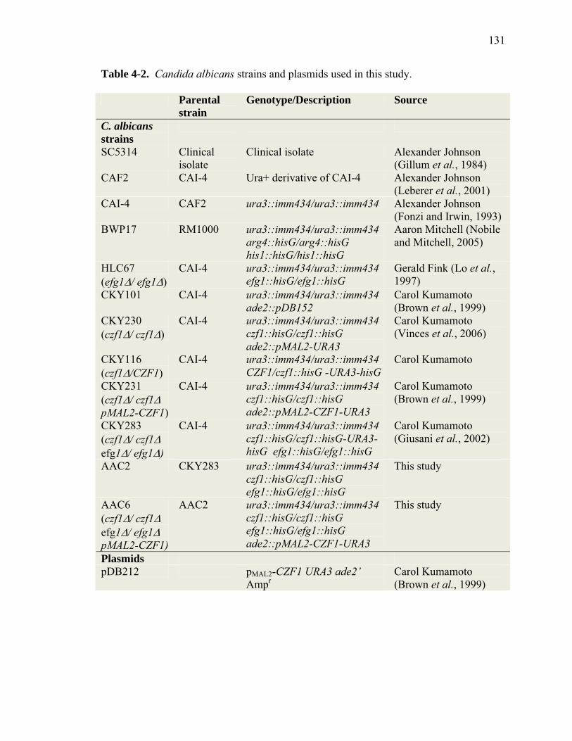
131
Table 4-2. Candida albicans strains and plasmids used in this study.
Parental strain
Genotype/Description Source
C. albicans strains
SC5314 Clinical isolate
Clinical isolate Alexander Johnson (Gillum et al., 1984)
CAF2 CAI-4 Ura+ derivative of CAI-4 Alexander Johnson (Leberer et al., 2001)
CAI-4 CAF2 ura3::imm434/ura3::imm434 Alexander Johnson (Fonzi and Irwin, 1993)
BWP17 RM1000 ura3::imm434/ura3::imm434 arg4::hisG/arg4::hisG his1::hisG/his1::hisG
Aaron Mitchell (Nobile and Mitchell, 2005)
HLC67 (efg1Δ/ efg1Δ)
CAI-4 ura3::imm434/ura3::imm434 efg1::hisG/efg1::hisG
Gerald Fink (Lo et al., 1997)
CKY101 CAI-4 ura3::imm434/ura3::imm434 ade2::pDB152
Carol Kumamoto (Brown et al., 1999)
CKY230 (czf1Δ/ czf1Δ)
CAI-4 ura3::imm434/ura3::imm434 czf1::hisG/czf1::hisG ade2::pMAL2-URA3
Carol Kumamoto (Vinces et al., 2006)
CKY116 (czf1Δ/CZF1)
CAI-4 ura3::imm434/ura3::imm434 CZF1/czf1::hisG -URA3-hisG
Carol Kumamoto
CKY231 (czf1Δ/ czf1Δ pMAL2-CZF1)
CAI-4 ura3::imm434/ura3::imm434 czf1::hisG/czf1::hisG ade2::pMAL2-CZF1-URA3
Carol Kumamoto (Brown et al., 1999)
CKY283 (czf1Δ/ czf1Δ efg1Δ/ efg1Δ)
CAI-4 ura3::imm434/ura3::imm434 czf1::hisG/czf1::hisG-URA3-hisG efg1::hisG/efg1::hisG
Carol Kumamoto (Giusani et al., 2002)
AAC2 CKY283 ura3::imm434/ura3::imm434 czf1::hisG/czf1::hisG efg1::hisG/efg1::hisG
This study
AAC6 (czf1Δ/ czf1Δ efg1Δ/ efg1Δ pMAL2-CZF1)
AAC2 ura3::imm434/ura3::imm434 czf1::hisG/czf1::hisG efg1::hisG/efg1::hisG ade2::pMAL2-CZF1-URA3
This study
Plasmids pDB212 pMAL2-CZF1 URA3 ade2’
Ampr Carol Kumamoto (Brown et al., 1999)

132
Supplementary Table 4-1. List of mutants screened in this study.
Mutant ORF# orf19.1795 orf19.7291 orf19.6760 orf19.4669 orf19.5037 orf19.11450 orf19.9364 orf19.6970 orf19.10248 orf19.9791 orf19.6952 orf19.9364 orf19.4966 orf19.7479 orf19.7412 orf19.5328 orf19.1857 orf19.8907 orf19.14178 orf19.4746 orf19.5235 orf19.3995 orf19.8635 orf19.3171 orf19.2723 orf19.768 orf19.3208 orf19.1409 orf19.564 orf19.856 orf19.11598 orf19.4284 orf19.4369 orf19.7389 orf19.4668 orf19.9508 orf19.5011 orf19.4658

133
orf19.1509 orf19.1593 orf19.695 orf19.6036 orf19.3014 orf19.3701 orf19.4763 orf19.5495 orf19.1805 orf19.13950 orf19.12265 orf19.5571 orf19.7614 orf19.10359 orf19.2990 orf19.4257 orf19.2237 orf19.771 orf19.6729 orf19.3396 orf19.2237 orf19.3563 orf19.2033 orf19.11973 orf19.3171 orf19.769 orf19.5251 orf19.7381 orf19.3202 orf19.1825 orf19.5571 orf19.580 orf19.3764 orf19.9081 orf19.5365 orf19.3012 orf19.12351 orf19.5445 orf19.6261 orf19.3009 orf19.10169

134
orf19.2763 orf19.2392 orf19.4893 orf19.2348 orf19.2653 orf19.5292 orf19.4457 orf19.6194 orf19.5094 orf19.271 orf19.1759 orf19.12603 orf19.6032 orf19.7201 orf19.9115 orf19.1005 orf19.4369 orf19.7389 orf19.4519 orf19.2660 orf19.4529 orf19.7472 orf19.6344 orf19.4428 orf19.1614 orf19.4369 orf19.3818 orf19.8837 orf19.11598 orf19.3906 orf19.7016 orf19.7400 orf19.9331 orf19.6185 orf19.3678 orf19.4535 orf19.7194 orf19.5001 orf19.4244 orf19.658 orf19.8326

135
orf19.3530 orf19.6194 orf19.6100 orf19.1510 orf19.4214 orf19.4285 orf19.3125 orf19.7208 orf19.5866 orf19.2938 orf19.9115 orf19.12706 orf19.6850 orf19.1252 orf19.5352 orf19.7451 orf19.7401 orf19.271 orf19.1394 orf19.2901 orf19.6737 orf19.811 orf19.2808 orf19.769 orf19.7207 orf19.530 orf19.4979 orf19.7381 orf19.7400 orf19.6950 orf19.14148 orf19.4409 orf19.4872 orf19.4513 orf19.6971 orf19.2270 orf19.3190 orf19.7410 orf19.4440 orf19.13704 orf19.6365

136
orf19.4116 orf19.3678 orf19.7381 orf19.3844 orf19.1331 orf19.1276 orf19.5761 orf19.7576 orf19.813 orf19.1907 orf19.4846 orf19.4543 orf19.4518 orf19.4823 orf19.7447 orf19.7324 orf19.2350 orf19.10841 orf19.5664 orf19.7610 orf19.248 orf19.425 orf19.4243 orf19.1628 orf19.3707 orf19.829 orf19.3254 orf19.1291 orf19.6365 orf19.4969 orf19.2061 orf19.1793 orf19.3122 orf19.5887 orf19.4188 orf19.4139 orf19.1795 orf19.5100 orf19.6267 orf19.7583 orf19.11659

137
orf19.7451 orf19.4474 orf19.3012 orf19.6036 orf19.4743 orf19.6763 orf19.4010 orf19.7207 orf19.2508 orf19.5016 orf19.5023 orf19.998 orf19.7355 orf19.814 orf19.411 19.6817 19.3809 19.7247 19.11410 19.4433 19.5908 19.7150 19.12215 19.12786 19.4972 19.173 19.4125 19.4766 19.3187 19.3986 19.6680 19.5026 19.4670 19.4318 19.7359 19.723 19.7518 19.2745 19.7583 19.5729 19.7570

138
19.3127 19.4568 19.1135 19.1187 19.5548 19.3308 19.2647 19.6182 19.431 19.7318 19.5380 19.1497 19.1255 19.3305 19.4573 19.9191 19.1007 19.2280 19.5338 19.4767 19.4776 19.4778 19.13396 19.2356 19.9326 19.909 19.2054 19.2260 19.798 19.889 19.2331 19.2315 19.1178 19.2393 19.2399 19.2458 19.3088 19.2612 19.2674 19.1358 19.1496

139
19.3193 19.3300 19.1589 19.1565 19.3405 19.3407 19.1694 19.1729 19.1826 19.3683 19.3753 19.3835 19.6781 19.6845 19.6850 19.7025 19.10266 19.10478 19.1227 19.1822 19.2064 19.2077 19.2432 19.2623 19.3390 19.4649 19.487 19.567 19.6985 19.718 19.735 19.976 orf19.3966 orf19.3966 orf19.299 orf19.299 orf19.4887 orf19.4887 orf19.2623 orf19.2623 orf19.1563

140
orf19.1563 orf19.1563 orf19.3010.1 orf19.3010.1 orf19.3010.1 orf19.4884 orf19.4884 orf19.3434 orf19.3434 orf19.2613 orf19.2613 orf19.2613 orf19.5412 orf19.4981 orf19.4981 orf19.4981 orf19.1277 orf19.1277 orf19.5867 orf19.5867 orf19.3869 orf19.3869 orf19.3869 orf19.1490 orf19.1490 orf19.1490 orf19.2476 orf19.2476 orf19.2476 orf19.5674 orf19.893 orf19.532 orf19.532 orf19.1714 orf19.3642 orf19.7251 orf19.3193 orf19.7374 orf19.166 orf19.2647 orf19.3188

141
orf19.1358 orf19.1499 orf19.6817 orf19.681 orf19.4662 orf19.5848 orf19.6407 orf19.5975 orf19.1032 orf19.2356 orf19.6985 orf19.7371 orf19.4766 orf19.3012 orf19.5133 orf19.5917 orf19.6203 orf19.861 orf19.6038 orf19.5849 orf19.4649 orf19.3294 orf19.2623 orf19.2315 orf19.1069 orf19.3190 orf19.6102 orf19.723 orf19.2540 orf19.3434 orf19.4961 orf19.5953 orf19.6124 orf19.173 orf19.6121 orf19.217 orf19.1035 orf19.1718 orf19.2423 orf19.255 orf19.2748

142
orf19.2753 orf19.2808 orf19.3252 orf19.3405 orf19.3876 orf19.4145 orf19.4166 orf19.4225 orf19.4288 orf19.4450 orf19.4524 orf19.5924 orf19.5940 orf19.7372 orf19.6227 orf19.6713 orf19.7319 orf19.5995 orf19.5759 orf19.4979 orf19.2395 orf19.6626 orf19.7523 orf19.1813 orf19.2217 orf19.702 orf19.7388 orf19.7388 orf19.896 orf19.896 orf19.7001 orf19.7001 orf19.4866 orf19.4866 orf19.5181 orf19.5181 orf19.4252 orf19.4252 orf19.4084 orf19.4084 orf19.469

143
orf19.469 orf19.5350 orf19.5350 orf19.2605 orf19.2605 orf19.5224 orf19.5224 orf19.663 orf19.663 orf19.451 orf19.451 orf19.1874 orf19.1874 orf19.4242 orf19.4242 orf19.2395 orf19.2395 orf19.3047 orf19.3047 orf19.794 orf19.794 orf19.4892 orf19.4892 orf19.5408 orf19.5408 orf19.223 orf19.223 orf19.3256 orf19.3256 orf19.1196 orf19.1196 orf19.835 orf19.835 orf19.469 orf19.469 orf19.4084 orf19.4084 orf19.2341 orf19.2341 orf19.35 orf19.35

144
orf19.4001 orf19.4001 orf19.4867 orf19.4867 orf19.7281 orf19.7281 orf19.3744 orf19.6369 orf19.4144 orf19.4269 orf19.4909 orf19.4909 orf19.4890 orf19.4890 orf19.4308 orf19.4308 orf19.4432 orf19.4432 orf19.3545 orf19.3545 orf19.7510 orf19.7510 orf19.2678 orf19.6913 orf19.6913 orf19.5068 orf19.5068 orf19.3854 orf19.3854 orf19.5357 orf19.4347 orf19.4347 orf19.7510 orf19.7510 orf19.5224 orf19.5224 orf19.3751 orf19.3049 orf19.7044 orf19.7044 orf19.5253

145
orf19.5253 orf19.2277 orf19.2277 orf19.7451 orf19.7451 orf19.3530 orf19.3530 orf19.4297 orf19.4297 orf19.4518 orf19.4518 orf19.2268 orf19.2102 orf19.3256 orf19.3256 orf19.844 orf19.844 orf19.3841 orf19.3841 orf19.3049 orf19.3049 orf19.2436 orf19.846 orf19.846 orf19.7355 orf19.3720 orf19.3720 orf19.2910 orf19.2910 orf19.4002 orf19.428 orf19.428 orf19.1341 orf19.3415 orf19.3415 orf19.3415 orf19.6243 orf19.7523 orf19.7523 orf19.6889 orf19.6889

146
orf19.5901 orf19.5901 orf19.1341 orf19.1341 orf19.130 orf19.130 orf19.2222 orf19.1283 orf19.1283 orf19.1283 orf19.1283 orf19.7510 orf19.7510 orf19.5911 orf19.5911 orf19.7164 orf19.7164 orf19.5325 orf19.5325 orf19.895 orf19.895 orf19.5162 orf19.5162 orf19.7652 orf19.7652 orf19.460 orf19.460

147
CHAPTER 5
Summary and Future Directions
With a brief excerpt from:
Langford, M.L., Atkin, A.L., and Nickerson, K.W. (2009) Cellular Interactions of
Farnesol, a Quorum Sensing Molecule Produced by Candida albicans. Future Microbiol.
4: 1353-62.

148
Summary
Farnesol has effects on all eukaryotic cells tested, and for many cell types, a low
level of farnesol is sufficient to inhibit growth or induce cell death. In contrast, C.
albicans has evolved mechanisms to produce and tolerate higher levels of farnesol and
use it as a quorum sensing molecule. A current challenge is to develop a unifying model
for the mode of action for farnesol in eukaryotes and elucidate the mechanisms used by
C. albicans to evade the adverse effects of farnesol and respond to farnesol as a signaling
molecule. In this study, we established environmental conditions in which C. albicans is
able to resist farnesol-mediated cell death: stationary phase cells provided with an energy
source. Conversely, we have defined conditions that stimulate sensitivity to farnesol in
C. albicans: energy-deprived, log phase cells (Chapter 2). Using the aforementioned
environmental conditions that allow C. albicans to tolerate farnesol and use it as a
signaling molecule, two novel factors were identified that are involved in the
morphological response to farnesol: Tup1 and Czf1 (Chapters 3, 4). It seems that
farnesol suppression of filamentation during C. albicans quorum sensing works through
both positive and negative regulators of filamentation (Czf1 and Tup1, respectively) to
achieve the resulting change in morphology. Czf1 also played a dual role in the farnesol
response, as it was partially responsible for farnesol tolerance in quorum sensing
conditions.
Farnesol Tolerance and Sensitivity in C. albicans
The precise mechanism for C. albicans white cell farnesol tolerance during
quorum sensing remains unclear. Since the effects of farnesol on other organisms have

149
been studied in seventeen different species (summarized in Chapter 1), this is an
important mechanism to understand in the field of interspecies communication.
Furthermore, as exogenous farnesol has many effects in the mammalian host during
infection such as preventing infection of mucosal surfaces, increasing virulence in a
disseminated infection and interference of the host cytokine response [Chapter 1 and
(Navarathna et al., 2007)], understanding the farnesol tolerance mechanism in C.
albicans may prove useful therapeutically.
It is unknown how C. albicans white cells are resistant to the toxic effects of
farnesol. Do the cells have a type of passive resistance, such as changes in membrane
composition, or do they undergo active resistance, such as conversion of farnesol into a
non-toxic compound? In order to look for genes that may be involved in farnesol
tolerance, a screening approach was used (Chapter 4). However, the screen was
unsuccessful in identifying mutants that were growth-inhibited (sensitive) in the presence
of farnesol, even at levels as low as 10 µM. Even so, the mutant library collection used
in this study is by no means a complete representation of the entire collection of C.
albicans predicted open reading frames (ORFs); there are 6197 predicted ORFs in the
genome, compared with the 560 mutants contained within the mutant library collection
used, leaving plenty of room for future identification of farnesol tolerance factors as more
mutants become available.
The identification of Czf1 and Efg1 as important players in farnesol survival is
intriguing because a farnesol-sensitive phenotype was observed only when both genes
were deleted. Ectopic expression of CZF1 in the efg1Δ/efg1Δ czf1Δ/czf1Δ double mutant
could not provide complete protection from farnesol killing yet the single efg1Δ/efg1Δ

150
and czf1Δ/czf1Δ mutants were unharmed (Chapter 4). Perhaps Czf1 functions upstream
of Efg1 in this situation, or the normal regulation of CZF1 might be especially important
in the absence of EFG1. In the reverse situation, could ectopic expression of EFG1 in the
double mutant restore partial or full farnesol resistance? Czf1 and Efg1 likely regulate a
redundant gene or set of genes for farnesol tolerance because the single mutants do not
show a farnesol sensitive phenotype while the double mutant exhibits a synthetic farnesol
sensitivity phenotype. While changes in gene expression have been extensively studied
in the efg1Δ/efg1Δ mutant compared to wild type/parental strains (Braun and Johnson,
2000; Harcus et al., 2004; Sohn et al., 2003), there is no matching data for Czf1
available. Microarray analyses designed to complement existing data on Efg1 may reveal
genes that are commonly regulated by Czf1 and Efg1, either directly or indirectly. Genes
whose expression was altered in cells lacking Czf1 and Efg1 could also be compared to
existing microarray data comparing gene expression between white and opaque cells
(Lan et al., 2002; Tsong et al., 2003; Zhao et al., 2005). Genes that are differentially
expressed in opaque cells, efg1Δ/efg1Δ, and czf1Δ/czf1Δ mutants would comprise a list of
strong candidates for potential farnesol resistance factors. One further step that may be
taken would be to look at other farnesol-sensitive organisms to see if homologs to the
candidate farnesol resistance genes are present in those species.
The role of temperature in farnesol tolerance (the efg1Δ/efg1Δ czf1Δ/czf1Δ double
mutant was more sensitive at 30°C than at 37°C) may be correlated to evidence
suggestive of slightly elevated farnesol production levels at higher temperatures (Hornby
et al., 2001). It would be informative to examine changes in gene expression and to
measure farnesol production in the efg1Δ/efg1Δ czf1Δ/czf1Δ double mutant at both 30°C

151
and 37°C, compared to wild type and parental strains. Differentially regulated genes may
be partly responsible for the cellular morphology and farnesol resistance changes
observed at these two temperatures. One would also predict that farnesol production may
be significantly lowered at 30°C based on the degree of farnesol sensitivity for the double
mutant cells grown at this temperature. Analysis of the efg1Δ/efg1Δ single mutant would
need to be included in this study; initial results using this mutant resulted in high
variation between replicates (M.L. Langford, data not shown) and further replicates will
need to be conducted to determine whether Efg1 plays a role in farnesol production.
Furthermore, a possible role for Tup1 in farnesol resistance should not be ignored. As
tup1Δ/tup1Δ mutants produced 17-fold more farnesol compared to wild type and parental
strains, these cells may have heightened abilities to tolerate farnesol. However, it will be
difficult to study higher tolerance to farnesol in these mutant cells since they are locked
in the filamentous morphology (unless secondary mutations are introduced), unlike the
efg1Δ/efg1Δ and czf1Δ/czf1Δ mutants. Preliminary experiments must first be conducted
to better understand the tup1Δ/tup1Δ farnesol resistance, such as a comparison of farnesol
tolerance between yeast, pseudohyphal, and hyphal cells. If the loss of Tup1 results in
higher farnesol tolerance, would a triple efg1Δ/czf1Δ/tup1Δ mutant have restored
resistance? What would the farnesol production levels in that mutant be? It is intriguing
that all three of these factors play a prominent role in white-opaque switching, farnesol
production and/or farnesol tolerance, as well as the morphological response to farnesol.
152
Signaling During the Morphological Response to Farnesol in C. albicans
Because the presence of a single farnesol receptor has still not been identified, it
remains a possibility that there may be multiple targets for farnesol in the cell. Two
models (network and central processor) have been proposed with regard to C. albicans
signaling in general (Braun and Johnson, 2000) and are worth considering when
examining the farnesol response. That study suggested C. albicans follows the network
signaling model, whereby many individual connections are formed between the different
regulatory pathways in the cell. This conclusion was reached when they observed that
several hyphal-specific genes are not regulated in unison in different hyphal inducing
conditions but in fact respond as individuals. If network signaling is indeed occurring
during the farnesol response, farnesol could help identify many of the different branches
in these pathways.
The Ras1-cAMP pathway has been identified as a critical, even a possible direct
target for farnesol inhibition during yeast to filament inhibition by farnesol (Davis-Hanna
et al., 2008; Deveau et al., 2010). Although a recent study (Davis-Hanna et al., 2008)
showed the inability of the ras1Δ/ras1Δ mutant to respond to farnesol in aerobic
conditions, it remains undetermined if it is capable of responding to farnesol under
different environmental conditions, specifically during embedded growth. The
ras1Δ/ras1Δ mutant is defective in filamentation under aerobic conditions and embedded
in YPS (yeast extract, peptone, sucrose) agar (Maidan et al., 2005), but it has not been
tested in our GPP embedded media. If filamentation can be achieved in our embedded
assay, the morphological response to farnesol can be tested; this would both complement

153
the existing aerobic data and determine the importance of Ras1 to the farnesol response in
other environmental conditions.
A recently characterized protein, Ras2 (Zhu et al., 2009), plays an antagonistic
role with Ras1 regarding stationary phase entry, stress resistance, and cellular cAMP
levels. Although Ras2 shares poor sequence homology with Ras1 and phylogenetic
analyses have suggested it belongs to a divergent group of fungal Ras proteins (Ras2 has
a homolog with high sequence similarity to C. dubliniensis), it still has functional
GTPase activity (Zhu et al., 2009). More importantly, since Ras2 has an antagonistic
effect on many of the physiological roles of Ras1, it may somehow be involved in
farnesol signaling as well. The ras2Δ/ras2Δ mutant maintains the ability to make
filaments in aerobic and embedded conditions, making it an ideal candidate for testing the
morphological response to farnesol. Might Ras2 play an antagonistic response to Ras1
during farnesol signaling as well?
While one new factor involved in the morphological response to farnesol, Czf1,
has a known connection to the Ras1-cAMP pathway in C. albicans, it remains unknown
how another factor involved, Tup1, fits into this model (Chapters 3, 4). Further studies
using farnesol as a tool may decipher whether Tup1 is somehow connected to the cAMP
pathway or is independently involved in the farnesol response. Construction of a triple
efg1Δ/czf1Δ/tup1Δ mutant may prove useful, not only to study farnesol tolerance as
mentioned earlier, but to study farnesol signaling as well. If this mutant is viable, what
would its morphology be in different growth conditions? Would this mutant be capable
of responding to farnesol? Vectors that can be used to delete EFG1, CZF1, and TUP1
have already been constructed, as well as ectopic expression constructs for all three genes

154
(Bockmuhl and Ernst, 2001; Braun and Johnson, 1997; Brown et al., 1999; Lo et al.,
1997), a couple of which have already been obtained by the Atkin laboratory. Therefore,
all the necessary tools are available to create not only a triple mutant, but the remaining
double mutant combinations (i.e. tup1Δ/czf1Δ, efg1Δ/tup1Δ) and ectopically express each
gene in the desired mutant strains. Construction and analysis of these strains can provide
valuable insight into C. albicans morphological signaling networks. For example, if
TUP1 was ectopically expressed in the efg1Δ/efg1Δ czf1Δ/czf1Δ double mutant
background and no differences in farnesol response were observed, Tup1 would most
likely be acting upstream of these two factors.
Other ways to test for a possible connection between Tup1 and the Ras1-cAMP
pathway would be to determine whether Czf1 and/or Efg1 are responsible for the increase
in TUP1 mRNA levels and/or Tup1 protein levels observed during the morphological
response to farnesol. For example, are the TUP1 mRNA levels in a czf1Δ/czf1Δ mutant
background still increased during farnesol treatment? If the mRNA levels do not increase
in response to farnesol, it would suggest that Czf1 is either directly or indirectly
responsible for the regulation of Tup1 during the farnesol response, and this information
could potentially provide the first data regarding the regulation of Tup1 since regulatory
factors upstream of Tup1 remain unknown.
Regardless of whether Czf1 would specifically regulate TUP1 expression, follow-
up experiments designed to better understand which genes are controlled by Czf1 are
desirable for the C. albicans research field. While it has been shown to bind its own
promoter and negatively regulate its expression, it is unknown what other genes are
directly regulated by Czf1. Moreover, because it controls so many processes in C.
155
albicans: morphogenesis, white-opaque switching, farnesol tolerance, and the
morphological response to farnesol, it is of considerable interest to identify genes that are
directly regulated by this unique transcription factor, both without and in the presence of
farnesol. ChIP (chromatin immunoprecipitation)-Seq, would be an ideal procedure to
determine direct targets of Czf1. Prior to this experiment, however, Czf1 antibodies
would ideally be raised (all protein work concerning Czf1 has utilized an HA-tagged
Czf1 protein, which would have the potential to slightly alter the natural function of the
protein). The ChIP-Seq method would first cross-link DNA (chromatin)-protein
complexes and shear the DNA into smaller pieces. An antibody against Czf1 can be used
to immunoprecipitate DNA bound with Czf1, and deep sequencing could subsequently be
used to sequence the precipitated DNA. Sequence analyses would finally provide the
location of the Czf1 binding sites in the C. albicans genome.
In addition to CZF1, the mutant screen from Chapter 4 identified 7 other genes
that may play a role in the farnesol response. Of note, identification of STP2 was a bit of
a surprise, as it is the only gene identified with no direct ties to morphogenesis or stress
responses. Either the acquisition of amino acids plays a more important role during
farnesol signaling than is currently appreciated, or Stp2 may participate in farnesol
signaling in an undiscovered manner. The RLM1 and YCK2 genes are both attractive
candidates for further analysis, particularly when examining the farnesol survival
mechanism, due to their known roles in stress resistance.

156
Final Thoughts
One common theme that can be discerned from studying the farnesol response in
C. albicans is that farnesol affects a variety of regulatory factors within the polymorphic
fungus. This unique feature has allowed us to study the interrelatedness of some of the
signaling networks in an effort to better understand the genetic pathways present in the
opportunistic pathogen. Farnesol can be used as an instrument to dissect the complicated
and intertwined pathways present in C. albicans, including pathways yet to be fully
understood, such as the regulation of Tup1, Hog1 signaling, and stress response
signaling. Although farnesol was initially discovered as a quorum sensing molecule that
only regulates morphogenesis, the discovery of its many other functions will hopefully
allow for a more complete understanding of C. albicans signaling as a whole.

157
References
Bockmuhl, D.P., and Ernst, J.F. (2001) A potential phosphorylation site for an A-type
kinase in the Efg1 regulator protein contributes to hyphal morphogenesis of
Candida albicans. Genetics 157: 1523-1530.
Braun, B.R., and Johnson, A.D. (1997) Control of filament formation in Candida
albicans by the transcriptional repressor TUP1. Science 277: 105-109.
Braun, B.R., and Johnson, A.D. (2000) TUP1, CPH1 and EFG1 make independent
contributions to filamentation in Candida albicans. Genetics 155: 57-67.
Brown, D.H., Jr., Giusani, A.D., Chen, X., and Kumamoto, C.A. (1999) Filamentous
growth of Candida albicans in response to physical environmental cues and its
regulation by the unique CZF1 gene. Mol Microbiol 34: 651-662.
Davis-Hanna, A., Piispanen, A.E., Stateva, L.I., and Hogan, D.A. (2008) Farnesol and
dodecanol effects on the Candida albicans Ras1-cAMP signalling pathway and
the regulation of morphogenesis. Mol Microbiol 67: 47-62.
Deveau, A., Piispanen, A.E., Jackson, A.A., and Hogan, D.A. (2010) Farnesol induces
hydrogen peroxide resistance in Candida albicans yeast by inhibiting the Ras-
cAMP signaling pathway. Eukaryot Cell Epub.
Harcus, D., Nantel, A., Marcil, A., Rigby, T., and Whiteway, M. (2004) Transcription
profiling of cyclic AMP signaling in Candida albicans. Mol Biol Cell 15: 4490-
4499.
Hornby, J.M., Jensen, E.C., Lisec, A.D., Tasto, J.J., Jahnke, B., Shoemaker, R., Dussault,
P., and Nickerson, K.W. (2001) Quorum sensing in the dimorphic fungus
158
Candida albicans is mediated by farnesol. Appl. Environ. Microbiol. 67: 2982-
2992.
Lan, C.Y., Newport, G., Murillo, L.A., Jones, T., Scherer, S., Davis, R.W., and Agabian,
N. (2002) Metabolic specialization associated with phenotypic switching in
Candida albicans. Proc Natl Acad Sci U S A 99: 14907-14912.
Lo, H.J., Kohler, J.R., DiDomenico, B., Loebenberg, D., Cacciapuoti, A., and Fink, G.R.
(1997) Nonfilamentous Candida albicans mutants are avirulent. Cell 90: 939-949.
Maidan, M.M., De Rop, L., Serneels, J., Exler, S., Rupp, S., Tournu, H., Thevelein, J.M.,
and Van Dijck, P. (2005) The G protein-coupled receptor Gpr1 and the Galpha
protein Gpa2 act through the cAMP-protein kinase A pathway to induce
morphogenesis in Candida albicans. Mol Biol Cell 16: 1971-1986.
Navarathna, D.H.M.L.P., Nickerson, K.W., Duhamel, G.E., Jerrels, T.R., and Petro, T.M.
(2007) Exogenous Farnesol Interferes with the Normal Progression of Cytokine
Expression during Candidiasis in a Mouse Model. Infect. Immun. 75: 4006-4011.
Sohn, K., Urban, C., Brunner, H., and Rupp, S. (2003) EFG1 is a major regulator of cell
wall dynamics in Candida albicans as revealed by DNA microarrays. Mol
Microbiol 47: 89-102.
Tsong, A.E., Miller, M.G., Raisner, R.M., and Johnson, A.D. (2003) Evolution of a
combinatorial transcriptional circuit: a case study in yeasts. Cell 115: 389-399.
Zhao, R., Daniels, K.J., Lockhart, S.R., Yeater, K.M., Hoyer, L.L., and Soll, D.R. (2005)
Unique aspects of gene expression during Candida albicans mating and possible
G(1) dependency. Eukaryot Cell 4: 1175-1190.

159
Zhu, Y., Fang, H.M., Wang, Y.M., Zeng, G.S., Zheng, X.D., and Wang, Y. (2009) Ras1
and Ras2 play antagonistic roles in regulating cellular cAMP level, stationary-
phase entry and stress response in Candida albicans. Mol Microbiol 74: 862-875.