fast uptake and long-lasting binding of methamphetamine in the human brain
TRANSCRIPT
NeuroImage 43 (2008) 756–763
Contents lists available at ScienceDirect
NeuroImage
j ourna l homepage: www.e lsev ie r.com/ locate /yn img
Fast uptake and long-lasting binding of methamphetamine in the human brain:Comparison with cocaine
Joanna S. Fowler a,b,c,⁎, Nora D. Volkow d,e, Jean Logan a, David Alexoff a, Frank Telang e, Gene-Jack Wang a,b,Christopher Wong a, Yeming Ma e, Aarti Kriplani f, Kith Pradhan g, David Schlyer a,f, Millard Jayne e,Barbara Hubbard a, Pauline Carter a, Donald Warner a, Payton King a, Colleen Shea a, Youwen Xu a,Lisa Muench e, Karen Apelskog a
a Brookhaven National Laboratory, Upton, NY 11973, USAb Mount Sinai School of Medicine, New York, NY 10029, USAc Department of Chemistry, State University of New York at Stony Brook, Stony Brook, NY 11794, USAd National Institute on Drug Abuse, Rockville, MD 20892, USAe National Institute on Alcohol Abuse and Alcoholism, Rockville, MD 20892, USAf Department of Biomedical Engineering, State University of New York at Stony Brook, Stony Brook, NY 11974, USAg Department of Applied Mathematics and Statistics, State University of New York at Stony Brook, Stony Brook, NY 11974, USA
⁎ Corresponding author. Medical Department, BldLaboratory, Upton, NY, USA. Fax: +1 631 344 5815.
E-mail address: [email protected] (J.S. Fowler).
1053-8119/$ – see front matter © 2008 Elsevier Inc. Alldoi:10.1016/j.neuroimage.2008.07.020
a b s t r a c t
a r t i c l e i n f oArticle history:
Methamphetamine is one of Received 12 April 2008Revised 21 June 2008Accepted 5 July 2008Available online 22 July 2008the most addictive and neurotoxic drugs of abuse. It produces large elevations inextracellular dopamine in the striatum through vesicular release and inhibition of the dopamine transporter.In the U.S. abuse prevalence varies by ethnicity with very low abuse among African Americans relative toCaucasians, differentiating it from cocaine where abuse rates are similar for the two groups. Here we reportthe first comparison of methamphetamine and cocaine pharmacokinetics in brain between Caucasians andAfrican Americans along with the measurement of dopamine transporter availability in striatum.Methamphetamine's uptake in brain was fast (peak uptake at 9 min) with accumulation in cortical andsubcortical brain regions and in white matter. Its clearance from brain was slow (except for white matterwhich did not clear over the 90 min) and there was no difference in pharmacokinetics between Caucasiansand African Americans. In contrast cocaine's brain uptake and clearance were both fast, distribution waspredominantly in striatum and uptake was higher in African Americans. Among individuals, those with thehighest striatal (but not cerebellar) methamphetamine accumulation also had the highest dopaminetransporter availability suggesting a relationship between METH exposure and DAT availability. Metham-phetamine's fast brain uptake is consistent with its highly reinforcing effects, its slow clearance with its long-lasting behavioral effects and its widespread distribution with its neurotoxic effects that affect not onlystriatal but also cortical and white matter regions. The absence of significant differences between Caucasiansand African Americans suggests that variables other than methamphetamine pharmacokinetics andbioavailability account for the lower abuse prevalence in African Americans.
© 2008 Elsevier Inc. All rights reserved.
Introduction
Methamphetamine (METH) is considered among the most addic-tive of the drugs of abuse. Its powerful addictive properties, coupledwith its broad availability have led to significant increases in both itsabuse and in the number of associated medical complications in manyareas of theworld (Rawson and Condon, 2007; Barr et al., 2006). In theUnited States the prevalence varies by region and ethnicity, being verylow among African Americans (Sexton et al., 2005; Iritani et al., 2007).This is in contrast to cocaine for which the prevalence rates for abuse
g 555, Brookhaven National
rights reserved.
and dependence are equivalent in Caucasians and African Americans(SAMHSA, 2003). Among the drugs of abuse, METH, which elevatesdopamine (DA) by both inhibiting its reuptake as well as releasing itfrom vesicles (Rothman et al., 2001), is one of the most potent. Itproduces very large increases in DA concentration in the nucleusaccumbens (NAc) (Camp et al., 1994), which is the mechanismbelieved to underlie the reinforcing effects of addictive drugs (DiChiara and Imperato, 1988). However, the reinforcing effects of drugsare not just a function of their potency in increasing DA in NAc but alsoof the rate at which they increase it; that is, the faster the increases,the stronger their reinforcing effects (Balster and Schuster, 1973). Wehave previously shown that cocaine (measured with [11C]cocaine)entered the brain very rapidly, which would explain why it is such areinforcing drug (Fowler et al., 1989). Moreover we corroborated a
757J.S. Fowler et al. / NeuroImage 43 (2008) 756–763
temporal correspondence between the fast uptake of the drug in brainand the subjective experience of the ‘high’ (Volkow et al., 1997).
Here we use PET and [11C]d-methamphetamine administered attracers doses to measure the pharmacokinetics and distribution ofMETH in the human brain. We also investigated whether differencesin the pharmacokinetics (PK) of METH in the brain could explain thelarge differences in the prevalence rate of abuse of METH betweenCaucasians and African Americans. We hypothesized, based on ourprior studies with [11C]d-methamphetamine in the baboon brain(Fowler et al., 2007), that METH uptake in the brainwould be very fastand similar to that of cocaine and that, as per our prior studies withstimulant drugs, its fast uptake would be associated with itsreinforcing effects (assessed by subjective descriptors of ‘high’)(Volkow et al., 1995). We also predicted that uptake of METH inbrain would be higher and faster in Caucasians than in AfricanAmericans. In this same group of subjects, we also measured cocainePK with [11C]cocaine for comparison with METH PK and to test thehypothesis that dopamine transporter (DAT) availability (assessedwith the distribution volume ratio (DVR) of striatum to cerebellumobtained from the [11C]cocaine images (Logan et al., 1990) wouldcontribute to the intersubject variability in METH uptake. Specificallywe predicted that the higher DATavailability would be associatedwithgreater METH uptake in brain.
Materials and methods
Volunteers
This study was carried out at Brookhaven National Laboratory andapproved by the local Institutional Review Board (Committee onResearch Involving Human Subjects, Stony Brook University). Writteninformed consentwas obtained from all of the participants prior to thestudy. Nineteen normal healthy men (9 Caucasians and 10 AfricanAmericans), average age 37±7 years (range 24–49) were recruited byword of mouth and newspaper advertisements and enrolled in thisstudy. Inclusion criteria were non-smoking males able to understandand give informed consent, age 18–50 years. Excluded were thosesubjects who were urine positive for psychoactive drugs (includingPCP, cocaine, amphetamine, opiates, barbiturates, benzodiazepinesand THC); who had clinically significant abnormal laboratory values;past or present history of medical illness or neurological or psychiatricdisease; use of antidepressant, antipsychotic, stimulants or chronicanxiolytic medications in the past one month; head trauma with lossof consciousness for more than 30 min; past or present or past historyof substance abuse including illicit substances, alcohol and nicotine.
Radiotracer synthesis
[11C]d-methamphetamine was prepared from D-amphetamine and[11C]methyl iodide using an adaptation of the literaturemethod (Inoueet al., 1990) as described previously (Fowler et al., 2007). Dynamic PETimaging was carried out on a Siemen's HR+ high resolution, wholebody PET scanner (4.5×4.5×4.8 mm FWHM at center of field of view)in 3D acquisition mode, 63 planes. For all scans, a transmission scanwas obtained with a 68Ge rotating rod source before the emission scanto correct for attenuation before each radiotracer injection. Thespecific activity of [11C]d-methamphetamine was 0.32±0.28 mCi/nmol at time of injection and the dose injected averaged 7.1±0.37 mCi). The radiochemical purity wasN98%. Scanning was carriedout for 90 min with the following time frames (1×10 s; 12×5 s;1×20 s; 1×30 s; 8×60 s; 4×300 s; 8×450 s).
[11C](−)-Cocainewas synthesized from nor-cocaine (NIDA ResearchTechnology Branch) according to the literature method (Fowler et al.,1989). Radiochemical purity wasN98% and specific activity was 0.53±0.36 mCi/nmol at end of synthesis and the dose injected averaged6.72±0.58 mCi. Scanning was carried out for 54 min with the
following time frames (1×10 s; 12×5 s; 1×20 s; 1×30 s; 4×60 s;4×120 s; 8×300 s).
Typically, the two studies were performed 2 h apart. For both [11C]d-methamphetamine and [11C]cocaine the concentration of parentradiotracer in the arterial plasma was measured (Fowler et al., 2007;Alexoff et al., 1995).
Image processing and data analysis
Time frames were summed over the experimental period (90 minfor [11C]d-methamphetamine and 54 min for [11C](−)-cocaine), thesummed images were resliced along the AC–PC line and planes weresummed in groups of two for the purpose of region of interest (roi)placement. For [11C]d-methamphetamine, roi's were placed on thecaudate, dorsal putamen, ventral striatum, thalamus and the frontal,temporal and parietal cortices, cingulate gyrus, cerebellum and whitematter and then projected onto the dynamic images. For the [11C]cocaine scans, roi's were placed over the caudate, putamen, thalamus,cerebellum and white matter and then projected onto the dynamicimages to obtain time–activity curves. Regions occurring bilaterallywere averaged. Carbon-11 concentration in each region of interest wasdivided by the injected dose to obtain the concentrations of C-11 vs.timewhich, along with the time course of the arterial concentration ofthe radiotracer, were used to calculate the DV's using a graphicalanalysis method for reversible systems (Logan et al., 1990). For [11C]cocaine we computed the ratio of the DV ratio (DVR) in caudate,putamen and thalamus to that in the cerebellum. The DVR-1 forputamen, which corresponds to Bmax/Kd, was used as an estimate ofDAT availability. The averaged time–activity curves for the putamen,thalamus, cerebellum and white matter were compared for [11C]methamphetamine and [11C]cocaine. For [11C]d-methamphetamine,the area under the time–activity curve (% injected dose/cm3 vs time)(AUC) in the ventral striatum (normalized to the AUC for the plasma)was also obtained.
In order to examine whether there were PK differences forCaucasians vs African Americans, we compared the followingparameters for [11C]d-methamphetamine and for [11C]cocaine: DV'sfor the caudate, putamen, thalamus and cerebellum; time to peak andthe % clearance at the end of study; areas under the time–activitycurve for plasma. For all subjects, correlation analyses were alsoperformed between [11C]d-methamphetamine uptake (AUC for theputamen), normalized to the AUC for the plasma and DAT availability(DVR-1) in the putamen as measured with [11C]cocaine (Logan et al.,1990). In order to compare the time course of the uptake to the timecourse of the ‘high’ reported previously (Newton et al., 2006), thetime–activity curve (% injected dose/cm3) in the ventral striatum wasdivided by the peak uptake value so that the values for each time pointcould be presented as % of peak. The time course for the ‘high’ for apharmacological dose of methamphetamine reported previously(Newton et al., 2006) was normalized to the peak value forcomparison to the C-11 uptake.
Comparisons between groups were made using Student's t-test(unpaired) and differences within individuals were compared withStudent's t-test (paired). Pearson product moment correlations wereused to assess the association between METH AUC in putamen andDAT availability.
Results
Distribution of and kinetics of METH and cocaine
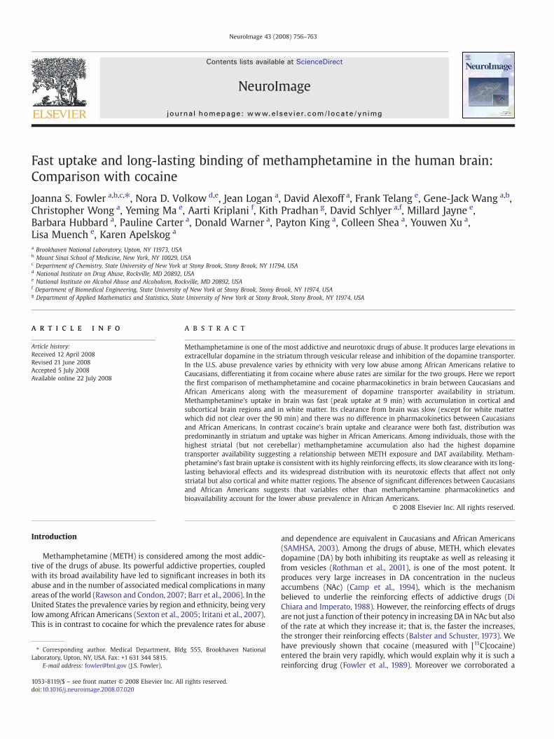
Carbon-11 was widely distributed to both cortical and subcorticalbrain regions after the intravenous administration of [11C]d-metham-phetamine in contrast to [11C]cocaine which was predominantlyconcentrated in the striatum (see Figs. 1A, B for averaged images andFig. 2 comparing distribution volumes (DV) for the two drugs in
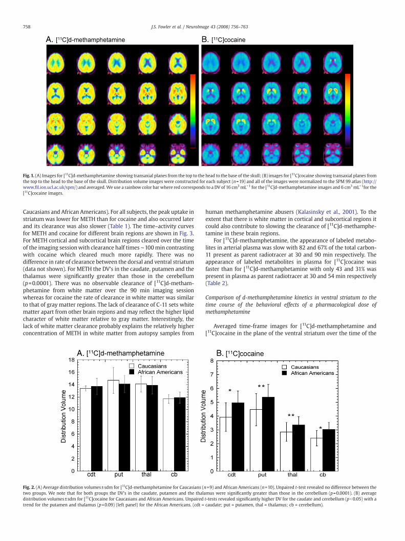
Fig.1. (A) Images for [11C]d-methamphetamine showing transaxial planes from the top to the head to the base of the skull; (B) images for [11C]cocaine showing transaxial planes fromthe top to the head to the base of the skull. Distribution volume images were constructed for each subject (n=19) and all of the images were normalized to the SPM 99 atlas (http://www.fil.ion.ucl.ac.uk/spm/) and averaged.We use a rainbow color bar where red corresponds to a DV of 16 cm3mL−1 for the [11C]d-methamphetamine images and 6 cm3mL−1for the[11C]cocaine images.
758 J.S. Fowler et al. / NeuroImage 43 (2008) 756–763
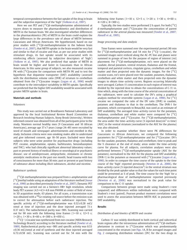
Caucasians and African Americans). For all subjects, the peak uptake instriatumwas lower for METH than for cocaine and also occurred laterand its clearance was also slower (Table 1). The time–activity curvesfor METH and cocaine for different brain regions are shown in Fig. 3.For METH cortical and subcortical brain regions cleared over the timeof the imaging sessionwith clearance half times ∼100min contrastingwith cocaine which cleared much more rapidly. There was nodifference in rate of clearance between the dorsal and ventral striatum(data not shown). For METH the DV's in the caudate, putamen and thethalamus were significantly greater than those in the cerebellum(p=0.0001). There was no observable clearance of [11C]d-metham-phetamine from white matter over the 90 min imaging sessionwhereas for cocaine the rate of clearance in white matter was similarto that of gray matter regions. The lack of clearance of C-11 sets whitematter apart from other brain regions and may reflect the higher lipidcharacter of white matter relative to gray matter. Interestingly, thelack of white matter clearance probably explains the relatively higherconcentration of METH in white matter from autopsy samples from
Fig. 2. (A) Average distribution volumes±sdm for [11C]d-methamphetamine for Caucasians (two groups. We note that for both groups the DV's in the caudate, putamen and the thaladistribution volumes±sdm for [11C]cocaine for Caucasians and African Americans. Unpairedtrend for the putamen and thalamus (p=0.09) (left panel) for the African Americans. (cdt =
human methamphetamine abusers (Kalasinsky et al., 2001). To theextent that there is white matter in cortical and subcortical regions itcould also contribute to slowing the clearance of [11C]d-methamphe-tamine in these brain regions.
For [11C]d-methamphetamine, the appearance of labeled metabo-lites in arterial plasma was slow with 82 and 67% of the total carbon-11 present as parent radiotracer at 30 and 90 min respectively. Theappearance of labeled metabolites in plasma for [11C]cocaine wasfaster than for [11C]d-methamphetamine with only 43 and 31% waspresent in plasma as parent radiotracer at 30 and 54 min respectively(Table 2).
Comparison of d-methamphetamine kinetics in ventral striatum to thetime course of the behavioral effects of a pharmacological dose ofmethamphetamine
Averaged time-frame images for [11C]d-methamphetamine and[11C]cocaine in the plane of the ventral striatum over the time of the
n=9) and African Americans (n=10). Unpaired t-test revealed no difference between themus were significantly greater than those in the cerebellum (p=0.0001). (B) averaget-tests revealed significantly higher DV for the caudate and cerebellum (pb0.05) with acaudate; put = putamen, thal = thalamus; cb = cerebellum).
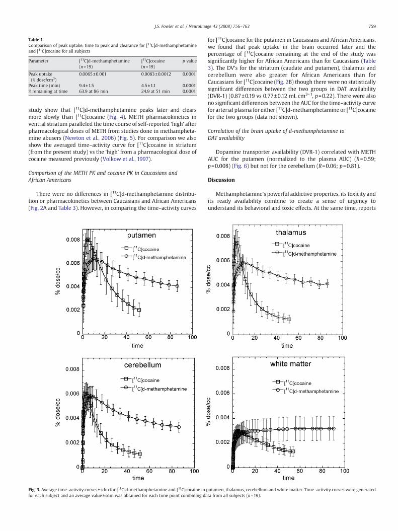
Table 1Comparison of peak uptake, time to peak and clearance for [11C]d-methamphetamineand [11C]cocaine for all subjects
Parameter [11C]d-methamphetamine(n=19)
[11C]cocaine(n=19)
p value
Peak uptake(% dose/cm3)
0.0065±0.001 0.0083±0.0012 0.0001
Peak time (min) 9.4±1.5 4.5±1.1 0.0001% remaining at time 63.9 at 86 min 24.9 at 51 min 0.0001
759J.S. Fowler et al. / NeuroImage 43 (2008) 756–763
study show that [11C]d-methamphetamine peaks later and clearsmore slowly than [11C]cocaine (Fig. 4). METH pharmacokinetics inventral striatum paralleled the time course of self-reported ‘high’ afterpharmacological doses of METH from studies done in methampheta-mine abusers (Newton et al., 2006) (Fig. 5). For comparison we alsoshow the averaged time–activity curve for [11C]cocaine in striatum(from the present study) vs the ‘high’ from a pharmacological dose ofcocaine measured previously (Volkow et al., 1997).
Comparison of the METH PK and cocaine PK in Caucasians andAfrican Americans
There were no differences in [11C]d-methamphetamine distribu-tion or pharmacokinetics between Caucasians and African Americans(Fig. 2A and Table 3). However, in comparing the time–activity curves
Fig. 3. Average time–activity curves±sdm for [11C]d-methamphetamine and [11C]cocaine in pfor each subject and an average value±sdm was obtained for each time point combining da
for [11C]cocaine for the putamen in Caucasians and African Americans,we found that peak uptake in the brain occurred later and thepercentage of [11C]cocaine remaining at the end of the study wassignificantly higher for African Americans than for Caucasians (Table3). The DV's for the striatum (caudate and putamen), thalamus andcerebellum were also greater for African Americans than forCaucasians for [11C]cocaine (Fig. 2B) though there were no statisticallysignificant differences between the two groups in DAT availability(DVR-1) (0.87±0.19 vs 0.77±0.12 mL cm3−1, p=0.22). There were alsono significant differences between the AUC for the time–activity curvefor arterial plasma for either [11C]d-methamphetamine or [11C]cocainefor the two groups (data not shown).
Correlation of the brain uptake of d-methamphetamine toDAT availability
Dopamine transporter availability (DVR-1) correlated with METHAUC for the putamen (normalized to the plasma AUC) (R=0.59;p=0.008) (Fig. 6) but not for the cerebellum (R=0.06; p=0.81).
Discussion
Methamphetamine's powerful addictive properties, its toxicity andits ready availability combine to create a sense of urgency tounderstand its behavioral and toxic effects. At the same time, reports
utamen, thalamus, cerebellum and white matter. Time–activity curves were generatedta from all subjects (n=19).
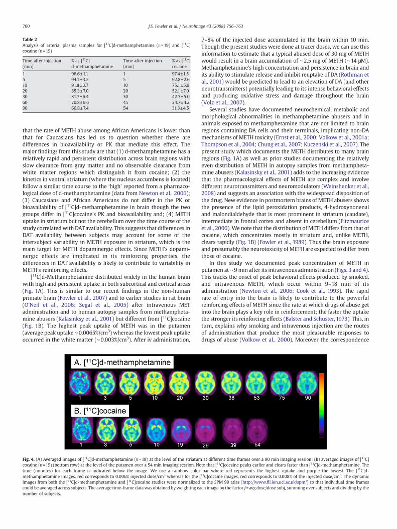
Table 2Analysis of arterial plasma samples for [11C]d-methamphetamine (n=19) and [11C]cocaine (n=19)
Time after injection(min)
% as [11C]d-methamphetamine
Time after injection(min)
% as [11C]cocaine
1 96.6±1.1 1 97.4±1.55 94.1±3.2 5 92.8±2.610 91.8±3.7 10 75.1±5.920 85.3±7.0 20 52.1±7.030 81.7±6.4 30 42.7±5.060 70.8±9.6 45 34.7±4.290 66.8±7.4 54 31.3±4.5
760 J.S. Fowler et al. / NeuroImage 43 (2008) 756–763
that the rate of METH abuse among African Americans is lower thanthat for Caucasians has led us to question whether there aredifferences in bioavailability or PK that mediate this effect. Themajor findings from this study are that (1) d-methamphetamine has arelatively rapid and persistent distribution across brain regions withslow clearance from gray matter and no observable clearance fromwhite matter regions which distinguish it from cocaine; (2) thekinetics in ventral striatum (where the nucleus accumbens is located)follow a similar time course to the ‘high’ reported from a pharmaco-logical dose of d-methamphetamine (data from Newton et al., 2006);(3) Caucasians and African Americans do not differ in the PK orbioavailability of [11C]d-methamphetamine in brain though the twogroups differ in [11C]cocaine's PK and bioavailability and; (4) METHuptake in striatum but not the cerebellum over the time course of thestudy correlated with DATavailability. This suggests that differences inDAT availability between subjects may account for some of theintersubject variability in METH exposure in striatum, which is themain target for METH dopaminergic effects. Since METH's dopami-nergic effects are implicated in its reinforcing properties, thedifferences in DAT availability is likely to contribute to variability inMETH's reinforcing effects.
[11C]d-Methamphetamine distributed widely in the human brainwith high and persistent uptake in both subcortical and cortical areas(Fig. 1A). This is similar to our recent findings in the non-humanprimate brain (Fowler et al., 2007) and to earlier studies in rat brain(O’Neil et al., 2006; Segal et al., 2005) after intravenous METadministration and to human autopsy samples from methampheta-mine abusers (Kalasinksy et al., 2001) but different from [11C]cocaine(Fig. 1B). The highest peak uptake of METH was in the putamen(average peak uptake ∼0.0065%/cm3) whereas the lowest peak uptakeoccurred in the white matter (∼0.003%/cm3). After iv administration,
Fig. 4. (A) Averaged images of [11C]d-methamphetamine (n=19) at the level of the striatumcocaine (n=19) (bottom row) at the level of the putamen over a 54 min imaging session. Notime (minutes) for each frame is indicated below the image. We use a rainbow colormethamphetamine images, red corresponds to 0.006% injected dose/cm3 whereas for the [images from both the [11C]d-methamphetamine and [11C]cocaine studies were normalizedcould be averaged across subjects. The average time-frame datawas obtained byweighting eanumber of subjects.
7–8% of the injected dose accumulated in the brain within 10 min.Though the present studies were done at tracer doses, we can use thisinformation to estimate that a typical abused dose of 30 mg of METHwould result in a brain accumulation of ∼2.5 mg of METH (∼14 μM).Methamphetamine's high concentration and persistence in brain andits ability to stimulate release and inhibit reuptake of DA (Rothman etal., 2001) would be predicted to lead to an elevation of DA (and otherneurotransmitters) potentially leading to its intense behavioral effectsand producing oxidative stress and damage throughout the brain(Volz et al., 2007).
Several studies have documented neurochemical, metabolic andmorphological abnormalities in methamphetamine abusers and inanimals exposed to methamphetamine that are not limited to brainregions containing DA cells and their terminals, implicating non-DAmechanisms of METH toxicity (Ernst et al., 2000; Volkow et al., 2001a;Thompson et al., 2004; Chung et al., 2007; Kuczenski et al., 2007). Thepresent study which documents the METH distributes to many brainregions (Fig. 1A) as well as prior studies documenting the relativelyeven distribution of METH in autopsy samples from methampheta-mine abusers (Kalasinsky et al., 2001) adds to the increasing evidencethat the pharmacological effects of METH are complex and involvedifferent neurotransmitters and neuromodulators (Weinshenker et al.,2008) and suggests an association with the widespread disposition ofthe drug. New evidence in postmortem brains of METH abusers showsthe presence of the lipid peroxidation products, 4-hydroxynonenaland malondialdehyde that is most prominent in striatum (caudate),intermediate in frontal cortex and absent in cerebellum (Fitzmauriceet al., 2006). We note that the distribution of METH differs from that ofcocaine, which concentrates mostly in striatum and, unlike METH,clears rapidly (Fig. 1B) (Fowler et al., 1989). Thus the brain exposureand presumably the neurotoxicity of METH are expected to differ fromthose of cocaine.
In this study we documented peak concentration of METH inputamen at ∼9min after its intravenous administration (Figs. 3 and 4).This tracks the onset of peak behavioral effects produced by smoked,and intravenous METH, which occur within 9–18 min of itsadministration (Newton et al., 2006; Cook et al., 1993). The rapidrate of entry into the brain is likely to contribute to the powerfulreinforcing effects of METH since the rate at which drugs of abuse getinto the brain plays a key role in reinforcement; the faster the uptakethe stronger its reinforcing effects (Balster and Schuster, 1973). This, inturn, explains why smoking and intravenous injection are the routesof administration that produce the most pleasurable responses todrugs of abuse (Volkow et al., 2000). Moreover the correspondence
at different time frames over a 90 min imaging session; (B) averaged images of [11C]te that [11C]cocaine peaks earlier and clears faster than [11C]d-methamphetamine. Thebar where red represents the highest uptake and purple the lowest. The [11C]d-
11C]cocaine images, red corresponds to 0.008% of the injected dose/cm3. The dynamicto the SPM 99 atlas (http://www.fil.ion.ucl.ac.uk/spm/) so that individual time framesch image by the factor f=avg dose/dose subj, summing over subjects and dividing by the
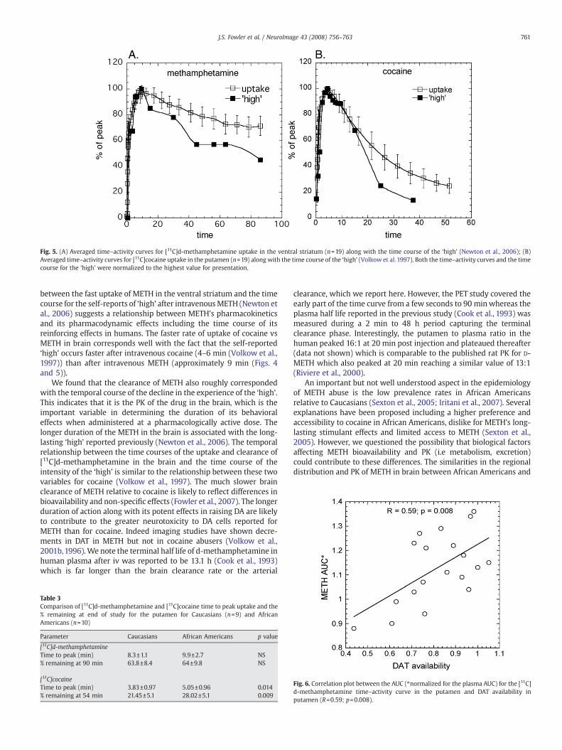
Fig. 5. (A) Averaged time–activity curves for [11C]d-methamphetamine uptake in the ventral striatum (n=19) along with the time course of the ‘high’ (Newton et al., 2006); (B)Averaged time–activity curves for [11C]cocaine uptake in the putamen (n=19) along with the time course of the ‘high’ (Volkow et al. 1997). Both the time–activity curves and the timecourse for the ‘high’ were normalized to the highest value for presentation.
761J.S. Fowler et al. / NeuroImage 43 (2008) 756–763
between the fast uptake of METH in the ventral striatum and the timecourse for the self-reports of ‘high’ after intravenousMETH (Newton etal., 2006) suggests a relationship between METH's pharmacokineticsand its pharmacodynamic effects including the time course of itsreinforcing effects in humans. The faster rate of uptake of cocaine vsMETH in brain corresponds well with the fact that the self-reported‘high’ occurs faster after intravenous cocaine (4–6 min (Volkow et al.,1997)) than after intravenous METH (approximately 9 min (Figs. 4and 5)).
We found that the clearance of METH also roughly correspondedwith the temporal course of the decline in the experience of the ‘high’.This indicates that it is the PK of the drug in the brain, which is theimportant variable in determining the duration of its behavioraleffects when administered at a pharmacologically active dose. Thelonger duration of the METH in the brain is associated with the long-lasting ‘high’ reported previously (Newton et al., 2006). The temporalrelationship between the time courses of the uptake and clearance of[11C]d-methamphetamine in the brain and the time course of theintensity of the ‘high’ is similar to the relationship between these twovariables for cocaine (Volkow et al., 1997). The much slower brainclearance of METH relative to cocaine is likely to reflect differences inbioavailability and non-specific effects (Fowler et al., 2007). The longerduration of action along with its potent effects in raising DA are likelyto contribute to the greater neurotoxicity to DA cells reported forMETH than for cocaine. Indeed imaging studies have shown decre-ments in DAT in METH but not in cocaine abusers (Volkow et al.,2001b,1996).We note the terminal half life of d-methamphetamine inhuman plasma after iv was reported to be 13.1 h (Cook et al., 1993)which is far longer than the brain clearance rate or the arterial
Table 3Comparison of [11C]d-methamphetamine and [11C]cocaine time to peak uptake and the% remaining at end of study for the putamen for Caucasians (n=9) and AfricanAmericans (n=10)
Parameter Caucasians African Americans p value
[11C]d-methamphetamineTime to peak (min) 8.3±1.1 9.9±2.7 NS% remaining at 90 min 63.8±8.4 64±9.8 NS
[11C]cocaineTime to peak (min) 3.83±0.97 5.05±0.96 0.014% remaining at 54 min 21.45±5.1 28.02±5.1 0.009
clearance, which we report here. However, the PET study covered theearly part of the time curve from a few seconds to 90 minwhereas theplasma half life reported in the previous study (Cook et al., 1993) wasmeasured during a 2 min to 48 h period capturing the terminalclearance phase. Interestingly, the putamen to plasma ratio in thehuman peaked 16:1 at 20 min post injection and plateaued thereafter(data not shown) which is comparable to the published rat PK for D-METH which also peaked at 20 min reaching a similar value of 13:1(Riviere et al., 2000).
An important but not well understood aspect in the epidemiologyof METH abuse is the low prevalence rates in African Americansrelative to Caucasians (Sexton et al., 2005; Iritani et al., 2007). Severalexplanations have been proposed including a higher preference andaccessibility to cocaine in African Americans, dislike for METH's long-lasting stimulant effects and limited access to METH (Sexton et al.,2005). However, we questioned the possibility that biological factorsaffecting METH bioavailability and PK (i.e metabolism, excretion)could contribute to these differences. The similarities in the regionaldistribution and PK of METH in brain between African Americans and
Fig. 6. Correlation plot between the AUC (⁎normalized for the plasma AUC) for the [11C]d-methamphetamine time–activity curve in the putamen and DAT availability inputamen (R=0.59; p=0.008).

762 J.S. Fowler et al. / NeuroImage 43 (2008) 756–763
Caucasians (Fig. 2A; Table 3) indicates that factors other thandifferences in bioavailability underlie the lower use of METH amongAfrican Americans. In contrast, there were significant differencesbetween the ethnic groups for cocaine; cocaine peaked later andcleared more slowly in African Americans than in Caucasians andbrain distribution volumes (all regions) for cocaine were also higherfor African Americans (Table 3 and Fig. 2B). Although we do not knowwhether there is any clinical significance due to these differences, thissurprising observation raises the question of whether differences inbioavailability between Caucasians and African Americans have anyepidemiological or clinical manifestation. It also merits furtherinvestigation in a larger group of subjects and highlights theimportance of considering and reporting ethnicity as a variable inclinical research studies and in matching ethnicity between controland experimental subjects.
The heterozygous deletion of DAT attenuates the behavioral effectsof METH (Fukushima et al., 2007) suggesting that DAT (as well asVMAT2) play a role in its neurotoxicity (Fumagalli et al., 1998, 1999).Given the role of the DAT in the behavioral effects of METH andassuming that higher METH exposure is an important variable in thebehavioral effects of the drug, we examined whether there would bean association between DAT availability (as measured using the DVR-1with [11C]cocaine) andMETH exposure using the area under the time–activity curve (AUC) for the putamen as the measure of METHexposure. Interestingly, the AUC varied by almost 2-fold for allindividuals. Subjects with the highest AUC for METH in the putamenalso had the highest DAT availability (Fig. 6). We note that the AUC forthe cerebellum for METH does not correlate with DAT availabilitysuggesting a specific association with striatum and not a global effect.These data suggest that individual DAT availability in striatum mayplay a role in the variability in individual's METH exposure.
There are potential study limitations that need to be addressed. Forexample, we measured METH PK at tracer doses whereas METHabusers use the drug at a typical dose of 0.5 mg/kg. This raises aquestion as towhether the PK of METHmeasuredwith a tracer dose of[11C]d-methamphetamine mimics the PK of a pharmacological dosethat has behavioral effects. However, there is evidence that this is avalid assumption based on the fact the PK of [11C]d-methampheta-mine in the baboon at tracer and at pharmacological doses did notdiffer (Fowler et al., 2007). For this study we also studied healthy, non-abusing controls rather than METH abusers. We felt that it wasimportant to investigate these relationships in a control, non-abusingpopulation to avoid introducing other variables such as the structuraland neurochemical abnormalities (including long-lasting decreases inDAT), which are known to occur in the METH abuser (Volkow et al.,2001b). We note that this study methodology could be adapted to theMETH abuser after an adequate washout period to assure that the DATavailability measurement would not be influenced by METH occu-pancy of the DAT.
Another issue that needs to be addressed is the extent towhich theC-11 in the brain reflects METH and not labeled metabolites or acombination of [11C]d-methamphetamine and its labeled metabolites.We analyzed the arterial plasma of each subject for the percent of thetotal C-11 that was in the form of the parent compound. We note thatthe appearance of labeled metabolites in plasma is slow so that inputto the brain is mostly [11C]d-methamphetamine (Table 2). In addition,a major metabolite of METH is amphetamine, which arises from N-demethylation. Since [11C]d-methamphetamine is labeled in the N-methyl group, amphetamine would not be detected. Similarly, for [N-11C-methyl]cocaine, the only labeled molecule that can penetrate thebrain is [11C]cocaine (Fowler et al., 1989).
In summary, in the first study of METH PK in the normal humanbrain, we found widespread and long-lasting distribution of METH,which parallels its long-lasting behavioral effects. Widespreaddistribution of METH in brain is also consistent with reports thatMETH's effects on brain chemistry and structure go beyond brain
regions highly innervated with DA (i.e parietal cortex, white matter).Contrary to our original hypothesis, we found no difference in METHPK and bioavailability between Caucasians and African Americanssuggesting that other variables need to be considered in accountingfor the lower rate of METH abuse in African Americans. However,these comparative studies also revealed significant differencesbetween Caucasians and African Americans in [11C]cocaine PK andbioavailability in brain which merit further investigation. Our findingthat individuals with the highest striatal METH exposure also had thehighest DAT availability suggests that DAT levels regulate the brainuptake of METH.
Acknowledgments
This research was carried out at Brookhaven National Laboratoryunder contract DE-AC02-98CH10886 with the U. S. Department ofEnergy and supported by its Office of Biological and EnvironmentalResearch and byNIH K05DA020001, the NIAAA Intramural program byGCRC grant #MO1RR10710. We are grateful to Richard Ferrieri andMichael Schueller for cyclotron and laboratory operations and to AnatBiegon for helpful discussions. We also thank the individuals whovolunteered for these studies.
References
Alexoff, D.L., Shea, C., Fowler, J.S., King, P., Gatley, S.J., Schlyer, D.J., Wolf, A.P., 1995.Plasma input function determination for PET using a commercial laboratory robot.Nucl. Med. Biol. 22, 893–904.
Balster, R.L., Schuster, C.R., 1973. Fixed-interval schedule of cocaine reinforcement:effect of dose and infusion duration. J. Exp. Anal. Behav. 20, 119–129.
Barr, A.M., Panenka, W.J., MacEwan, G.W., Thornton, A.E., Lang, D.J., Honer, W.G.,Lecomte, T., 2006. The need for speed: an update onmethamphetamine addiction. J.Psychiatry Neurosci. 31, 301–313 Review.
Camp, D.M., Browman, K.E., Robinson, T.E., 1994. The effects of methamphetamine andcocaine on motor behavior and extracellular dopamine in the ventral striatum ofLewis versus Fischer 344 rats. Brain Res. 668, 180–193.
Chung, A., Lyoo, I.K., Kim, S.J., Hwang, J., Bae, S.C., Sung, Y.H., Sim, M.E., Song, I.C., Kim, J.,Chang, K.H., Renshaw, P.F., 2007. Decreased frontal white-matter integrity inabstinent methamphetamine abusers. Int. J. Neuropsychopharmacol. 10, 765–775.
Cook, C.E., Jeffcoat, A.R., Hill, J.M., Pugh, D.E., Patetta, P.K., Sadler, B.M., et al., 1993.Pharmacokinetics of methamphetamine self-administered to human subjects bysmoking S-(+)-methamphetamine hydrochloride. Drug Metab. Dispos. 21, 717–723.
Di Chiara, G., Imperato, A., 1988. Drugs abused by humans preferentially increasesynaptic dopamine concentrations in the mesolimbic system of freely moving rats.Proc. Natl. Acad. Sci. U. S. A. 85, 5274–5278.
Ernst, T., Chang, L., Leonido-Yee, M., Speck, O., 2000. Evidence for long-term neuro-toxicity associated with methamphetamine abuse: a 1H MRS study. Neurology 54,1344–1349.
Fitzmaurice, P.S., Tong, J., Yazdanpanah, M., Liu, P.P., Kalasinsky, K.S., Kish, S.J., 2006.Levels of 4-hydroxynonenal and malondialdehyde are increased in brain of humanchronic users of methamphetamine. J. Pharmacol. Exp. Ther. 319, 703–709.
Fowler, J.S., Volkow, N.D., Wolf, A.P., Dewey, S.L., Schlyer, D.J., Macgregor, R.R.,Hitzemann, R., Logan, J., Bendriem, B., Gatley, S.J., et al., 1989. Mapping cocainebinding sites in human and baboon brain in vivo. Synapse 4, 371–377.
Fowler, J.S., Kroll, C., Ferrieri, R., Alexoff, D., Logan, J., Dewey, S.L., Schiffer, W., Schlyer, D.,Carter, P., King, P., Shea, C., Xu, Y., Muench, L., Benveniste, H., Vaska, P., Volkow, N.D.,2007. PET studies of d-methamphetamine pharmacokinetics in primates: compar-ison with L-methamphetamine and (−)-cocaine. J. Nucl. Med. 48, 1724–1732.
Fukushima, S., Shen, H., Hata, H., Ohara, A., Ohmi, K., Ikeda, K., Numachi, Y., Kobayashi, H.,Hall, F.S., Uhl, G.R., Sora, I., 2007.Methamphetamine-induced locomotor activity andsensitization in dopamine transporter and vesicular monoamine transporter 2double mutant mice. Psychopharmacology (Berl) 193, 55–62.
Fumagalli, F., Gainetdinov, R.R., Valenzano, K.J., Caron, M.G., 1998. Role of dopaminetransporter in methamphetamine-induced neurotoxicity: evidence from micelacking the transporter. J. Neurosci. 18, 4861–4869.
Fumagalli, F., Gainetdinov, R.R., Wang, Y.M., Valenzano, K.J., Miller, G.W., Caron, M.G.,1999. Increased methamphetamine neurotoxicity in heterozygous vesicularmonoamine transporter 2 knock-out mice. J. Neurosci. 19, 2424–2431.
Inoue, O., Axelsson, S., Lundqvist, H., Oreland, L., Långström, B., 1990. Effect of reserpineon the brain uptake of carbon 11methamphetamine and its N-propargyl derivative,deprenyl. Eur. J. Nucl. Med. 17, 121–126.
Iritani, B.J., Hallfors, D.D., Bauer, D.J., 2007. Crystal methamphetamine use among youngadults in the USA. Addiction 102, 1102–1113.
Kalasinsky, K.S., Bosy, T.Z., Schmunk, G.A., Reiber, G., Anthony, R.M., Furukawa, Y., Guttman,M., Kish, S.J., 2001. Regional distribution of methamphetamine in autopsied brain ofchronic human methamphetamine users. Forensic. Sci. Int. 116, 163–169.
Kuczenski, R., Everall, I.P., Crews, L., Adame, A., Grant, I., Masliah, E., 2007. Escalatingdose-multiple binge methamphetamine exposure results in degeneration of theneocortex and limbic system in the rat. Exp. Neurol. 207, 42–51.
763J.S. Fowler et al. / NeuroImage 43 (2008) 756–763
Logan, J., Fowler, J.S., Volkow, N.D.,Wolf, A.P., Dewey, S.L., Schlyer, D.J., MacGregor, R.R.,Hitzemann, R., Bendriem, B., Gatley, S.J., et al., 1990. Graphical analysis ofreversible radioligand binding from time-activity measurements applied to [N-11C-methyl]-(−)-cocaine PET studies in human subjects. J. Cereb. Blood FlowMetab. 10, 740–747.
Newton, T.F., Roache, J.D., De La Garza 2nd, R., Fong, T., Wallace, C.L., Li, S.H., Elkashef, A.,Chiang, N., Kahn, R., 2006. Bupropion reduces methamphetamine-inducedsubjective effects and cue-induced craving. Neuropsychopharmacology 31,1537–1544.
O'Neil, M.L., Kuczenski, R., Segal, D.S., Cho, A.K., Lacan, G., Melega, W.P., 2006.Escalating dose pretreatment induces pharmacodynamic and not pharmacoki-netic tolerance to a subsequent high-dose methamphetamine binge. Synapse 60,465–473.
Rawson, R.A., Condon, T.P., 2007. Why do we need an addiction supplement focused onmethamphetamine? Addiction 102 (Suppl. 1), 1–4.
Riviere, G.J., Gentry, W.B., Owens, S.M., 2000. Disposition of methamphetamine and itsmetabolite amphetamine in brain and other tissues in rats after intravenousadministration. J. Pharmacol. Exp. Ther. 292, 1042–1047.
Rothman, R.B., Baumann, M.H., Dersch, C.M., Romero, D.V., Rice, K.C., Carroll, F.I., Partilla,J.S., 2001. Amphetamine-type central nervous system stimulants release norepi-nephrine more potently than they release dopamine and serotonin. Synapse 39,32–41.
Segal, D.S., Kuczenski, R., O'Neil, M.L., Melega, W.P., Cho, A.K., 2005. Prolonged exposureof rats to intravenous methamphetamine: behavioral and neurochemical charac-terization. Psychopharmacology (Berl) 180, 501–512.
Sexton, R.L., Carlson, R.G., Siegal, H.A., Falck, R.S., Leukefeld, C., Booth, B., 2005.Barriers and pathways to diffusion of methamphetamine use among AfricanAmericans in the rural South: preliminary ethnographic findings. J. Ethn. Subst.Abuse 4, 77–103.
Substance Abuse and Mental Health Services Administration (SAMHSA), 2003 Resultsfrom the National Survey on Drug Use and Health: National Findings. Office of AppliedStudies, NHSDA Series H-22, DHHS Publication no. SMA 03–3836. Rockville, MD:SAMHSA.
Thompson, P.M., Hayashi, K.M., Simon, S.L., Geaga, J.A., Hong, M.S., Sui, Y., Lee, J.Y., Toga,A.W., Ling, W., London, E.D., 2004. Structural abnormalities in the brains of humansubjects who use methamphetamine. J. Neurosci. 24, 6028–6036.
Volkow, N.D., Ding, Y.S., Fowler, J.S., Wang, G.J., Logan, J., Gatley, J.S., Dewey, S., Ashby, C.,Liebermann, J., Hitzemann, R., et al., 1995. Is methylphenidate like cocaine? Studieson their pharmacokinetics and distribution in the human brain. Arch. Gen.Psychiatry 52, 456–463.
Volkow, N.D.,Wang, G.J., Fowler, J.S., Logan, J., Hitzemann, R., Gatley, S.J., MacGregor, R.R.,Wolf, A.P., 1996. Cocaine uptake is decreased in the brain of detoxified cocaineabusers. Neuropsychopharmacology 14, 159–168.
Volkow, N.D., Wang, G.J., Fischman, M.W., Foltin, R.W., Fowler, J.S., Abumrad, N.N.,Vitkun, S., Logan, J., Gatley, S.J., Pappas, N., Hitzemann, R., Shea, C.E., 1997.Relationship between subjective effects of cocaine and dopamine transporteroccupancy. Nature 386, 827–830.
Volkow, N.D., Wang, G.J., Fischman, M.W., Foltin, R., Fowler, J.S., Franceschi, D.,Franceschi, M., Logan, J., Gatley, S.J., Wong, C., Ding, Y.S., Hitzemann, R., Pappas, N.,2000. Effects of route of administration on cocaine induced dopamine transporterblockade in the human brain. Life Sci. 67, 1507–1515.
Volkow, N.D., Chang, L., Wang, G.J., Fowler, J.S., Franceschi, D., Sedler, M.J., Gatley, S.J.,Hitzemann, R., Ding, Y.S., Wong, C., Logan, J., 2001a. Higher cortical and lowersubcortical metabolism in detoxified methamphetamine abusers. Am. J. Psychiatry158, 383–389.
Volkow,N.D., Chang, L.,Wang,G.J., Fowler, J.S., Leonido-Yee,M., Franceschi, D., Sedler,M.J.,Gatley, S.J., Hitzemann, R., Ding, Y.S., Logan, J., Wong, C., Miller, E.N., 2001b.Association of dopamine transporter reduction with psychomotor impairment inmethamphetamine abusers. Am. J. Psychiatry 158, 377–382.
Volz, T.J., Fleckenstein, A.E., Hanson, G.R., 2007. Methamphetamine-induced alterationsin monoamine transport: implications for neurotoxicity, neuroprotection andtreatment. Addiction 102 (Suppl. 1), 44–48 Review.
Weinshenker, D., Ferrucci, M., Busceti, C.L., Biagioni, F., Lazzeri, G., Liles, L.C., Lenzi, P.,Pasquali, L., Murri, L., Paparelli, A., Fornai, F., 2008. Genetic or pharmacologicalblockade of noradrenaline synthesis enhances the neurochemical, behavioraland toxic effects of methamphetamine. J. Neurochem. 105, 471–483.