feline mucopolysaccharidosis
DESCRIPTION
FELINE MUCOPOLYSACCHARIDOSIS . Christina Copple, DVM Radiology Resident NCSU CVM-VTH April 13, 2009 Accession #57629. Feline Mucopolysaccharidosis (MPS). Uncommon, autosomal recessive inherited disorder = reduction or absence of glycosaminoglycan (GAG) dermatan sulfate catabolism - PowerPoint PPT PresentationTRANSCRIPT

FELINE MUCOPOLYSACCHARIDO
SIS Christina Copple, DVM
Radiology ResidentNCSU CVM-VTHApril 13, 2009
Accession #57629

Feline Mucopolysaccharidosis (MPS)
►Uncommon, autosomal recessive inherited disorder = reduction or absence of glycosaminoglycan (GAG) dermatan sulfate catabolism
►> 10 forms are recognized in humans due to various enzyme defects Many of the same forms have been
recognized in both canines and felines

Feline Mucopolysaccharidosis (MPS)
► Lysosomal degradation of mucopolysaccharides is necessary for normal growth
►Undegraded product is stored in tissues and excreted in the urine
►Abnormal catabolism → chronic, progressive, and multisystemic disease Stimulation of inflammation and apoptosis in cartilage and
synovial cells► Two subclasses have been identified in cats (esp. those
of Siamese ancestry) MPS I MPS VI

Feline Mucopolysaccharidosis (MPS)MPS I
►Deficiency of alpha1-iduronidase►DSH cat►Similar clinical features as MPS VI
Lacks bony proliferative changes, spinal cord compression and neurologic deficits
MPS VI►Deficiency of lysosomal enzyme:
arylsulfatase B►Siamese cats
http://www.dkimages.com/discover/previews/928/60025166.JPG
Clinical Features of MPS VI► Physical deformities noticeable by 8 weeks of age
(facial dysmorphia & disproportionate dwarfism): Small head Flat and broad face Wide spaced eyes Depressed bridge of nose Small ears Large forepaws Pectus excavatum Generally smaller than littermates
► Corneal opacification► Cardiac valvular thickening► Skeletal changes:
Fusion of cervical vertebrae Variable fusion of thoracic and lumbar vertebrae Hyperextension of distal extremity joints due to laxity
► Bony proliferation: In the intervertebral foramina causes nerve root
compression Into the vertebral canal causes compression of the
spinal cord

MPS VI►Bony proliferative changes and associated
spinal cord compression occur prior to or at the time of epiphyseal closure (~ 9 mths). These changes are probably nonprogressive after this point.
►Neurologic deficits (indicative of a transverse T3-L3 myelopathy) are seen between 4 -7 mths of age with progression over the next 2 -4 weeks. Loss of conscious proprioception in the pelvic
limbs Normal to increased pelvic limb reflexes Decreased pain perception in the pelvic limbs

Accession #57629
Radiographic changes of both the axial and appendicular skeleton:
• generalized epiphyseal dysplasia of long bones and vertebral endplates
• delayed and incomplete mineralization of the epiphyseal cartilage model
• ossified regions of the epiphyses are smaller than normal & have a nonuniform opacity with a granular appearance
• vertebral bodies are shorter than normal and cuboid in shape
• maxilla is short and flat• frontal sinuses are small or absent• hip subluxation or luxation may result from
femoral head epiphysis remodeling

Diagnosis and Treatment of MPS VI
►Signalment, clinical signs and radiographic abnormalities of the spine
►Measure arylsulfatase B activity in leukocytes
►Potential for decompressive surgery of the spine after 9 mths of age since disease is nonprogressive Possible to have multiple sites of compression
►No treatment for the lysosomal enzyme deficit Bone marrow transplantation?? Enzyme replacement therapy??
Radiographically Visualized Skeletal Changes Associated With Mucopolysaccharidosis VI In
CatsKonde, Thrall, Gasper, Dial, McBiles, Colgan, and
HaskinsVetRad, 28:6, 1987. pp223-228► Characterized radiographic abnormalities of 11 cats
of Siamese ancestry with MPS VI► Definitive diagnosis via:
Measurement of increased urinary GAG excretion Presence of typical Alder-Reilly bodies in neutrophils Absence of measurable leukocyte arylsulfatase-B activity
► Most severe changes centered in areas of enchondral ossification: accumulation of GAG inhibits normal transition from cartilage to bone in the physis and on bone formation in epiphyseal ossification centers
► Diseases with similar radiographic appearance: congenital hypothyroidism, epiphyseal dysplasia, hypervitaminosis A, and hyperparathyroidism

Radiographically Visualized Skeletal Changes Associated With Mucopolysaccharidosis VI In
CatsKonde, Thrall, Gasper, Dial, McBiles, Colgan, and
HaskinsVetRad, 28:6, 1987. pp223-228► All affected cats had severe decreased bone opacity
with thin cortices and a coarse trabecular pattern► Epiphyseal dysplasia► Abnormal nasal turbinate development► Coxofemoral subluxation► Impaired development of skeletal growth► Pectus excavatum► Hyoid hypoplasia► Aplasia, hypoplasia, and fragmentation or abnormal
ossification of the dens► Aplasia or hypoplasia of frontal and sphenoid
sinuses
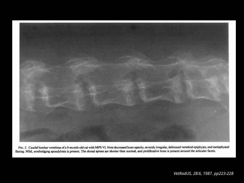
VetRadUS, 28:6, 1987. pp223-228

VetRadUS, 28:6, 1987. pp223-228
Normal
MPS VI

References► Ettinger, Stephen J., DVM and Edward C. Feldman, DVM. Textbook of
Veterinary Internal Medicine Diseases of the Dog and Cat. 6th ed. Volume I. Elsevier, St. Louis, MO. 2005. 872-873.
► Thrall, Donald E., DVM, PhD. Textbook of Veterinary Diagnostic Radiology. 5th ed. Saunders, St. Louis, MO. 2007. 276-277.
► Konde, Linda J., DVM, Mary Anna Thrall, DVM, MS, Peter Gasper, DVM, PhD, Sharon M. Dial, DVM, Kit McBiles, Sean Colgan, Mark Haskins, VMD, PhD. Radiographically Visualized Skeletal Changes Associated With Mucopolysaccharidosis VI In Cats. Veterinary Radiology, 28:6, 1987. 223-228.
► Yogalingam, Gouri, Tom Litjens, Julie Bielicki, Allison C. Crawley, Vivienne Muller, Donald S. Anson, and John J. Hopwood. Feline Mucuopolysaccharidosis Type VI: Characterization of Recombinant N-Acetylgalactosamine 4-Sulfatase and Identification of a Mutation Causing The Disease. The Journal Of Biological Chemistry. 271:44, November 1996. 27259-27265.
► Simonaro, Calogera M., Marina D’Angelo, Xingxuan He, Efrat Eliyahu, Nataly Shtraizent, Mark E. Haskins, and Edward H. Schuchman. Mechanism of Glycosaminoglycan-Mediated Bone and Joint Disease: Implications for the Mucopolysaccharidoses and Other Connective Tissue Diseases. The American Journal of Pathology. 172:1, January 2008. 112-122.