female reproductive system. i. general structure of the female reproductive tract a. two ovaries, b....
TRANSCRIPT

FEMALE REPRODUCTIVE SYSTEM
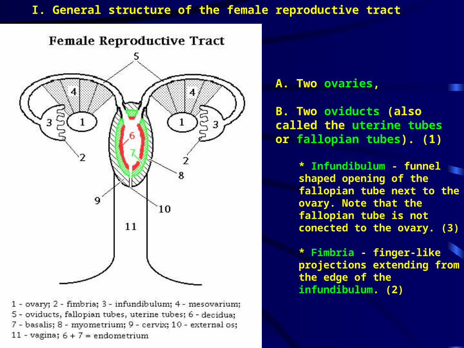
I. General structure of the female reproductive tract
A. Two ovaries,
B. Two oviducts (also called the uterine tubes or fallopian tubes). (1)
* Infundibulum - funnel shaped opening of the fallopian tube next to the ovary. Note that the fallopian tube is not conected to the ovary. (3)
* Fimbria - finger-like projections extending from the edge of the infundibulum. (2)

C. Uterus
* Uterine horns - points where the fallopian tubes connect on the left and right sides.
* Fundus or body of uterus
* The uterine wall (6,7,8)
* Cervix - bulbus end of the uterus that projects into the vagina (9)
* Os - a passageway from the vaginal canal into the uterine cavity. (10)
D. Vagina (11)
E. External genitalia, the labial lips and clitoris.
II. The ovary
A. Covered by modified mesothelium called germinal epithelium.
1. This name is a misnomer since the germ cells (i.e. the oocytes) do not arise from it.
2. This mesothelial covering is a simple cuboidal epithelium in young woman, and becomes a simple squamous epithelium in older woman.
http://www.finchcms.edu/anatomy/histology/organology/female/o_f_1.html

B. Just below germinal epithelium is a poorly defined region of dense connective tissue called the tunica albuginea of the ovary
1. Recall that a tunica albuginia is also present in the testis and penis.
2. These tunics have the same developmental derivation in the gonads of males and females
C. The ovarian tissue beneath the tunica albuginia can be divided into cortical and medullary regions.
http://anatomy.utmb.edu/microanatomy/Female_reproductive/Ovary_preovulation.htm
http://www.finchcms.edu/anatomy/histology/organology/female/o_f_2.html

1. Cortex
a. Contains ovarian follicles that are surrounded by a primitive connective tissue composed of spindle shaped fibroblasts.
* The follicles are spherical structures that are each composed of somatic cells (called follicle cells) that surround an oocyte.
* In addition to the spindle shaped fibroblasts, the stroma of the cortex also contains collagen and reticular fibers.

2. Medulla
a. Blood vessels (arteries and veins) and nerves in a dense stroma of supporting tissue.
b. The blood vessels and nerves enter/leave the ovary through a hilus of connective tissue.

D. Cortex/Follicles -There are four types of follicles.
1. Primordial follicles
a. One layer of flattened follicle cells - not complete - surrounding a central oocyte (immature egg)
b. These follicles are located just beneath the tunica albuginia and form groups of primordial follicles called egg nests.
c. The oocyte is about 40 m in diameter in humans.
http://www.finchcms.edu/anatomy/histology/organology/female/o_f_2.html

http://www.finchcms.edu/anatomy/histology/organology/female/o_f_6.html
2. Primary follicles
a. 1 or more layers of cuboidal to low columnar follicle cells.
b. No fluid filled spaces between cells.

2. More primary follicles
http://www.finchcms.edu/anatomy/histology/organology/female/o_f_6.html
c. During the primary to secondary follicle stage the oocyte grows to about 110 um in diameter.
* It develops a thick acellular layer of glycoproteins, called the zona pellucida, that completely surrounds the oocyte.
* The plasmalemma of the oocyte and the adjacent plasmalemma of the the adjacent layer of follicle cells develop many microvilli that extend into the zona pellucida and interdigitate with one and other.

** This interdigitation of microvilli increases the surface area of contact between the oocyte and its adjacent follicle cell layer.
** Thus, the area over which nutrients can pass into and wastes out of the oocyte is increased.
** The layer of follicle cells that is directly adjacent to the zona pellucida is sometimes called the corona radiata.
d. Theca interna forms from stromal cells that surround the original follicle cell layers.
http://www.finchcms.edu/anatomy/histology/organology/female/o_f_8.html
http://www.finchcms.edu/anatomy/histology/organology/female/o_f_6.html

3. Secondary follicles (also called an antral or vesicular follicle)
a. Consist of 8 - 12 layers of follicle cells surrounding the central oocyte
b. Major characteristic is that fluid-filled spaces have formed between some of the layers of follicle cells.
* These spaces will enlarge and fuse with each other to form the antrum of the mature follicle.
http://www.finchcms.edu/anatomy/histology/organology/female/o_f_7.html

c. Theca folliculi is identifiable
* Consists of two major stratified layers of cells that are derived from the stromal cells
* These layers are the theca interna and theca externa
http://www.finchcms.edu/anatomy/histology/organology/female/o_f_8.html

4. Tertiary or Graafian follicle
a. Has a well developed antrum filled with a fluid called liquor folliculi (or follicular liquor).
b. Oocyte located within a hillock of multiple layers of follicle cells that projects into the antral cavity - called the cumulus oophorus.
c. The follicle cells directly adjacent to oocyte form the corona radiata that will remain associated with oocyte after ovulation.
http://med-ed.med.virginia.edu/med-ed/histology/display.cfm?keyID=22&click=1
http://www.udel.edu/Biology/Wags/histopage/colorpage/cfr/cfrgf.GIF
http://www.finchcms.edu/anatomy/histology/organology/female/o_f_8.html
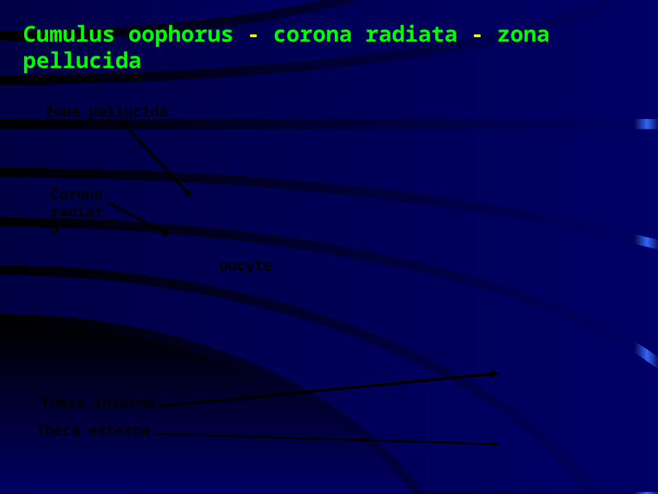
Cumulus oophorus - corona radiata - zona pellucida
Zona pellucida
oocyte
Theca interna
Theca externa
Corona radiata
E. Ovulation - release of oocyte from ovary.
1. Takes place about the 14th day of menstral cycle in humans.
2. Occurs in response to a peak in production of leutinizing hormone by cells called gonadotrophs that are located in the anterior lobe (pars distalis) of the pituitary gland.
a. As the Graafian follicle reaches full maturity it presses against the tunica albuginia surrounding the ovary.
b. Approximately 24 hr after a peak in the production of luteinizing hormone by the pituitary gland, the follicle and the portion of the tunica albuginia it abutts against (called the stigma), rupture and the oocyte, including a few surrounding layers of follicle cells, is expressed into the peritoneal cavity near the infundibulum of the adjacent oviduct.
http://www.meddean.luc.edu/lumen/MedEd/Histo/frames/h_fram21.html
3. Exactly how the follicle is ruptured and the oocyte expressed into the peritoneal cavity is not fully understood, but may involve,
a. An increase in fluid pressure in the antrum.
b. Contraction of smooth muscle cells located in the theca folliculi.
c. A weakening of ovarian tissues overlying point of rupture.
4. At ovulation, the oocyte is released into the peritonial cavity and nearly always enters the infundibulum of the oviduct.
a. This movement of the oocyte into the infundibulum and oviduct proper is aided by currents produced by cilia within the oviduct.
http://www.meddean.luc.edu/lumen/MedEd/Histo/frames/h_fram21.html

5. If fertilization occurs, it will take place in the upper oviduct, sperm having been transported there by means of ciliary currents and perhaps smooth muscle contractions of oviduct.
6. The fertilized egg (now diploid in terms of genetic content) is called a zygote (earliest embryonic stage).
F. Events in the ovary following ovulation
1. Once ovulation has occurred, this does not spell the end of the follicle cells of the Graafian follicle.
2. The cells of the stratum granulosum and theca interna increase in size and undergo differentiation to form an endocrine gland called the corpus luteum.
3. This glandular tissue within the ovary secretes progesterone and estrogen that are necessary to maintain the uterine lining in readiness for implantation of the embryo should the oocyte be fertilized.

4. If fertilization and implantation do occur, cells that are part of the developing placenta of the embryo will produce a hormone called chorionic gonadotropin.
a. This hormone will support the continued existance of the corpus luteum. This structure will degenerate considerably after the 6 month of pregnancy.
http://www.finchcms.edu/anatomy/histology/organology/female/o_f_10.html

5. If fertilization does not occur, the corpus luteum will degenerate by the beginning of the next menstrual cycle.
a. After pregnancy, corpus luteum will degenerate completely leaving scar of dense connective tissue called the corpus albicans.
G. Most ovarian follicles and their contained oocytes degenerate as a female matures (only a small fraction are ever ovulated). This process of degeneration is called atresia. The degenerating follicles are called atretic follicles.
http://www.finchcms.edu/anatomy/histology/organology/female/o_f_10.html
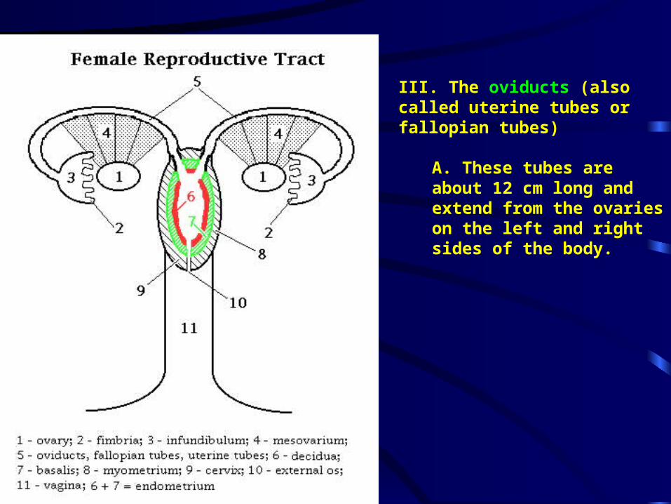
III. The oviducts (also called uterine tubes or fallopian tubes)
A. These tubes are about 12 cm long and extend from the ovaries on the left and right sides of the body.
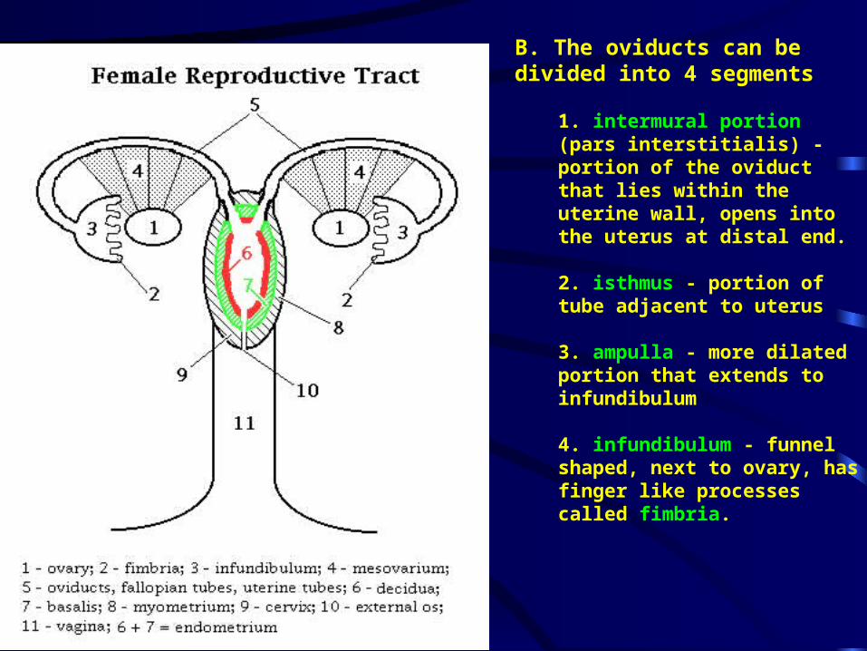
B. The oviducts can be divided into 4 segments
1. intermural portion (pars interstitialis) - portion of the oviduct that lies within the uterine wall, opens into the uterus at distal end.
2. isthmus - portion of tube adjacent to uterus
3. ampulla - more dilated portion that extends to infundibulum
4. infundibulum - funnel shaped, next to ovary, has finger like processes called fimbria.

C. The wall of the oviduct has 3 layers
1. A mucosa adjacent to the oviduct lumen.
a. Consists of long, longitudinal folds that are most numerous in the ampulla.
http://www.ttuhsc.edu/courses/cbb/histo/femalerep/intro.html

b. The mucosal epithelium is simple columnar and contains two types of cells.
* Columnar ciliated cells
* Non-ciliated secretory cells - peg cells.
** Thought to secrete mucus
http://www.finchcms.edu/anatomy/histology/organology/female/o_f_12.htmlhttp://www.medsch.wisc.edu/anatomy/histo/htm/tfmbase.htm

2. A muscularis - muscular layers
a. Inner circular and outer longitudinal layer of smooth muscle.
3. An outer serosa
a. A layer of connective tissue
b. An outer mesothelium that covers the oviduct.
http://www.ttuhsc.edu/courses/cbb/histo/femalerep/intro.html
http://www.finchcms.edu/anatomy/histology/organology/female/o_f_12.html
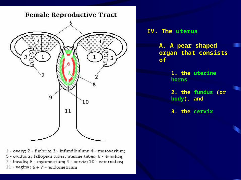
IV. The uterus
A. A pear shaped organ that consists of
1. the uterine horns
2. the fundus (or body), and
3. the cervix
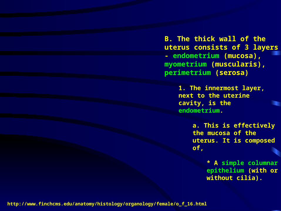
B. The thick wall of the uterus consists of 3 layers - endometrium (mucosa), myometrium (muscularis), perimetrium (serosa)
1. The innermost layer, next to the uterine cavity, is the endometrium.
a. This is effectively the mucosa of the uterus. It is composed of,
* A simple columnar epithelium (with or without cilia).
http://www.finchcms.edu/anatomy/histology/organology/female/o_f_16.html

* A thick lamina propria.
* Tubular glands that extend deep into lamina propria. Lined by a columnar secretory epithelium.
http://www.finchcms.edu/anatomy/histology/organology/female/o_f_16.html
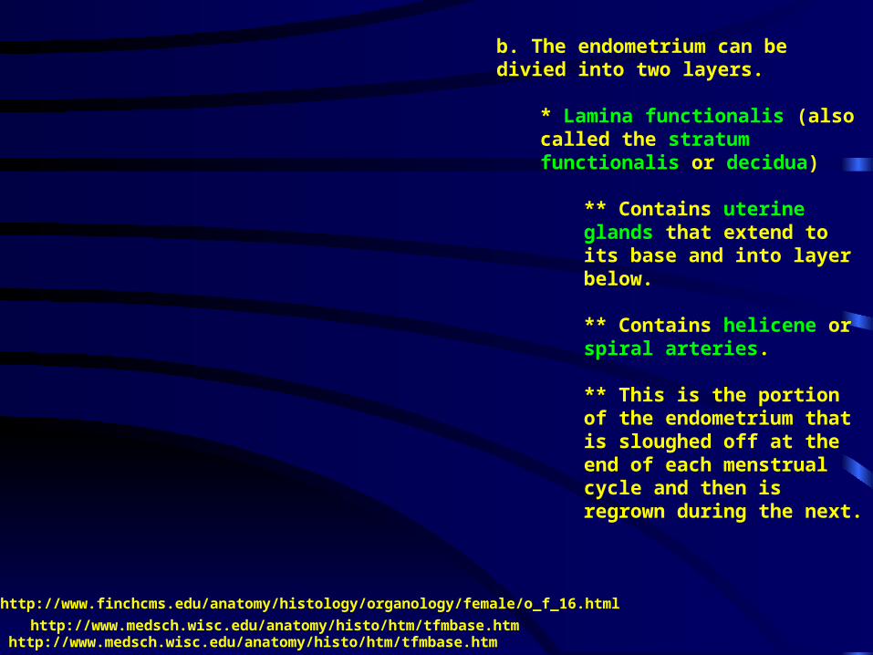
b. The endometrium can be divied into two layers.
* Lamina functionalis (also called the stratum functionalis or decidua)
** Contains uterine glands that extend to its base and into layer below.
** Contains helicene or spiral arteries.
** This is the portion of the endometrium that is sloughed off at the end of each menstrual cycle and then is regrown during the next.
http://www.finchcms.edu/anatomy/histology/organology/female/o_f_16.html
http://www.medsch.wisc.edu/anatomy/histo/htm/tfmbase.htmhttp://www.medsch.wisc.edu/anatomy/histo/htm/
tfmbase.htm

* Lamina basalis (also called the stratum basalis or basalis)
** Contains straight arteries.
** Decidua regenrates from cells in the bases of glands in this layer.
c. The lamina functionalis will interact with tissues of the developing embryo to form the placenta. Placental structure is described in your text.
http://www.medsch.wisc.edu/anatomy/histo/htm/tfmbase.htm
2. The myometrium
a. A tunic of smooth muscle that surrounds the endometrium.
b. Thickest layer of the uterine wall.
c. Consists of 3 poorly defined layers of smooth muscle.
*inner - longitudinal*middle - circular and oblique*outer - longitudinal
http://www.finchcms.edu/anatomy/histology/organology/female/o_f_15.html

3. An outer serosa called the perimetrium
http://www.finchcms.edu/anatomy/histology/organology/female/o_f_14.html

C. The cervix
1. This is the tapering portion of the uterus that bulges into the vaginal canal.
2. The wall differs from the rest of the uterus in that it is mainly composed of dense collagenous and elastic fibers.
3. Only about 15% of the wall is smooth muscle
4. Contains the narrow os (also called the cervical canal). Lined by simple columnar epithelium.
5. The mucosa of the cervix contains mucous glands and is thrown into deep branching folds called plicae palmatae.
6. The cervix undergoes tremendous dilation during birth as the fetus passes from the uterus into the vaginal canal.
http://www.medsch.wisc.edu/anatomy/histo/htm/tfmbase.htm

IV. The vaginaA. The wall of the vagina is devoid of glands. Fluid secretions during sexual stimulation result from blood plasma that seeps through the epithelial lining into the vaginal cavity from an engorged venous plexus in the lamina propria.
B. The wall of the vaginal canal consists of 3 layers of tissue
1. A mucosaa. A nonkeratinized, stratified, squamous epithelium.
http://www.finchcms.edu/anatomy/histology/organology/female/o_f_21.html

c. A lamina propria of loose connective tissue.
* abundant lymphocytes
* contains numerous elastic fibers (this is important for dilation that occurs during birth.
* Richly vascularized
http://www.finchcms.edu/anatomy/histology/organology/female/o_f_21.html
2. A muscularis
a. Mainly longitudinal bundles of smooth muscle
b. Some circular bundles next to the mucosa.
3. An adventitia
a. A layer of dense connective tissue that merges with surrounding tissues
http://www.finchcms.edu/anatomy/histology/organology/female/o_f_21.html

THE END!