femoral hernia – a diagnostic dilemma. hernias are less common than inguinal hernias, but more...
TRANSCRIPT
DOI: 10.21276/aimdr.2018.4.3.SG3
Case Report ISSN (O):2395-2822; ISSN (P):2395-2814
Annals of International Medical and Dental Research, Vol (4), Issue (3) Page 8
Section: S
urgery
Femoral Hernia – A Diagnostic Dilemma. Raj Kumar1, Giles Fanny A2, Romy B Chengazhacherril2 1Additional Chief Health Director, Dr. Babasaheb Ambedkar Memorial Hospital, Central Railway, Byculla, Mumbai.
2Resident, General Surgery, Dr. Babasaheb Ambedkar Memorial Hospital, Central Railway, Byculla, Mumbai.
Received: April 2018 Accepted: April 2018
Copyright: © the author(s), publisher. Annals of International Medical and Dental Research (AIMDR) is an Official Publication of “Society for Health Care & Research Development”. It is an open-access article distributed under the terms of the Creative Commons Attribution Non-Commercial License, which permits unrestricted non-commercial use, distribution, and reproduction in any medium, provided the original work is properly cited.
ABSTRACT Femoral hernias are less common than inguinal hernias, but more prone to strangulation. Accurate diagnosis and prompt surgical correction are extremely important to prevent bowel ischemia and necrosis in an incarcerated femoral hernia. Clinical presentation is generally a mildly painful, irreducible groin bulge below the inguinal ligament. However, differentiation from an inguinal hernia on physical exam is not entirely reliable, regardless of the examining surgeon’s experience. Even Ultrasonography and CT findings may be equivocal and sometimes femoral hernia presents a diagnostic dilemma. Keywords: Femoral hernia, Incarceration.
INTRODUCTION Femoral Hernia is a type of groin hernia with lesser incidence than inguinal hernia. The femoral hernia repairs constitute less than 3 % of the groin hernia repairs.[1] Mean age of presentation is 46.84 years with a predominance of female gender (71%).[2] Around 40% of the patients having femoral hernia present with incarceration or strangulation, which may require emergency surgeries.[2] Patients present with lump in groin region, pain in the groin region or signs of strangulation. Clinical examination may or may not give a clue to the accurate diagnosis. The diagnosis in most cases will be confirmed by using CT study. CT images are helpful in differentiating femoral hernia from inguinal hernia and also will provide information regarding extension of the sac, content of the sac and other features.[3] Despite being an important diagnostic tool, CT imaging may fail to identify the type of hernia or may even mislead. This brief report documents a case of an elderly female presented with a groin swelling and diagnosed clinically as inguinal hernia. USG and CT reported inguinal hernia but on operation it was found to be a case of incarcerated femoral hernia.
CASE REPORT
A 61 years old lady presented with a painful swelling in the right groin region noticed by her for Name & Address of Corresponding Author Dr. Raj Kumar, Additional Chief Health Director, Department of Surgery, Dr.Babasaheb Ambedkar Memorial Hospital, Central Railway, Byculla, Mumbai – 400027.
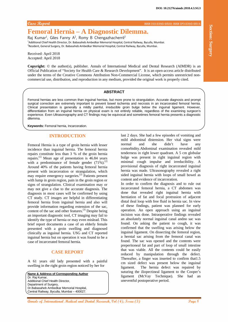
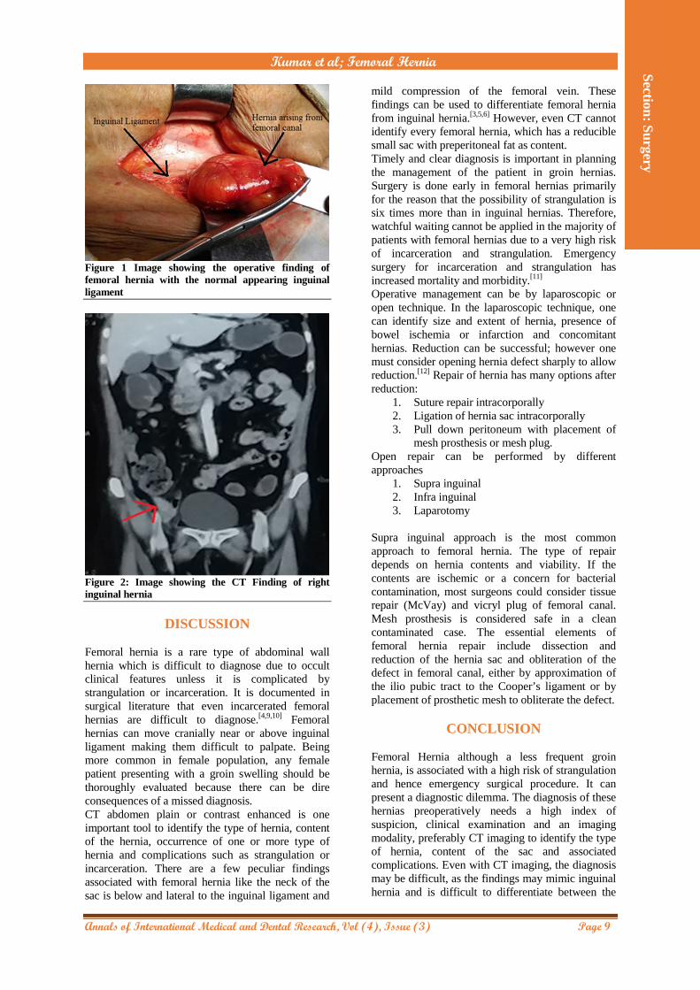
last 2 days. She had a few episodes of vomiting and mild abdominal distension. Her vital signs were normal and she didn’t have any comorbidity.Abdominal examination revealed mild tenderness in right lower quadrant. A 5 cm globular bulge was present in right inguinal region with minimal cough impulse and irreducibility. A provisional diagnosis of right incarcerated inguinal hernia was made. Ultrasonography revealed a right sided inguinal hernia with loops of small bowel as content and evidence of incarceration. In order to confirm the diagnosis and to rule out incarcerated femoral hernia, a CT abdomen was done that revealed right inguinal hernia with herniation of fat and focal protrusion of adjacent distal ileal loop with free fluid in hernia sac. In view of these findings, patient was planned for early operation. An open approach using an inguinal incision was done. Intraoperative findings revealed an absolutely normal inguinal canal andno sac was found. On asking the patient to cough, it was confirmed that the swelling was arising below the inguinal ligament. On dissecting the femoral region, a hernial sac arising from the femoral canal was found. The sac was opened and the contents were preperitoneal fat and part of loop of small intestine that was viable. All the contents could be easily reduced by manipulation through the defect. Thereafter, a finger was inserted to confirm that1.5 cm sized defect was present below the inguinal ligament. The hernia defect was repaired by suturing the iliopectineal ligament to the Cooper’s ligament (McVay Technique). She had an uneventful postoperative period.
Kumar et al; Femoral Hernia
Annals of International Medical and Dental Research, Vol (4), Issue (3) Page 9
Section: S
urgery
Figure 1 Image showing the operative finding of femoral hernia with the normal appearing inguinal ligament
Figure 2: Image showing the CT Finding of right inguinal hernia
DISCUSSION Femoral hernia is a rare type of abdominal wall hernia which is difficult to diagnose due to occult clinical features unless it is complicated by strangulation or incarceration. It is documented in surgical literature that even incarcerated femoral hernias are difficult to diagnose.[4,9,10] Femoral hernias can move cranially near or above inguinal ligament making them difficult to palpate. Being more common in female population, any female patient presenting with a groin swelling should be thoroughly evaluated because there can be dire consequences of a missed diagnosis. CT abdomen plain or contrast enhanced is one important tool to identify the type of hernia, content of the hernia, occurrence of one or more type of hernia and complications such as strangulation or incarceration. There are a few peculiar findings associated with femoral hernia like the neck of the sac is below and lateral to the inguinal ligament and
mild compression of the femoral vein. These findings can be used to differentiate femoral hernia from inguinal hernia.[3,5,6] However, even CT cannot identify every femoral hernia, which has a reducible small sac with preperitoneal fat as content. Timely and clear diagnosis is important in planning the management of the patient in groin hernias. Surgery is done early in femoral hernias primarily for the reason that the possibility of strangulation is six times more than in inguinal hernias. Therefore, watchful waiting cannot be applied in the majority of patients with femoral hernias due to a very high risk of incarceration and strangulation. Emergency surgery for incarceration and strangulation has increased mortality and morbidity.[11]
Operative management can be by laparoscopic or open technique. In the laparoscopic technique, one can identify size and extent of hernia, presence of bowel ischemia or infarction and concomitant hernias. Reduction can be successful; however one must consider opening hernia defect sharply to allow reduction.[12] Repair of hernia has many options after reduction:
1. Suture repair intracorporally 2. Ligation of hernia sac intracorporally 3. Pull down peritoneum with placement of
mesh prosthesis or mesh plug. Open repair can be performed by different approaches
1. Supra inguinal 2. Infra inguinal 3. Laparotomy
Supra inguinal approach is the most common approach to femoral hernia. The type of repair depends on hernia contents and viability. If the contents are ischemic or a concern for bacterial contamination, most surgeons could consider tissue repair (McVay) and vicryl plug of femoral canal. Mesh prosthesis is considered safe in a clean contaminated case. The essential elements of femoral hernia repair include dissection and reduction of the hernia sac and obliteration of the defect in femoral canal, either by approximation of the ilio pubic tract to the Cooper’s ligament or by placement of prosthetic mesh to obliterate the defect.
CONCLUSION
Femoral Hernia although a less frequent groin hernia, is associated with a high risk of strangulation and hence emergency surgical procedure. It can present a diagnostic dilemma. The diagnosis of these hernias preoperatively needs a high index of suspicion, clinical examination and an imaging modality, preferably CT imaging to identify the type of hernia, content of the sac and associated complications. Even with CT imaging, the diagnosis may be difficult, as the findings may mimic inguinal hernia and is difficult to differentiate between the
Kumar et al; Femoral Hernia
Annals of International Medical and Dental Research, Vol (4), Issue (3) Page 10
Section: S
urgery
two. Both open and laparoscopic surgical approaches are available, usually preferred with the use of prosthetics.
REFERENCES
1. Burcharth J, Pedersen M, Bisgaard T, Pedersen C, Rosenberg J (2013) Nationwide Prevalence of Groin Hernia Repair. PLoS ONE 8(1): e54367.
2. Alimoglu, O., Kaya, B., Okan, I. et al. Hernia (2006) 10: 70-73.
3. S Shigeru, F Shigeru, O Kota, S Tsutomu, M Jun et al. Differentiation of Femoral Versus Inguinal Hernia: CT Findings, American Journal of Roentgenology 2007 189:2, W78-W83.
4. Naude, Gideon P et al. Femoral hernia: The dire consequences of a missed diagnosis. The American Journal of Emergency Medicine , Volume 15 , Issue 7 , 680 - 682
5. Richard J Wechsler, Alfred B Kurtz, Laurence Needleman, Bradley W Dick, Rick I Field, Pamela L Hilpert. Cross sectional imaging of abdominal wall hernias. AJR 153:517-521, September 1989
6. Lee GH, Cohen AJ. CT imaging of abdominal hernias. AJR. American journal of roentgenology. 1993 Dec;161(6):1209-13.
7. Rosenberg J, Bisgaard T, Kehlet H, Wara P, Asmussen T, Juul P, Strand L, Andersen FH, Bay-Nielsen M. Danish Hernia Database recommendations for the management of inguinal and femoral hernia in adults. Dan Med Bull. 2011 Feb;58(2):C4243.
8. Kingsnorth AN, LeBlanc KA, editors. Management of abdominal hernias. Springer Science & Business Media; 2013 Jan 30.
9. Nehme AE. Groin hernias in elderly patients: management and prognosis. The American journal of surgery. 1983 Aug 1;146(2):257-60.
10. Yoo HG, Lee KM, Choi UJ. The diagnostic concordance of femoral hernia and the factors influencing diagnosis. Journal of the Korean Surgical Society. 2009 Mar 1;76(3):179-86.
11. Gallegos NC, Dawson J, Jarvis M, Hobsley M. Risk of strangulation in groin hernias. British Journal of Surgery. 1991 Oct 1;78(10):1171-3.
12. Hachisuka T. Femoral hernia repair. Surgical Clinics of North America. 2003 Oct 1;83(5):1189-205. How to cite this article: Kumar R, Fanny AG, Chengazhacherril RB. Femoral Hernia – A Diagnostic Dilemma. Ann. Int. Med. Den. Res. 2018; 4(3):SG08-SG10.
Source of Support: Nil, Conflict of Interest: None declared