fermented pueraria lobata extract ameliorates dextran … · 2016-09-30 · fermented pueraria...
TRANSCRIPT

151
Lab Anim Res 2016: 32(3), 151-159
http://dx.doi.org/10.5625/lar.2016.32.3.151
ISSN 1738-6055 (Print)
ISSN 2233-7660 (Online)
Fermented Pueraria Lobata extract ameliorates dextran sulfate sodium-induced colitis by reducing pro-inflammatorycytokines and recovering intestinal barrier function
Seungho Choi1, Jong-Kyu Woo1, Yeong-Su Jang2, Ju-Hee Kang2,3, Jung-Eun Jang2, Tae-Hoo Yi4,5,Sang-Yong Park4, Sun-Yeou Kim2, Yeo-Sung Yoon1, Seung Hyun Oh2,*
1Department of Anatomy and Cell Biology, College of Veterinary Medicine, Seoul National University, Seoul, Korea2Gachon Institute of Pharmaceutical Sciences, Gachon University, Incheon, Korea
3Research Institute, National Cancer Center, Gyeonggi-do, Korea4Graduate School of Biotechnology, College of Life Science, Kyung Hee University, Gyeonggi-do, Korea
5Department of Oriental Medicine Biotechnology, College of Life Science, Kyung Hee University Global Campus, Gyeonggi-do, Korea
Inflammatory bowel disease is a chronic inflammatory disorder occurring in the gastrointestinal track.However, the efficacy of current therapeutic strategies has been limited and accompanied by side effects.In order to eliminate the limitations, herbal medicines have recently been developed for treatment ofIBD. Peuraria Lobata (Peuraria L.) is one of the traditional herbal medicines that have anti-inflammatoryeffects. Bioavailability of Peuraria L., which is rich in isoflavones, is lower than that of their fermentedforms. In this study, we generated fermented Peuraria L. extracts (fPue) and investigated the role of fPuein inflammation and intestinal barrier function in vitro and in vivo. As the mice or intestinal epithelialcells were treated with DSS/fPue, mRNA expression of pro-inflammatory cytokines was reduced and thearchitecture and expression of tight junction proteins were recovered, compared to the DSS-treatedgroup. In summary, fPue treatment resulted in amelioration of DSS-induced inflammation in the colon,and the disrupted intestinal barrier was recovered as the expression and architecture of tight junctionproteins were retrieved. These results suggest that use of fPue could be a new therapeutic strategy fortreatment of IBD.
Keywords: IBD, herbal medicines, Pueraria Lobata, inflammation, intestinal barrier
Received 10 May 2016; Revised version received 29 June 2016; Accepted 30 June 2016
Inflammatory bowel disease (IBD), including ulcerative
colitis (UC) and Crohn’s disease (CD), are autoimmune
diseases, characterized by chronic inflammation in the
gastrointestinal (GI) tract, and recent reports indicated
that the incidence and prevalence of IBD have increased
worldwide [1]. Studies on pathogenesis of IBD have
reported various factors that cause both types of IBD,
including genetic susceptibility, environmental factors,
infectious agents, and abnormality of immune system
function [2]. IBD is accompanied by other diseases such
as psychiatric disorders [3,4] and colon cancer development
[5,6].
In the pathogenesis of IBD, the expression and
secretion of pro-inflammatory cytokines, such as tumor
necrosis factor-α (TNFα) and interleukin-1β (IL-1β), are
increased and the expression of cytokines is regulated by
nuclear factor-kappa B (NF-κB) [7]. In addition,
dysfunction of the gastrointestinal barrier is a major step
in the pathogenesis of IBD [8-10]. The major components
forming the gastrointestinal barrier are tight junction
(TJ) proteins in the apical junction complex, which
regulate paracellular permeability. It has been reported
*Corresponding author: Seung Hyun Oh, Gachon Institute of Pharmaceutical Sciences, Gachon University, 191 Hambakmoero,Yeonsu-gu, Incheon 21936, KoreaTel: +82-32-820-4929; Fax: +82-32-820-4829; E-mail: [email protected]; [email protected]
This is an Open Access article distributed under the terms of the Creative Commons Attribution Non-Commercial License (http://creativecommons.org/licenses/by-nc/3.0) which permits unrestricted non-commercial use, distribution, and reproduction in any medium, provided the original work is properly cited.

152 Seungho Choi et al.
Lab Anim Res | September, 2016 | Vol. 32, No. 3
that the structure and function of TJ proteins are
disrupted in IBD patients [11], which leads to intestinal
bacterial translocalization into mesenteric lymph nodes
[12].
There are several therapeutic strategies for remission
of IBD, including use of anti-inflammatory medicines
and immunomodulators, however, their clinical efficacy
has been limited and accompanied by side effects,
including fluid retention, insomnia and vomiting [13].
The overall remission rate of IBD patients with therapies
is only approximately 50% [14,15]. Among the therapeutic
strategies, targeting the adoptive immune system, has
been the most common for treatment of IBD, such as
monoclonal antibodies against TNFα [13,16]. However,
treatment with the agents is successful only in less than
one third of IBD patients and a proportion of patients
experience loss of response to the agents [14,17-18].
Therefore, development of new therapeutic strategies for
treatment of IBD is needed.
Complementary and alternative medicines (CAMs)
have recently been developed for treatment of IBD and,
the use of herbal medicines is the most common for
treatment of IBD [19,20]. Herbal medicines have a
number of beneficial effects, including anti-bacterial,
anti-oxidant, and anti-inflammatory effects [19]. Pueraria
L., a common Chinese herbal medicine, also has various
beneficial effects, including anti-diarrhetic and anti-
emetic effects, as well as an anti-oxidant effect [21,22].
Pueraria L. is rich in isoflavones, including peurarin,
daidzein and genistein [21]. Isoflavones are known for
their anti-inflammatory effect and anti-oxidant effect
[23]. However, bioavailability of isoflavones is lower
than that of their fermented forms, isoflavone aglycones
[24].
In this study, for development of more effective herbal
medicine for treatment of IBD, we generated the
fermented Pueraria L. extract (fPue) and compared its
anti-inflammatory effect with that of Pueraria L. extract
(Pue). In addition, in vivo experiments were performed
on a dextran sulfate sodium (DSS)-induced colitis model,
which mimics human ulcerative colitis [25].
Materials and Methods
Generation of fPue
For generation of fPue, Pueraria L. was purchased
from Omniherb Inc.; 400 g of that was crushed and
boiled in 4 L of water at 121°C for 30 minutes and the
aqueous extract was filtered using Whatman filter paper
2 (GE Healthcare, Little Chalfont, United Kingdom).
Dextrose (2%) and yeast extract (1%) were autoclaved
and added to the Pue extract with 1% Lactobacillus
brevis, aD110T, and the Pue extract was incubated at
30°C for 5 days. Following incubation, the Pue extract
was centrifuged at 12,000 rpm for 15 minutes, and the
supernatant was filtered. The filtered extract was
concentrated at 40°C and 80 rpm, using a rotary
evaporator (Eyela, Irvine, CA).
Animals
Specific pathogen-free female ICR mice (5 weeks old)
were purchased from Orient Inc., Gyeonggi-do, Korea
and the mice were acclimatized for 1 week prior to the
experiment. All mice were maintained under 12 hours
light/dark cycles at 22±5°C and 60±5% humidity and
provided with food (Orient Inc.) and water ad libitum.
The information about components and calorie of the
food is supported by Orient Inc. All experiments with
animals were approved by the Institutional Animal Care
and Use Committee at Lee Gil Ya Cancer and Diabetes
Institute (IACUC No., LCDI-2014-0010).
Cell culture and treatment
Raw264.7 and Caco-2 cell lines were purchased from
Korea Cell Line Bank, Seoul, Korea. The cell lines were
cultured in 10% FBS-supplemented DMEM (WelGene,
Daegu, Korea) with streptomycin (100 μg/mL)/penicillin
(100 units/mL) at 37°C in a humidified chamber with
5% CO2. Raw264.7 and Caco-2 cells were treated with
Pue or fPue for western blot, RT-PCR and immuno-
fluorescence analysis. For detection of NF-κB signaling
by western blot, Raw264.7 cells were treated with Pue
or fPue (0, 25, 100, 400 μg/mL) for 4 hours, followed by
treatment with lipopolysccharide (LPS) for 1 hour. Total
protein was isolated and western blot was performed.
For determination of transcriptional level of cytokines in
Raw264.7 cells, the cells were treated with Pue or fPue
(0, 25, 100, 400μg/mL) for 4 hours, followed by treatment
with LPS for 3 hours. Total RNA was isolated using
TriZol (Invitrogen, Waltham, MA) and RT-PCR was
performed. Caco-2 cells were seeded in 12-well plates
and cultured with or without a cover slip for immuno-
fluorescence assay or western blot, respectively. The
cells were incubated until they become 100% confluent,
followed by incubation for an additional 7 days. The
cells were treated with fPue (0, 25, 100, or 400 μg/mL)

Fermented Pueraria Lobata extract ameliorates DSS-induced colitis in animal model 153
Lab Anim Res | September, 2016 | Vol. 32, No. 3
for 48 hours, followed by treatment with 2% DSS for 24
hours. Then, immunofluorescence assay and membrane
fractionation for western blot were performed.
Induction of colitis by DSS
The mice were divided into 3 groups (7 mice in each
group): (1) no treatment group; (2) 5% DSS-treated
group; (3) 5% DSS with fPue (100 mg/kg)-treated group.
DSS (MP Biomedical, Burlingame, CA) and fPue were
dissolved in distilled water. Five percent DSS was
administered to the mice in the drinking water for 7 days
and normal distilled water was then supplied for 4 days
for recovery (Figure 2A). During the recovery period,
the mice were treated with fPue, or distilled water daily
by gavage (Figure 2A). At the end of the recovery
period, disease activity index (DAI) score was determined
as described in the previous report [26] and necropsy
was then performed.
Histological evaluation of colitis
The mice were sacrificed at the end of the recovery
period. Following excision of the whole colon from anus
to cecum, the length was measured. Approximately 0.5
cm of the distal colon was excised and frozen in liquid
nitrogen for RT-PCR and histological analysis, and the
remaining colon was Swiss-rolled and fixed in 10%
formalin. The tissue samples were then paraffin-embedded
and sectioned at 5μm-thickness. The sections were stained
with hematoxylin and eosin to evaluate the severity of
colitis, according to three parameters described in the
previous report [26]. The severity of colitis was scored
blindly by 2 researchers.
Western blot
Twenty micrograms of each protein sample was used
for western blot analysis. The primary antibodies are as
followed: anti-rabbit ZO-1, anti-mouse Occludin, 1:1,000,
anti-mouse Claudin-1 (Invitrogen, Waltham, MA); anti-
rabbit p-P65 (Cell Signaling, Danvers, MA), anti-rabbit
P65, anti-goat Lamin B (Santa Cruz, CA) and anti-
mouse GAPDH (Merck Millipore, Billerica, MA).
Reverse transcription-PCR
Using 3 μg of RNA, cDNA was synthesized using the
PrimeScript RT reagent Kit (TaKaRa, Shiga, Japan) as
described in the manufacturer’s instructions. RT-PCR
was performed, using the primers listed on Supplementary
Table 1 (Table S1).
Immunofluorescence analysis
The 5 μm-thick frozen colon tissue and cells on the
slides were fixed in an acetone-methanol (1:1) mixture
at −20°C. The slides were incubated with primary
antibodies (anti-rabbit ZO-1, anti-mouse Occludin, anti-
mouse Claudin-1; Invitrogen, Waltham, MA) overnight
at 4°C and secondary antibodies (anti-rabbit IgG FITC,
Santa Cruz; Alexa Fluor 488 goat anti-mouse IgG;
Invitrogen) for 2 hours at room temperature. After
mounting the slides with VectaShield Mounting Medium
with DAPI (Vector Laboratories, Burlingame, CA), the
slides were sealed with nail polish and stored at −20°C
until observation, using a confocal microscope (Nikon
Instruments Inc., Melville, NY).
Statistical analysis
Numerical data for all graphs were expressed as mean
±standard deviation. Statistical changes in all data were
determined using one-way analysis of variance (ANOVA).
Scheffe was used for the post hoc test. The significance
level was limited to 5% (P<0.05). SPSS 22.0 was used
as a tool for statistical analysis.
Results
Anti-inflammatory effect of Pue is enhanced though
fermentation in Raw264.7 cells
To determine whether fPue can enhance anti-inflammatory
effect, compared to Pue, the activity of P65, a subunit of
NF-κB, was examined in murine macrophage cell line
Raw264.7. The cells were treated with Pue or fPue,
followed by activation by LPS, which induces phosphory-
lation of P65 in Raw264.7 cells. Results of western blot
showed that treatment with fPue significantly inhibited
phosphorylation of P65 while Pue slightly inhibited that
of P65 (Figure 1A). Activation of NF-κB leads to
upregulated transcription of pro-inflammatory cytokines,
including IL-1β, TNFα, IL-6, and IL-12. Treatment of
LPS resulted in increased transcriptional levels of IL-1β,
TNFα, IL-6 and IL-12 (Figure 1B). On the other hand,
when pre-treated with fPue, the expression levels of the
cytokines were decreased while treatment with Pue did
not affect them (Figure 1B). These results suggest that
fPue would improve the anti-inflammatory effect.

154 Seungho Choi et al.
Lab Anim Res | September, 2016 | Vol. 32, No. 3
fPue has a therapeutic effect on DSS-induced colitis in
vivo
Because fPue exerted an anti-inflammatory effect in
Raw264.7 cell line (Figure 1), we examined whether
fPue could improve DSS-induced colitis. DSS-induced
colitis is characterized by disruption of epithelial structure
and infiltration of inflammatory cells and loss of goblet
cells [27]. To evaluate the effect of fPue on DSS-induced
colitis, DAI was assessed by fecal analysis, to examine
how much water and blood the fecal pellet contains and
histological analysis was performed by H&E staining on
the paraffin block sections to assess the severity of
colitis. In the control group, DAI score was nearly 0 and
that of the DSS-treated group showed a significant
increase (Figure 2B), indicating that colitis was induced
by DSS. On the other hand, DAI score of the DSS+
fPue-treated group was significantly decreased, suggesting
that fPue treatment improved the colonic function
(Figure 2B). Histological analysis demonstrated that
DSS treatment induced severe inflammation in mucosa,
edema in submucosa and epithelial cell loss while they
were significantly lower in the DSS+fPue-treated group,
compared with the DSS+Vehicle-treated group (Figure
2C, 2D). In addition, alcian blue staining showed that
while goblet cells were lost in the DSS-treated group,
they were restored by fPue treatment (Figure 2E). These
results suggest that treatment with fPue resulted in
amelioration of colonic inflammation and promoted
recovery of epithelial structure in the colonic mucosa,
following recovery from diarrhea.
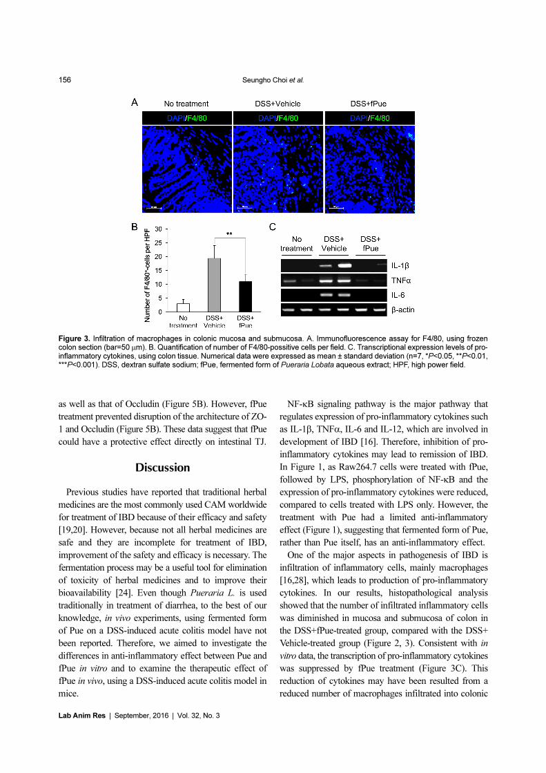
fPue treatment reduced infiltration of macrophages
and pro-inflammatory cytokines in DSS-induced colitis
In a DSS-induced colitis model, inflammatory cells,
mainly macrophages, are infiltrated into submucosa and
lamina propria of the inflamed colon [16,28]. To
determine whether the number of infiltrated macrophages
is decreased by treatment with fPue, immunostaining
was performed for F4/80, a marker for macrophages, on
frozen sections of colon tissue. Immunofluorescence
analysis revealed that while the number of F4/80-
positive cells present in mucosa and submucosa of the
inflamed colon was significantly increased, that of F4/
80-positive cells was markedly reduced in the colon of
the DSS+fPue-treated group (Figure 3A, B), indicating
that macrophage infiltration was reduced by fPue treatment.
In addition, decrease in transcriptional expression of pro-
inflammatory mediators, IL-1β, TNFα and IL-6 was
observed by performing RT-PCR (Figure 3C).
DSS-induced disruption of intestinal epithelial barrier
was recovered by treatment with fPue
As previously mentioned, epithelial dysfunction is a
major aspect in the pathogenesis of IBD. To determine
whether the epithelial barrier, disrupted in DSS-induced
colitis, is recovered by treatment with fPue, the expression
levels and the arrangement of ZO-1, Claudin1 and
Occludin, were examined by immunofluorescence assay,
using cryosections of colon tissue. As shown in Figure
4A, all three TJ proteins were expressed along with the
inner lining of the columnar epithelium of normal colon
tissue. While the expression levels of TJ proteins were
decreased and their architecture was disrupted in DSS-
Figure 1. Anti-inflammatory effect of Peuraria L. extract and itsfermented form. A. Western blot analysis for phosphorylation ofNF-κB in Raw264.7 cells after treatment of Pue or fPue withLPS. B. RT-PCR for transcriptional expression levels of pro-inflammatory cytokines in Raw264.7 cells after treatment ofPue or fPue with LPS. Pue, Pueraria Lobata aqueous extract;fPue, fermented form of Pueraria Lobata aqueous extract.Intensity of each band in western blot and RT-PCR results wasquantified by using ImageJ and normalized to GAPDH (A) andβ-actin (B).

Fermented Pueraria Lobata extract ameliorates DSS-induced colitis in animal model 155
Lab Anim Res | September, 2016 | Vol. 32, No. 3
induced colitis, they were recovered as DSS-induced
colitis was ameliorated by fPue treatment (Figure 4A). In
addition, to investigate whether the physiological function
of the epithelial barrier was recovered, bacterial trans-
localization was examined by blood agar assay, using
cells from mesenteric lymph nodes. As shown in Figure
4B and C, bacterial translocalization was induced as the
epithelial barrier was disrupted by DSS treatment. On
the other hand, in the DSS+fPue-treated group a decreased
number of bacteria was present in the lymph node
(Figure 4B, C), indicating that bacterial translocalization
was reduced as disrupted tight proteins were rescued by
fPue treatment.
fPue directly recovers DSS-induced TJ damage in
Caco-2 cells
To determine whether DSS-induced disruption of TJ
would be recovered by fPue treatment, an in vitro
experiment was performed using colonic epithelial cell
line Caco-2. Caco-2 cells form a monolayer and TJ
between the cells, and the TJ is disrupted by DSS
treatment [29]. Consistent with the previous study,
treatment of Caco-2 cells with DSS resulted in a
decrease in protein expression of Occludin, which was
detected by western blot, while that of ZO-1 did not
change (Figure 5A). Although ZO-1 expression level
was not affected by DSS, its architecture was disrupted
Figure 2. Effect of fPue on DSS-induced colitis in mice. A. Schedule for administration of 5% DSS and fPue. B. Assessment ofdisease activity index (DAI) by fecal analysis. C. Histological evaluation of DSS-induced colitis according to degree of epithelial cellloss, crypt damage, and infiltration of inflammatory cells. D. Histological figures of colon stained by H&E. (a, mucosa; b,submucosa; c, muscularis; d, crypt) E. Histological figures to observe goblet cell loss by Alcian Blue staining. Numerical data wereexpressed as mean±standard deviation (n=7, *P<0.05, **P<0.01, ***P<0.001). DSS, dextran sulfate sodium; fPue, fermented formof Pueraria Lobata aqueous extract; H&E, hematoxylene and eosin.
156 Seungho Choi et al.
Lab Anim Res | September, 2016 | Vol. 32, No. 3
as well as that of Occludin (Figure 5B). However, fPue
treatment prevented disruption of the architecture of ZO-
1 and Occludin (Figure 5B). These data suggest that fPue
could have a protective effect directly on intestinal TJ.
Discussion
Previous studies have reported that traditional herbal
medicines are the most commonly used CAM worldwide
for treatment of IBD because of their efficacy and safety
[19,20]. However, because not all herbal medicines are
safe and they are incomplete for treatment of IBD,
improvement of the safety and efficacy is necessary. The
fermentation process may be a useful tool for elimination
of toxicity of herbal medicines and to improve their
bioavailability [24]. Even though Pueraria L. is used
traditionally in treatment of diarrhea, to the best of our
knowledge, in vivo experiments, using fermented form
of Pue on a DSS-induced acute colitis model have not
been reported. Therefore, we aimed to investigate the
differences in anti-inflammatory effect between Pue and
fPue in vitro and to examine the therapeutic effect of
fPue in vivo, using a DSS-induced acute colitis model in
mice.
NF-κB signaling pathway is the major pathway that
regulates expression of pro-inflammatory cytokines such
as IL-1β, TNFα, IL-6 and IL-12, which are involved in
development of IBD [16]. Therefore, inhibition of pro-
inflammatory cytokines may lead to remission of IBD.
In Figure 1, as Raw264.7 cells were treated with fPue,
followed by LPS, phosphorylation of NF-κB and the
expression of pro-inflammatory cytokines were reduced,
compared to cells treated with LPS only. However, the
treatment with Pue had a limited anti-inflammatory
effect (Figure 1), suggesting that fermented form of Pue,
rather than Pue itself, has an anti-inflammatory effect.
One of the major aspects in pathogenesis of IBD is
infiltration of inflammatory cells, mainly macrophages
[16,28], which leads to production of pro-inflammatory
cytokines. In our results, histopathological analysis
showed that the number of infiltrated inflammatory cells
was diminished in mucosa and submucosa of colon in
the DSS+fPue-treated group, compared with the DSS+
Vehicle-treated group (Figure 2, 3). Consistent with in
vitro data, the transcription of pro-inflammatory cytokines
was suppressed by fPue treatment (Figure 3C). This
reduction of cytokines may have been resulted from a
reduced number of macrophages infiltrated into colonic
Figure 3. Infiltration of macrophages in colonic mucosa and submucosa. A. Immunofluorescence assay for F4/80, using frozencolon section (bar=50 μm). B. Quantification of number of F4/80-possitive cells per field. C. Transcriptional expression levels of pro-inflammatory cytokines, using colon tissue. Numerical data were expressed as mean ± standard deviation (n=7, *P<0.05, **P<0.01,***P<0.001). DSS, dextran sulfate sodium; fPue, fermented form of Pueraria Lobata aqueous extract; HPF, high power field.

Fermented Pueraria Lobata extract ameliorates DSS-induced colitis in animal model 157
Lab Anim Res | September, 2016 | Vol. 32, No. 3
epithelium. In vitro data shown in Figure 1 also suggest
that fPue could have a suppressive effect on production
of pro-inflammatory cytokines in macrophages as it
inhibits NF-κB activation. However, it remains unclear
how fPue reduced macrophage infiltration and its cytokine
production.
Another aspect of IBD is dysfunction of gastrointestinal
barrier by destruction of TJ in colonic mucosa [8-10].
The gastrointestinal barrier is responsible for uptake of
nutrients, ions, and water, and for preventing translocation
of pathogens across tissue [11]. Our in vivo experiment
demonstrated that as acute colitis was induced by DSS
administration in mice, the structure of the TJ complex
was disrupted (Figure 4A). In contrast, the disrupted TJ
complex was restored by fPue treatment (Figure 4A). In
addition, while bacterial translocalization was induced
due to disrupted TJ complex, bacteria were not trans-
localized into mesenteric lymph nodes as the TJ
complex was recovered by treatment with fPue (Figure
4B, C), indicating that the function of intestinal barrier
was recovered. Interestingly, treatment with fPue inhibited
collapse of TJ proteins (Figure 5), suggesting that fPue
may has a direct effect on recovery of the intestinal
barrier function in acute colitis by rescuing TJ protein
distribution.
In conclusion, in vitro experiment with Raw264.7
suggested that fPue has a more beneficial effect on
inflammatory activity than the extract itself. The anti-
Figure 4. Effect of fPue on recovery of colonic epithelial barrier. A. Immunofluorescence assay for tight junction proteins, ZO-1,Claudin-1, and Occludin, on frozen colon tissue. B. Blood agar assay, using cells isolated from mesenteric lymph nodes. C.Representative figures for blood agar assay. Numerical data were expressed as mean ± standard deviation (n=7, *P<0.05,**P<0.01, ***P<0.001). DSS, dextran sulfate sodium; fPue, fermented form of Pueraria Lobata aqueous extract.

158 Seungho Choi et al.
Lab Anim Res | September, 2016 | Vol. 32, No. 3
inflammatory effect of fPue was also demonstrated in an
in vivo experiment. fPue ameliorated DSS-induced acute
colitis and recovered gastrointestinal barrier function. In
addition, there was no side effect in the use of fPue, such
as weight loss or liver injury (data not shown). However,
investigation to determine whether there are additional
beneficial effects of fPue is needed. In this study, we
suggest that the fPue has a therapeutic potential for
treatment of IBD without undesirable effects.
Acknowledgments
This work was conducted under the industrial
infrastructure program (No. N0000888) for fundamental
technologies, which is funded by the Ministry of Trade,
Industry, and Energy (MOTIE, Korea), and also supported
by High Value-added Food Technology Development
Program 114006-04, Ministry of Agriculture, Food and
Rural Affairs.
Conflict of interests The authors declare that there is
no financial conflict of interests to publish these results.
References
1. Molodecky NA, Soon IS, Rabi DM, Ghali WA, Ferris M,Chernoff G, Benchimol EI, Panaccione R, Ghosh S, BarkemaHW, Kaplan GG. Increasing incidence and prevalence of theinflammatory bowel diseases with time, based on systematicreview. Gastroenterology 2012; 142(1): 46-54.
2. Baumgart DC, Carding SR. Inflammatory bowel disease: causeand immunobiology. Lancet 2007; 369(9573): 1627-1640.
3. Walker JR, Ediger JP, Graff LA, Greenfeld JM, Clara I, Lix L,Rawsthorne P, Miller N, Rogala L, McPhail CM, Bernstein CN.The Manitoba IBD cohort study: a population-based study of theprevalence of lifetime and 12-month anxiety and mood disorders.Am J Gastroenterol 2008; 103(8): 1989-1997.
4. Goodhand JR, Wahed M, Mawdsley JE, Farmer AD, Aziz Q,Rampton DS. Mood disorders in inflammatory bowel disease:relation to diagnosis, disease activity, perceived stress, and otherfactors. Inflamm Bowel Dis 2012; 18(12): 2301-2309.
5. Itzkowitz SH, Yio X. Inflammation and cancer IV. Colorectalcancer in inflammatory bowel disease: the role of inflammation.Am J Physiol Gastrointest Liver Physiol 2004; 287(1): G7-G17.
6. Jess T, Rungoe C, Peyrin-Biroulet L. Risk of colorectal cancer in
Figure 5. Evaluating a direct effect of fPue on recovery of tight junction proteins, using Caco-2 cells. A. Western blot analysis forexpression of tight junction proteins, ZO-1 and Occludin after treatment of fPue with 2% DSS. B. Immunofluorescence assay forZO-1 and Occludin on Caco-2 cells. DSS, dextran sulfate sodium; fPue, fermented form of Pueraria Lobata aqueous extract.

Fermented Pueraria Lobata extract ameliorates DSS-induced colitis in animal model 159
Lab Anim Res | September, 2016 | Vol. 32, No. 3
patients with ulcerative colitis: a meta-analysis of population-based cohort studies. Clin Gastroenterol Hepatol 2012; 10(6):639-645.
7. Reinecker HC, Steffen M, Witthoeft T, Pflueger I, Schreiber S,MacDermott RP, Raedler A. Enhanced secretion of tumournecrosis factor-alpha, IL-6, and IL-1 beta by isolated laminapropria mononuclear cells from patients with ulcerative colitis andCrohn's disease. Clin Exp Immunol 1993; 94(1): 174-181.
8. Fries W, Renda MC, Lo Presti MA, Raso A, Orlando A, Oliva L,Giofre MR, Maggio A, Mattaliano A, Macaluso A, Cottone M.Intestinal permeability and genetic determinants in patients, first-degree relatives, and controls in a high-incidence area of Crohn’sdisease in Southern Italy. Am J Gastroenterol 2005; 100(12):2730-2736.
9. Reuter BK, Pizarro TT. Mechanisms of tight junctiondysregulation in the SAMP1/YitFc model of Crohn’s disease-likeileitis. Ann N Y Acad Sci 2009; 1165: 301-307.
10. Zeissig S, Burgel N, Gunzel D, Richter J, Mankertz J,Wahnschaffe U, Kroesen AJ, Zeitz M, Fromm M, Schulzke JD.Changes in expression and distribution of claudin 2, 5 and 8 leadto discontinuous tight junctions and barrier dysfunction in activeCrohn's disease. Gut 2007; 56(1): 61-72.
11. Bruewer M, Samarin S, Nusrat A. Inflammatory bowel diseaseand the apical junctional complex. Ann N Y Acad Sci 2006; 1072:242-252.
12. Zareie M, Johnson-Henry K, Jury J, Yang PC, Ngan BY, McKayDM, Soderholm JD, Perdue MH, Sherman PM. Probioticsprevent bacterial translocation and improve intestinal barrierfunction in rats following chronic psychological stress. Gut 2006;55(11): 1553-1560.
13. Fakhoury M, Negrulj R, Mooranian A, Al-Salami H.Inflammatory bowel disease: clinical aspects and treatments. JInflamm Res 2014; 7: 113-120.
14. Peyrin-Biroulet L, Lemann M. Review article: remission ratesachievable by current therapies for inflammatory bowel disease.Aliment Pharmacol Ther 2011; 33(8): 870-879.
15. Clark M, Colombel JF, Feagan BC, Fedorak RN, Hanauer SB,Kamm MA, Mayer L, Regueiro C, Rutgeerts P, Sandborn WJ,Sands BE, Schreiber S, Targan S, Travis S, Vermeire S. Americangastroenterological association consensus development conferenceon the use of biologics in the treatment of inflammatory boweldisease, June 21-23, 2006. Gastroenterology 2007;133(1): 312-339.
16. Neurath MF. Cytokines in inflammatory bowel disease. Nat RevImmunol 2014; 14(5): 329-342.
17. Gisbert JP, Panes J. Loss of response and requirement ofinfliximab dose intensification in Crohn’s disease: a review. Am JGastroenterol 2009; 104(3): 760-767.
18. Billioud V, Sandborn WJ, Peyrin-Biroulet L. Loss of response andneed for adalimumab dose intensification in Crohn’s disease: asystematic review. Am J Gastroenterol 2011; 106(4): 674-684.
19. Langmead L, Rampton DS. Review article: complementary andalternative therapies for inflammatory bowel disease. AlimentPharmacol Ther 2006; 23(3): 341-349.
20. Jackson LN, Zhou Y, Qiu S, Wang Q, Evers BM. Alternativemedicine products as a novel treatment strategy for inflammatorybowel disease. Am J Chin Med 2008; 36(5): 953-965.
21. Jun M, Hong J, Jeong WS, Ho CT. Suppression of arachidonicacid metabolism and nitric oxide formation by kudzu isoflavonesin murine macrophages. Mol Nutr Food Res 2005; 49(12): 1154-1159.
22. Bebrevska L, Foubert K, Hermans N, Chatterjee S, Van Marck E,De Meyer G, Vlietinck A, Pieters L, Apers S. In vivo antioxidativeactivity of a quantified Pueraria lobata root extract. JEthnopharmacol 2010; 127(1): 112-117.
23. Jiang RW, Lau KM, Lam HM, Yam WS, Leung LK, Choi KL,Waye MM, Mak TC, Woo KS, Fung KP. A comparative study onaqueous root extracts of Pueraria thomsonii and Pueraria lobata byantioxidant assay and HPLC fingerprint analysis. J Ethnopharmacol2005; 96(1-2): 133-138.
24. Izumi T, Piskula MK, Osawa S, Obata A, Tobe K, Saito M,Kataoka S, Kubota Y, Kikuchi M. Soy isoflavone aglycones areabsorbed faster and in higher amounts than their glucosides inhumans. J Nutr 2000; 130(7): 1695-1699.
25. Boismenu R, Chen Y. Insights from mouse models of colitis. JLeukoc Biol 2000; 67(3): 267-278.
26. Nishiyama Y, Kataoka T, Yamato K, Taguchi T, Yamaoka K.Suppression of dextran sulfate sodium-induced colitis in mice byradon inhalation. Mediators Inflamm 2012; 2012: 239617.
27. Stillie R, Stadnyk AW. Role of TNF receptors, TNFR1 andTNFR2, in dextran sodium sulfate-induced colitis. InflammBowel Dis 2009; 15(10): 1515-1525.
28. Stevceva L, Pavli P, Husband AJ, Doe WF. The inflammatoryinfiltrate in the acute stage of the dextran sulphate sodium inducedcolitis: B cell response differs depending on the percentage ofDSS used to induce it. BMC Clin Pathol 2001; 1(1): 3.
29. Samak G, Chaudhry KK, Gangwar R, Narayanan D, Jaggar JH,Rao R. Calcium/Ask1/MKK7/JNK2/c-Src signalling cascademediates disruption of intestinal epithelial tight junctions bydextran sulfate sodium. Biochem J 2015; 465(3): 503-515.