fever in the icu - ucsf medical education fever in the icu... · fever in the icu: epidemiology...
TRANSCRIPT
2/4/2014
1
FEVER IN THE ICUInfectious Diseases in Clinical PracticeFebruary 2014
Jennifer Babik, MD, PhDDivision of Infectious DiseasesUniversity of California, San Francisco
Disclosures
• NONE
Learning Objectives
• To know the differential diagnosis for fever in a patient in the ICU
• To know the common clinical presentation, diagnosis, and management of common infections in the ICU
• To recognize the common non-infectious etiologies for fever in the ICU
Fever: Definition and Measurement
• Definition of fever is arbitrary• ≥38.3°C (101°F) commonly used, IDSA/ACCCM guidelines• Use a lower threshold in immunocompromised patients • T < 36.0°C should also prompt an investigation for infection• Note that patients on CRRT or ECMO may not mount a fever
O’Grady et al, Crit Care Med 2008, 35:1330.
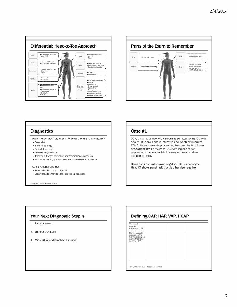
Fever in the ICU: Epidemiology
Niven et al, J Intensive Care Med 2012, 27:290.
• At least 50% of febrile episodes are non-infectious!• Most common infectious etiologies: PNA, bloodstream, abdominal infections• Most common non-infectious etiologies: post-op fever, central fever
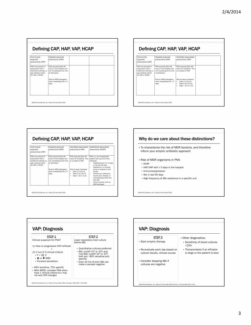
Differential: Head-to-Toe Approach
•Nosocomial meningitis (post-NSG)
•Nosocomial meningitis (post-NSG)CNS
•Nosocomial Sinusitis•URI (hospital-acquired)•Nosocomial Sinusitis•URI (hospital-acquired)
HEENT
•Hospital-acquired PNA•Empyema•ARDS
•Hospital-acquired PNA•Empyema•ARDS
Pulmonary
•Endocarditis•Pericarditis •Endocarditis•Pericarditis
Cardiac
•Abdominal abscess•SBP•Acalculous cholecystitis•Pancreatitis•C. difficile•CA-UTI
•Abdominal abscess•SBP•Acalculous cholecystitis•Pancreatitis•C. difficile•CA-UTI
GI/GU
•Osteomyelitis/septic arthritis
•Gout
•Osteomyelitis/septic arthritis
•GoutMSK
•Cellulitis at PIV/CVC •Infected decubitus ulcer •Surgical site infection
•Cellulitis at PIV/CVC •Infected decubitus ulcer •Surgical site infection
Skin
•CLABSI•Candidemia•CLABSI•Candidemia
Systemic
•Drug Fever/Withdrawal•DVT/PE•Malignancy•Rheumatologic•Central fever•Post-op fever•Transfusion reaction•Transplant rejection•Adrenal insufficiency
•Drug Fever/Withdrawal•DVT/PE•Malignancy•Rheumatologic•Central fever•Post-op fever•Transfusion reaction•Transplant rejection•Adrenal insufficiency
Other non-infectiousetiologies
2/4/2014
2
Differential: Head-to-Toe Approach
•Nosocomial meningitis (post-NSG)
•Nosocomial meningitis (post-NSG)CNS
•Nosocomial Sinusitis•URI (hospital-acquired)•Nosocomial Sinusitis•URI (hospital-acquired)
HEENT
•Hospital-acquired PNA•Empyema•ARDS
•Hospital-acquired PNA•Empyema•ARDS
Pulmonary
•Endocarditis•Pericarditis •Endocarditis•Pericarditis
Cardiac
•Abdominal abscess•SBP•Acalculous cholecystitis•Pancreatitis•C. difficile•CA-UTI
•Abdominal abscess•SBP•Acalculous cholecystitis•Pancreatitis•C. difficile•CA-UTI
GI/GU
•Osteomyelitis/septic arthritis
•Gout
•Osteomyelitis/septic arthritis
•GoutMSK
•Cellulitis at PIV/CVC •Infected decubitus ulcer •Surgical site infection
•Cellulitis at PIV/CVC •Infected decubitus ulcer •Surgical site infection
Skin
•CLABSI•Candidemia•CLABSI•Candidemia
Systemic
•Drug Fever/Withdrawal•DVT/PE•Malignancy•Rheumatologic•Central fever•Post-op fever•Transfusion reaction•Transplant rejection•Adrenal insufficiency
•Drug Fever/Withdrawal•DVT/PE•Malignancy•Rheumatologic•Central fever•Post-op fever•Transfusion reaction•Transplant rejection•Adrenal insufficiency
Other non-infectiousetiologies
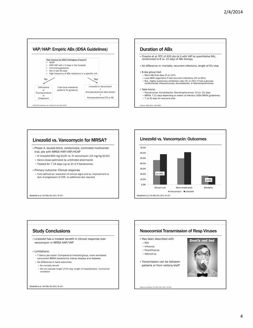
Parts of the Exam to Remember
•Careful neuro exam•Careful neuro examCNS
•Look for nasal discharge•Look for nasal dischargeHEENT
•Back and joint exam•Back and joint examMSK
•Examine line sites•Take off bandages•Peri-anal exam•Look for (drug) rashes
•Examine line sites•Take off bandages•Peri-anal exam•Look for (drug) rashes
Skin
Diagnostics
• Avoid “automatic” order sets for fever (i.e. the “pan-culture”)• Expensive• Time-consuming• Patient discomfort• Unnecessary radiation• Transfer out of the controlled unit for imaging/procedures• With more testing, you will find more colonizers/contaminants
• Use a rational approach• Start with a history and physical• Order labs/diagnostics based on clinical suspicion
O’Grady et al, Crit Care Med 2008, 35:1330.
Case #1
35 y/o man with alcoholic cirrhosis is admitted to the ICU with severe influenza A and is intubated and eventually requires ECMO. He was slowly improving but then over the last 2 days has starting having fevers to 38.3 with increasing O2 requirement. He has trouble following commands when sedation is lifted.
Blood and urine cultures are negative. CXR is unchanged. Head CT shows pansinusitis but is otherwise negative.
Your Next Diagnostic Step is:
1. Sinus puncture
2. Lumbar puncture
3. Mini-BAL or endotracheal aspirate
Defining CAP, HAP, VAP, HCAP
Community-acquired pneumonia (CAP)
PNA not acquired in association with a healthcare setting (i.e. not meeting criteria for HAP or HCAP)
IDSA/ATS Guidelines, Am J Resp Crit Care Med 2005.
2/4/2014
3
Defining CAP, HAP, VAP, HCAP
Community-acquired pneumonia (CAP)
Hospital-acquired pneumonia (HAP)
PNA not acquired in association with a healthcare setting (i.e. not meeting criteria for HAP or HCAP)
PNA acquired after 48 hours in the hospital and not incubating at the time of admission.
Risk for MDR pathogens when hospitalized for ≥ 5 days.
IDSA/ATS Guidelines, Am J Resp Crit Care Med 2005.
Defining CAP, HAP, VAP, HCAP
Community-acquired pneumonia (CAP)
Hospital-acquired pneumonia (HAP)
Ventilator-associated pneumonia (VAP)
PNA not acquired in association with a healthcare setting (i.e. not meeting criteria for HAP or HCAP)
PNA acquired after 48 hours in the hospital and not incubating at the time of admission.
Risk for MDR pathogens when hospitalized for ≥ 5 days.
PNA acquired after 48 hours of intubation. This is a subset of HAP.
Risk by days intubated:• Days 1-5 (3%/d) • Days 5-10 (2%/d)• Days > 10 (1 %/d)
IDSA/ATS Guidelines, Am J Resp Crit Care Med 2005.
Defining CAP, HAP, VAP, HCAP
Community-acquired pneumonia (CAP)
Hospital-acquired pneumonia (HAP)
Ventilator-associated pneumonia (VAP)
Healthcare-associated pneumonia (HCAP)
PNA not acquired in association with a healthcare setting (i.e. not meeting criteria for HAP or HCAP)
PNA acquired after 48 hours in the hospital and not incubating at the time of admission.
Risk for MDR pathogens when hospitalized for ≥ 5 days.
PNA acquired after 48 hours of intubation. This is a subset of HAP.
Risk by days intubated:• Days 1-5 (3%/d) • Days 5-10 (2%/d)• Days > 10 (1 %/d)
PNA in a non-hospitalized patient with any one of the following:- Hospitalization for ≥2 days
in the last 90 days- Residence in a nursing
home or long-term care facility
- Intravenous antibiotics, wound care, dialysis, or chemotherapy within the last 30d
- Family member with an MDR pathogen
IDSA/ATS Guidelines, Am J Resp Crit Care Med 2005.
• To characterize the risk of MDR bacteria, and therefore inform your empiric antibiotic approach
• Risk of MDR organisms in PNA:• HCAP• HAP/VAP with ≥ 5 days in the hospital• Immunosuppression• Abx in last 90 days• High frequency of ABx resistance in a specific unit
Why do we care about these distinctions?
IDSA/ATS Guidelines, Am J Resp Crit Care Med 2005.
VAP: Diagnosis
STEP 1Clinical suspicion for PNA?
(1) New or progressive CXR infiltrate +
(2) 2 out of 3 clinical criteria: • F > 38˚C• or WBC• Purulent secretions
• 69% sensitive, 75% specific• With ARDS: consider PNA when
have 1 clinical criteria b/c may not see CXR changes
STEP 2Lower respiratory tract culture before ABx
• Quantitative cultures preferred • BAL (cutoff 104 or 105) and
mini-BAL (cutoff 103 or 104) both are ~80% sensitive and specific
• Even 24 hrs of prior ABx can make a sample negative
IDSA/ATS Guidelines, Am J Resp Crit Care Med 2005. Klompas, JAMA 2007, 297:1583.
VAP: Diagnosis
STEP 3• Start empiric therapy
• Re-evaluate each day based on culture results, clinical course
• Consider stopping ABx if cultures are negative
• Other diagnostics:• Sensitivity of blood cultures
<25%• Thoracentesis if an effusion
is large or the patient is toxic
IDSA/ATS Guidelines, Am J Resp Crit Care Med 2005. Raman, Crit Care Med 2013, 41:0,
2/4/2014
4
VAP/HAP: Empiric ABx (IDSA Guidelines)
Ceftriaxoneor
Fluoroquinoloneor
Ertapenem
Risk Factors for MDR Pathogens Present?• HCAP• HAP/VAP with ≥ 5 days in the hospital• Immunosuppression• Abx in last 90 days• High frequency of ABx resistance in a specific unit
No Yes
Linezolid or Vancomycin+
Anti-pseudomonal beta lactam+
Anti-pseudomonal FG or AG
IDSA/ATS Guidelines, Am J Resp Crit Care Med 2005.
*Use local resistance patterns for guidance
• Chastre et al: RTC of 400 pts dx’d with VAP by quantitative BAL, randomized to 8 vs. 15 days of ABx therapy
• No difference in: mortality, recurrent infections, length of ICU stay
• 8-day group had:• More ABx-free days (9 vs 13%)• Less MDR organisms if had recurrent infections (42 vs 65%)• But…higher pulmonary reinfection rate (41 vs 25%) if had a glucose
nonfermenter (Pseudomonas, Acinetobacter, or Stenotrophomonas)
• Take-home: • Pseudmonas, Acinetobacter, Stenotrophomonas: 14 (or 15) days• MRSA: 7-21 days depending on extent of infection (IDSA MRSA guidelines)• 7 (or 8) days for everyone else
Duration of ABx
Chastre, JAMA 2003, 290:2588.
Linezolid vs. Vancomycin for MRSA?
• Phase 4, double-blind, randomized, controlled multicenter trial, pts with MRSA HAP/VAP/HCAP• IV linezolid 600 mg Q12h vs. IV vancomycin (15 mg/kg Q12h)• Vanco dose-optimized by unblinded pharmacist • Treated for 7-14 days (up to 21 d if bacteremia)
• Primary outcome: Clinical response • Cure defined as: resolution of clinical signs and sx, improvement or
lack of progression of CXR, no additional abx required
Wunderink et al, Clin Infect Dis 2012; 54: 621.
Linezolid vs. Vancomycin: Outcomes
Wunderink et al, Clin Infect Dis 2012; 54: 621.
Study Conclusions
• Linezolid has a modest benefit in clinical response over vancomycin in MRSA HAP/VAP
• Limitations:• ? Vanco pts sicker: Compared to linezolid group, more ventilated,
concurrent MRSA bacteremia, kidney disease and diabetes • No difference in hard outcomes:
• No mortality benefit
• Did not evaluate length of ICU stay, length of hospitalization, mechanical ventilation
Wunderink et al, Clin Infect Dis 2012; 54: 621.
Nosocomial Transmission of Resp Viruses
• Has been described with:• RSV• Influenza• Parainfluenza• Adenovirus
• Transmission can be between patients or from visitors/staff
Aitken and Jeffries, Clin Micro Rev 2001, 14:528.
2/4/2014
5
HAP/VAP: Take Home Points
• Think about risk factors for MDR pathogens and use that to guide empiric therapy
• Diagnosis is based on a combination of clinical and microbiologic paramters
• Duration of therapy 7 days with the exception of the glucose nonfermenters +/- MRSA
• Consider linezolid for MRSA if not responding to vancomycin
Nosocomial Sinusitis• Epidemiology:
• Radiographic sinusitis in 25-75% of ICU pts• But etiology of nosocomial fever in ~5%• Radiographic sinusitis ≠ infectious sinusitis
• Micro: Pseudomonas, S. aureus, can be polymicrobial
• Clinical: classic signs/sx of sinusitis often absent
• Dx: CT, aspirate by ENT to confirm dx and guide ABx therapy
• Treatment duration: 7 days
O’Grady et al, Crit Care Med 2008, 35:1330. George et al, Clin Infect Dis 1998, 27:463. Talmor et al, Clin Infect Dis 1997, 25:1441. Borman et al, JAMA 1992, 164:412. Stein and Kaplan, Curr Opin Infect Dis 2005, 18:147.
A 65 y/o M is admitted with an STEMI. 4 days into his hospitalization he spikes a fever to 39, drops his SaO2 to the low 90s on RA, and becomes altered. He has a foley. He is started on vancomycin and pip/tazo and improves.
Work-up reveals:• CXR with a new LLL infiltrate• Blood cultures and sputum culture negative at 48h• UA (from his catheter) shows 30 WBC, Urine cx >100K VRE
Case #2 What would you do with his ABx?
1. Continue pip-tazo and d/c vanco
2. Continue pip-tazo and change vancomycin to linezolid to cover VRE
CA-UTI is a Diagnosis of Exclusion!
• Catheter-associated bacteriuria is common (up to 25% of patients with short term catheters)
• CA-bacteriuria usually represents colonization (asymptomatic bacteriuria = ASB) and NOT infection
• What about pyuria?• Pyuria is common in catheterized patients (up to 75% with short term
catheters)!• The presence or degree of pyuria cannot differentiate ASB from UTI• But, the absence of pyuria suggests an alternative diagnosis
Hooton, CID 2010. Nicolle, CID 2005. Tambyah, Arch Intern Med 2000.
1. Patient with a catheter currently or within the last 48 h2. Symptoms or signs c/w UTI3. No other source of infection (i.e., diagnosis of exclusion)4. ≥103 cfu of ≥1 bacterial species in a urine culture
Catheter-associated UTI: Definition
Hooton, CID 2010.
2/4/2014
6
• What are “signs and symptoms compatible w/UTI” in a patient with a catheter?• New onset or worsening of fever, rigors, altered mental status, malaise
or lethargy with no other identified cause• Flank pain or CVAT• Acute hematuria• Pelvic discomfort• Spinal cord injury patients: increased spasticity, autonomic dysreflexia,
or sense of unease
The million dollar question…
Hooton, CID 2010.
• First, STOP: do you need a UA/urine culture if you have a high suspicion for an alternative source (eg PNA)?
• Second: if you are sending a urine culture, always get a UA
• How to get the best culture?• A catheter culture may not accurately reflect what is in the bladder• If a catheter has been in place for >2 weeks, change it and get a Ucx
from the newly placed catheter• In a patient with a condom cath, get a specimen from a freshly placed
condom cath after cleaning the glans because skin can be colonized
Diagnosis
Hooton, CID 2010.
Signs and Symptoms of Systemic Infection without classic UTI signs/ sxAlternate Diagnosis Likely?
(Signs/ sx of other illness present)
Yes
Do not order U/A, urine cx
No
Send U/A, urine cx
U/A, urine cx (-)
Do not treat for UTI
U/A (-), urine cx (+)
Asymptomatic bacteriuria- Do not Rx for UTI*- Look for alternate dx
U/A (+), urine cx (+)
Treat for UTI-If no alternate dx identified
*Exceptions: pregnancy, pre-urological procedure, neutropenic host
U/A (+), urine cx (-)
On abx No abx
Do not Rx for UTI
Empiric Therapy for Complicated UTI
• Community acquired:• Ertapenem• Ceftriaxone
• Healthcare associated:• Pip/tazo• Ertapenem• Ceftriaxone
• Duration:• 7 days if there is prompt resolution of symptoms• 10-14 days if response is delayed
• Catheter change? • Yes, if the catheter has been in for >2 weeks, change it• This has been associated with:
• CA-bacteriuria and CA-UTI at 28d• time to resolution of sx
Treatment
Hooton, CID 2010.
• Asymptomatic candiduria:• In general, don’t treat!• Change the foley: can eliminate candiduria in 20-40%• Exceptions: Patients at high risk of dissemination
• Neutropenia• Patients undergoing urologic procedures
• Symptomatic candiduria: treat
Candiduria: Who to Treat?
Pappas, CID 2009.
2/4/2014
7
1st line: Fluconazole• Excellent urine levels, 10-fold higher than serum levels
• Can get concentrations in the urine that are higher than the MIC for organisms that are intermediate or resistant
• 200-400mg PO daily
Candida UTI: Treatment Options
Fisher, CID 2011.
• Can try fluconazole and re-check Ucx (if not systemically ill)
• Other options:• Flucytosine• Amphotericin B • Ampho bladder washes: Resolve candiduria in >90% but relapses
• Other azoles? • Vori, posa, itra have poor urinary penetration
• Caspofungin? • Poor urinary penetration, but use if suspect systemic disease
Fluconazole-Resistant Candida UTI
Fisher, CID 2011.
CA-UTI: Take-home points
• ASB and pyuria are common in patients with a foley
• To diagnose a CA-UTI, the patient must have:• Signs and symptoms compatible with UTI • No other source for infection
• Treat for 7-14 days depending on clinical response
• Candiduria is almost always asymptomatic and does not require treatment
Case #3
A 55 year old man is admitted to the ICU with severe gallstone pancreatitis. He has been afebrile and slowly improving on TPN and with conservative measures. He spikes a fever to 39 associated with new hypotension and rigors. Blood cultures drawn from his triple lumen subclavian line turn positive 6 hours before his peripheral blood culture. Both are growing E. coli.
The most sensitive test to determine the source of the E coli is:1. Differential time to positivity
2. Inflammation around the central line exit site
3. Remove the catheter and culture the tip
The IDSA Guidelines recommend:
1. Pull the line
2. Ok to attempt line salvage with ABx lock therapy
3. Ok to attempt line salvage by giving systemic ABx through the line
2/4/2014
8
CLABSI: Diagnosis
• Clinical findings unreliable: • Inflammation at the exit site is extremely insensitive (<3%)
• Positive peripheral bcx and > 15 CFU/plate of same organism from catheter tip• 79% sensitive, 92% specific• But >80% of catheters withdrawn b/c of clinical suspicion of CLABSI are
removed unnecessarily
• Quantitative blood cultures (line vs peripheral) may be most sensitive/specific, but not routinely available
Mermel et al CID 2009. Safdar and Maki, Crit Care Med 2002, 30:2632.
CLABSI: Differential Time to Positivity• Allows for diagnosis without removing the line
• Draw culture from central line and peripheral blood at the same time
• CLABSI = blood culture drawn from central line turns positive at least 2 hrs before peripheral culture turns positive
• Test characteristics• 95% sensitive• 90% specific (but only ~40% specific for yeast)
Liñares, Clin Infect Dis 2007, 44:827. Bouza et al, Clin Infect Dis 2007, 44:820. Bouza et al, Clin MicrobiolInfect 2013, 19: E129.
CLABSI: Management
• Possible scenarios:• Line (+)/ peripheral (+)
• DTTP ≥ 2 hrs CLABSI remove line and treat• DTTP < 2 hrs look for another source and treat
• Line (+)/ peripheral (-) colonization/ contaminant
• Duration of treatment (with line removal): • 5-7 days: coagulase negative Staphylococci
• 7-14 days: GNRs, Enterococcus
• 14 days: S. aureus, Candida
Mermel et al CID 2009
CLABSI: Line Management
• Remove line whenever possible:• Guidewire exchange only if no alternative insertion site• If septic, hemodynamically unstable, persistent bacteremia remove line • Long term catheters: remove if S aureus, Pseudomonas, Candida• Short term catheters: remove if S aureus, GNRs, Candida, Enterococcus
• Line salvage• Antibiotic lock therapy + systemic antibiotic therapy• Studied primarily in long-term catheters
Mermel et al CID 2009
Case #4
85 y/o man is admitted with fever and respiratory failure to the ICU and treated with vanc/pip-tazo. He initially responds but then 5 days into therapy he began spiking high fevers up to 39˚C daily. His respiratory status is unchanged. He is escalated to vanc/meropenem with no change in his fever or respiratory status after another 5 days. Extensive work-up for other sources of infection is negative.
What is Your Next Step?
1. Change vanco to linezolid
2. Add tobramycin
3. Stop antibiotics
2/4/2014
9
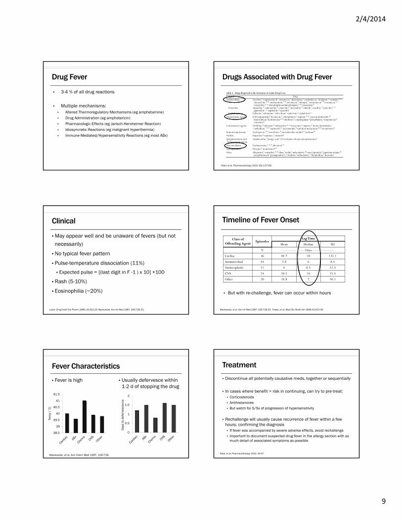
Drug Fever
• 3-4 % of all drug reactions
• Multiple mechanisms:• Altered Thermoregulatory Mechanisms (eg amphetamine)• Drug Administration (eg amphotericin)• Pharmacologic Effects (eg Jarisch-Herxheimer Reaction)• Idiosyncratic Reactions (eg malignant hyperthermia)• Immune-Mediated/Hypersensitivity Reactions (eg most ABx)
Drugs Associated with Drug Fever
Patel, et al. Pharmacotherapy 2010; 30(1):57-69.
Clinical
• May appear well and be unaware of fevers (but not necessarily)
• No typical fever pattern
• Pulse-temperature dissociation (11%)
• Expected pulse = [(last digit in F -1 ) x 10] +100
• Rash (5-10%)
• Eosinophilia (~20%)
Labor. Drug Intell Clin Pharm 1986; 20:413-20. Mackowiak, Ann Int Med 1987; 106:728-33. Mackowiak, et al. Ann Int Med 1987; 106:728-33. Foster, et al. Med Clin North Am 1966;42:523-39
Class of Offending Agent
EpisodesLag Time
Mean Median SD
N --------------------Days--------------------
Cardiac 36 44.7 10 131.1
Antimicrobial 44 7.8 6 8.4
Antineoplastic 11 6 0.5 12.3
CNS 24 18.5 16 15.4
Other 20 18.8 7 34.1
• But with re-challenge, fever can occur within hours
Timeline of Fever Onset
Fever Characteristics
• Fever is high • Usually defervesce within 1-2 d of stopping the drug
Mackowiak, et al, Ann Intern Med 1987, 106:728.
38.5
39
39.5
40
40.5
41
41.5
0
0.5
1
1.5
2
Tem
p (˚
C)
Day
s to
def
erve
scen
ce
Treatment
• Discontinue all potentially causative meds, together or sequentially
• In cases where benefit > risk in continuing, can try to pre-treat: • Corticosteroids• Antihistamines• But watch for S/Sx of progression of hypersensitivity
• Rechallenge will usually cause recurrence of fever within a few hours, confirming the diagnosis• If fever was accompanied by severe adverse effects, avoid rechallenge• Important to document suspected drug fever in the allergy section with as
much detail of associated symptoms as possible
Patel, et al, Pharmacotherapy 2010, 30:57.
2/4/2014
10
Cross-Reactivity of Antibiotics?
Change to another class if possible (i.e. Beta-lactam to fluoroquinolone
No studies exist which address drug fever cross reactivity specifically – focus is on all symptoms of hypersensitivity
Joint Task Force on Practice Parameters. Ann Allergy Asthma Immunol 1999; 83:665-700.
Drug Fever: Take Home Points
• Always consider it in the ddx for fever in the hospital
• Look for eos, temp-pulse dissociation, rash although remember these are present in <20% of cases
• Consider stopping the ABx or swtiching classes if you really suspect it
• Remember to document drug fever as an allergy!
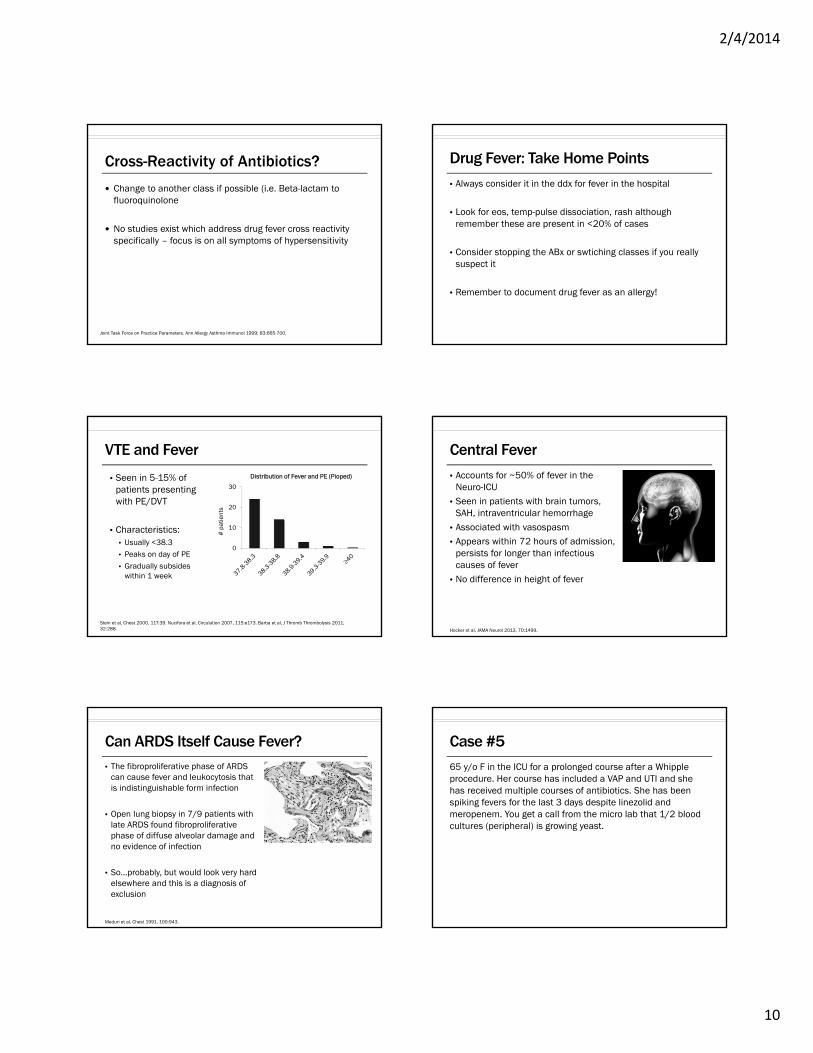
VTE and Fever
• Seen in 5-15% of patients presenting with PE/DVT
• Characteristics:• Usually <38.3• Peaks on day of PE• Gradually subsides
within 1 week
0
10
20
30
Distribution of Fever and PE (Pioped)
Stein et al, Chest 2000, 117:39. Nucifora et al, Circulation 2007, 115:e173. Barba et al, J Thromb Thrombolysis 2011, 32:288.
# p
atie
nts
Central Fever
• Accounts for ~50% of fever in the Neuro-ICU
• Seen in patients with brain tumors, SAH, intraventricular hemorrhage
• Associated with vasospasm• Appears within 72 hours of admission,
persists for longer than infectious causes of fever
• No difference in height of fever
Hocker et al, JAMA Neurol 2013, 70:1499.
Can ARDS Itself Cause Fever?• The fibroproliferative phase of ARDS
can cause fever and leukocytosis that is indistinguishable form infection
• Open lung biopsy in 7/9 patients with late ARDS found fibroproliferativephase of diffuse alveolar damage and no evidence of infection
• So…probably, but would look very hard elsewhere and this is a diagnosis of exclusion
Meduri et al, Chest 1991, 100:943.
65 y/o F in the ICU for a prolonged course after a Whipple procedure. Her course has included a VAP and UTI and she has received multiple courses of antibiotics. She has been spiking fevers for the last 3 days despite linezolid and meropenem. You get a call from the micro lab that 1/2 blood cultures (peripheral) is growing yeast.
Case #5
2/4/2014
11
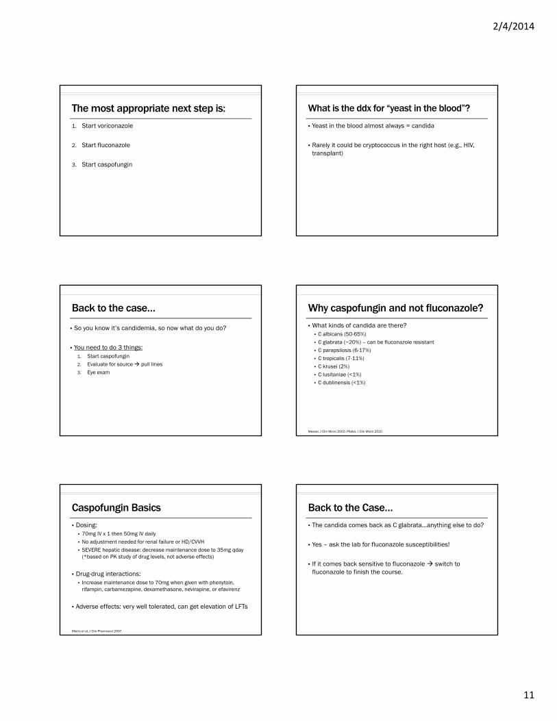
The most appropriate next step is:
1. Start voriconazole
2. Start fluconazole
3. Start caspofungin
• Yeast in the blood almost always = candida
• Rarely it could be cryptococcus in the right host (e.g., HIV, transplant)
What is the ddx for “yeast in the blood”?
• So you know it’s candidemia, so now what do you do?
• You need to do 3 things:1. Start caspofungin2. Evaluate for source pull lines3. Eye exam
Back to the case…
• What kinds of candida are there?• C albicans (50-65%)• C glabrata (~20%) – can be fluconazole resistant• C parapsilosis (6-17%)• C tropicalis (7-11%)• C krusei (2%)• C lusitaniae (<1%) • C dublinensis (<1%)
Why caspofungin and not fluconazole?
Messer, J Clin Micro 2003. Pfaller, J Clin Micro 2010.
• Dosing:• 70mg IV x 1 then 50mg IV daily• No adjustment needed for renal failure or HD/CVVH• SEVERE hepatic disease: decrease maintenance dose to 35mg qday
(*based on PK study of drug levels, not adverse effects)
• Drug-drug interactions:• Increase maintenance dose to 70mg when given with phenytoin,
rifampin, carbamezapine, dexamethasone, nevirapine, or efavirenz
• Adverse effects: very well tolerated, can get elevation of LFTs
Caspofungin Basics
Mistry et al, J Clin Pharmacol 2007.
• The candida comes back as C glabrata…anything else to do?
• Yes – ask the lab for fluconazole susceptibilities!
• If it comes back sensitive to fluconazole switch to fluconazole to finish the course.
Back to the Case…
2/4/2014
12
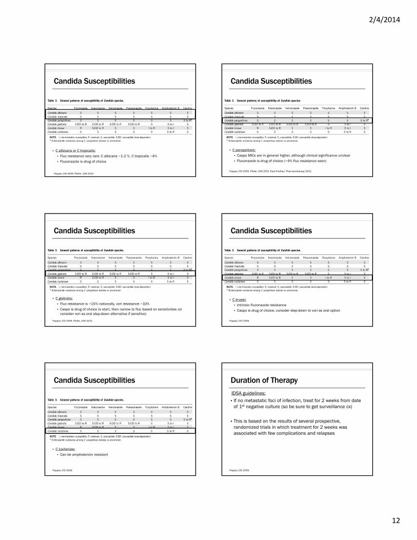
Candida Susceptibilities
• C albicans or C tropicalis:• Fluc resistance very rare: C albicans ~1-2 %, C tropicalis ~4% • Fluconazole is drug of choice
Pappas, CID 2009. Pfaller, JCM 2010.
• C parapsilosis:• Caspo MICs are in general higher, although clinical significance unclear• Fluconazole is drug of choice (~4% fluc resistance seen)
Candida Susceptibilities
Pappas, CID 2009. Pfaller, JCM 2010. Kale-Pradhan, Pharmacotherapy 2010.
• C glabrata:• Fluc resistance is ~15% nationally, vori resistance ~10% • Caspo is drug of choice to start, then narrow to fluc based on sensitivities (or
consider vori as oral step-down alternative if sensitive)
Candida Susceptibilities
Pappas, CID 2009. Pfaller, JCM 2010.
• C krusei:• Intrinsic fluconazole resistance• Caspo is drug of choice, consider step-down to vori as oral option
Candida Susceptibilities
Pappas, CID 2009.
• C lusitaniae:• Can be amphotericin resistant
Candida Susceptibilities
Pappas, CID 2009.
IDSA guidelines:• If no metastatic foci of infection, treat for 2 weeks from date
of 1st negative culture (so be sure to get surveillance cx)
• This is based on the results of several prospective, randomized trials in which treatment for 2 weeks was associated with few complications and relapses
Duration of Therapy
Pappas, CID 2009.
2/4/2014
13
• Remove cathethers if possible:• Often difficult to tell in an individual patient if catheter or GI is the
source• Exception: C parapsilosis is often catheter-associated• Removal is associated with more rapid clearance of blood cultures and
decreased mortality • Note that this data is less compelling in neutropenic patients, so
recommendation to remove catheters is less strong in this population
Pull the line!
• Rule out chorioretinitis (seen in ~10%) or endophthalmitis(seen in 1-2%)
• This is not an emergency (unless having visual symptoms)
• In fact, may increase your sensitivity by waiting ~1 week after starting therapy
Get an Eye Exam
Oude Lashof , CID 2011.
• Intravitreal injections (ampho)
• Longer duration of therapy (4-6 weeks)
• Choose an agent with good eye penetration • Azoles (voriconazole>fluconazole)• Ampho + 5-FC• NOT echinocandins (have poor ocular penetration)
Why does this matter?
1. Start an echinocandin empirically• Check surveillance cx in 48hr• Get susceptibilities if it’s C glabrata and change to fluc if sensitive• Change to fluc if it’s a susceptible species (albicans, tropicalis,
parapsilosis)• Treat for 2 weeks from the date of the 1st negative culture
2. Pull the line
3. Eye exam• If positive, use vori if sensitive• Duration of therapy 4-6 weeks for eye involvement
Candidemia: Take Home Points
What is the role of procalcitonin in the ICU?
• Diagnosis of sepsis vs. non-infectious SIRS• Meta-analysis of 18 studies suggests low diagnostic accuracy (sensitivity/
specificity of ~ 70%)
• Aid in antibiotic discontinuation• Meta-analysis of 14 RCTs investigating PCT algorithms for antibiotic
therapy decisions • Reduced antibiotic exposure by 20-37% • No mortality difference
• Decr LOS in some studies, not in others
Tang et al Lancet ID 2007; Schuetz Arch Intern Med 2011.
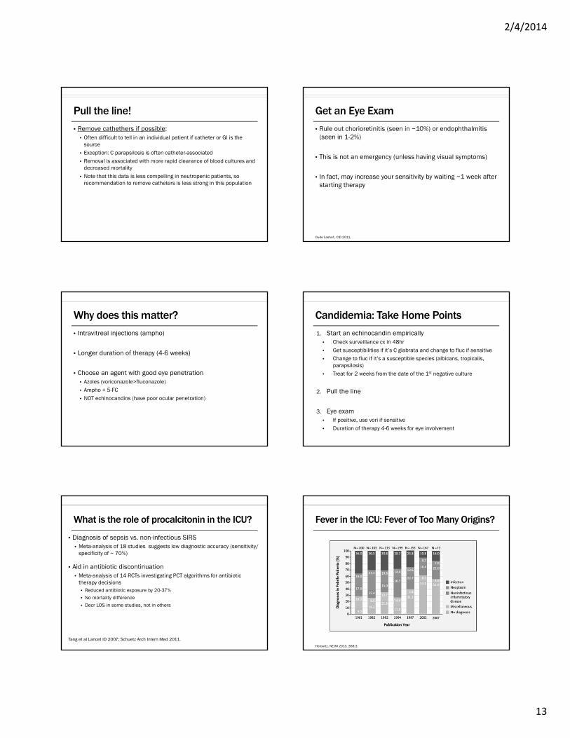
Fever in the ICU: Fever of Too Many Origins?
Horowitz, NEJM 2013, 368:3.
2/4/2014
14
Thank you!