($fi(./ - inflibnetshodhganga.inflibnet.ac.in/bitstream/10603/6162/16/16_paper... · icffect of...
TRANSCRIPT


Icffect of Brah~na Rasayana 011 Antioxitlirnt Systems and Cy- tokine Levels in Mice During Cyclophospt~amide Adrninis- tration
I l i t r ; r~~~r iLoneal administration or cyclophosphamide (( 'TX) 711r;'L;: h \ ~ t . ~ l i~sc imousc for 10 days u s l i , r ~ l r c l to supprcss the tissue ;111d serum level o f red~lced f l u l a l l ~ i o~ i c iGSI I ) . blood glu~athione peroxidase ([; l 'X) and tissue levels o f sllpcroxide dislnutase (SOD) alrd c:~rill;~.;v (CAT) Tlssue levcls o f g lu t~ t l i i one 1~011c1:rsc (GR) and glutatliiolie-S-irnnsf'erase (GST) were ~rnaltcrccl hv C'IX ircalmcnt wl i i lc serum and 11s- .,IIC lipid peroxicle levcls \!,ere signi f icant ly increased. 01:rI , i i i ~ i i ~ l i i s t ~ a t ~ i ~ n 01. Rralima Rasayana RR- 50n r~~ .~ i~ose l1nou re for 10 days a n d 3 0 days significantly enlrnlirc(1 t l ~ c lissrlc lcvels of- SOD. CAT. (;ST. CiI'X. i i r u r n and tissue GSI1 and signi f icant ly reduced thc s e l i i l r l .11iiI i lss~ ic l ip id peroxidation. B R treat- niclit was also found to enliance tlie seruln cytokine level o i i r ~ ~ c r l c l r ~ l l - ~ ( l l - N . i ) iliterleukin-2 (1L-2) and granlilocytc rnaciophagc- colony st i lnulat ing factor(GM-CSF) 111 ~nor~ l ia l arid C1.X treated mice. The re- ~LIIIS are indicative of' the usc o f B K to rcduce tlie oxidn~it slrt.s:> ~~ iduccc l h) C T X treatment and its ~:ili.ct I ccllular function.
I(L,~ Lt'ords: Bral ima Rasayana, (~yclophosphamide, Antioxldnlit eliryn1cs. l i p i d pcrouidalion, Cytokines
Cancer cl icmotherapy generally produces non- selcctlve cel l k i l l i ng as i t affects normal as wel l :IS c:lnccrous tlssues. Cyc lo l~ l iosphamide ( C T X ) is an a l k ~ l . ~ t l n g asellt used as 3 clicmotherape~rtic agent. Micr~ iso l i ia l oxidase systcln in l ivcr converts the C l X to 4- l iydroxy cyclopliosphnrnide, wl i ich re- CI~ICIS (lie c l r cu la t io~ i arid is suliscquently taken rlpto pcrip1icr;ll tissues and tumor. wl iele i t under- :iils.> ~ . n ~ t o ~ i i c ~ i s a t ~ o ~ i to aldnpliospliarnidc. Sponta- null15 ~lcgr:rcl;~lion o f a l d ~ > l ~ l ~ o s ~ ~ l l ; i r ~ ~ i c l ~ ~ to hrln a
. . i c ; ~ c t ~ \ c rc;lgcnl ac ro l c~ r~ . I S caliahlc o f t lcpleling cc l lo l i~ r glulatl i ionc (GSt1) and cnasing DNA alky- l i l t l l l l l (! ) .
/\yrlrvcda, the tradit ional I l idien system o f med- i c I givcri great ernllhasls or1 thc promotion 111 I~c;i lrh. Drahma I<asayana (R IO is a non-toxic po lv l~c~-ba l preparation. w l ~ i c l i is made b y the ellracts f rom plants w i th vnnous biological i~ct lv l ics including anr~oxidant, immunomotlulation ctc. I<asayana has been showri to stimulate both I ~ I I I I I I ~ 2 and cell-medi;~lccl imni l ln iry in inice
(3) . and \\a, 1n1i11d to iedtici. the leukocytopcnin produced t)y cyclol)liospliamide (4). Recently we have rcportcci tlr:ir RI< \+as t i x~nd to be a potent oxygen fret, I I sca\ lc~~gcr in V I I ~ O and in 1.iv11 (5). DR u n , also i o r ~ ~ i i l to reduce the radiation induced c l i~sto$el~ ic i ly 111 eupcrilnental animals (6) atid cotlld I>II>IV~.I lllr. I~;~cmarological systcm i r i patlclils rlnilcrforn$ rl icniotl irrapy and radiation t i c 1 1 0 I ' I I I g r e d n t in RR is 1 1 1 1 / I \\Ii,clr I 101111d to l iavc vcr? I ~ i g h I I I I I I ~ I c ~ i i I I O (8). In the lire- scnl s~udy \\.? liavc c\;~Iilat:d [lie e rec t o f BI< on clidogcnous n i i t l ~ x i ( l ; ~ ~ i t CIIZYII~CC and cytokine pro- ductioli i r i mlcc rreihcd \\'it11 C T X .
Mater ia ls a1111 h l r t l ~ o d s
Brnhlna I<c~; .~y,~~~,~ (UI11 w i ~ s purchased fiom Vaidy:rrailla~il 0 1 l 1 1 l l i i s i l i Ollur. Kerala, I l l i l i a . N i i o l l ~ ~ C I I ~ I I I I I I I ) ~~ i co t i nam ide ;~dcnlnc

i l~r~uclcot ide phosphate reduced ictmsodium salt (b~l)I'Ii Na,), reduced glutath~one (<?SII). oxidised Klu~nt l~ ione (GSSG), 5-5' (lithiobis (2-nitrohcnzolc ac~d)(l)TNB), I - a l i ~ i i i e 4-aminoantipyrennc and r~-keloglut;~r~c acld were p~~rcl iased r r o ~ n Sisco I(c!,carcl~ l.ahoratories (SItL) Pvl.Lld.. Mumhai. I- chli,rti-2.4-d11iitr<)hcnze1ie (CDNB) ucas obtained l?um 1.aha Clicniic Pvt. Ltd.. Mumh;i~. 1 ,I .3.3-Tetra inctlioxy propane (09%) was obtained i'rom M T M ltc\c;~rcli C'liemicals. Eastgate. England. T1iloharh1- t u ~ ~ c acid (TI3A) was obta~~icd lrum U D H Lahoralo- ry Supplies. Pocile, England. Plienyl phosphate dis- ot i~um salt diliydrate (99%) obtained f rom Lancast- cr Synthesis, 15astgate. England. Cyrlophosphamide ( ( ' I X ) was obtained fioni Dahur Pliar~naceuticals. New, Dellii, India. Glurath~one peroxidase (GI'X) kit was obta~ned fi-orn Itandox Laboratories Ltd.. Ard- tnorc, U.K. Mousc cytokine elise hits (IFN-y, IL-2, (;M-CSI') were obtainecl lion1 Endogen, I ISA. A l l citl~cr cl~en,~cals and rcagerirs used were o r irnalyt- 1c.d ~ rade . Inbred strains o l Swiss albino mice (4-
wccks old, 20-25gi \\ere p~~rc l iascd rrom our :in~rnal rann and were lioused 111 ventilated cages in all- controlled rooms and fed wi lh normal mouse cliow (I-lpton, India) arld water ad l i h ~ r u n ~ .
I I~IIIII~I I 1 / ( R on ~17(luge- ~io i rs ~ ~ l r i o x i d o n ~ ~nzv~n(!.~ ond lipid peroxidorion. Swiss alblno niice were d ~ v ~ d e d into five groups of six animals ecleh as given belo\r;
(;roup 'Treat~ne~~t \
I Normal co~itrol I I Cyclophosphan~ide(C'TX)control
251ny/kg.h.wt./mouse, I 0 days ip. I l l CTX control-:!5mg/kg.b.\vt./n1ouse.
I 0 days ip and \vas kept ior 30 days. IV (~TX+l3K-5011igidose/1nouse, 1Cldays po. V CTX+6R-5On~~idose/1nouse, 30days po. The nnilnals o f group I. ll, and I V were sucri-
ticed on [he I l t h day and other group5 on the 3lst d.1) by celvical dislocatio~~. Blood was collected by Ihcan l n ~ n c t ~ ~ r e irnmedl.llcl! a ~ i d Iiber \\as excised : ~ i ~ i l t l io ro~~ghly waslicd 111 ice-cold saline (0.9%). L ~ \ c r lio~nogenate \%'as preparcd in ice-cold Tris- 1iC1 hult'cr (pH 7.4) and cytosol~c sample o f liver hornogenatc was prepar-ctl by centrifuging at I(1.000rprn for 30min. at .IYC. 'The hlood, serum, I Iiomojienate and cytosol were used for the h ~ i ~ c l i e l ~ ~ i c a l analysis.
The lbl lowing biocl,e~i,ical parameters \rere a11:llpsed. I'rotein n8:ls est~lnatcd by Lo\vry3s mctliod ('1'1. Superoxidc d~.;nintasc (SOD) act~vi ty o f liver
tissue was detc~mincd by NR'I' leduction method o f Mc Cord and I :ndo\~cl~ ( L O ) . C'atalase (CA-I) activity was estimated hy the riicthod of' Aebi et al ( I I ) by measuring the rate o f dccompos~tion o f hydrogen
aL 240nm. Reduced glutathione. (GSH) act iv i~y in serum and l~\,er tissue were lneasured hy the method o r Moron c~ al ( I 2) hased on the reac- tion w ~ t l ~ 5 -5 'd1 t111o l i1~(2 -n1 t ro l~e1~zo ic acid). Glu- talh~onc pcroxidasc (GI'X) acttvtty o f l iver tiss~jc was dcterm~li&l hy t l ~ e mctliod of Hafernan11 et al (13) based 011 r l~c degradation o f hydrogen perox- ide III the presence o f reduced glurathionc.
GPX activity o f blood w;ls cs~imated by Paglia and Valentine's method (14) (kit supplied by Randox Laboratories, U.K.) based on curnene hydroperoxide mediated oxidatio~i o r rcduccd glutathione by GPX. Glutath~one-S-tr;rnskrase ((;SI-) activity ofcytosolic liver- sa~~ ip l c was est~lllatcil by the method o f l labip e l a l ( l 5 ) hased on r:hc or i!icrease in con,jugate for- rna~ io~ i bct\vee~i I-educed g l~~ ta t l~ ionc and I-chloro- 2.4-dinitrobenzcrie. The a c t ~ \ i ~ y o f cytosolic liver ,ample o f gl~itat l~ione reductase (CiR) was ~neasured by Racker's method (10) tl:ised on the amount o f reduced rorm 01 r~icoiina~iiidc adenine dinuclcotide phospllate consun~i,d dul-in: the conversion o f oxi- dised gl l~tat l~ione to educed glutathio~~e. Lipid per- oxide (LPO) level In scrurn and liver were eslimat- ed using tliiobarbilurlc ac~d ( T B A ) method o f Ol~kawa et a1 (1 7).
Delern~irlarlon ofrhi, ($fi(./ r ~ f OR on cyrokine producrio~l. llibrcd srralns o f 6alb/c mice were used f o ~ the experllneni. l'11e a111mals were divided into four gl-oups. Group I \\.as treated as normal. Group I1 was treated i \ ,~fh I 0 doscs o f UR (SOmddoseiani- nlal, lbr I 0 days po) Group Ill al~irnals were treat- ed with 10 doses o f C'TX (25mglkg.b.wt.. ip) Cor I 0 days. Group I\/ was treatcd w i ~ h same dose o f C'TX as above lind UR (50mg/dose/animal, for 10 days po) A l l ani~lials wcrc sacriliced on thc l l t h day. l i lood w;is collected hy hearc punclure aild ser-unl w:~:, st.pal;rted, cytokilie lcvcls of interlbron- y (IFN- y), i i~tcrlcukin-?(lL-2) and granuloeyte macrophage- colony stimulat~ng factor (CiM-CSF) were dctermincd lby illoust Elisa kits, Endogen, USA.
S ~ J I I I I I . I \vas expressed as mean + slandal-d <le\,iation (SD). Significance lev- els for compariso~~ u f d~t'l'rl-r~,ces were determined usins Student's t tesi.
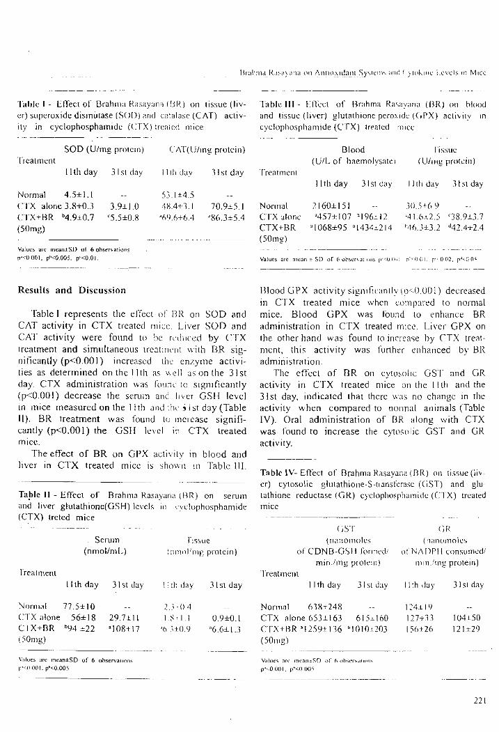
I c I - Cll'ecr o f Drali111.1 1K;liayan;l l l3K) 011 tissue ( l iv- cr) superoxide dislnu~ase (SO[)) ;in0 c;~talasc (CAT) activ- ity in cyclophospha~nidc ( t : l 'X) 1rt:;licd mice
~ ~
SOD (Uirn_r protein) C ' A ~ l ( . l J i ~ n ~ protein) Trealment
I I tl i day -7 I sl day I i 111 i l ;~v 3 I sr day
Nnrnial 4.511.1 .. 53. l i 4 .5 ..
('1X alonc 3.810.3 3.9*1.0 1X.4t3.1 70.915.1 (: lX+HR b4.9i0.7 '5.53~0.8 ^hq.h+h.4 '86.315.4 ( 5 0 ~ )
~~~ ~ ~ ~ ~ . ~
Vdlurr rrc n!eamSIJ or 6ohrci\alio8~3 p'.:O 001. ph-:000S. p'<U.Ol . ~ ~~- .. ~- ~ - ~ -
R e s ~ r l l s a n d Discuss ion
Table I represents the e rec t of D R or1 SOD and C A T act iv i ty i n C T X trcatcil mice. L i v e r SOD and C A T act iv i ty were found to bc r~. i I i lccd by C T X treatment and simultaneous trcatmc,lil w i t h BK sig- ni f icanl ly (p i0 .001) increased [he enzyme act iv i - ties as determined o n the I l th as i\ c l l as o n the 3 I st day. C T X administrat ion was ii111nd to s i g n ~ f i c a n t l y (p<0.001) decrease the senlin ant lhver G S H level In Inice measured o n the I I ti) and tic 3 1 st day (Table 11). BI< treatment was round lo lnclease signif i- cant ly (p<0.001) the G S I I I c \ c l in C T X treated mice.
' l ' ~ ~ l ) l c 111 - Ell>ct of h r n R : I I ) I blood and t~ssuc ( l ivcr) ~ l u t a r l i ~ o n e peroklilc ((;[ 'X) acl i \ , l~" in cyclopliospliamide (CTX) treated nilcc .- ~- ...~ ~
Blood IISS~IC ( U I L o f liaemolyi:~tei (Ui ing protc i l~)
Treatmenl I l t h day 31sl d:~y I l ~ l i day 71sr day
N o r ~ r ~ a l ?1603Z151 -~ 3ii.5+0 9 ..
(:rX alonc "4571107 "196=12 141.6i2.5 '35.93Z3.7 CTX+RR '1068195 "434r2 14 46..li3.2 "424+2.4 (50mg)
~ -- ~~ - ~~ . ~ - ~~
Valucr arc n l u m = SI) or b o ~ . r c i \ a t ~ ~ . t n I,':" f 1 t . i ?',O 01 v. 11 0 7 , p " ~ f 1 0 ~
-- ~ ~ ~ ~ ~ ~~ ~ ~~ ~ --
Blood G P X act iv i ty s iyn i f ican l l j ip~~:.O.OOI) decreased i n C I - X treated rnice whcn ci)t~?pa!-cd to normal mice. B l o o d G P X was found i o mhance BK admin isk i t i on i n C.rX treated ~ i i ~ c e . I.ivcr G P X on the other hand was found to increase lhy C T S treat- ment, this a c t ~ v i t y was furlher elihanced b y till administration.
The etyect o f RR on cy~oso l lc G S I and GR ac l iv i ty in C T X treated mice or1 the l l t l i arid the 31st day, indicated that thcrc was no change In ihc act iv i ty \\'hen compared to oorinal animals (Table IV) . Ora l administrat ion o f BR irlong w i th C T X was round to increase the cytosc>lic G S T arid GR activity.
The effect o r BR o n G P X act ix i ty i n b l o o d and - ~ ~ ~~ -~ ~- ~ ~
l iver i n CTX treated rnicc is shown i n Tablc 111. Tablr I L - Effect o f Drahrna Kasavan;~ il3R) 011 tissue i l i r~
Table I 1 - Ef fcc~ of Rrah~iia Kasayana ( R R ) on serurii arid liver glutathionc(GSH) levrls 111 cycl<~phospharnide (CTX) lreted mice
~ ~ ~ ~
I re;llniclil I l t h day 31sr day I l r l l rlay 3 1 s day
er) cytosolic glutathione-S-transicrasc ((;ST) and g l u ~ [athione reductase (GR) cyclopl~ospti;~~iiirIc (C l -X ) ~reatcd rnice
~ - - - ~ - - ~ ~ ~ -~ ~ ~
(i51~ ( ; ri ( ~ ~ ; ~ n o m ~ ~ l c r ( ~ ~ n ~ ~ i ~ ~ i l o l c s
o l CDND-GSI I I h r ~ ~ i e d i ill' N h I)PI I con~umcdl n i i n l m g prolt' l~ii 111n.'!ng prolelrl)
Treatiiienl I l t h day 31s d:~y I l th day 31sr day
Nornlal 638*24X -- l?4L lc) ..
0.93Z0.1 CTX alone 653k163 hl51lhO 1 2 7 4 3 104t50 %.611.3 CTS+HR '1259i 136 b10101?03 ISht26 121129
(50rng) ~~ .... - ~ -
V;mlilc< I,,. n>ran;St> o l i,oh\crr.t,#,,w.
OU I . 1,*<0 W'

~ ~ - ~ ~~~ -
Table \'- I!ttcc~ 01' 13rahliia Rasayalia (lil0 i r r l wrllln ~ I I I ~
tissuc ( I~vcr) li111d pcr~~xld:ltio~r in C~CIOPIIOI~II~~!~I~C. (CTX) lrealcd ~nicc
- - ~.~ ~ ~~ ~
Malonaldcliyde(MUA) l i~rmcd Serum (n1nol11nL) Tiss~rc (nn~n l ln~p prolcln)
'I~rcatmcnl 11th day 31s1 day I l r l ~ <la) i 1 s 1 day
Nor111al 1.8i0.3 .. 2 2i0.4 ..
CTX alone q.3 i0 .6 2.3i0.4 d3.7:0.6 "5110.9 CTX+BR 9.3i0.3 "1.6*0.3 '2910.4 "i.8+0.4 (501ng)
~ ~ -~~~~ -
The elicct ol' B R on seruilr a ~ l d l i i c r Ihpici pcr- osidation is given in Table V DOIII si.rllnl i i l i c i l ~ v e r l ip id peroxide lcvel u e r v l o u ~ ~ d to be s ~ g n ~ l i c ; % ~ l r l v (pc0.001) increased 01, ihc Ill11 and tlie 3151 day 111 CTX treated micc. B R treaili1elll s i y ~ l l i c ; ~ n t l y reduced the serum and lissue l i p i ~ l pcru?idnllon i n C'I'X treated mice. Effect o f BK on cv t t ~h~ r l e pro- duction in CTX treated rnice is shcoiin ill lahli, V I Administrat ion o f C T X was found lo dccrcase the serum cytokine Icvels ( IFN-% I L - 2 and (';hl-('ST) and simultaneous B R treatmcnl 1nc1-eased r l ~ c Icv- els. ,
I n the present study we werc sl1oir.11 (ha1 C f X administration suppress the levels o i gltitathione systeli] (GSH) i n serum and l iver iissuc i n n o ~ m a l mice. Tlie decreased level o f G S H may he duc to t l l r cfTect o f elcctroplrilie bul-d?n and i,\ i~l; i t i i ,c
-~ --- .
Table \ ' I - CCTcct o f Urahrna Rasaya~ia ( 1 1 l l 1 LNI c,loh~nc Icv?ls in normal and cyclophosphani~d~~ i C l \I ircarcd lnicr
- ~ ~
2980 Nc~rlnal ~ 1 . 5 3-11) HK(5Onig) 3205 22.5 ( 1 . 3 C'TX alone 2 130 5.1 14.2 CTX-RR 3070 25.4 4 5 . 0
(50nrg) -p~
~ ~ ~ - ~. ..~
\'~Iuc, a x II,~.~,, ,,i; ,>l,,,~r~:,~,,,,,~ ~~ ~ ~~~
~ ~-
suess induced by C T X ' l ' r ? i ~ t ~ l ~ c n l w i l t ~ C ' I X also dccreascd thc activity o f l ~ v c r a i ~ i ~ o x i d a r ~ i cnzymcs such as SOD, C A T and bloocl (;IIX wl i~lc. C S ' I and G R act iv i ty were unaltcrcd. f u n a ~ ~ o ~ t o f SOD is lu protect the cells against ~ h c high clrcmical rcacrl ir- ities of varoius oxygen derived radicals (18). 11
catalyses conversion oF superox~dcs (0, ) to lr>dro- gen peroxide (Ii,O,), w l r ~ c l ~ ~II~II~II IS eilher dctl,x~- l i ed by C A T or G S H dc1lc11ci11 ~eact ic~r~s. (;IJX may also react wi th l i p id perox~des to prcvent l ip id pcr- oxidat ion (19). G P X plays an imponant role in rcducing the potential tor ox ida~ ivc cell damage (20). Decreased SOD. C A T and GPX increases tllc oxidat ion stress already induced by CTX Ircalmetrt. Th i s was reflected in the increased serum and \is- sue peroxides produced aficr C I X ireatmcnl. 1'1ca1~ lnent w i t h BR signi1ic;inily i~lcrcascd 111c sel-ulil and l iver GSH, l iver SOI). C A I n ~ ~ d cylosolic G S I ' and G R activity m a k ~ ~ i g thc cclls Inore rcac1ii:c against clccrruphilic dl-up metal,ol~tes and rcactivc oxygen spccier (ROS). f a i l ~ c r \ re repofled rl lni RR \%,as found to be an a i ~ d I I 5 ) . Rl< treatment was also l'ound lo decrcasc thc seluni and l iver l i p i d l~c rox i t i~ . I i i l i i c l ~ \\,,\ L,Ic\;IIL~?~ b y C T X . B R stimulated I p roduc r i i ~ l ~ 111'
cytokines ( IFN-y, I L -2 arid GM-(:SF) in (:TX trcsl- ed irlise. These experimental t i nd~ngs clcarly reveal the possibil i ty o f reducing the ox ida~ ivc stress associated side elTecls produced by CIX by the treatment wi th BR.
I. M c Iliarmid M . 4 . lypc I'I . Kc>lc~<loc~ h.. .I.~c,il,~i~n-K:trir! I).. S~rickl;~t~d IJ..[.: Evidc~~cc i ' t ~ ~ ~ ~ t ~ ~ l ~ ~ o . ~ l l ~ ~ d ~ I i ~ ~ l ONA lpcripheral bluod lcukocy~cs 0 1 ' c o n c c ~ l ~ . i l i e ~ i ~ ~ IIU:IIC~I \111I1
c~clopl~ospha~nidc. Mutnr. Krs. 24X.q.;99. 1991 2. Pravcen Kurnar V . Kurtan K.. Kuunll c;.:Elir.r.~ 0 1 t<asny;~~
Inas. a hcrbal drug prcparalinn on III~IIILI~~C i~q~nnscs and i l s rlgnllicance in canccr ircalmcni , I I IJI . I~ I I(xp Riol 37: 27~31, lYcJ')
3. I ' ravccn Ku~nr: V.. K u t ~ d ~ , R.. I ! I,.cI 1 1 1
Kasayanas or) ccII mcdi;llctl !II,~~CIIIC ~CI~IIII\C< 1~1111(11
bearing mtcs Ind~ao J. Lsp tliol. 37: 23-?b, itit)') 4. Pmvccn Ku~ im V.. h~rllau H . I ; i : Cl~i .~nr~pruler l l i~~
aclmri o l Kasaynnal arainsl ( ' y ~ \ o 1 ~ \ ~ n s p h ~ i ~ ~ ~ ~ \ c lo\iclr\. '1-umori 80: 306~308. 194-1.
5 . R e I S I G , I K . I I I I .ICII~II!
or Uralit~la Kar;!y;~na Indi:tn J. I.xp 1 3 ~ 1 . \ I n [?rcssl 6. Rekha P.S.. K11113<1 (i.. K K A I I I .~i>d 21111
claslogc~iic t t k a I?r;!l>~na K;lr;l!.,i>.l An~.!l:l l<cs 1 3 k t I Ietln. 17: <h-4>. lL)VY
7. Joseph C.D.. Prert~.,~ kun,:r, V.. ~IIII~III (;. K l l i l i ~ n I< M y r l n p r ~ ~ l r c i ~ ~ ~ ~ c i k c l 3 ,U~I,,.I,WC ,IXII:CII~W, !~R,I,;IV:~~ lioli Rasayana in canrl, i pa~q~nts ~ C C C I Y I I I ~ CIICIIIOIIIC~:I- phy and r~d i ;$r la~r I l l c r i lp l~y J. I . Clin i',lnccr K C * IX:.;, 1999.
8. Jose J K., 1 1 I . A $ ~ I I O A I ~ ~ I I I i l c l i i ~ l ) ill i . ~ n l ~ l 1 ~ 8

~ ~ l l i ~ ~ ~ ~ n l l s . (~;ilcnn. 1 I I l~ochcm Nutr 1'1. 01-70. 1991. 9. 1-nwry H 1). K o c l ~ h e f t : N I . I arr A l . , Randa R . I . Pro-
lcln rncasulerncnl w ~ l h l o l ~ r l phcl~oi rcagrnt I . Biol. (Jhcm. 193: 265-275, I<J51
10. Mc Cord J.M., F r ~ d u v i c l ~ I : Superor~ilcd~smulase,an e n q - mauc funcl!un for ery throcr~prc~~i I l i io l . CIlem 244: 6049-6055, 1969.
I I. Achl I IE. , In: Bcrgmcycr I i . 0 , r:ds. Calalasc. Methuds in enzymatic analysis New York Academtr I'ress, 1983. 3: 273- 276.
12. Morun M.A., D r Picnc J.W.. Manncrvlck 5 . : I.euels 01' glulathiunc. glulathione rcducld\c, glt l lalhiunc~S~tranz- lcrace acl iv i t~es in ral l ~ v c r Bmchcrn Biopllys Acta 582: 67-68, 1979.
13. Halhrnann r1.G.. Sunde R . A . , Houcslla W.(;: . J. Nutr. 104: 580.1974.
14. Paglia D.E., Valentine WN: Studlcs on the qualitative and quantitative characler~atlun o f erythrocytes glu- wlhlonc peroxidnse. I . Lab. (:l>n Msd. 70: 158-159, \ ? h i .
15. H a h ~ g W.H., Pabst M.J., lakohl \+'.R . C;luta~l~~onc-S-trans- rrrasc, the first rnrynlatlc stcp III i~,i,rcsptc~rir acid l i lr- maliun. J. R i u l Cllrm 1.49. 71 : O 713'1. l t 7 4 .
16. Keckcr E.: 111. Sndltcy 1'1 . \.t111,!11 1 ) k . cds. i;littnth~nne rrai~>ctnsr ( l ~ v r r snrl y c : ~ ) Mi,tI>c,di \ i \ r n ~ y ~ w l o g y . NEW Yolk Academic Prcsc. 1'155, : 721-727
17. Ohhawa H.. O l ~ i s l ~ ~ N.. Yagl h Assay li)r l ~ p i d perox- idc in animal lissucs 1,). I l ~ ~ c ~ ! ~ . ~ r l h ~ t o r ~ ~ n c ~ d reacllon Anal Blochem. 95: 351-358. 1'2 1'1
18. Frldovich J.: The hi i~logy 0 1 O \ Y I < C I I radicals. Seience 201 875-880. 1979.
19. Charles B.. Sies H.. B n v r r ~ s A liydroprroxtde n>etaboliam ~n mammalian organs. Phy51ill l<rr 59 527-605. 1979
20. Tappel A.L.: Selellfitlrn c l ~ ~ l a ~ l h , ~ o c pcro\~d;tse and vltainln E. Am. J. C l ~ n . Nulr 2 7 . 91~1l-'lh~. 1074
Received: Novemtrer 2. 2000
Dr. Kamadasan Kutlan. Ph D Research Direclur Amala Cancer Research Crlllrc Amala Nagar l'l~r>ssnrr. Kcr.da India 680553

Indian Journal of Experimental Biology Vol. 38, October 2000, pp. 999-1002
Effect of herbal preparation, brahma rasayana, in amelioration of radiation induced damage
P S Rekha, Girija Kuttan & Ramadasan Kuttan* Amala Cancer Research Centre, Amala Nagar P.0, Thrissur 680553, India
Received 7 September 1999: revised 8 July 2WO
Oral administration of hrahma rasayana (BR; 10 and 50 mgldosdanimal) for 15 days increased significantly total leukocyte count and percentage of polymorphonuclear cells in irradiatcd micc. Bone marrow cellularity and a-esterase positive cells also increased significantly in radiation-treated animals after BR administration. Number of nodula colonies on the surface of splccn on day scvcn incrcascd significantly in lethally irradiated recipients receiving hone marrow cclls horn animals treated with BR. Oral administration of BR also enhanced in serum Icvel of interferon-y (IFNq ), interleukin-2 (IL-21, and granulocyte macrophage-colony stimulating factor(GM-CSF) in normal and irradiated mice. These results indicated that proliferation of stem cells induced hy BR in irradiated mice may he relatcd to its stimulation of cytokine production.
Cancer treatment with chemotherapeutic agents and ionizing radiation have considerable effect on haemopoietic system. In fact, myelosuppression which is a major side effect of these treatments can at times produce even life threatening situation. Although growth factors could reduce these side effects, the treatment is costly and may not be immediately useful to patients in several developing countries. ,
Herbal drugs often contain constituents which are mitogenic and imrnunomodulatory. Indigenous medical practice in India (Ayurveda) often makes use of these medicines to boost immunity in normal and sick people. Rasayana a polyherbal preparation stimulates both humoral' and cell-mediated response in Rasayana also reduces the side effects of cyclophosphamide4. Brahma rasayana (BR) containing nearly 60 herbal plants inhibits metastasis induced by B16F-I0 melanoma cells in miceS and reduces carcinogenesis induced by 20- methylcholanthrene6. In a pilot study of cancer patients undergoing chemotherapy and radiation therapy administration of BR significantly improves the total white blood cell count, especially neutrophilsl. Maharishi amrith kalash, a modified indigenous herbal preparation, possesses many of activities produced by BR' .
In the present paper, it has been tried to study the mechanism of radioprotecting and immunostimulating activity of BR in mice.
Materials and Methods Brahma rasayana (BR) was purchased from
Vaidyaratnam Oushadhasala, Ollur. Aqueous suspension of BR was used for all experiments. Inbred strains of BalWc and Swiss albino mice (4-5 weeks old, 20-25g) were purchased from National Centre for Laboratory Animal Sciences, Hyderabad. They were housed in ventillated cages in air controlled rooms and fed with normal mouse chow (Lipton, India) and water ad libitum.
p-Rosaniline hydrochloride and a-naphthyl acetate were obtained from Loba Chemie, Bombay. Harris haematoxylin was purchased from Glaxo India Ltd.,Bombay. Cytokine mouse Elisa kits (FN-y, I L-2 and GM-CSF) were obtained from Endogen,USA. All other chemicals and reagents were of analytical grade.
Whole body radiation was given using Cobalt 60 teletherapy unit (Theratron 780, Canada). Animals were kept in specially constructed restraining boxes with a capacity of holding ten mice and irradiated by gamma rays (1GyImin).
Effect of BR on haeinatological parameters- Male Swiss albino mice (4-5 weeks, 20g) were divided into three groups (6 animalslgroup). Group I received single whole body radiation (6 Gylanimal) and served as control. Group I1 and III received whole body radiation and daily oral administration of BR (10 and 50 mg/dose/animal) which was started 3days prior to radiation and continued for 15 days. Blood was collected from caudal vein, and total leukocyte count ( ~ a e m o c ~ t o m e t e r ~ ) and differential count were

1000 INDIAN J EXP BIOL. OCTOBER 2000
recorded prior to drug administration, 24 hr after radiation and continued on every third day for 30 days.
Effect of BR on body weight, organ weight, bone marrow cellularify and a-ester-ase activiry-Four groups of male Swiss albino mice (4-5 weeks old, 12 micelgroup) were used for the experiment. Group I was kept as normal control. Other groups (Group 11- IV) received single exposure of whole body radiation (6 Gylanimalj. Group I1 served as radiation treated control. Group I11 and 1V were treated orally with 10 and 50 mg/dose/aninial respectively. Administration of BR was started 3 days prior to radiation and continued for 15 days. Three micc from each group were sacrificed on day 3, 9, 16 and 21 for analysis of bone marrow cellularity and y-esterase activity.
Bonc marrow cellularity was done according to the method of Sredni et a1 "". Bone marrow was collected lrom femur into the medium containing 2% goat serum and made into a single cell suspension. The number of cells wcre determined using a hacmocylometcr and expressed as total live cells (trypan blue exclusion) per femur. Bone marrow cells from the above preparations were smeared on clear glass slides and stained with p-rosaniline and Harris haema~oxylin to determine the non-specific a-esterase activity by simultaneous azo dye coupling method".
Deteriilina~ion of effect of BA 011 spleen colorzy assq-Inbred strains of female B~lb lc mice (4-5 weeks) were dividcd into three groups (6 animalslgroup). All groups were exposed to single whole body radiation (4 Gylanimal). At this dose the radiation related mortality could be significantly reduced ( L D s ~ 6Gy) and there was a significant depletion of bone marrow cellsLz. Group I received bone marrow cells ( 1 ~ 1 0 ~ e l l s l m o u s e ) from normal mice through caudal vein (ivj which served as normal control. Groups I1 and 111 received bone marrow cells from BR trealed mice (daily dose of 50mgldoselmouse for 10 days, po). Group 111 continued receiving BR for five more days (50mg/dose/mouse, po). Maximum number of spleen colonies are seen by 7-9daysL3. Hence all the animals were sacrificed on day 7 and the number of nodular colonies on the surface of spleens'4 were counted. Each colony formed was derived from a single precursor stem cell designated as colony forming unitspleen (CFU-S).
Effect of BR on cytokine productio~z-Four groups of female Balblc mice (4-5 weeks old) were used to carry out this study. Group I was treated as normal.
Group I1 was treated with BR(dai1y dose of 50 m~doselniouse for 10 days, po). Group 111 and IV were exposed to whole body rad~ation (6 Gylanimal). Group 111 was kept as radiation treated control. Group rV was treated with daily dose of BR (50 mgldoselmouse for 10 days. po). All the animals were sacrificed on day I I. Blood was collected and serum was separated, levels of interferon-y (LFN-y), interleukin-2 (IL-2) and granulocyte macrophage colony stiniulating factor(GM-CSF) were determined by mouse Elisa Kits Endogen, USA.
Statistical annlysis-Data was expressed as mean +standard deviation (SD). Significance levels for conlparison of differences were determined using Student's t test.
Results Effect of RR on total white blood cells a?~.dper cerll
of polyn~orphonuclrt~r (PMN) cells iiz irradinred nzice-Irradiation significantly reduced total WBC in mice within 24hr and conlinued to be low (< 3500) up to 12th day and hereafter increased. It did no1 reach the nom~al value even on 30th day (Fig.lA). BR
Fig.1 -Erect of brahma rasayana on total leukocyte count und ,
differential count in irradiated mice -- (A) total WBC counr/mm3; and (B) perccntagc PMN.

REKHA er a/.: BRAHMA RASAYANA IN AMELIORATION OF RADIATION INDUCED DAMAGE 1001
treated animals had lower WBC initially, however the Effect of BR on spleen colony assay -Irradiated values increased significantly thereafter. Values were animals which received bone marrow cells from BR higher than 3500 cells/mm3 on 6th day and continued treated animals showed significant increase in the to be higher than untreated controls. On 30th day number of nodular colonies on the surface of spleens while total WBC in control was 6513 cells/mm3, BR (6.220.75) compared to those animals received bone treated animals had a total WBC between 8000-9400 marrow cells from normal animals (3.83 + 1.07). cells/rnm3. Group of animals with continued BR treatment for
Polymorphonuclear cells (Fig. 18) were also low in five days after irradiation showed a significantly radiation treated animals (13.5%) on 3rd day and higher number of nodular colonies on spleens values increased thereafter and on 30th day it was (9.83+ 1.07). 19.3%. In BR treated animals PMN values were 20- Effect of BR on cytokine production-The 22% on 3rd day and thereafter increased and reached concentration of IFN-y in serum increased maximum(30%) on 18th day. These experiments significantly in BR treated normal animals and indicated that BR treatment increased the total count irradiated animals treated with BR (Table 3). IL-2 and and PMN cells in radiation treated animals. GM-CSF levels also enhanced in BR treated normal
Effect of BR on 6one marrow cellulmrity ant1 a- and irradiated animals. es?era.re activity-There was also a significant reduction in bone marrow cellularity which reduced Discussion significantly in radiation treated animals (3 .8~10" Administration of BR significantly increases total cells/femur) as compared with normal ( 1 2 . 7 ~ 1 0 ~ WBC cells and per cent of polymorphonuclear cells in cells/femur; Table 1). BR treatment significantly both nor ma^'^ and irradiated mice, indicating that BR increased bone marrow cellularity which was stimulates the hacmopoietic system. In normal and comparable to normal animal or higher on various irradiated animals, BR treatment significantly days. increased the bone marrow cellularity and a-esterase
Effect of BR on a-esterase positive cclls is given in positive cells which indicated proliferation of stem Table 2. a-Esterase positive cells in bone marrow of cells and its differentiation. radiation treated .animals were low (24214000 cells) Administration of bone marrow cells from BR and did not reach the normal level (1046/4000 cells) treated animals to irradiated recipients increased the even after 2lst day. In the case o f BR trealed number of nodular colonies on the surface of spleens. irradiated animals, there was significant increase in a - As we have used the radiation dosage of 4Gy for estcrasc positive cells (Table 2). depleting the stem cells from recipient mice, part of
Table 1 -Effect of hrahmarasayana (BR) on bonc marrow cellularity in mice treated with radiation
[Values are mean 2 SD of 3 observations]
Trcatmcn t Bone marrow cellularitylfemur day3 day9 day16 day21
Radiation alone 3.8 x lo6 k 0.8 1 1 . 8 ~ 1 0 ~ 1 . 2 8 . 0 ~ lo't1.2 1 1 . 6 ~ 1 0 ~ f 1.5 Radiation+ BR (IOmgj 98.8 x 10% 6.4 2 1 . 8 x l 0 ~ i 3 . 0 ' 1 7 . 2 ~ 106?2.6 1 9 . 3 ~ 1 0 ~ ~ 1 . 1 Radiation+ BR (50mg) 2 2 . 6 ~ 106+0.8 ' 1 8 . 8 ~ 10%2.0 "17.8 x 10⁶+0.4 b15.0x 106t0.8
Normal hone marrow cellularitylfemur was 12.7 ~ ~ 1 0 ~ k 0 . 7 Significanl at '<0.001, '<0.005 andC<0.0l
Table 2-ESrect o l hrahma rasayana (BR) on a-esterase activity in irradiated mice
[Values are mean? SD of 3 observations]
Treatment Numbcr of a-eslerase positive cells14000 day3 day9 day16 day21
Radiation alone 242 ? 23 212+14 405?41 393 k 7 Radiation + BR (IOmgj '887? 104 "930? 25 "1136?36 "1262k 105 Radiation + BR (50rngj "178 ? 24 9 6 5 +30 '1263226 '1298k46
Numher of rx-esteme positive cells in normal animal wah 1046?7914000 cells Sipf icant at 'd .001

1002 INDIAN J EXP BIOL . OCTOBER 20W
Table 3 -Effect ol brahma raayana (BR) on cytokine levels in nonnal and imadiated mice
[Values are mean af 3 observations]
Treatment Cylokines (pg/mL serum) IFN-y I L 2 GM-CSF
Normal 2980 7.5 32.0 BR (50mg) 3205 22.5 56.3 Radiation alone 690 7.2 26.3 Radiation + BR (50rng) 1770 24.3 37.1
the colony may be derived from recipients own stem cells. However, the data indicated a significant difference between bone marrow of BR treated animals and that of normal animals.
BR stimulated the production of cytokine, such as IFN-y, IL-2 and GM-CSF in normal and irradiated mice. IL-2 stimulates specific receptors situated on the surface of T-lymphocytes and induce vigorous proliferation of T-cell clone in parallel with mitogenic stimulation. I F N y enhances the immune response by increasing THcell function which can promote expression of IL-2 on T-cells. GM-CSF promotes differentiation of activated B-cells that secrete IgM, IgGh and IgG, (Ref. 16).
These results indicated that BR, a nontoxic herbal drug preparation, has immunopotentiating activity in stem cell production, its differentiation and proliferation. The biological products obtained from plant sources such as polysaccbarides, lectins, peptides etc. have been shown to stimulate the immune system". Mechanism of action of many of the plant materials present in BR is largely unknown. As the preparation given a multitude of biological activity, it should be inferred that activity of BR is a combined effect of several plant derived compounds. Active principle involved in it, is yet to be confirmed.
References I h v e e n Kumar V, Kuttan R & K u m G, Effect of raayanas, a
herbal drug preparation on immune responses and its significance in canccr Lrealmmt Indian J E r p Biol, 37 (1999) 27.
2 Praveen Kumar V. Kuttan R & Kuttan G, lmmunomodulatory and chcmoprotective effects of raayanas, Proceedings of Kerala Sciencc Congress. 1'1 (Science. Technology and Environment Departmcnl. Covt. of Kcrala. India) 1994,219.
3 Praveen Kumar V, Kuttan R & Kuttan G, Effect of rasayana on cell mediated immune responses in tumor bearing mice, bldian J Exp Biol , 3 7 (1999) 23.
4 Praveen Kumar V. Kuttan R & Kuttan G, Chemoproteclive action af rasayanas against cyclophospl~amidc toxicity. Tumouri, 80 (1994) 306.
5 Menon L G, Kuttan R & Kuttan G, EFfect of raayanas in the inhibition of lung metastasis induced hy B16F-I0 melanoma cells, J Exp Clin Cancer Res, 16 (1997) 365.
6 Menon L G. Kuttan R & Kuuan G, Inhibition of chcmical induced carcinognesis by rasayana J Exp Clin Can<:er Res. IS (1996)241.
7 Sharma H M, Hanna A N. Kauffman E M & Newman H A. Inhibillon of human low-density lipoprotejn oxidation in vifro by maharishi ayurveda herbal mixtures, Pharmacol Bioci~em Behav, 43 (1992) 1175.
8 Chessbrough M & Mac-Arlhur J, A laborarory manual for rural iropical hospital.~, (Edinburgh, Churchill Living Stone, London) 1976,35.
9 Sreedni B, Albeck M, Kazimmirsky G & Shalet F, The immunomodulator administered orally as a radiopmtective agent, In11 Immunopharmacol. 14 (1992) 619.
10 Mehara E & Vaidya M C, in A hand book of practical and clinical immunology, edited by G P Talwar and S K Gupla, (CBS Publishers, New Delhi) 1984, 44.
I1 Bancroft 1 D & Cook H C. Manual of histologic techniques, (Churchill Living Stone, London) 1984. 171.
12 Pravcen Kumar V. Kuttan R & Kullan G, Radioprotective efkct of rasayanas, Indian J E x p Aiol, 34 (1996) 848.
13 Robert M C, Mary F L & Raymond E S, Cells and tissues of the immune system Fundamental immunology. (2nd edition), (Wm C. Brown Publishers, USA) 1989. 36.
14 Till J E, Mc Cullocl~ E A, A direct measurement of the radialion sensitivity of normal mouse bone marrow cells, Radial Res, 14 (1961) 213.
15 Pravcen Kumar V. Kuttan R & Kuttan G, Effect of rasayanas in normal and tumour beaing mice, J Exp Clin Cancer Res. 13 (1994) 67.
I6 Robert M C, Mary F L & Raymond E S, Cells and tissues of the immune system, Fundnmenral immunology (2nd edition), (Wm C, Brown Publishers, USA) 1989, 120.
17 Vasudevan D M & Sreekumari S, Text book of biochemstry for medical sludents Val. 8 (Jayapee Brothers Medical Publishers [PI Ltd, Daryaganj, New Delhi) 1995. 195.

Indian Juumal o l Expcrimenlal Biology Vol 39. May 2001. pp. 447-452
Antioxidant activity of brahma rasayana
Rekha P S, Girija Kuttan & Ramadasan Kuttan* Amala Canccr Research Centre, Amala Nagar P 0 , Thrissur, 680553, India.
Receivrd I1 April 2000; revised 6 Febnrory ?001
Prcc oxygen radical scavenging aclivity o f hrahn~a rasayana(BR) %,as studied by ill virm and irr vivo models. Addition of aqueous exlract of BR was found to scavenge thc lipid peroxides already prcscnt i n rat liver homoge11ate(lC~~700pdml) and inhibit the lipid pcroxide gcr~erated by FcZf- ascorbatc(lCs~ 2600pg!ml) and Fry*- ADP - acorhate system(1Cso 1200pgIlnl). BR was round in scavengc the hydroxyl radical gcncrated hy Rnton reaction (ICsa 7400pg!1nI) and superoxide penemled by pholorcduction of ribotlavin (ICjo 180pg/ml). BR was also found to inhibit the nitric oxidc radical generaled in vilro from sodium nitroprusside (ICso 5.5pglml). Oral adminialmtion of BR(SOmgIdose/animal) was found Lo inhibit the PMA induccd superoxide generation in lnicc peritoneal macrophages. Oral administration of BR;10 and 50mg/dose/anirnal was also Found to inhihil thc nitrite pruductinn in peritoneal macrophages and percentage inhibition was 25.2%. and 37.8'10 respectively. These results indicate significant antioxidant activity of BR br iirro and in vivo.
Reactive oxygen species(R0S) and free radicals which are formed in the body as a consequence of normal metabolic reactions, exposure to ionising radiation and by the influence of many xenobiotics are indicated in the causation of several diseases. Antioxidants, which can scavenge free radicals have an important role in biological system and their use is implicated in the prevention o i cancer, heart diseases, aging etc. Human body has an inherent mechanism to reduce the free radicals induced injury by enzymatic or non-enzymatic methods'. When the normal level of antioxidant defense mechanism is not sufficient for the eradication of free radicals induced injury, administration of antioxidants have a protective role to play. Several antioxidants of plant origin are experimentally proved and used as effective protective agents against oxidative stress2.
Indigenous medicines in .India have several preparations which are implicated in preventive medicine. Rasayanas are a group of non-toxic polyherbal drug preparation, which are imrnunostimulatory and thereby prevent the causation of the diseases3. We have earlier reported that rasayanas could reduce the side effects of radiation and c h e m ~ t h e r a ~ ~ ~ ' ~ and could stimulate the immune cells"'. It was also found to significantly reduce the methylcholanthrene induced sarcoma in miceR and transplanted tumors in animals9. In the present study
we have evaluated the potency of brahma rasayana as an antioxidant.
Materials and Methods Brahma rasayana(BR) was purchased from
Vaidyaratnam Oushadhasala, Ollur, India (composition of BR and mode of preparation is given in Table 1). Nitroblue tetrazoliurn (NBT), adenosine diphosphate (ADP) and N(1-naphthylethylenediamine dihydro- chloride were purchased from Sisco Research Laboratories Pvt. Ltd. Bombay. 2deoxy-D-ribose was obtained from Sigma Chemical Co. St. Louis M.O. Phorbol 12- myristate-13-acetate (PMA) was a gift from Dr. Allan Conney, USA. Tissue culture medium KPMI-1640 was obtained from Hi-media Laboratories, Bombay. Foetal calf serum(FCS) was obtained from Biological Industries, Kibbutz Beit Haemek, Israel. All other chemicals and reagents used were of analytical grade. Aqueous extract of BR was prepared by freshly stirring BR in water for lhr and centrifuged and the supernatant was used for the assay.
Inbred strains of BalWc mice(20-25g, 4-5weeks old) were purchased from National Centre for Laboratory Animal Sciences, Hyderabad. They were housed in ventilated cages in air controlled rooms and fed with normal mouse chow (Lipton, India) and water ad libitum.
Determination of antioxidant activily of brahma rasavana in vitro - ~~ ~~ -
*Correspondent author : Fax : 009 1487 21 1020.E-mail: director Effect of BR on inhibition of lipid peroxide @amda.com formation induced by ~e'+-ascorbate system-

448 INDIAN I EXP BIOL., MAY 2001
Reaction mixture (0.5ml) containing 25% rat liver homogenate (0. I ml) w/v in tris-HCI buffer(40mM. pH 7.0), potassium chloride(30mM), ferrous iron(0.16mM) and ascorbic acid(0.06mM) was incubated for Ihr at 3 7 ' ~ in presence and absence of different concentrations of BR. The lipid peroxide formed was measured by the method of Ohkawa et al l0 . For this 0.4ml of reaction mixture was treated with sodium dodecyl sulphate (SDS-0.2m1,8.1%), thiobarbituric acid(TBA-1.5ml,O.8%) and acetic acid (1.5m1,2.5% of pH 3.5). The mixture(4ml) was then kept in a water bath at 9 5 ' ~ for lhr. After cooling, Iml of distilled water and 5ml of a mixture of 11-
butanol and pyridine(l5:l v/v) were added and shaken vigorously. After centrifugation, thc chromophore was measured at 532nm. The percentage inhibition of lipid prroxidation was dctermined by comparing the result of control and test compounds.
Effect of BR on inhibition of lipid peroxide formation induced by ~ e ~ + - ~ ~ ~ - a s c o r b u t e system-
The incubation mixture contained 10% rat liver homogenate (OSml), ferric iron (O.ImM), ADP(1.7mM). ascorbic acid(0.5mM) and the final volume was made up to 1.5ml with KC1 (0.15M). Mixture was incubated for 20min at 3 7 ' ~ in presence and absence of different concentration of BR. After incubation O.bml of reaction mixture was taken and inhibition of lipid peroxidation was determined by estimation of thiobarbituric acid reacting substances (TBARS) as described by Ohkawa et al.10 as given above.
Effecr of BR on hydroxyl radical scavertgir~g activity-Hydroxyl radical scavenging activity was measured by studying the competition bctween deoxyribose and test compounds for hydroxyl radical generated by F c ' + - a s c o r b a t e - ~ ~ ~ ~ - ~ f i (Fcnton reaction1'). The hydroxyl radicals attack deoxyribose that eventually result in TBARS formation.
The reaction mixture contained deoxyribose(2.8mM), FeCl,(O.lmM), EDTA(O.ImM),
Tahle 1- Composition of hrahma rasayana(BR) and modc of preparalion'P
1. Entblico qfficinoli~(20%) 19. Desrnosfuchya bipinnara(0.470)
2. Terfninolio cl:ebuln(6.67%) 20. Soccharun~ oficinarurn(0.49li)
3. Urun'rd pitcd(0.4%) 2 1. O~yzo ,rwlan~puzhensis(0.4% j
4. Desmodirim gangericunt(0.44b) 22. Cinnnmomum iners(O.l6%)
5. Gmelina arborea(0.4%) 23. Elerraria cardamomun1(0.l6%) ,
6. Solanurn nigrunt(0.4%) 24. Cyperus rolundus(O.l6%)
7. Tribulus r~rresrris(0.4%) 25. Curcumo longo(O.l6%)
8. Aegle marmelos(0.4%) 26. Piper longurn(O.I6%)
9. Premfm ron1eraosa(0.4%) 27. Aqrtilorio ogallocha(O.l6%~)
10. S r ~ r ~ o x p ~ r n t u m suw~olens(0.4%) 28. Sonrolu~~: albion(0.16%)
1 1. Sido rl1orr,bil/oliu(0.4~%) 29. Ce-nr~ilo asciarica(O.l6%)
12. Boerhaavia diffusa(0.4%) 30. Mesua ferrea(O.l6%,)
13. Ricinlrs communis(0.4%,) 31. Clitoria r~rnu1a(O.l6%)
14. Viyna r~e+ilo~o(0.4%) 32. Acorus catamas(O.l6%)
15. Phaseolus adenanrltus(0.4%) 33. Scirpus crossus(O.I6%)
16. Asperagtrs racemosus(0.4%,) 34. Glycyrrhira glabra(O.l6%j
17. Iloloslemn~a anne lo r~(0 .48) 35. Embeliu ribes(O.l6%)
18. Lepfadenia reticlrlom(0.470)
Items I - 21 were made inlo small picces and washed it well. 16part of watrr was added lo the total quantity of drugs and allowed to boll to gct onc fuur!h of original volume. The seeds from fruit of Efr~blica oficinrrlis and Terminalia chebula were removed and the pulp wa.s roasted well by adding sulficient quantity of ghee and scsamc oil. l l c n suflicicnt quantity of sugarwas added to the ahove decoction to gct a paste like formation. The plants 22-35 were cleaned and dricd wcll and was made into line powder and tlus powdcr was mixed with ahove paste and stirred well. When the preparation comes to normal temperamre, sufficient quantity of honey wils addcd. mixcd wcll and scored at room temperature.

REKHA el 0 1 . : ANTIOXIDANT ACTIVITY OF BRAHMA RASAYANA 449
H202( lmM), ascorbic acid(0. lmM), KHIPOI-KOH buffcr(20mM, pH 7.4) and various concentrations of BR in a final volume of lml. The reaction mixture was incubated for lhr at 3 7 ' ~ . Deoxyribose degradation was measured as TBARS by the method of Ohkawa et a/''. The percentage inhibition was determined by comparing the results of test compounds and control.
Effect of BR on il~lzibition of superoxide radical generation-Superoxide scavenging was determined by the NBT reduction method of McCord and ~r idov ich '~ . The reaction mixture contained EDTA(6.6mM) containing 3 ~ g NaCN, riboflavin(2~M). NBT(5OpM), various concentrations of BR and phosphate buffer (67mM, pH 7.8) in a final volume of 3ml. The tubes were uniformly illuminated with an incandescent lamp for 15min. and the optical density was measured at 530nm before and after the illumination. The percentage inhibition of superoxide generation was measured by comparing the absorbance values of control and those of test compounds.
Effect of BR on inhibition of nitric oxide radical generation-Aqueous solution of sodium nitroprusside spontaneously generates nitric oxide (NO) at physiological pH, which interacts with oxygen to produce nitrite ions and which was measured c o l ~ r i m e t r i c a l l ~ ' ~ ~ ' ~ . 3ml of reaction mixture containing sodium nitroprusside(l0mM) in phosphate buffered saline(PBS) and various concentrations of water extract of BR was incubated at 2 5 ' ~ for 150min. Controls without test compound was kept in an identical manner. After incubation 0.51111 of reaction mixture was removed and 0.5ml of Griess reagent (1% sulphanilamide, 2% HH,PO~ and 0.1% naphthylethylenediamine dihydrochloride) was added. The absorbance of the chromophore formed was read at 546nm. The percentage inhibition of nitric oxide generation was measured by comparing the absorbance values of control and those of test compounds.
Effect of BR on nitrite production in peritoneal macrophages-Nitric oxide(N0) produced by macrophages quickly reacts with oxygen to produce nitrite ions. The nitrite concentration in the cell free culture supernatant of peritoneal macrophages were measured spectrophotometrically'5"6. Inbred strains of female Balblc mice(20-25g, 4-5 weeks old) were used for the experiment. Peritoneal macrophages were elicited in all animals by injecting a 5% solution of sodium caesinate (0.2ml) intraperitoneally. Five days
after injection, macrophages were harvested by peritoneal lavage using sterile phosphate buffered saline(PBS). Cells were washed, centrifuged and 1x10~ cells were cultured in 96 well flat bottom titre plates in RPMI-1640 supplemented with 5% FCS for 24hr at 37°C in presence and absence of various concentrations of methanolic extract of BR. Culture plates were centrifuged and medium from five wells of treated and untreated wells were pooled. Iml of medium (in triplicate) was mixed with sulphosalicylic acid (O.lml, 70%), vortexed, centrifuged and the supernatant was mixed with 5% NHJCl solution (0.8ml) containing sodium borate buffer pH9, NaOH (0,21111, 10%) and Griess reagent (0.5ml). The reaction mixture was incubated at 6 0 ' ~ for lOmin and at 4 ' ~ for 5min. The optical density was measured at 546nm. which is a measure of the amount of nitrite ions produced by the peritoneal macrophages. In all the experiments the nitrite content in the well containing medium without cells alone and with drug were determined as controls and substracted from the corresponding normal values.
Determination of antioxidant activity of brahma rasayana in vivo
Effect of BR on nitrite production in peritoneal macrophages-Inbred strains of female Balbtc mice(20-25g, 4-5weeks old) were used for this study. They were divided into three groups (3animalslgroup). Peritoneal macrophages were elicited in all animals by injecting a 5% solution of sodium caesinate (0.2ml) intraperitoneally. Group I served as untreated control. Group I1 was treated with daily single dose of BR(lOmg/animal for 5 days, po). Group111 was treated with daily single dose of BR (50mg/animal, po) for 5 days. Peritoneal macrophages were harvested on fifth day after drug administration and cells were cultured ( 1 x 1 0 ~ cellslwell10.25ml) in 96 well flat titre plates in RPMI -1640 supplemented with 5% FCS for 24hr. The concentration of nitrite ions formed in the cell free culture supernatant was determined by the method of Ding et a1.15 as given above.
Effect of BR on PMA induced superoxide generation in peritoneal macrophages-Inbred strains of female BalWc mice(20-25g. 4-5 weeks old) were divided into three groups(3animalslgroup). All the animals were injected(ip) with 0.2ml of sodium caesinate(5%) to elicit macrophages. Group I kept as untreated control. Group I1 was treated with daily single dose of BR(5Omg/animal for 5 days. po).

450 INDIAN J EXP BIOL.
Group I11 was ~reated with single dose of BR(5Omglanimal on dayl). Peritoneal macrophages elicited by sodium caesinate were activated in vivo on 5" day by injecting PMA (100 ng/animal, ip). Three hour after activation, peritoneal macrophages were harvested. The effect of the test compounds on the inhibition of superoxide generation i n the macrophages were measured by inhibition in the reduction of NBT to formazan by the method of Dwivedi er clll'. The reaction mixture (1.0ml) contained in a ratio of 6:2: 1 of NBT (0.2% in PBS, pH 7.4). dextrose(5%) and Hank's balanced salt solution(pH 7.4) was mixed with 0.5ml of peritoneal macrophages (1x10~ cellslml) and incubated for 45niinutes at room temperature. The mixture was centrifuged and cell pellet was boiled with 2ml of pyridine for lominute. The optical density of the supernatant was measured at 515 nm, which is a measure of the superoxides produced by the activated peritoneal macrophages. The percentage inhibition was determined by comparing the absorbance values of untreated and treated animals.
Results
In vitro study lirlzibition of lipid peroxidation by BR - Addition
of BR was found to inhibit peroxides generated by ~e~'-ascorbate and ~ e ~ + - ~ ~ ~ - a s c o r b a t ~ in rat liver homogenate (Fig. 1A). The concentration of BR needed for 50% inhibition was found to 2600 pdml, 1200pglml respectively. Addition of BR was also found to scavenge lipid peroxides already present and the concentration needed for 50% inhibition was found to be 700 pg/ml(Fig LA). Time course of lipid peroxidation induced by ~e~'-ascorbate as shown by TBARS in the absence and presence of BR(5mgIml) is shown i n Fig. IB.
Inllibirion of'llydro-lyl radical by BR-Degradation of deoxyribose by hydroxyl radical generated from ~ e ~ + - a s c o r b a t e - ~ ~ ~ ~ - ~ . 0 2 system was found to be inhibited by BR. Concentration of BR needed for 50% inhibition was found to be 7400 pg/ml(Fig. 2A).
Inlzibition of superoxide radical by BR - BR was found to scavenge the superoxide radical generated by photoreduction of riboflavin. Concentration of BR needed for 50% inhibition was found to be 180 pg!ml(Fig 2B).
lnlzibitio~~ of nitric oxide radical by BR -Nitric oxide radical generated from sodium nitroprusside at physiological pH was found to be inhibited by BR.
-. . .- Ferrous (Fat) - armrbale - Fen!. (Fe3t) - FDP - aro*da
c A ... A Mlhoulwindudbn
S 0 0 .- / : LO
4 - s 0
0 1 2 3 L 5 6 7 0
01 ' 0 15 3 0 4 5 6 0 9 0
Time, min
F ig .14 l fcc t of b r h m i rasayana on lipid proxidarion in vilro -- ( A ) lipid peroxidation induced by various process; and (B) time
course uf lipid peroxidation.
Concentration of BR ( pg/ml)
Table 2.- ESSect of hraiima rasayana(BR) on nitrite production and PMA induced superoxide generation in micc
peritoneal macrophagcs (bl vivo). [Valucs are mean r SD of 3 observations]
Treatment Percent inhihition
Nitrite Superoxide produclion gcncratiun
Pcrccntagc inhibition was calculated from control peritoneal macrophages on day 5.

REKHA el nl.: ANTIOXIDANT ACTIVITY O F BRAHMA RASAYANA 45 1
Conccnlration of BR needcd for 50% inhibition was found to bc 5.5 pg/ml(Fig 2C). Mcthanolic extract of BR was found lo scavenge the nitric oxidc radical produced by peritoneal macrophages. Concentration needed for 50% inhibition was found to be 4pglml.
In vivo study Inhibition of' nirrk: uxirle radical production in
perilunenl nlacrophages by OR -In vivo treatment with BR was found to reduce the nitrite ions produced by macrophagcs(Table 2). Animals were treated with five doses of BR(1O and 50mg/dose/animal, po) was found to produce lesser nitrite ions; the percent inhibition observed was found to be 25.2 and 37.8 respectively.
lnhibiliun uf PMA induced superoxide generation irr peritoneal macrophages by BR -BR was found to scavcnge the superoxide generation in the macrophages(Tab1e 2). Percent inhibition of supcroxide generated by macrophages Crom animals treated with 5daily doses of BR was 44.4% and from animals treated with one dose of BR inhibition was 21%.
Discussion
Even though herbal drugs and products derived from plants are slill being used in medical practice, the mechanism of action of many herbal drugs are unknown. Active principle in these dr& are seldom identificd. In the case of polyherbal preparations this problem is more acute as many drugs may synergistically or antagonistically act together to give the final activity of the preparation. Herbal preparations in Indian medicines(Ayurveda) have withstood the test of the time and are being practiced in India along with the modern medicine. Herbal preparations in India are especially useful against of autoimmune diseases and many are immunorestorative.
Brahma rasayana(BR) is a polyherbal preparations with nearly 60 plant extracts of various concentrations being used as a medicine to combat immunodeficiency. A systemic adminislration of BR was found to improve the cell mediated humoral immunity in mice. Conventional therapy of cancer always produce side effect and thc most important being myelosuppression, which at times produce life threatening consequences. Brahma rasayana has been shown to protect the tissues from the undesirable side- effects of radiation4 and was found to reduce
myelosuppression in cancer patients undergoing chemotherapy18.
The present study indicate that BR could inhibit the oxygen radicals as sccn from the inhibition of lipid peroxidation, scavenging of superoxide, hydroxyl radical and nitric oxide radical in viiro and in vivo. It was also found to scavenge the lipid peroxide already present in the tissue. BR was also found to scavenge both superoxide radical and nitritc ions (in vitru and in vivo) produced in mice peritoneal macrophages. Present studies indicates that BR could reduce the oxygen radicals and subsequently reduce the harmful effects produced by the oxygen free radicals mediated injuries.
References
1 Sun Y, Free radicals, anlioxidanl enLymes and carcinogenes~s, Frce radical Biol Med, 8(1990)583.
2 Nilhigalri I, Kuttan R. Oku II, Ashmri F, Abe I 1 & Yagi K, Suppresssive effecl of curcumin on lipid pcmxidalion induced in rats by carbon tetrachloride or Cobalt- hO irradiation, J Clin Riochem Nulr. 13(1992)23.
3 Singh K. Concep of ojaq and effect of raqayanaq in the management of cancer, Proceeding of Ayurveda s e m i ~ r on cancer, Thrissur loth March, 15(1990).
5 Praveen Kumar V. Kullan R & Kulm G. Chcmopmlcctive action of m y a n a s against cyclophosphamide toxicity, Tumori. 80( 1994)306.
6 Raveen K u m V. Kutlan R & Kulm G. Effccl of rasayanas, s hemal drug prepantion on immune responses and iLq
significance in cancer lrealmenl. Indian J Erp Biol, 37(1999)27. 7 h v e e n K u m V, KuMn R & KuMn G. Effect of mayanaq on
cell medialed immune responses in lumor bearing mice. I n h J % niol, 37(1999)23.
8 Menon L G, Kutlan R & Kunan G. Inhibition of chcmical induced carcinogenesis by mayma. J bkp Clin Cancer Hes, 15(1996)241.
9 Praveen K u m V, K u m R & Kutm G, Effect of mayanas in n o d and lumor bearing micc. J Erp Clin Cancer Res. 13(1994)67.
10 Ohkawa H. Ohishi N & Yagi K, Assay lor lipid pcmxides in animal tissucs by tbiobdihuic acid reaction, Anal Bwchem, 95(1979)351.
I I Halliwll B, How lo charactcrizc a biological anlioxidanl, Free Hadic Res C m u n . 9(1990)1.
I2 Mc Cord I M & Fridovich I. Supcmxidc dismurasc, an enzymalic function for erythwprein, J Bid Chem, 244(1969)6049.
13 Green L C , Wagner D A, Glogowski J, Skipper P 1 Wishnok J S & Tannenbaum S R. Analysis of nilrdle, nilrile and ("N) nitrate in biological fluids. Am1 Biochem, 126(1982)131.
14 M m i L. Maguire J J, b y - I ~ f a i x M T & Packer 1 l h e nilric oxidc xavcnging pmpcfly Ginkgo biloha exlracl EGO 761,

452 INDIAN I EXP BIOL.. MAY 2WI
Biochem Biophys Res Commun, 201(1934a)748. IS Ding A H, Nathan C F & Stuehr D 1. Release of reanive
nitrogen intermediates and reanivc oxygen intermediates fmm mouse peritoneal macrophages. J Imrnuml, 141(1988)2407.
16 Brouet I & Ohshima H. Curcumin, an anli-[umor promotor and anti-inflammatory agent. inhibits induction of nitric oxide synthase in activated macrophages, Biochem Biophys Res Cornmun, 206(1995)533.
17 Dwivedi P D, Verma A S & Ray P E, Induction of immune rejection of tumors by protein A in mice bearing
transplantable solid (issue Dalton's lymphoma tumors, Immunophannacol Immunoroxicol, 14(1992)105.
18 Joseph C D, Praveen Kumar V, Kuttan G & Kuuan R, Mycloprotective effccl of a non-toxic indigenous preparation rasayana in cancer patients receiving chemotherapy and radiation therapy, a pilot study. J Exp Clirr Conccr Kes, 18(1999)3.
19 Vaidya Iadavaji Trikanji Acharya, Rasayama part(chaptcr I ) , Charakasamhita-Chikilsa sthanam (4'h edilion). (Munshirarn Manoharlal Publishers (P) LLd, New Delhi). 1981,41.

Indian Jonmal of Experimental Biology Vol. 39, November 2001, pp. 1173-1 175
Effect of brahma rasayana on antioxidant system after radiation
P S Rekha, Ginja Kuttan & Ramadasan Kuttan* Amala Cancer Research Centre, Amala Nagar P.O. Thrissur 680 553, India
\.. Received 17 October 2000; revised 31 July 2Wl
Oral administration of brahma rasavana (BR: 50 melanimal for 10 and 30 davs) sienificantlv increased the liver , . . ~ - , . 0
antioxidant enzymes such as superoxide dismutase (SOD). catalase(CAT) and tissue and serum levels of reduced glutathione (GSH). Whole body irradiation~uppressed the levels of SOD, CAT and GSH. Reduced activity of SOD. CAT and GSH was significantly elevated bv treatment with BR after radiation treatment. Similarlv radiation exDosure induced increase in serum and liver lipid peroxides was significantly reduced by further treatment with BR. Th; results indicate that BR could ameliorate lhc oxidative damage produced in the body by radiation and may be useful as an adjuvant during radiation therapy.
Rasayanas are a group of non-toxic polyherbal preparation commonly used in indigenous medical practice (Ayurveda) in India to improve the health and longevity. Rasayana has been shown to stimulates both humoral' and cell-mediated immunity in micez. It has antioxidant property in vitro3 and reduced the radiation induced clastogenicity in experimental animals4. Brahma rasayana (BR) has been found to protect the haematological system in patients undergoing radiation and chemotherapy5. One of the main ingredient in BR is Emblica oficinalis which has very high antioxidant potential in vilroh. In the present study t$e effect of BR has been evaluated on the endogenous antioxidant enzymes in normal mice and mice treated with radiation.
Brahrna raqayana (BR) was purchased from Vaidyaratnam Oushadhasala, Ollur. Majority of chemicals used in this study were of analytical grade and procured from standard chemical laboratories. Glutathione peroxidase(GPX) kit was obtained from Rmdox Laboratories Ltd., Ardmore, U.K.
Inbred strains of Swiss albino mice (4-5 weeks old, 20-25g). reared in Centre's animal house were housed in ventillated cages in air controlled rooms and fed with normal mouse chow (Lipton, India) and water ad libitum. One dose whole body radiation (600 rads) was given using Cobalt 60 teletherapy unit (Theratron 780, Canada) at the rate of 100 raddmin.
Group Treatment 1 Normal conbvl I1 BR-SOmgIanimal (day 1- day 10, po). I11 BR-SOmgIanimal (day 1- day 30, po). N & V Radiation control (600 raddanimal). VI Radiation (day I) followed by BR-
SOmg/animal (day I -'day 10, po). W Radiation (day I) followed by BR-
50rnglanimal (day 1 -day 30, po).
The animals of group I, 11, IV, VI were sacrificed on 1 lth day and other groups on 31st day by cervical dislocation. Blood was collected by heart puncture immediately and liver' was excised and thoroughly washed in ice-cold saline (0.9%). Liver homogenate was prepared in ice-cold Tris-HC1 buffer @H 7.4) and cytosolic sample of liver homogenate was prepared by centrifuging at 10,000 rpm for 30 min. at 4 ' ~ . The blood, serum, liver homogenate and cytosol were used for the biochemical analysis.
The biochemical parameters estimated were , superoxide dismutases, catalase9, glutathione'', glutathione peroxidasel', glutathione-~-transferaselz, glutathione reductase" and lipid peroxidation".
Statistical analysis-Data were expressed as mean + SD. Significance levels for comparison of differences were determined using Student's r test.
Table I represents the effect of BR on the biochemical parameters in normal and irradiated animals. The interaction of cells with radiation produces
The animals were divided into following groups of 6 generation of oxygen free radicals such as superoxide animals each as given below. radical, hydroxyl radical, peroxide radical etc.and these
radicals are scavenged by the endogenous antioxidant 'Correspondent author enzymes15. Superoxide dismutase (SOD) catalyses

1174 INDIAN J EXP BIOL. NOVEMBER 2001
Table 1-Effect of brahma rasayana (BR) on various biochemical parameters
[Values are mean t SD of 6 observations, except in blood glutathione pemridase, where 4 observations are from 4 animal1
Normal BR (50mg) Radiation alone Radiation + BR(5Omg)
SOD" A 4.5+_1.1 5.420.4" 3.9iO.8 4.8~1.0
B - 5.9d.6 " l . M . 8 2.3+0.5'
CAT* A 53.1~4.5 64.023.1' 37.6~6.1 48.3+5.6** B - 80.8~6.7' 62.225.8 77.6?5.8**
Serum A 77.5~10 158221' 30.0~7.0 1 1 5 ~ 1 2 . 6 ~
G S H ~ B - 146.8~12' 25.623.1 85223'
Liver A 2 3 0 . 4 6.6iO.9* l.6H.2 4.6iO.6'
GSHE B - 4.2+1.0* 0.9s.4 2.2+0.8**
Blood A 2160*151 1467250' 149&169* 1756~95'
CPX" B - 2427D0 1 223367.6 2429t133'
Liver A 30.56.9 3 1.9~4.0' 34.6~6.6 40.66.1"
GPX" 6 - 29.625.3' 28.Oi6.9 29.6~3.7
Liver A 6382248 l580+377* 1807?321* 3140031'
GST B - 78W216 464+341 553+331
Liver A 124t19 233262' 2 0 ~ 9 " 3082107~
GR' B - 139~25 93*26 176+63***
Serum lipid A 1.8~0.3 1.8H.2 5.5iO.5* 3.M.3'
peroxidationb B - 1.8~0.5 4.4d.4' 2.0HJ.3'
Liver lipid A 2.2~0.4 0.2H.1* 2.8iO.4' 0.5iO.L+
PeroxidationC B - 2.0H.2 4.6H.6' 2 . 0 . 4 ' A-- day 11; B-- day 31 Pvalues *<0.001, **<0.005. ***<0.01, "<0.02, "<0.05 Units: ' : U img protein : U /I. of haemolysate
: n mol /ml ' : n moles CDNB-GSH conjugate ' : n mol img protein formed imin img protein ' : n moles NADPH consumed imin img proteill
conversion of superoxides to hydrogen peroxide, which in turn either detoxified by catalase (CAT) or reduced by glutathione (GSH) dependent reactions. In the present study animals exposed to radiation (600 rads) had suppressed endogenous antioxidant enzymes such as SOD, CAT and serum and tissue GSH. Treatment with BR significantly increased the SOD and CAT levels making the cells more capable of scavenging the oxygen radicals.
Other than the direct activity of BR on the oxygen radicals in vitro3 and significant increase in the levels of antioxidant enzymes, modulation of the levels of GSH is perhaps most important in reducing the oxidative stress produced by BR. GSH level in tissue and serum were significantly reduced by radiation which was increased by BR administration. The decreased level of GSH may be due to effect of electrophilic burden and oxidative stress: induced by radiation. BR also produced an increase in glutathione reductase (GR) which reduces the oxidised glutathione (GSSG) and reduces
the activity of blood glutathione peroxidase (GPX) conserving the glutathione (CSH) levels. In fact radiation alone also increased GR in tissue and reduced GPX in blood thus reducing the depletion of GSH in the cell similarly levels of GST were also enhanced after irradiation and BR treatment, summarizing
BR treatment enhanced the level of SOD, CAT, GSH, GST and GR and reduced GPX activity in n o d and irradiated animals indicating that BR could be able to act as effective protector against exogenous oxidant insult i e , radiation) by inducing the endogenous antioxidant enzymes level. Irradiation significantly increased the serum and liver lipid peroxide (LPO) level and simultaneous BR administration significantly decreased the level.
Similar results were also seen using normal (non- irradiated) animals. Administration of BR was found to increase the levels of SOD and CAT (Tablel). indicating that BR increases the oxidative defense mechanism in normal animals. More significantly, bere

was a two fold increase in GSH level in serum and in liver. GSH foms one of the major reducing agent in the body and is a protector of several enzymes. Increase in GSH was reflected in the increased efficiency of the body to control not only the body defense against oxidative insult but also its efficiency to reduce xenobiotic injury.
BR is made with plant extract available indigenously. A detailed composition of BR has been reported earlie? . Enlblica oficimlis wa5 one of the major component which have very high concentration of polyphenolic compounds which can undergo spontaneous and enzymatic oxidation generating reducing potential in the body. Moreover increased reducing potential can generate increased NADPH levels thus increasing ATP production and may be one of the reason for the adaptogenic properties of Rasayana.
This data also suggest that many indigenous products such as BR can actively reduce the radiation induced damage. Radiation has been reported to produce several biological effects after its exposure including myelosuppression, immunosuppression, genetic damage etc.. The finding as reported in this paper that BR could reduce radiation damage is highly significant as many other antioxidants needed to be given before the radiation exposure. Preparation is highly cost effective and could be given as a adjuvant during radiation treatment as well as in condition such as accidental exposwe to radiation. . References
I Raveen Kumar V, Kuttan R & Kunan G, Effen of rasayanas, a hnbal drug preparation on immune responses and its significance in cancer Lreatment, Indian J & Biol, 37 (1999) 27.
2 Raveeo Kumar V, Kutm R & Kullan G, Effect of rasayanas on oell mediated immune responses in tumor bearing mice. ldim J Erp Biol .37 (1999) 23.
3 ReUa P S. Kutlan G & KuMn R, Antioxidant activity of brahma rasayana. Indian J& Biol, 39 (2001) 447.
4 ReUa P S. KuMn G & KuMn R, Antioxidant and anticlastogenic effect of brahma rasayana, Amala Res BulIefin. 19 (1999) 36.
5 Joseph C D, Praveen Kumar V. KuMn G & Kunan R, MyelopmteEtive effecl of a non-loxic indigenous preparation rasayana in cancer patienls rseiving chemolheraphy and radiation lheraphy, J Exp Clin Cancer Res, 18 (1999) 3.
6 lose 1 K & Kutran R, Antioxidant activity of Emblica o&inalis, Gaem, J Clin Biochem Nurr, 19 (1995) 63.
7 Lowry 0 H, Rosenbmugh N 1, Farr A L & Randall R J, Protein measurement wilh folin phenol reagent J Biol Chem, 193 (1951) 265.
8 Mc Cord 1 M & Fridovich 1, Supemxide dismurase, an enzymatic function for erythrocuprein, J Biol Chem. 244 (1969) 6049.
9 Acbi HE. Catalase, in Methods in enrymMmMc mlysis , Vo1.3. edited by H 0 Bergmeyer (Academic Press, New York) 1983,276.
10 Momn M A, De Pierre 1 W & Manne~vick B, Levels of glutathionc, glutathione reductase, glutathionc-S-wnsf~ase activities in rat liver, Biochem Biophys Ada, 582 (1979) 67.
11 Paglia D E & Valentine W N, Studies on rhe qualitative and quantitative characterisation of erythrocytes glutathione pemxidase, J Lab Clin Med, 70 (1967) 158.
12 Habig W H, Pabst M 1 & J&oby W B, Glutathione-S- uansfensz, lhe fmt enzymatic step in mercaphlric acid formation. JBiol Chem 249 (1974) 7130.
13 Racker E. In Sidney P C & Nathan 0 K, (UK ed.) Glutathione reductase (liver and yeast), Methods in enrymology, Vol. 2 (Academic Press, New York) 1955,722.
14 Ohkawa H, Ohishi N & Yagi K, Assay for lipid peroxide in animal tissues by thiobarbituric acid reaction, Anal Biochem, 95 (1979) 351.
15 Ryor W A, The role of free radical reactions in biologicals systems. In free mdicds in biology. Vol. l (Academic Press New York) 1976, 1.